Serum Trimethylamine N-Oxide Level Is Associated with Peripheral Arterial Stiffness in Advanced Non-Dialysis Chronic Kidney Disease Patients


Abstract
:1. Introduction
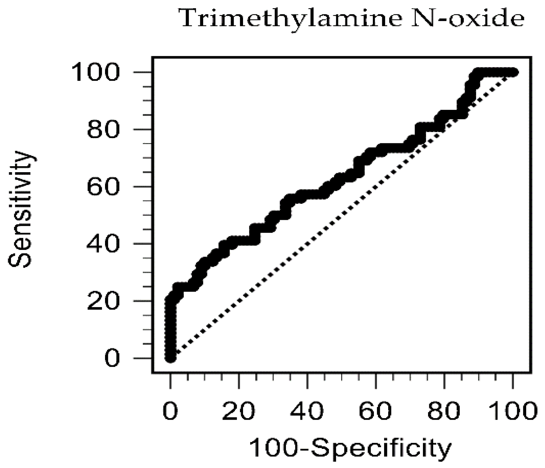
2. Results
3. Discussion
4. Conclusions
5. Materials and Methods
5.1. Patients
5.2. Anthropometric and Biochemical Examination
5.3. Determination of Serum TMAO Levels by High-Performance Liquid Chromatography–Mass Spectrometry
5.4. Examination of Left and Right Brachial-Ankle PWV
5.5. Statistical Analysis
Author Contributions
Funding
Institutional Review Board Statement
Informed Consent Statement
Data Availability Statement
Conflicts of Interest
References
- Tomlinson, J.A.P.; Wheeler, D.C. The role of trimethylamine N-oxide as a mediator of cardiovascular complications in chronic kidney disease. Kidney Int. 2017, 92, 809–815. [Google Scholar] [CrossRef] [PubMed] [Green Version]
- Wu, I.W.; Hsu, K.H.; Lee, C.C.; Sun, C.Y.; Hsu, H.J.; Tsai, C.J.; Tzen, C.Y.; Wang, Y.C.; Lin, C.Y.; Wu, M.S. p-Cresyl sulphate and indoxyl sulphate predict progression of chronic kidney disease. Nephrol. Dial. Transpl. 2011, 26, 938–947. [Google Scholar] [CrossRef] [PubMed] [Green Version]
- Lin, C.J.; Liu, H.L.; Pan, C.F.; Chuang, C.K.; Jayakumar, T.; Wang, T.J.; Chen, H.H.; Wu, C.J. Indoxyl sulfate predicts cardiovascular disease and renal function deterioration in advanced chronic kidney disease. Arch. Med. Res. 2012, 43, 451–456. [Google Scholar] [CrossRef]
- Barreto, F.C.; Barreto, D.V.; Liabeuf, S.; Meert, N.; Glorieux, G.; Temmar, M.; Choukroun, G.; Vanholder, R.; Massy, Z.A.; European Uremic Toxin Work, G. Serum indoxyl sulfate is associated with vascular disease and mortality in chronic kidney disease patients. Clin. J. Am. Soc. Nephrol. 2009, 4, 1551–1558. [Google Scholar] [CrossRef] [Green Version]
- Troseid, M.; Ueland, T.; Hov, J.R.; Svardal, A.; Gregersen, I.; Dahl, C.P.; Aakhus, S.; Gude, E.; Bjorndal, B.; Halvorsen, B.; et al. Microbiota-dependent metabolite trimethylamine-N-oxide is associated with disease severity and survival of patients with chronic heart failure. J. Int. Med. 2015, 277, 717–726. [Google Scholar] [CrossRef] [PubMed]
- Dambrova, M.; Latkovskis, G.; Kuka, J.; Strele, I.; Konrade, I.; Grinberga, S.; Hartmane, D.; Pugovics, O.; Erglis, A.; Liepinsh, E. Diabetes is associated with higher trimethylamine N-oxide plasma levels. Exp. Clin. Endocrinol. Diabetes 2016, 124, 251–256. [Google Scholar] [CrossRef] [Green Version]
- Rhee, E.P.; Clish, C.B.; Ghorbani, A.; Larson, M.G.; Elmariah, S.; McCabe, E.; Yang, Q.; Cheng, S.; Pierce, K.; Deik, A.; et al. A combined epidemiologic and metabolomic approach improves CKD prediction. J. Am. Soc. Nephrol. 2013, 24, 1330–1338. [Google Scholar] [CrossRef] [Green Version]
- Farhangi, M.A.; Vajdi, M. Novel findings of the association between gut microbiota-derived metabolite trimethylamine N-oxide and inflammation: Results from a systematic review and dose-response meta-analysis. Crit. Rev. Food Sci. Nutr. 2020, 60, 2801–2823. [Google Scholar] [CrossRef]
- Tang, W.H.; Wang, Z.; Kennedy, D.J.; Wu, Y.; Buffa, J.A.; Agatisa-Boyle, B.; Li, X.S.; Levison, B.S.; Hazen, S.L. Gut microbiota-dependent trimethylamine N-oxide (TMAO) pathway contributes to both development of renal insufficiency and mortality risk in chronic kidney disease. Circ. Res. 2015, 116, 448–455. [Google Scholar] [CrossRef] [Green Version]
- Fang, Q.; Zheng, B.; Liu, N.; Liu, J.; Liu, W.; Huang, X.; Zeng, X.; Chen, L.; Li, Z.; Ouyang, D. Trimethylamine N-oxide exacerbates renal inflammation and fibrosis in rats with diabetic kidney disease. Front. Physiol. 2021, 12, 682482. [Google Scholar] [CrossRef] [PubMed]
- Gupta, N.; Buffa, J.A.; Roberts, A.B.; Sangwan, N.; Skye, S.M.; Li, L.; Ho, K.J.; Varga, J.; DiDonato, J.A.; Tang, W.H.W.; et al. Targeted inhibition of gut microbial trimethylamine N-oxide production reduces renal tubulointerstitial fibrosis and functional impairment in a murine model of chronic kidney disease. Arterioscler. Thromb. Vasc. Biol. 2020, 40, 1239–1255. [Google Scholar] [CrossRef]
- Zeng, Y.; Guo, M.; Fang, X.; Teng, F.; Tan, X.; Li, X.; Wang, M.; Long, Y.; Xu, Y. Gut microbiota-derived trimethylamine N-oxide and kidney function: A systematic review and meta-analysis. Adv. Nutr. 2021, 12, 1286–1304. [Google Scholar] [CrossRef] [PubMed]
- Li, T.; Gua, C.; Wu, B.; Chen, Y. Increased circulating trimethylamine N-oxide contributes to endothelial dysfunction in a rat model of chronic kidney disease. Biochem. Biophys. Res. Commun. 2018, 495, 2071–2077. [Google Scholar] [CrossRef] [PubMed]
- Abbasalizad Farhangi, M.; Vajdi, M. Gut microbiota-associated trimethylamine N-oxide and increased cardiometabolic risk in adults: A systematic review and dose-response meta-analysis. Nutr. Rev. 2021, 79, 1022–1042. [Google Scholar] [CrossRef] [PubMed]
- Farhangi, M.A. Gut microbiota-dependent trimethylamine N-oxide and all-cause mortality: Findings from an updated systematic review and meta-analysis. Nutrition 2020, 78, 110856. [Google Scholar] [CrossRef] [PubMed]
- Chen, G.; He, L.; Dou, X.; Liu, T. Association of trimethylamine-N-oxide levels with risk of cardiovascular disease and mortality among elderly subjects: A systematic review and meta-analysis. Cardiorenal Med. 2022, 12, 39–54. [Google Scholar] [CrossRef]
- Zhou, Z.; Jin, H.; Ju, H.; Sun, M.; Chen, H.; Li, L. Circulating trimethylamine-N-oxide and risk of all-cause and cardiovascular mortality in patients with chronic kidney disease: A systematic review and meta-analysis. Front. Med. 2022, 9, 828343. [Google Scholar] [CrossRef]
- Stevens, P.E.; Levin, A.; Kidney Disease: Improving Global Outcomes Chronic Kidney Disease Guideline Development Work Group Members. Evaluation and management of chronic kidney disease: Synopsis of the kidney disease: Improving global outcomes 2012 clinical practice guideline. Ann. Intern. Med. 2013, 158, 825–830. [Google Scholar] [CrossRef] [Green Version]
- Vlachopoulos, C.; Aznaouridis, K.; Stefanadis, C. Prediction of cardiovascular events and all-cause mortality with arterial stiffness: A systematic review and meta-analysis. J. Am. Coll. Cardiol. 2010, 55, 1318–1327. [Google Scholar] [CrossRef] [PubMed] [Green Version]
- Karras, A.; Haymann, J.P.; Bozec, E.; Metzger, M.; Jacquot, C.; Maruani, G.; Houillier, P.; Froissart, M.; Stengel, B.; Guardiola, P.; et al. Large artery stiffening and remodeling are independently associated with all-cause mortality and cardiovascular events in chronic kidney disease. Hypertension 2012, 60, 1451–1457. [Google Scholar] [CrossRef] [Green Version]
- Chen, S.C.; Chang, J.M.; Liu, W.C.; Tsai, Y.C.; Tsai, J.C.; Hsu, P.C.; Lin, T.H.; Lin, M.Y.; Su, H.M.; Hwang, S.J.; et al. Brachial-ankle pulse wave velocity and rate of renal function decline and mortality in chronic kidney disease. Clin. J. Am. Soc. Nephrol. 2011, 6, 724–732. [Google Scholar] [CrossRef] [PubMed] [Green Version]
- Wang, S.C.; Lai, Y.H.; Liu, C.H.; Wang, C.H.; Hsu, B.G.; Tsai, J.P. Association between serum indoxyl sulfate levels with carotid-femoral pulse wave velocity in patients with chronic kidney disease. Ren. Fail. 2021, 43, 796–802. [Google Scholar] [CrossRef] [PubMed]
- Wang, C.H.; Lai, Y.H.; Kuo, C.H.; Lin, Y.L.; Tsai, J.P.; Hsu, B.G. Association between serum indoxyl sulfate levels and endothelial function in non-dialysis chronic kidney disease. Toxins 2019, 11, 589. [Google Scholar] [CrossRef] [PubMed] [Green Version]
- Lai, Y.H.; Wang, C.H.; Kuo, C.H.; Lin, Y.L.; Tsai, J.P.; Hsu, B.G. Serum p-cresyl sulfate is a predictor of central arterial stiffness in patients on maintenance hemodialysis. Toxins 2019, 12, 10. [Google Scholar] [CrossRef] [PubMed] [Green Version]
- Tomiyama, H.; Shiina, K. State of the art review: Brachial-ankle PWV. J. Atheroscler. Thromb. 2020, 27, 621–636. [Google Scholar] [CrossRef] [PubMed]
- Tanaka, A.; Tomiyama, H.; Maruhashi, T.; Matsuzawa, Y.; Miyoshi, T.; Kabutoya, T.; Kario, K.; Sugiyama, S.; Munakata, M.; Ito, H.; et al. Physiological diagnostic criteria for vascular failure. Hypertension 2018, 72, 1060–1071. [Google Scholar] [CrossRef] [PubMed]
- Brunt, V.E.; Casso, A.G.; Gioscia-Ryan, R.A.; Sapinsley, Z.J.; Ziemba, B.P.; Clayton, Z.S.; Bazzoni, A.E.; VanDongen, N.S.; Richey, J.J.; Hutton, D.A.; et al. Gut microbiome-derived metabolite trimethylamine N-oxide induces aortic stiffening and increases systolic blood pressure with aging in mice and humans. Hypertension 2021, 78, 499–511. [Google Scholar] [CrossRef]
- Dou, L.; Jourde-Chiche, N.; Faure, V.; Cerini, C.; Berland, Y.; Dignat-George, F.; Brunet, P. The uremic solute indoxyl sulfate induces oxidative stress in endothelial cells. J. Thromb. Haemos. 2007, 5, 1302–1308. [Google Scholar] [CrossRef]
- Sun, C.Y.; Chang, S.C.; Wu, M.S. Uremic toxins induce kidney fibrosis by activating intrarenal renin-angiotensin-aldosterone system associated epithelial-to-mesenchymal transition. PLoS ONE 2012, 7, e34026. [Google Scholar] [CrossRef] [Green Version]
- Ge, X.; Zheng, L.; Zhuang, R.; Yu, P.; Xu, Z.; Liu, G.; Xi, X.; Zhou, X.; Fan, H. The gut microbial metabolite trimethylamine N-oxide and hypertension risk: A systematic review and dose-response meta-analysis. Adv. Nutr. 2020, 11, 66–76. [Google Scholar] [CrossRef]
- Missailidis, C.; Hallqvist, J.; Qureshi, A.R.; Barany, P.; Heimburger, O.; Lindholm, B.; Stenvinkel, P.; Bergman, P. Serum trimethylamine-N-oxide is strongly related to renal function and predicts outcome in chronic kidney disease. PLoS ONE 2016, 11, e0141738. [Google Scholar] [CrossRef] [PubMed] [Green Version]
- Mishima, E.; Fukuda, S.; Mukawa, C.; Yuri, A.; Kanemitsu, Y.; Matsumoto, Y.; Akiyama, Y.; Fukuda, N.N.; Tsukamoto, H.; Asaji, K.; et al. Evaluation of the impact of gut microbiota on uremic solute accumulation by a CE-TOFMS-based metabolomics approach. Kidney Int. 2017, 92, 634–645. [Google Scholar] [CrossRef] [PubMed] [Green Version]
- Costabile, G.; Vetrani, C.; Bozzetto, L.; Giacco, R.; Bresciani, L.; Del Rio, D.; Vitale, M.; Della Pepa, G.; Brighenti, F.; Riccardi, G.; et al. Plasma TMAO increase after healthy diets: Results from 2 randomized controlled trials with dietary fish, polyphenols, and whole-grain cereals. Am. J. Clin. Nutr. 2021, 114, 1342–1350. [Google Scholar] [CrossRef] [PubMed]
- Crimarco, A.; Springfield, S.; Petlura, C.; Streaty, T.; Cunanan, K.; Lee, J.; Fielding-Singh, P.; Carter, M.M.; Topf, M.A.; Wastyk, H.C.; et al. A randomized crossover trial on the effect of plant-based compared with animal-based meat on trimethylamine-N-oxide and cardiovascular disease risk factors in generally healthy adults: Study with appetizing plantfood-meat eating alternative trial (SWAP-MEAT). Am. J. Clin. Nutr. 2020, 112, 1188–1199. [Google Scholar]
- Argyridou, S.; Davies, M.J.; Biddle, G.J.H.; Bernieh, D.; Suzuki, T.; Dawkins, N.P.; Rowlands, A.V.; Khunti, K.; Smith, A.C.; Yates, T. Evaluation of an 8-week vegan diet on plasma trimethylamine-N-oxide and postchallenge glucose in adults with dysglycemia or obesity. J. Nutr. 2021, 151, 1844–1853. [Google Scholar] [CrossRef] [PubMed]
- Miao, L.; Du, J.; Chen, Z.; Shi, D.; Qu, H. Effects of microbiota-driven therapy on circulating trimethylamine-N-oxide metabolism: A Systematic review and meta-analysis. Front. Cardiovasc. Med. 2021, 8, 710567. [Google Scholar] [CrossRef] [PubMed]
- Geng, J.; Yang, C.; Wang, B.; Zhang, X.; Hu, T.; Gu, Y.; Li, J. Trimethylamine N-oxide promotes atherosclerosis via CD36-dependent MAPK/JNK pathway. Biomed. Pharmacother. 2018, 97, 941–947. [Google Scholar] [CrossRef]
- Boini, K.M.; Hussain, T.; Li, P.L.; Koka, S. Trimethylamine-N-oxide instigates NLRP3 inflammasome activation and endothelial dysfunction. Cell. Physiol. Biochem. 2017, 44, 152–162. [Google Scholar] [CrossRef]
- Seldin, M.M.; Meng, Y.; Qi, H.; Zhu, W.; Wang, Z.; Hazen, S.L.; Lusis, A.J.; Shih, D.M. Trimethylamine N-oxide promotes vascular inflammation through signaling of mitogen-activated protein kinase and nuclear factor-kappaB. J. Am. Heart Assoc. 2016, 5, e002767. [Google Scholar] [CrossRef] [Green Version]
- Zhang, X.; Li, Y.; Yang, P.; Liu, X.; Lu, L.; Chen, Y.; Zhong, X.; Li, Z.; Liu, H.; Ou, C.; et al. Trimethylamine-N-Oxide Promotes Vascular Calcification Through Activation of NLRP3 (Nucleotide-Binding Domain, Leucine-Rich-Containing Family, Pyrin Domain-Containing-3) Inflammasome and NF-kappaB (Nuclear Factor kappaB) Signals. Arterioscler. Thromb. Vasc. Biol. 2020, 40, 751–765. [Google Scholar] [CrossRef]
- Yang, M.; Zhang, R.; Zhuang, C.; Wu, Y.; Yang, Q.; Yu, Z.; Liu, J.; Zha, B.; Gong, Q.; Yang, B.; et al. Serum trimethylamine N-oxide and the diversity of the intestinal microbial flora in type 2 diabetes complicated by diabetic kidney disease. Clin. Lab. 2022, 68, 210836. [Google Scholar] [CrossRef] [PubMed]

| Characteristics | All Patients (n = 157) | Control group (n = 89) | PAS Group (n = 68) | p Value |
|---|---|---|---|---|
| Age (years) | 67.90 ± 13.00 | 63.35 ± 12.04 | 73.85 ± 11.81 | <0.001 * |
| Body mass index (kg/m2) | 26.12 ± 3.95 | 26.27 ± 4.39 | 25.92 ± 3.30 | 0.587 |
| Female, n (%) | 76 (48.4) | 41 (46.1) | 35 (51.5) | 0.502 |
| TMAO (μg/L) | 27.43 (16.68–49.54) | 25.28 (15.56–42.47) | 35.83 (17.95–77.79) | 0.006 * |
| Left baPWV (m/s) | 17.30 ± 3.46 | 14.93 ± 1.54 | 20.40 ± 2.74 | <0.001 * |
| Right baPWV (m/s) | 17.26 ± 3.39 | 14.97 ± 1.80 | 20.26 ± 2.54 | <0.001 * |
| Diabetes mellitus, n (%) | 52 (33.1) | 23 (25.8) | 29 (42.6) | 0.027 * |
| Hypertension, n (%) | 97 (61.8) | 49 (55.1) | 48 (70.6) | 0.047 * |
| Hyperlipidemia, n (%) | 93 (59.2) | 55 (61.8) | 38 (55.9) | 0.555 |
| Smoking, n (%) | 16 (10.2) | 10 (11.2) | 6 (8.8) | 0.244 |
| SBP (mmHg) | 144.31 ± 17.84 | 140.53 ± 19.49 | 149.25 ± 14.09 | 0.002 * |
| DBP (mmHg) | 82.14 ± 10.21 | 82.06 ± 10.31 | 82.25 ± 10.15 | 0.907 |
| Total cholesterol (mg/dL) | 163.01 ± 43.75 | 165.22 ± 47.37 | 160.12 ± 38.64 | 0.470 |
| Triglyceride (mg/dL) | 121.0 (87.5–165.0) | 116.0 (88.5–166.5) | 128.50 (86.25–163.0) | 0.430 |
| LDL–C (mg/dL) | 92.63 ± 36.28 | 95.93 ± 39.41 | 88.31 ± 31.50 | 0.193 |
| Fasting glucose (mg/dL) | 104.0 (98.0–134.0) | 100.0 (97.0–118.5) | 110.0 (100.0–141.75) | 0.010 * |
| Blood urea nitrogen (mg/dL) | 33.00 (24.00–45.00) | 31.00 (24.50–44.00) | 35.50 (24.00–49.50) | 0.562 |
| Creatinine (mg/dL) | 1.90 (1.40–2.70) | 1.80 (1.40–2.60) | 2.05 (1.60–3.00) | 0.163 |
| eGFR (mL/min) | 31.37 ± 15.33 | 34.06 ± 15.42 | 27.84 ± 14.58 | 0.011 * |
| CKD stage 3, n (%) | 79 (50.3) | 51 (57.3) | 28 (41.2) | 0.119 |
| CKD stage 4, n (%) | 45 (28.7) | 23 (25.8) | 22 (32.4) | |
| CKD stage 5, n (%) | 33 (21.0) | 15 (16.9) | 18 (26.4) | |
| Albumin (g/dL) | 4.07 ± 0.32 | 4.10 ± 0.33 | 4.04 ± 0.29 | 0.256 |
| Hemoglobin (g/dL) | 11.68 ± 2.58 | 11.71 ± 2.03 | 11.64 ± 3.17 | 0.870 |
| CRP (mg/dL) | 0.13 (0.05–1.20) | 0.06 (0.05–1.12) | 0.34 (0.08–163) | 0.012 * |
| ARB use, n (%) | 88 (56.1) | 46 (51.7) | 42 (61.8) | 0.207 |
| β–blocker use, n (%) | 50 (31.8) | 25 (28.1) | 25 (36.8) | 0.248 |
| CCB use, n (%) | 60 (38.2) | 31 (34.8) | 29 (42.6) | 0.318 |
| α–adrenergic blocker use, n (%) | 21 (13.4) | 10 (11.2) | 11 (16.2) | 0.368 |
| Statin use, n (%) | 73 (46.5) | 41 (46.1) | 32 (47.1) | 0.902 |
| Fibrate use, n (%) | 25 (15.9) | 15 (16.9) | 10 (14.7) | 0.715 |
| Variables | Odds Ratio | 95% CI | p Value |
|---|---|---|---|
| Trimethylamine N-oxide, 1 μg/L | 1.016 | 1.002–1.029 | 0.021 * |
| Age, 1 year | 1.081 | 1.043–1.121 | <0.001 * |
| systolic blood pressure, 1 mmHg | 1.042 | 1.004–1.081 | 0.031 * |
| Hypertension, present | 0.596 | 0.171–2.072 | 0.415 |
| Diabetes mellitus, present | 3.062 | 0.970–9.660 | 0.056 |
| Fasting glucose, 1 mg/dL | 0.998 | 0.987–1.009 | 0.710 |
| Estimated glomerular filtration rate, 1 mL/min | 0.991 | 0.962–1.020 | 0.538 |
| C-reactive protein, 1 mg/dL | 1.085 | 0.889–1.325 | 0.422 |
| Variables | TMAO | Left PWV | Right PWV | Age | BMI | eGFR | CRP | Glucose | LDL–C | TG | SBP | DBP | |
|---|---|---|---|---|---|---|---|---|---|---|---|---|---|
| TMAO | r p | 0.256 0.001 * | 0.267 0.001 * | 0.219 0.006 * | −0.098 0.222 | −0.442 <0.001 * | 0.261 0.001 * | −0.015 0.856 | −0.082 0.305 | −0.079 0.325 | 0.029 0.716 | −0.049 0.546 | |
| Left PWV | r p | 0.256 0.001 * | 0.913 <0.001 * | 0.453 <0.001 * | −0.013 0.870 | −0.147 0.066 | 0.182 0.022 * | 0.132 0.100 | 0.038 0.637 | 0.035 0.666 | 0.328 <0.001 * | 0.103 0.200 | |
| Right PWV | r p | 0.267 0.001 * | 0.913 <0.001 * | 0.411 <0.001 * | −0.036 0.650 | −0.151 0.060 | 0.157 0.050 | 0.126 0.117 | 0.039 0.629 | 0.059 0.459 | 0.291 < 0.001 * | 0.084 0.294 | |
| Age | r p | 0.219 0.006 * | 0.453 <0.001 * | 0.411 <0.001 * | −0.006 0.941 | −0.110 0.171 | 0.147 0.066 | 0.013 0.867 | −0.024 0.767 | −0.129 0.109 | 0.041 0.611 | −0.346 <0.001 * | |
| BMI | r p | −0.098 0.222 | −0.013 0.870 | −0.036 0.650 | −0.006 0.941 | 0.166 0.038 | −0.072 0.373 | 0.125 0.118 | 0.028 0.724 | 0.128 0.109 | 0.221 0.006 * | 0.127 0.113 | |
| eGFR | r p | −0.442 <0.001 * | −0.147 0.066 | −0.151 0.060 | −0.110 0.171 | 0.166 0.038 * | −0.185 0.021 * | 0.126 0.116 | 0.088 0.270 | −0.033 0.680 | −0.184 0.021 * | −0.058 0.472 | |
| CRP | r p | 0.261 0.001 * | 0.182 0.022 * | 0.157 0.050 | 0.147 0.066 | −0.072 0.373 | −0.185 0.021 * | −0.034 0.673 | −0.088 0.272 | −0.006 0.937 | 0.050 0.537 | −0.072 0.370 | |
| Glucose | r p | −0.015 0.856 | 0.132 0.100 | 0.126 0.117 | 0.013 0.867 | 0.125 0.118 | 0.126 0.116 | −0.034 0.673 | −0.106 0.185 | 0.167 0.036 * | 0.028 0.729 | 0.031 0.697 | |
| LDL–C | r p | −0.082 0.305 | 0.038 0.637 | 0.039 0.629 | −0.024 0.767 | 0.028 0.724 | 0.088 0.270 | −0.088 0.272 | −0.106 0.185 | 0.196 0.014 * | −0.028 0.730 | 0.120 0.133 | |
| TG | r p | −0.079 0.325 | 0.035 0.666 | 0.059 0.459 | −0.129 0.109 | 0.128 0.109 | −0.033 0.680 | −0.006 0.937 | 0.167 0.036 * | 0.196 0.014 * | −0.075 0.352 | 0.043 0.592 | |
| SBP | r p | 0.029 0.716 | 0.328 <0.001 * | 0.291 <0.001 * | 0.041 0.611 | 0.221 0.006 * | −0.184 0.021 * | 0.050 0.537 | 0.028 0.729 | −0.028 0.730 | −0.075 0.352 | 0.566 <0.001 * | |
| DBP | r p | −0.049 0.546 | 0.103 0.200 | 0.084 0.294 | −0.346 <0.001 * | 0.127 0.113 | −0.058 0.472 | −0.072 0.370 | 0.031 0.697 | 0.120 0.133 | 0.043 0.592 | 0.566 <0.001 * | |
| Peripheral arterial stiffness | Odds ratio | 95% CI | p Value |
|---|---|---|---|
| Diabetes mellitus, yes | 1.007 | 0.995–1.020 | 0.249 |
| Diabetes mellitus, no | 1.035 | 1.016–1.054 | <0.001 * |
| Hypertension, yes | 1.016 | 1.004–1.028 | 0.011 * |
| Hypertension, no | 1.032 | 1.008–1.056 | 0.008 * |
| Hyperlipidemia, yes | 1.014 | 1.003–1.025 | 0.017 * |
| Hyperlipidemia, no | 1.038 | 1.011–1.066 | 0.006 * |
| Chronic kidney disease | |||
| Stage 3 | 1.006 | 0.992–1.019 | 0.432 |
| Stage 4 | 1.025 | 1.004–1.050 | 0.046 * |
| Stage 5 | 1.036 | 1.006–1.068 | 0.021 * |
Publisher’s Note: MDPI stays neutral with regard to jurisdictional claims in published maps and institutional affiliations. |
© 2022 by the authors. Licensee MDPI, Basel, Switzerland. This article is an open access article distributed under the terms and conditions of the Creative Commons Attribution (CC BY) license (https://creativecommons.org/licenses/by/4.0/).
Share and Cite
Hsu, B.-G.; Wang, C.-H.; Lin, Y.-L.; Lai, Y.-H.; Tsai, J.-P. Serum Trimethylamine N-Oxide Level Is Associated with Peripheral Arterial Stiffness in Advanced Non-Dialysis Chronic Kidney Disease Patients. Toxins 2022, 14, 526. https://doi.org/10.3390/toxins14080526
Hsu B-G, Wang C-H, Lin Y-L, Lai Y-H, Tsai J-P. Serum Trimethylamine N-Oxide Level Is Associated with Peripheral Arterial Stiffness in Advanced Non-Dialysis Chronic Kidney Disease Patients. Toxins. 2022; 14(8):526. https://doi.org/10.3390/toxins14080526
Chicago/Turabian StyleHsu, Bang-Gee, Chih-Hsien Wang, Yu-Li Lin, Yu-Hsien Lai, and Jen-Pi Tsai. 2022. "Serum Trimethylamine N-Oxide Level Is Associated with Peripheral Arterial Stiffness in Advanced Non-Dialysis Chronic Kidney Disease Patients" Toxins 14, no. 8: 526. https://doi.org/10.3390/toxins14080526
APA StyleHsu, B.-G., Wang, C.-H., Lin, Y.-L., Lai, Y.-H., & Tsai, J.-P. (2022). Serum Trimethylamine N-Oxide Level Is Associated with Peripheral Arterial Stiffness in Advanced Non-Dialysis Chronic Kidney Disease Patients. Toxins, 14(8), 526. https://doi.org/10.3390/toxins14080526