1. Introduction
Copper (Cu) is an essential trace element and a critical cofactor for a variety of key enzymes, such as cytochrome C oxidase, superoxide dismutase, and tyrosinase, and is involved in numerous physiological processes including redox reactions, energy metabolism, neurotransmitter synthesis, and connective tissue formation. It is crucial for maintaining the health of the nervous system, hematopoietic system, and bones [
1,
2]. Under physiological conditions, the body tightly regulates Cu homeostasis through intestinal absorption, hepatic storage, and biliary excretion [
3]. However, long-term dietary Cu deficiency, impaired intestinal absorption (e.g., due to Menkes disease or post-gastrectomy), or specific nutritional conditions (e.g., long-term parenteral nutrition) can induce Cu deficiency, potentially leading to multiple systemic dysfunctions including anemia, osteoporosis, neurodegenerative changes, and immunodeficiency [
4].
A tight bidirectional relationship exists between Cu and immune function. Under physiological conditions, adequate Cu supports normal immune responses through mechanisms such as maintaining ceruloplasmin synthesis and antioxidant defense [
5]. However, disruption of Cu homeostasis—whether deficiency or excess—can disturb immune balance. In Cu deficiency, the pro-inflammatory interaction between neutrophils and microvascular endothelial cells is enhanced, the expression of cyclooxygenase-2 (COX-2) in the liver is significantly upregulated in an SOD1-independent manner [
6], the expression of pro-inflammatory and fibrinogen-related genes is increased [
7], and nuclear factor kappa-B (NF-κB) protein levels are elevated [
8]. Conversely, Cu excess can also induce gut microbiota dysbiosis and inflammation [
9,
10]. This bidirectionality suggests that the maintenance of Cu homeostasis, rather than the absolute level of Cu itself, is key to immune regulation. Furthermore, in various inflammatory diseases, serum Cu levels are often compensatorily elevated to several times the normal physiological range [
11], further corroborating the close link between Cu and inflammatory regulation.
The intestinal tract serves as the primary site for dietary Cu digestion and absorption, and it is also a key organ for the body to regulate Cu homeostasis and excrete excessive Cu. In addition, Cu plays a complex and crucial role in maintaining intestinal homeostasis, which is essential for the stability of intestinal morphology and structure as well as the microbial community. Excessive Cu exposure can induce intestinal microbiota dysbiosis, characterized by a reduction in probiotic abundance, a decreased Firmicutes/Bacteroidetes (F/B) ratio, and altered populations of bacteria associated with lipid metabolism and intestinal inflammation, ultimately contributing to the development of intestinal inflammation [
12]. Dietary supplementation with a high dose of Cu (120 mg/kg) can lead to the accumulation of unabsorbed Cu in the intestinal lumen, which significantly reduces the α-diversity and species richness of the gut microbiota, notably decreasing the abundance of anaerobic commensals such as
Clostridium [
13]. Physiological Cu supplementation, particularly during the weaning period, has been shown to improve intestinal morphology and structure, potentially through promoting the proliferation of crypt epithelial cells and enhancing the self-renewal activity of intestinal stem cells [
14].
As a critical segment of the intestine, the colon serves as a key organ for digestion, immune defense, and microbial colonization, with its homeostasis reliant on an intact mucosal barrier, an optimal immune response, and a balanced gut microbiota. Given that the colon is particularly susceptible to dietary influences and microbial dysbiosis, understanding how Cu deficiency specifically affects colonic homeostasis is of considerable importance. Evidence suggests that Cu is associated with the occurrence and development of colon diseases such as inflammatory bowel disease, but systematic experimental evidence on how Cu deficiency affects colon morphology and structure, barrier function, and the gut microbiome is still limited. Therefore, this study was designed to establish a Cu-deficient mouse model to assess the impact of Cu deficiency on colonic histopathology, goblet cell function, mucosal barrier integrity, inflammatory responses, and the composition and structure of the intestinal microbiota. The findings aim to elucidate the role of Cu deficiency in colonic homeostasis disruption, providing a foundation for future studies on copper–gut interactions.
2. Methods
2.1. Animals and Treatments
Three-week-old healthy male ICR mice (16–18 g) were obtained from Dashuo Biological Technology Company (Chengdu, China). All mice were housed in a specific pathogen-free facility under controlled environmental conditions (temperature: 25 ± 2 °C; 12 h light/dark cycle) with unrestricted access to standard chow and water. Mice were excluded if they had pre-existing abnormalities (e.g., abnormal baseline body weight, congenital defects, or traumatic injury) or experienced non-experimental death during the study. The fifty-four mice were randomly divided into control, Cu-deficient (CuD), and CuD + CuSO
4 three groups, with 18 mice per group. To ensure experimental reproducibility and statistical robustness, each experimental group included three independent biological replicates (each replicate comprised one cage of six mice). To prevent selection bias, an independent investigator implemented random allocation through a computer-generated randomization process using Microsoft Excel. The sample size was calculated with G*Power software (version 3.1.9.7). Following Cohen’s established criteria for effect magnitudes in ANOVA, a prudent and moderately large effect size (f = 0.4) was applied to secure sufficient statistical power. Under these conditions, with a significance level (α) of 0.05 and 80% power, the analysis determined that a minimum of 22 mice per group would be needed. However, subsequent studies employing the same copper-deficiency protocol have detected significant differences in key outcome measures with as few as 6 mice per group [
15], indicating that the actual effect size in this model is considerably larger than the conservative f = 0.4 estimate. Therefore, in keeping with the Reduction tenet of the 3R principles and based on this empirical evidence, the final sample size was set at 18 mice per group. For each experimental assay, 6 mice per group (biological replicates) were used, and all findings are based on this sample size. The animal feeding intervention protocols and copper supplementation dosages and administration routes were designed and implemented in accordance with the methodological system established by Pan et al. [
16]. The control group was fed with a standard pellet diet for 5 weeks and injected with saline in the last week. During the final week, the CuD group received daily intraperitoneal injections of saline, while the CuD + CuSO
4 group received daily intraperitoneal injections of copper sulfate (CuSO
4, 10 μg/g body weight). The intraperitoneal route was selected to bypass potential intestinal absorption impairments that may persist after prolonged copper deficiency, thereby ensuring controlled systemic copper delivery. Our data confirmed that this one-week regimen restored serum copper to levels comparable to the Control group (
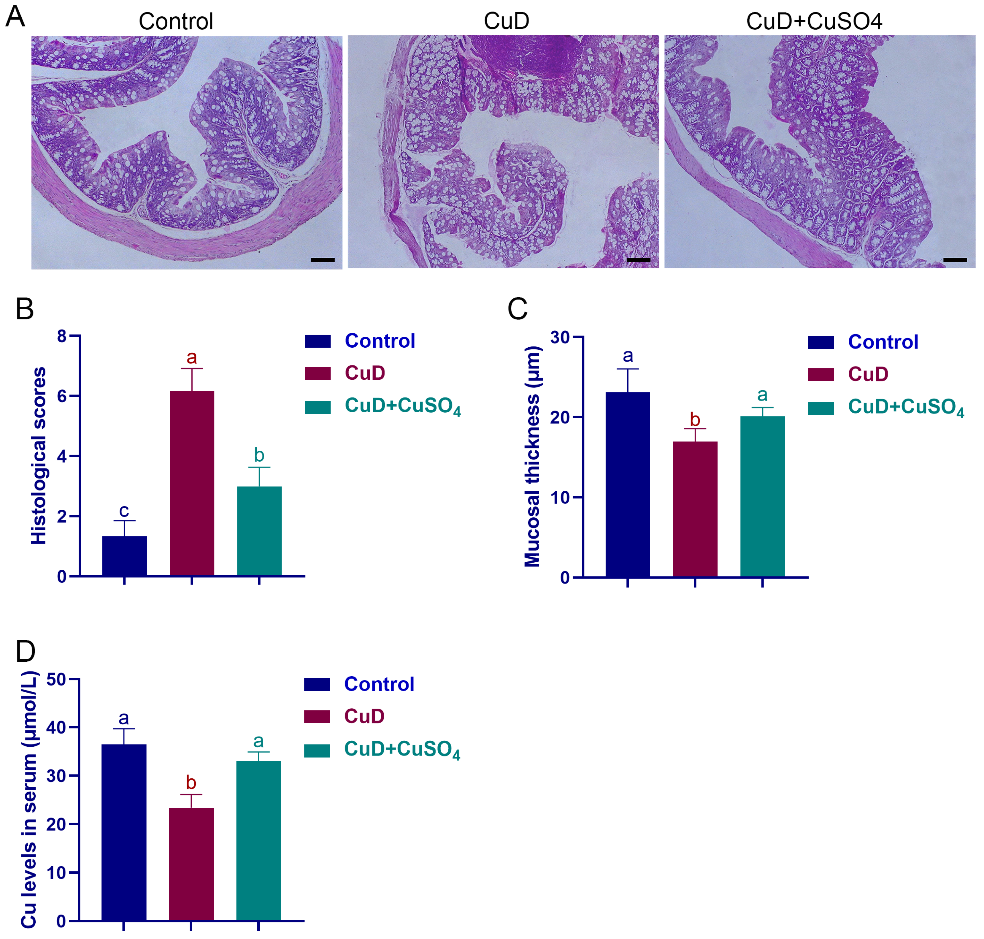
Figure 1D). The CuD + CuSO
4 group was included as a proof-of-concept rescue condition to assess the reversibility of copper-deficiency-induced changes, rather than to mimic dietary copper repletion. After the designated interventions, mice were weighed and then anesthetized with isoflurane. Blood samples were collected from the orbital sinus using sterile 1.5 mL centrifuge tubes. Subsequently, euthanasia was performed by cervical dislocation to minimize suffering, in accordance with the Refinement principle. For tissue allocation within each group of 18 mice, six mice provided colon tissues for H&E staining and immunofluorescence staining, six mice provided colon tissues and colonic contents for quantitative PCR and microbiome analysis, and the remaining six mice provided colon tissues for Western blotting. All procedures were conducted in strict compliance with institutional animal care and use guidelines approved by the Animal Ethics Committee of Southwest University of Science and Technology, Mianyang, China (Approval No.: L2024014). Due to the complexity of the in vivo physiological processes investigated in this study, no suitable non-animal alternatives were available to fully address the research questions, thereby necessitating the use of animal models in compliance with the Replacement principle. The experimental diets were supplied by SPF Biotechnology Co., Ltd. (Beijing, China) and fully met the nutritional standards specified in the AIN-93M maintenance diet. The results of Cu content analysis in the feed are shown in
Figure S1.
2.2. Histopathological Observation
Colon samples were fixed in 4% paraformaldehyde, then dehydrated through an ethanol series, embedded in paraffin, sliced into 5-μm sections, and stained with hematoxylin-eosin. Blinded histopathological analysis was carried out at 100× magnification using a Nikon DS-Ri1 microscope (Tokyo, Japan), with semi-quantitative ratings of 0 to 3 applied to four criteria: inflammatory cell infiltration, epithelial tissue loss, crypt structural damage, and reduction in mucosal thickness. The total histology injury score, ranging from 0 (no pathology) to 12 (severe damage), was determined by summing the four parameter scores, based on an adapted reference method [
17]. For each colon sample, five non-overlapping images were randomly captured at 100× magnification using a light microscope. Mucosal thickness was measured from the base of the crypts to the luminal surface using Image Pro Plus version 6.0 software (Media Cybernetics, Rockville, MD, USA). All measurements were performed by an investigator blinded to the experimental groups, and the mean value of five measurements per sample was used for statistical analysis.
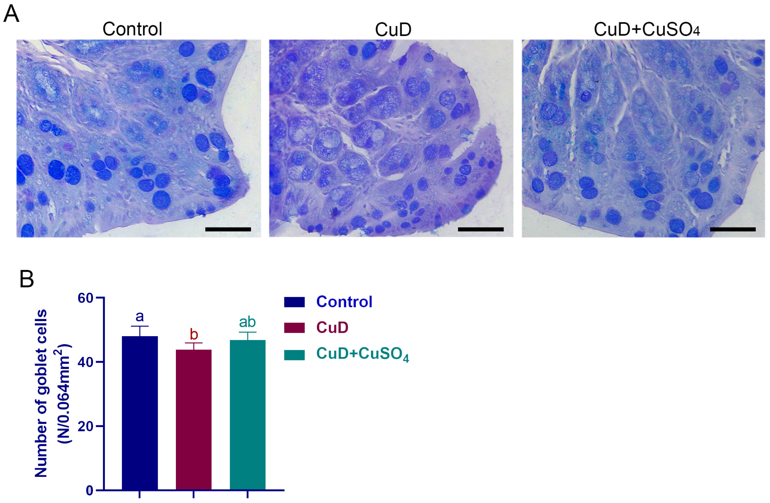
Goblet cells in colonic tissue sections were identified via combined Alcian Blue and periodic acid–Schiff (AB/PAS) histochemistry. Briefly, deparaffinized sections were incubated with 1% Alcian Blue solution (pH 2.5) for 5 min, rinsed thoroughly with distilled water, treated with 1% periodic acid for oxidation, washed again, and then exposed to Schiff’s reagent. After final water rinsing and dehydration, sections were coverslipped and examined under bright-field microscopy. Acid mucins in goblet cells appeared blue, enabling clear identification and assessment. Images were captured and analyzed in a blinded manner with respect to group allocation. From each colon specimen, five representative tissue sections were prepared. For each section, five non-overlapping fields were randomly selected and imaged at 400× magnification. Only goblet cells with well-defined morphology and distinct blue staining (indicative of acidic mucin content) were included in the quantification. Cell counting was performed manually by a blinded observer using standardized criteria, following a protocol adapted from Yin et al. [
18].
2.3. Serum Cu Concentration
Mice were anesthetized and blood was collected from the retro-orbital plexus. Clotted blood was centrifuged at 3000× g for 10–15 min to separate serum, which was then stored at −80 °C until analysis. Serum Cu concentration was determined using a commercially available colorimetric assay kit (E010-1-1, Nanjing Jiancheng Bioengineering Institute, Nanjing, China), following the manufacturer’s protocol.
2.4. Immunofluorescent Staining
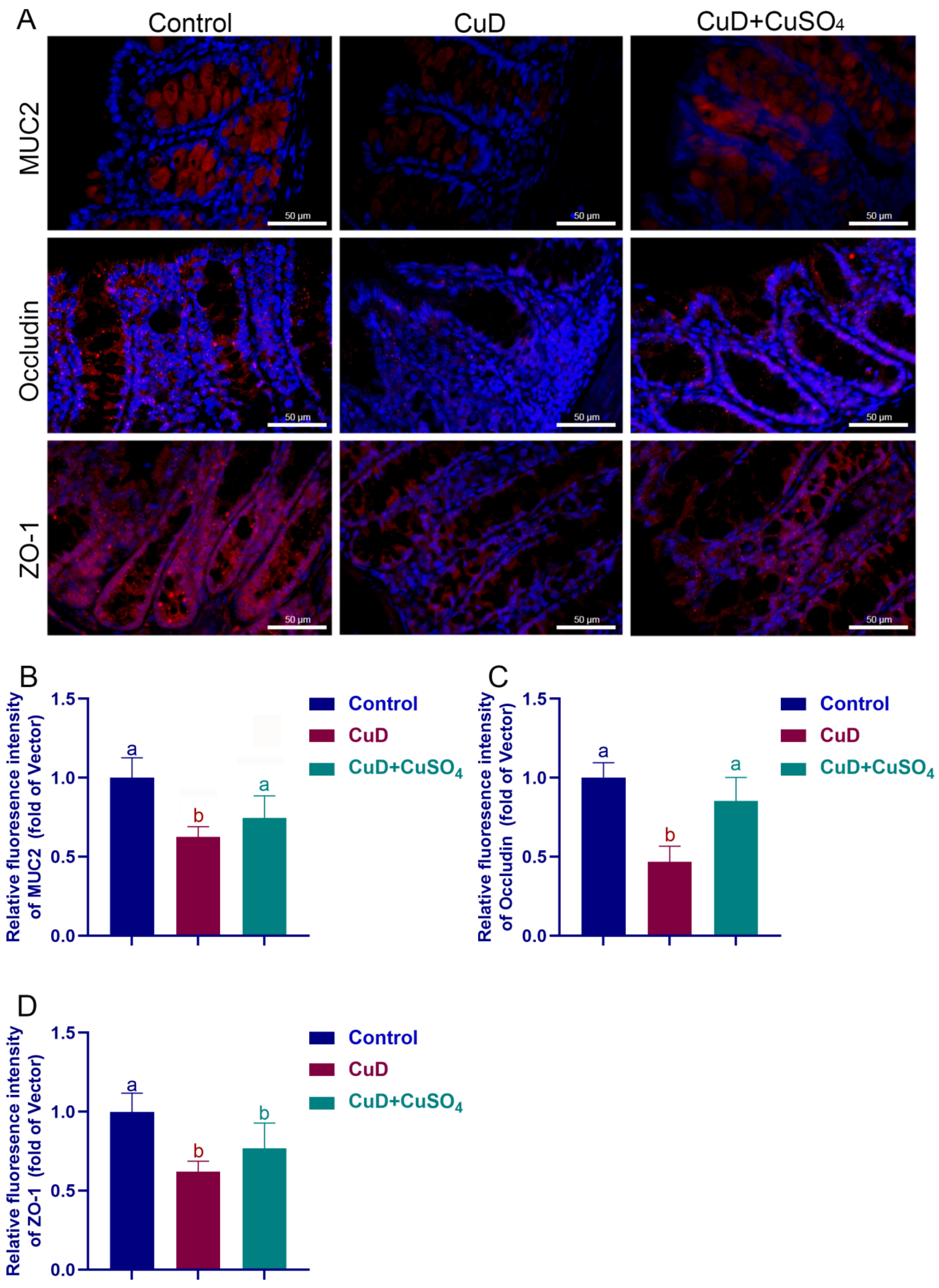
Mouse colon tissue sections underwent deparaffinization, antigen retrieval in citrate buffer (pH 6.0) via thermal treatment, equilibration to room temperature, and PBS rinsing. Following a 30 min blocking step with 5% goat serum, the sections were incubated at 4 °C overnight with primary antibodies targeting MUC2, ZO-1, CD11b, and occludin. On the following day, the tissue sections underwent three PBS washes and were subsequently exposed to fluorescently labeled secondary antibodies for 30 min at ambient temperature under light-protected conditions. Fluorescent signal was enhanced using a commercial development reagent (10 min, dark), nuclei were stained with DAPI for 10 min in the dark, and the slides were sealed with an antifade mounting medium prior to fluorescence imaging.
For each colon tissue sample, three non-adjacent sections were selected, and five random fields of view (400× magnification) were captured per section. All images were acquired using the same microscope (Olympus, Japan) with identical acquisition settings (exposure time, gain, and fluorescence intensity thresholds) across all samples to ensure comparability. Fluorescence intensity was quantified using ImageJ 1.8.0 software by an investigator blinded to the experimental groups. The mean intensity values from all fields per sample were used for statistical analysis to avoid pseudoreplication.
2.5. Cytokine Levels in Serum
Serum samples were processed identically to those described in
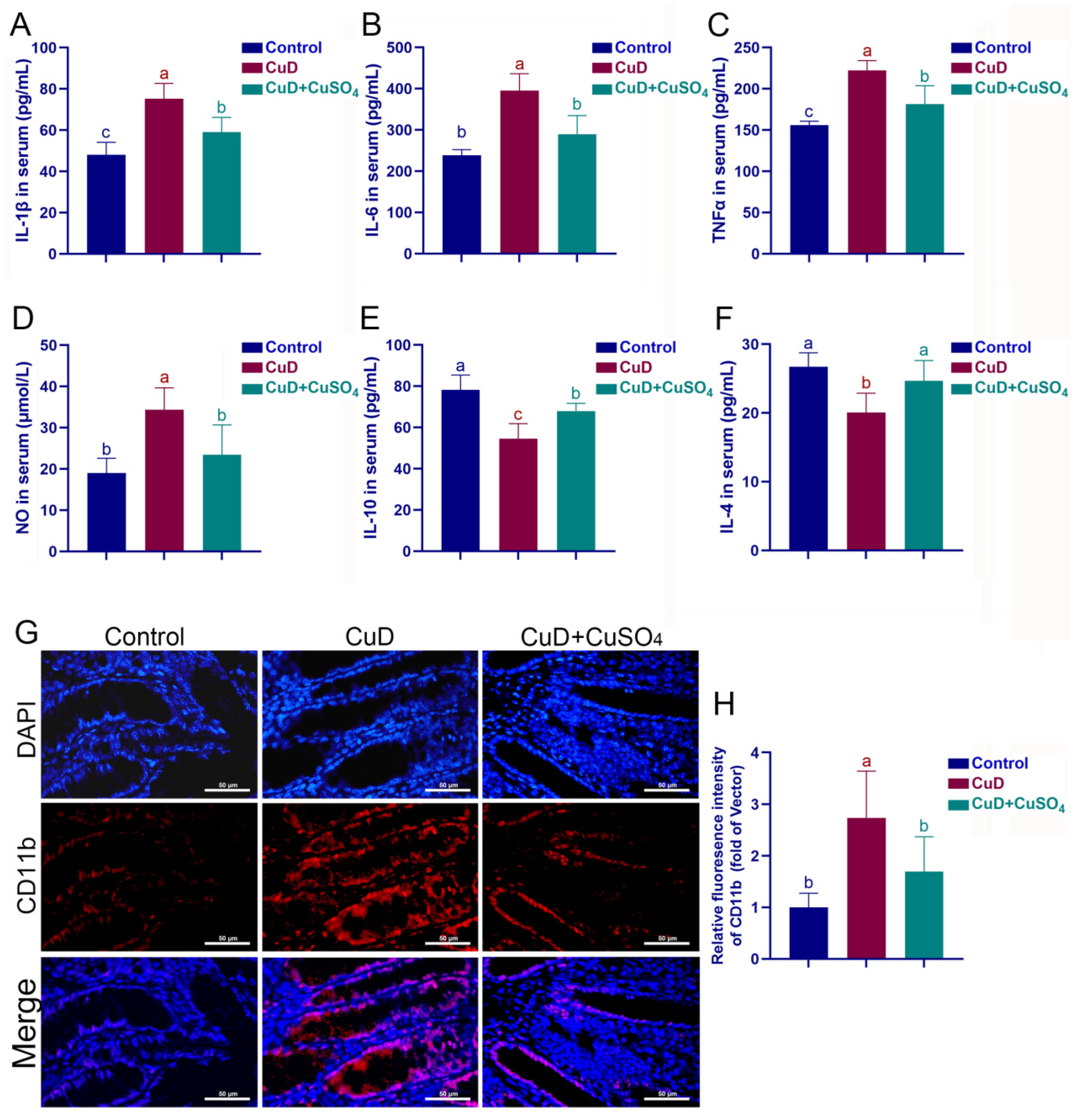
Section 2.3. IL-1β, IL-6, TNF-α, IL-4, IL-10, and nitric oxide (NO) concentrations were quantified using commercially available ELISA kits (Biosharp, Hefei, China), according to the supplier’s protocol. All assays were performed in technical triplicate, and mean values were calculated for statistical analysis.
2.6. Western Blotting Analysis
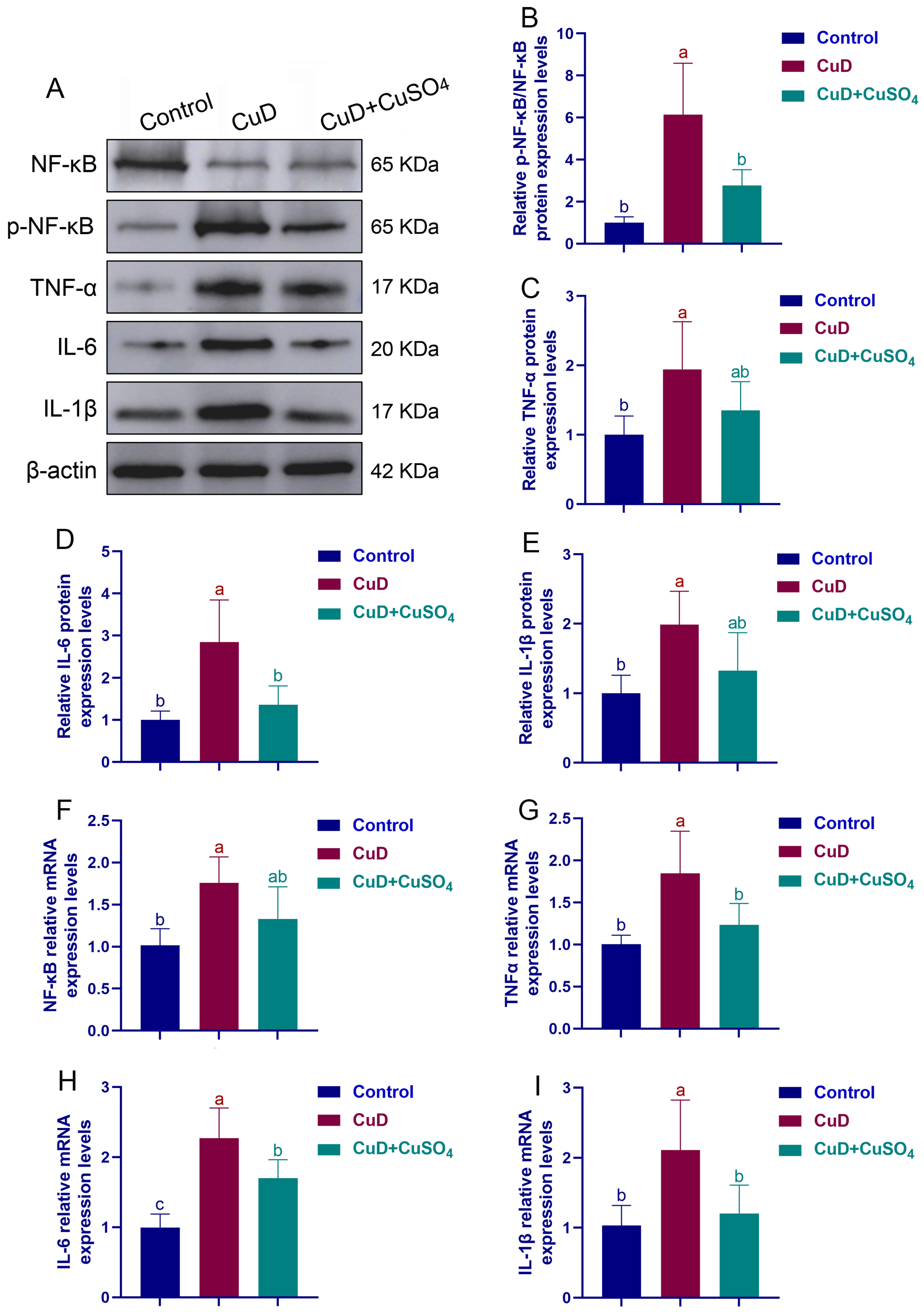
Colon tissue samples were homogenized in RIPA lysis buffer to extract total protein, and protein concentration was determined using the BCA method (P0010S, Beyotime, Shanghai, China). Equal protein quantities were separated by SDS-PAGE and transferred electrophoretically to nitrocellulose membranes. The membranes were blocked for 1 h at room temperature with 5% non-fat dry milk in TBST, then incubated overnight at 4 °C with primary antibodies specific for NF-κB (1:1000, #8242, CST), phospho-NF-κB (1:1000, #3033, CST), IL-1β (1:1000, A22257, Abclonal, Wuhan, China), IL-6 (1:1000, A0286, Abclonal), TNF-α (1:3000, A28059, Abclonal), and β-actin (1:3000, AC026, Abclonal). Afterwards, the membranes were probed for 1 h with horseradish peroxidase (HRP)-labeled secondary antibodies, and immunoreactive bands were detected using an enhanced chemiluminescence (ECL) kit (P0018A, Beyotime, China) on a ChemiDoc XRS imaging system. Image acquisition and quantification were performed blindly, with the analyst unaware of group assignments, and protein expression levels were normalized to β-actin.
2.7. qRT-PCR Analysis
Colon tissues were flash-frozen in liquid nitrogen, homogenized in a pre-chilled mortar and pestle, and stored at −80 °C. Total RNA was extracted from samples using RNAiso Plus (Takara, Shiga, Japan; Cat. No. 9108/9109) according to the supplier’s instructions. Subsequently, 1 µg of purified RNA was reverse-transcribed into cDNA with the PrimeScript RT Reagent Kit (Takara, Japan; Cat. No. RR047A). Quantitative real-time PCR (qRT-PCR) was carried out on a LightCycler
® 480 System (Roche, Basel, Switzerland) using SYBR Premix Ex Taq II (DRR820A, Takara, Japan). Primer sequences are provided in
Table 1. Target gene expression was normalized to β-actin as an internal reference, and relative mRNA expressions was quantified via the 2
−ΔΔCt comparative quantification method.
2.8. Determination and Analysis of Gut Microbiota
Colonic luminal contents were collected from mice and immediately frozen at −80 °C. Microbial DNA was extracted using a commercial stool DNA kit. DNA concentration and quality were measured with the Qubit dsDNA HS Assay Kit on a Qubit 4 Fluorometer. The V3–V4 hypervariable region of the bacterial 16S rRNA gene was amplified and subjected to high-throughput sequencing on the Illumina NovaSeq 6000 platform. Bioinformatic analysis was performed on the Novogene Cloud Platform (
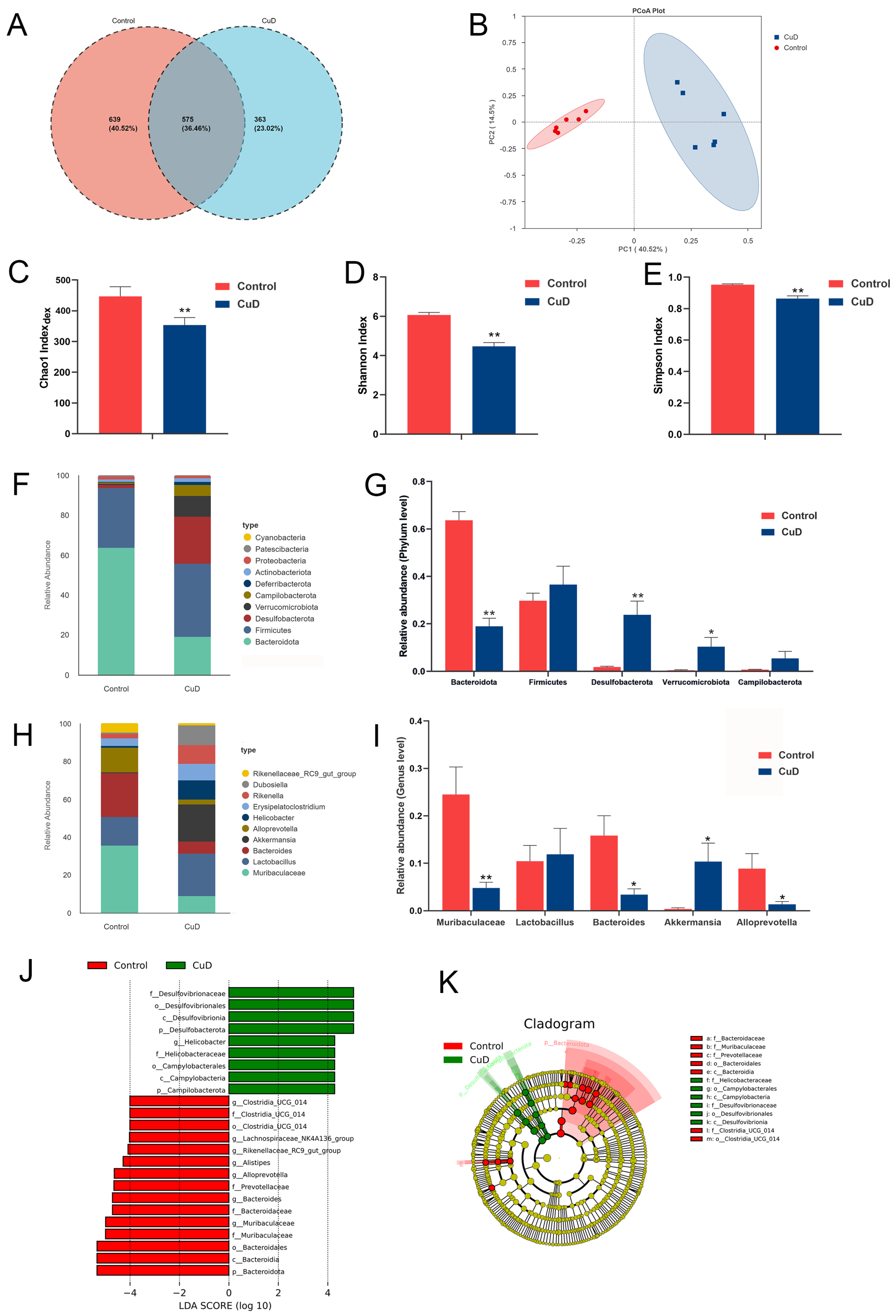
https://www.novogene.com/eu-en/technology/platforms/, accessed on 10 May 2024). Following read splicing, filtering, and sample demultiplexing, Operational Taxonomic Unit (OTU) clustering was performed. Based on the resulting OTU clusters, taxonomic classification was conducted, and the OTU abundance matrix was subsequently used for downstream analyses. The resulting valid data were subjected to species annotation and abundance analysis to determine the species composition of the samples. Multiple alpha diversity metrics were calculated based on the OTU data, and sequencing depth was assessed through rarefaction analysis. Specifically, alpha diversity metrics (observed species, Chao1 index, Shannon index, and Simpson index) as well as beta diversity based on Bray–Curtis distance were calculated using the R package vegan. Principal coordinate analysis (PCoA) was used to visualize the beta diversity results, and permutational multivariate analysis of variance (PERMANOVA) was adopted to statistically compare the differences in microbiota structure. Taxa with significantly differential abundance were identified by linear discriminant analysis effect size (LEfSe) analysis, with the screening criteria set as an LDA score > 4 and
p < 0.05.
2.9. Statistical Analysis
Data are presented as mean ± standard error of the mean (SEM). For each experimental group, 18 mice were housed in three cages (6 mice per cage). For each assay, a stratified random sampling approach was used: 2 mice were randomly selected from each cage, yielding a total of 6 mice per assay. This ensured representation from all cages and balanced potential cage effects. The experimental unit for all analyses was the individual mouse (n = 6 per group per assay). Differences between group means were assessed using one-way analysis of variance (ANOVA), followed by Tukey’s honestly significant difference (HSD) test for post hoc comparisons. Dunnett’s T3 test was applied when the assumption of homogeneity of variances was violated. All statistical tests were conducted with SPSS 22.0 (IBM Corp., Armonk, NY, USA), and differences were considered statistically significant at p < 0.05.
4. Discussion
A bidirectional physiological relationship exists between Cu and the gut. The intestine serves not only as the primary site for dietary Cu absorption but also as a key regulator of systemic Cu homeostasis. Conversely, Cu levels and speciation directly influence intestinal structure and function. By establishing a mouse model of dietary Cu deficiency, this study systematically examined the resulting multifaceted detrimental effects on the colon and elucidated the underlying mechanisms.
Histomorphological assessment showed that Cu deficiency was associated with impaired colonic development and pathological changes, most notably a significant decrease in mucosal thickness accompanied by inflammatory cell infiltration. Goblet cells, mucus-secreting epithelial cells abundant in the colon, produce a protective mucus layer that shields the epithelium from gastric acid, bile, proteolytic enzymes, and pathogens, thereby maintaining intestinal barrier integrity. A reduced number of goblet cells reflects mucosal atrophy and weakened mucin (e.g., MUC2) synthesis, thereby promoting intestinal barrier hyperpermeability and local immune dysregulation [
18]. The significant reduction in both goblet cell number and AB-PAS staining intensity in the CuD group suggests that copper deficiency may impair goblet cell differentiation or secretory function. Given that goblet cell maturation and mucin synthesis are energy-intensive processes, copper depletion—via its effects on mitochondrial respiration—may limit the metabolic capacity of these cells to produce and secrete mucins. Additionally, the observed reduction could partially reflect epithelial atrophy, as indicated by the decreased mucosal thickness in the CuD group
. To further explore the relationship between Cu deficiency and colonic barrier integrity, this study detected the expression of mucin MUC2 and tight junction proteins. MUC2 is a key structural component of the intestinal mucus layer, and its reduced expression indicates that the integrity of the mucus barrier may be compromised [
19]. Tight junctions, which are primarily composed of proteins including ZO-1 and Occludin, constitute essential structural elements of the intestinal epithelial barrier by regulating paracellular permeability and maintaining mucosal integrity [
20]. The downregulation of MUC2, Occludin, and ZO-1 in the CuD group suggests that copper deficiency may compromise the intestinal barrier at multiple levels. MUC2 reduction likely reflects impaired goblet cell secretory function, while the loss of tight junction proteins may result from either direct effects of copper depletion on epithelial cell homeostasis or secondary consequences of heightened local inflammation, given that pro-inflammatory cytokines such as TNF-α are known to disrupt tight junction assembly. The combined impairment of the chemical (mucus) and physical (tight junction) barriers may create a self-reinforcing cycle, where a weakened barrier permits greater luminal antigen translocation, further fueling inflammation. Interestingly, both copper deficiency and excess appear to compromise colonic integrity. Early studies found that long-term low dietary Cu significantly increased the incidence of colon tumors in rats and caused colonic abnormalities [
20]. Liao et al. [
21] reported that excessive Cu intake impairs the colonic barrier function in pigs, as evidenced by reduced expression of tight junction proteins (ZO-1, Occludin, Claudin-1, and JAM-1), Muc2, and mucus secretion-related genes. Taken together, disruption of Cu homeostasis, whether through deficiency or excess, may be associated with compromised colonic barrier integrity. Furthermore, this research provides experimental evidence supporting a potential role of Cu deficiency in relation to the development and maintenance of colonic barrier integrity.
The impairment of intestinal barrier function facilitates the translocation of luminal microbiota and antigens, which activate innate immunity and contribute to the development of inflammatory bowel disease [
22]. To investigate the impact of Cu deficiency on colonic inflammation, this study systematically assessed key inflammatory markers. The elevated systemic levels of pro-inflammatory cytokines (IL-1β, IL-6, TNF-α) and NO, coupled with suppressed anti-inflammatory cytokines (IL-4, IL-10), suggest that copper deficiency induces a systemic inflammatory state. The concurrent upregulation of CD11b in colonic tissue indicates that this inflammatory response is, at least in part, localized to the intestine. The activation of NF-κB phosphorylation observed in the CuD colon provides a plausible mechanistic link: copper deficiency may lower the threshold for NF-κB activation, as Cu-dependent antioxidant enzymes normally restrain redox-sensitive inflammatory signaling. The resulting pro-inflammatory milieu could then contribute to the barrier disruption discussed above. Moreover, the significant increase in both mRNA and protein expression of IL-1β and IL-6 in colon tissue further evidenced the activation of intestinal inflammation. Collectively, our results indicate that Cu deficiency can induce systemic inflammation accompanied by the occurrence of colonic inflammatory responses, while the causal relationship between the two remains to be further explored. NF-κB serves as a central regulator of inflammatory responses and is intricately involved in key pathological processes, including intestinal barrier impairment and oxidative stress [
18,
23]. This study confirmed that inflammatory markers (NF-κB and TNF-α) were significantly elevated in Cu-deficient colon tissues, suggesting that Cu deficiency is accompanied by intestinal inflammation. Currently, studies specifically addressing the impact of Cu deficiency on colonic inflammation remain scarce, with the existing literature predominantly centered on the effects of Cu overload or therapeutic supplementation. Zhang et al. [
13] reported that feeding high doses of Cu (120 and 240 mg/kg feed) could significantly increase the concentration of pro-inflammatory factors in rat serum, accompanied by disordered fecal microbiota structure, and indirectly promote intestinal inflammation. Similarly, Liao et al. [
21] found that excessive Cu leads to reduced secretion of secretory immunoglobulins A (SIgA) and G (SIgG) in the jejunum and colon, along with upregulated expression of inflammatory factors such as IL-1β and TNF-α. Lin et al. [
24] found that long-term Cu exposure significantly increased the mRNA levels of TLR3, TLR7, TLR8, NF-κB, I-κB, TNF-α and IL-8 in the gut of marsh eels, triggering intestinal biological damage and inflammatory responses. Notably, Fu et al. [
25] showed that supplementation with Cu ion-Lut nanocomplexes could relieve colitis symptoms by inhibiting NF-κB expression and exerting anti-inflammatory and intestinal barrier protective effects. However, the cross-sectional nature of this study precludes determining the temporal sequence of these events. Reduced tight junction protein expression and mucin depletion may facilitate luminal antigen translocation, thereby triggering inflammatory responses [
26]. Conversely, inflammation itself can directly disrupt tight junction integrity and goblet cell function through pro-inflammatory cytokines such as TNF-α and IL-1β [
27,
28]. It is therefore plausible that barrier dysfunction and inflammation operate as a self-amplifying feedback loop under Cu-deficient conditions, rather than following a simple unidirectional causality. Future time-course studies are needed to dissect the initiating events in this process.
Gut microbiota homeostasis is essential for intestinal health, and its disruption is recognized as a key driver of intestinal inflammation [
29,
30]. To determine whether Cu deficiency contributes to colonic pathological changes through modulation of the gut microbiota, we further examined structural alterations in the intestinal microbial community. The reduced α-diversity and altered community structure in the CuD group may reflect a selective pressure imposed by copper deficiency on the gut microbial ecosystem. Copper serves as an essential cofactor for numerous bacterial enzymes involved in energy metabolism and antioxidant defense [
31], and its scarcity may disproportionately affect species with higher copper requirements, thereby reducing community richness. At the phylum level, the marked decrease in Bacteroidetes—a dominant phylum in the healthy gut—and the concomitant expansion of Desulfobacterota suggest a shift toward a pro-inflammatory community configuration. This compositional shift could be driven by both direct effects of copper limitation on bacterial growth and indirect effects mediated by the altered host environment, including a compromised mucus barrier and heightened oxidative stress in the intestinal lumen [
32,
33]. Most studies report that intestinal inflammation is associated with a marked reduction in colonic Bacteroidetes abundance, and this reduction is negatively correlated with markers of inflammation [
34]. In addition, Desulfobacterota can release lipopolysaccharide (LPS) into the gut, which has been associated with promoting inflammatory responses and disrupting intestinal energy metabolism in other studies [
35]. Verrucomicrobiota, primarily residing in the inner mucosal layer of the intestine, contribute to energy and nutrient supply by degrading polysaccharides such as mucopolysaccharides and cellulose [
36]. Notably, Ren et al. [
37] observed a marked increase in Verrucomicrobiota abundance in the colon of mice with DSS-induced colitis. At the genus level, Cu deficiency led to a significant reduction in the relative abundance of
Muribaculaceae,
Bacteroides, and
Alloprevotella, whereas the abundance of
Akkermansia was markedly increased.
Muribaculaceae and
Bacteroides, both members of the
Bacteroidales order, produce short-chain fatty acids that supply energy to intestinal epithelial cells, and their increased abundance is significantly correlated with enhanced intestinal barrier integrity and reduced inflammation [
38,
39].
Alloprevotella is a gut commensal bacterium that metabolizes dietary proteins and carbohydrates, with butyrate as its primary metabolite, which exerts inhibitory effects on enteric pathogens and plays a critical role in sustaining intestinal homeostasis in the host [
40,
41]. While
Akkermansia muciniphila is widely recognized as a beneficial symbiont that promotes gut barrier integrity and metabolic health under normal conditions [
42], its role is highly context-dependent.
Akkermansia is a mucin-degrading specialist; under conditions of compromised mucosal barrier function—such as the reduced MUC2 expression and goblet cell depletion observed in our CuD mice—excessive
Akkermansia proliferation could hypothetically exacerbate mucus layer thinning by accelerating mucin degradation, potentially further weakening the barrier, although this remains to be directly tested. This is consistent with reports that
Akkermansia expansion has been observed in certain pathological states where its overgrowth is associated with, rather than protective against, barrier dysfunction [
43]. Copper deficiency resulted in a marked reduction in key anti-inflammatory commensals and an increase in pro-inflammatory taxa, which is generally consistent with the findings of Klevay et al. [
44], suggesting that copper deficiency is associated with reshaping of the gut microbiota and with colonic inflammation. Causative relationships between specific taxa and inflammatory outcomes cannot be established from our correlational data.



 and
and