Article

Enhancing Resistance to Enterococcus faecalis: Immunobiotic Lactiplantibacillus plantarum Strains as a Strategy for Malnourished Hosts

by
María Daniela Vera
1,2,
Lorena Paola Arce
1,2,
Melisa Florencia Müller
1,2,
Fernanda Raya Tonetti
3,
Ramiro Ortiz Moyano
3,
Héctor Luis Blanco
1,2,
Haruki Kitazawa
4,5,*,
María Guadalupe Vizoso-Pinto
1,2,* and
Julio Villena
3,*
1
Infection Biology Laboratory, Instituto Superior de Investigaciones Biológicas (INSIBIO), CONICET-UNT, Tucumán 4000, Argentina
2
Laboratorio Central de Ciencias Básicas, Facultad de Medicina, Universidad Nacional de Tucumán, Tucumán 4000, Argentina
3
Laboratory of Immunobiotechnology, Reference Centre for Lactobacilli (CERELA-CONICET), Chacabuco 145, Tucumán 4000, Argentina
4
Food and Feed Immunology Group, Laboratory of Animal Food Function, Graduate School of Agricultural Science, Tohoku University, Sendai 980-8572, Japan
5
Livestock Immunology Unit, International Education and Research Center for Food and Agricultural Immunology (CFAI), Graduate School of Agricultural Science, Tohoku University, Sendai 980-8572, Japan
*
Authors to whom correspondence should be addressed.
Nutrients 2025, 17(11), 1770; https://doi.org/10.3390/nu17111770
Submission received: 14 April 2025 / Revised: 19 May 2025 / Accepted: 21 May 2025 / Published: 23 May 2025

Abstract

:
Background: Enterococcus faecalis causes serious opportunistic infections in patients with weakened immune defenses, such as individuals suffering from malnutrition. We investigated the effects of Lactiplantibacillus plantarum MPL16 and CRL1506 on the resistance to E. faecalis infection in mice immunosuppressed by protein malnutrition. Methods: Male BALB/c mice received a protein-deficient diet for 21 days, followed by a 7-day renourishment period with a balanced conventional diet (BCD) with or without lactobacilli supplementation. Malnourished controls (MN) and renourished mice were treated with gentamicin for 3 days and then challenged orally with E. faecalis 102. Infection levels in the gut, liver, spleen, and blood, intestinal tissue damage, and the cytokine response were evaluated 2 days after the challenge. Results: Malnourished mice had an impaired ability to produce pro-inflammatory cytokines (TNF, IFN-γ, IL-1β, IL-6, IL-17, and KC) and the regulatory IL-10 in response to the infection compared to mice in the BCD group. The imbalance of inflammatory and regulatory mediators in MN mice favors colonization and invasion by E. faecalis and increases tissue damage, making the disease more severe than in animals renourished with BCD. Supplementing the BCD with L. plantarum strains significantly enhanced resistance to E. faecalis 102 infections, as evidenced by a marked reduction in bacterial loads and intestinal damage. The effect of lactobacilli was associated with enhanced levels of IFN-γ, IL-6, and IL-10 and reduced TNF, IL-1β, IL-17, and KC. Conclusions: Given their efficacy in enhancing host resistance, these Lactiplantibacillus strains hold great promise as a preventive strategy against E. faecalis infections in susceptible individuals.

Graphical Abstract

1. Introduction

Enterococcus faecalis is a commensal microorganism that inhabits the gastrointestinal tract of mammals without inducing adverse effects. However, this bacterium can cause opportunistic infections in immunocompromised hosts or patients undergoing prolonged antibiotic use [1]. In these cases, this bacterium can play a dual role as both a commensal and a pathogen. E. faecalis efficiently colonizes the intestinal mucosa; under certain conditions, it overgrows and can invade the host’s tissues. This bacterium can adapt its metabolic gene expression to the microenvironmental conditions of the intestinal tract [2] and pass from the intestinal lumen to the lamina propria through its internalization in intestinal epithelial cells (IECs) [3,4]. Although enterococcal virulence factors are necessary for the colonization of the intestinal epithelium, the status of the host is also crucial for the onset of infectious disease since alterations of the intestinal barrier, microbiota, and/or the immune system can enable the overgrowth and invasion of enterococci. Research in mice has shown that antibiotics capable of inducing dysbiosis of the intestinal microbiota, such as cephalosporins [5], clindamycin [6], and vancomycin [7], can promote E. faecalis overgrowth in the gut and its translocation into the bloodstream. The intestinal microbiota can control the overgrowth of E. faecalis through its metabolic activity, such as the production of antimicrobial substances or the modulation of metabolites in the intestinal lumen [8,9]. On the other hand, early studies demonstrated that the administration of the immunosuppressive drug cyclophosphamide significantly augmented the susceptibility of mice to bacteremia and mortality after the oral challenge with E. faecalis [10].
Secondary immunodeficiencies can develop because of several factors, including the use of immunosuppressive compounds, cancer, chronic infections, and inadequate nutrition [11]. Nutrient deficiencies alter the immune defenses, leading to enhanced frequency and severity of infections, while infectious diseases potentiate malnutrition as they often promote malabsorption and diminished food intake [12]. The increased susceptibility of malnourished hosts to pathogens, particularly those causing intestinal and invasive infections, has been attributed to alterations of the intestinal microbiota and barrier and the impairment of the immune system [12]. The high rates of cell division and proliferation within the intestinal mucosa make this tissue particularly vulnerable to the detrimental effects of malnutrition. Protein malnutrition influences caspase-dependent apoptosis of IECs, affecting cell turnover [13]. In humans, villous atrophy, increased intestinal permeability, and crypt hyperplasia were described in protein-malnourished hosts [12]. These histological and functional alterations of the intestinal mucosa by protein malnutrition were also found in pigs, rats, mice, and mouse organoids [14,15,16,17]. Protein malnutrition affects the integrity of the gastrointestinal mucosa and, with reduced gastric acid secretion and altered bile production, leads to enhanced susceptibility to infections. On the other hand, studies in animal models and clinical trials in malnourished children demonstrated that malnutrition affects several arms of the immune system including neutrophil chemotaxis and bactericidal capacity, macrophages phagocytic activity, dendritic cells maturation and the levels of interleukin (IL)-1β, IL-6, tumor necrosis factor (TNF), and complement proteins in blood [12]. Malnutrition also affects immune responses mediated by lymphocytes, inducing alterations in the numbers of T cells in the spleen and lymph nodes and circulating B cells and reducing the levels of secretory immunoglobulin A (IgA) [18]. Impairment in the number and function of B and T cells in Peyer’s patches, along with a reduced ability to produce appropriate IgA and IgM antibodies in response to antigens, has been described in malnourished hosts [19]. All these alterations induced by protein malnutrition on the intestinal microbiota, barrier, and immune system have been described to promote E. faecalis infection. However, enterococcal infections in protein-malnourished hosts have not been studied in depth despite of the fact that malnourished children demonstrate the intestinal overgrowth of pathobionts [20], including enterococci strains able to induce enteropathy [21], and that antibiotic treatments are usually included as part of the management of malnourished children, even in the absence of infection [22].
The use of functional foods containing probiotic microorganisms with immunomodulatory capacities (immunobiotics) has been proposed to enhance resistance to infections [23]. Studies in immunocompetent mice demonstrated that immunobiotic strains like Lactiplantibacillus plantarum CRL1506 [24,25] and L. plantarum MPL16 [26] increase immune defense mechanisms and protect against pathogens. Furthermore, we have previously demonstrated that protein-malnourished mice have impaired defenses against Salmonella [27], Candida albicans [28], and Streptococcus pneumoniae [29] and that the repletion of these immunocompromised hosts with a balanced conventional diet (BCD) supplemented with immunobiotic lactobacilli can accelerate the recovery of the immune system and improve the resistance against them.
Enterococcal infections in malnourished hosts remain poorly studied despite evidence that children with malnutrition often harbor an overabundance of intestinal pathobionts that trigger enteric dysfunction [20], that enterococci isolated from malnourished children can induce enteropathy in malnourished gnotobiotic mice [21], and that a broad-spectrum of antibiotics is routinely recommended in guidelines as part of the management of malnourished children, regardless of the presence of clinical symptoms of infection [22]. We hypothesize that protein malnutrition and antibiotic treatment enhance susceptibility and severity of E. faecalis intestinal–invasive infection and that the recovery of malnourished hosts with a dietary intervention including immunomodulatory lactobacilli improves mucosal and systemic defenses against opportunistic enterococci. In this work, we tested these hypotheses by evaluating the resistance of protein-malnourished gentamicin-treated mice to an oral challenge with pathogenic E. faecalis 102 and the influence of a BCD supplemented with the immunobiotic strains L. plantarum MPL16 or CRL1506 on the resistance to the infection and the immune response in the malnourished hosts.

2. Materials and Methods

2.1. Microorganisms

L. plantarum CRL1506 was obtained from the CERELA culture collection (CERELA-CONICET, Tucumán, Argentina) and had been previously isolated from goat milk [30]. L. plantarum MPL16 was obtained from the Food and Feed Immunology Group Culture Collection (Tohoku University, Sendai, Japan) and had been previously isolated from the feces of pigs fed with wakame (Undaria pinnatifida), a traditional Japanese fermented food [31]. Lactobacilli (stored at −70 °C) were activated and cultured for 12 h at 37 °C (final log phase) in Man-Rogosa-Sharpe (MRS, Britania, Argentina) broth. The bacteria were harvested by centrifugation at 3000× g for 10 min and washed three times with sterile 0.01 mol/L PBS, pH 7.2, and resuspended in sterile 15% non-fat milk.
E. faecalis 102 was obtained from the Culture Collection of the Faculty of Biochemistry, Chemistry and Pharmacy of the National University of Tucumán. This strain had been previously isolated from a wound of a 41-year-old patient from the Angel C. Padilla Hospital (San Miguel de Tucumán, Argentina) and was selected in preliminary experiments in malnourished mice because of its ability to induce intestinal–invasive infection. E. faecalis 102 was cultured in Brain Heart Infusion (BHI, Britania, Argentina) at 37 °C for 16 h and stored in 50% sterile glycerol at −70 °C for further use.

2.2. Survival of Lactic Acid Bacteria to Gastrointestinal Conditions In Vitro

Gastrointestinal (GI) conditions were simulated by following the methods reported before [32,33,34,35]. Briefly, to test the survival to gastrointestinal conditions, lactic acid bacteria (LAB) were grown for 16 h in MRS, washed twice with phosphate-buffered saline (PBS), and suspended in MRS or 15% reconstituted skim milk after centrifugation. Then, LAB were sequentially subjected to 1:1 dilution in a buffer mimicking saliva (NaCl 6.2 g/L; KCl 2.2 g/L; CaCl2 0.22 g/L; NaHCO3 1.2 g/L pH 7.2), a 3:5 dilution in artificial gastric fluid (NaCl 6.2 g/L; KCl 2.2 g/L; CaCl2 0.22 g/L; NaHCO3 1.2 g/L pH 2.5), and a 1:4 dilution in synthetic intestinal fluid (NaHCO3 6.4 g/L; KCl 0.239 g/L; NaCl 1.28 g/L; sodium deoxycholate monohydrate (Sigma-Aldrich, Buenos Aires, Argentina) 0.5%; pancreatin protease (Pankreoflat A.D., Munro, Argentina) (>1900 USP). Samples were incubated under constant shaking for 5, 60, and 120 min to simulate the digestion processes occurring in saliva, gastric fluids, and intestinal fluids, respectively [34,35]. Then, viable LAB cells were serially diluted (10-fold dilutions), plated onto MRS agar plates, and incubated at 37 °C for 48 h under anaerobic conditions. The assay was performed in triplicate twice.

2.3. Animals Feeding Procedures and Experimental Infection in Malnourished Mice

Mice and Diet. Three-week-old male BALB/c mice were obtained from CERELA (San Miguel de Tucumán, Argentina). After weaning, the mice were fed a low-protein diet for 21 days. This diet consisted mainly of carbohydrates supplemented with vitamins, essential fatty acids, and minerals [29,36,37]. As shown in Figure 1, mice were randomly divided into four groups of 5 animals each. Two control groups were included: malnourished mice without renourishment (MN group) and mice renourished with a conventional balanced diet (BDC) for 7 days without LAB supplementation (BDC group). The composition of BCD is provided in the Supplementary Table S1. L. plantarum MPL16 or L. plantarum CRL1506 were administered at a dose of 108 cells/mouse/day in drinking water (BDC + MPL16 and BDC + CRL1506 groups, respectively). Three days before the challenge with E. faecalis (day 26), the mice were treated with the antibiotic gentamicin (1.6 mg/kg) (Invitrogen, Dreieich, Germany) [38] supplemented in drinking water (Figure 1). The experimental unit was each animal. The total number of animals used was 20 mice, and none were excluded. The number of animals was decided based on previously published works [28,29,30]. To minimize potential confounders, treatments and challenges were performed in different orders. Samples were blinded for group members analyzing the samples and data. Animals were monitored daily in order to avoid suffering. None of the animals died during the experiment.
Oral challenge with E. faecalis. E. faecalis 102 was cultured on BHI agar for 18 h; freshly grown colonies were suspended in 5 mL of BHI broth and incubated at 37 °C for 16 h under shaking. The bacterial cells were harvested by centrifugation at 10,000 rpm for 10 min and washed three times with sterile PBS. The cell density was adjusted to 8 × 10⁸ cells/L.
On day 29, mice were orally challenged with a single dose of E. faecalis 102 (50 µL, 108 UFC/mL) and euthanized 48 h post-challenge. The mouse samples (blood, feces, intestinal fluid, liver, and spleen) were collected for subsequent analysis (Figure 1). To minimize animal suffering, predefined humane endpoints were established and approved by the Institutional Animal Care and Use Committee. Mice were monitored at least twice daily for clinical signs of distress, especially during infection. Animals were euthanized if they met any of the following criteria: (1) persistent lethargy or inability to access food or water; (2) severe diarrhea or signs of dehydration, (3) hunched posture, ruffled fur, and inactivity lasting more than 24 h; (4) signs of systemic infection or sepsis, including labored breathing; or (5) self-isolation or failure to engage in normal behaviors.

2.4. Counts of E. faecalis Colonies in Mouse Samples

Two days after the challenge with E. faecalis (31 days), mouse samples were aseptically collected and suspended in 5 mL of sterile PBS to determine viable E. faecalis counts. Fecal samples were disaggregated with a sterile pestle and homogenized in PBS. Bowel lavage was collected by flushing the small intestine with PBS. The liver and spleen were homogenized in 5 mL of sterile peptone water. Blood samples were obtained via cardiac puncture and collected in heparinized tubes.
Mouse samples were serially diluted, plated in duplicate on BHI agar, and incubated for 18 h at 37 °C. Due to the differences in growth kinetics, it was possible to differentiate enterococci from lactobacilli: the colonies of E. faecalis were visible and counted (UFC/mL) after the first 24 h, whereas the colonies of L. plantarum appeared later, after 48 h.

2.5. Determination of Cytokine Concentrations in Serum and Bowel Lavage

To evaluate the local and systemic immune response, as well as the level of inflammation caused by E. faecalis 102, cytokine concentration was measured in serum and intestinal lavage samples obtained from malnourished mice treated with LAB and subsequently challenged with E. faecalis 102. The concentrations of TNF, interferon (IFN)-γ, IL-1β, IL-6, keratinocyte chemoattractant (KC), IL-10, and IL-17 in serum and bowel lavage were measured using commercially available enzyme-linked immunosorbent assay (ELISA) kits, following the manufacturer’s recommendations (R&D Systems, Minnneapolis, MN, USA).

2.6. Histopathology

After 48 h of the challenge with E. faecalis 102, euthanasia was performed, and small intestine samples were collected for histological analysis. These samples were stored in 10% formalin (Biopack, Buenos Aires, Argentina) and kept at 4 °C until processing. Samples were first dehydrated by passing them through a series of alcohol solutions (Frau ethyl alcohol 96°, Argentina), with increasing concentrations: two passages through 70% alcohol, one through 80% alcohol, and two through 96% alcohol. The samples were transferred to butyl acetate (Cirarelli, San Lorenzo, Argentina) and a histological clearing agent (Pathoclear®Plus, Biopack, Argentina), each step involving 1 h incubation. After incubation, the samples were embedded in paraffin (Biopack, Argentina) through two additional 1 h incubation periods. The inclusion process was followed, during which tissue blocks were assembled. Serial sections were made using a rotary microtome, with section sizes ranging from 3 to 5 microns. Once sectioning was complete, the staining process began. The cut sections, already mounted on slides, were rehydrated and immersed in hematoxylin stain (Biopack, Argentina) for 5 min. They were rinsed with running water and subsequently immersed in an eosin stain (Biopack, Argentina) for another 5 min. After staining, the samples were again passed through alcohol solutions, butyl acetate, and a clearing agent. Finally, the mounting process was carried out by placing a drop of synthetic Canadian balsam (Biopack, Argentina) on the slide and covering it with a cover slip. Once prepared, the samples were examined under an optic microscope (Leica, Wetzlar, Germany).

2.7. Statistical Analysis

Experiments were performed in duplicates, and results were expressed as mean  ±  standard deviation (SD). Statistical analyses were performed using Prism 8.0 (GraphPad software, San Diego, CA, USA). After verification of the normal distribution of data, one-way ANOVA was used. Tukey’s test (for pairwise comparisons of the means) was used to test for differences between the groups. Differences were considered significant at p  <  0.05. Graphics were created using BioRender (https://www.biorender.com/, May 2025).

2.8. Ethical Statement

Experiments with mice were performed in accordance with the guidelines for the care and use of laboratory animals approved by the CERELA-CONICET Animal Care and Ethics Committee, protocol number CRL-CICUAL-IBT-2024/2A, approved 24 June 2024.

3. Results

3.1. Survival to In Vitro Gastrointestinal Conditions

The survival of L. plantarum strains MPL16 and CRL1506 under simulated GI conditions was evaluated. ON cultures were suspended in either fresh MRS broth or 15% reconstituted skimmed milk and subsequently subjected to sequential exposure to simulated saliva, gastric juice, and intestinal juice.
When MPL16 was cultured in MRS, it survived the passage over simulated saliva and gastric juice, maintaining the initial bacterial counts (~109 CFU/mL) (Figure 2A). However, exposure to simulated intestinal juice resulted in the complete loss of viability. To enhance its resistance, MPL16 was suspended in 15% reconstituted milk and retested. Under these conditions, MPL16 maintained the CFU levels at ~109 CFU/mL after exposure to saliva and gastric juice. However, in intestinal juice, CFU levels decreased by two log units, indicating sensibility but improved protection (Figure 2A).
In contrast, CRL1506 exhibited robust resistance to simulated GI conditions regardless of whether it was tested in MRS or 15% reconstituted milk, maintaining CFU levels at 109 CFU/mL throughout the experiment (Figure 2B).
Given the ability of both strains to resist gastrointestinal passage when suspended in 15% reconstituted milk, this medium was selected for subsequent in vivo assays. Specifically, the conditions were applied in a murine model of immunosuppression induced by malnutrition (Figure 1).

3.2. Effect of LAB on Resistance to Infection by E. faecalis

To evaluate the impact of renourishment with LAB (MPL16 or CRL1506) on resistance to E. faecalis infection, viable bacterial counts were assessed in different organs following the oral challenge with the pathogen. None of the animals showed any of the endpoint criteria. The BDC mice exhibited significantly lower E. faecalis loads in stool, blood, and spleen compared to the control group (MN group) (Figure 3). However, administering MPL16 or CRL1506 during renourishment (BDC + MPL16 and BDC + CRL1506 groups) significantly reduced the bacterial burden in all organs tested. Further, statistical analysis revealed that both MPL16 and CRL1506 treatments led to a marked decrease in E. faecalis counts in the liver (p < 0.0001) and spleen (p < 0.01). Additionally, bacterial levels in the blood and feces were significantly lower in the immunobiotic-treated groups than in the BDC group (p < 0.01) (Figure 3). These results suggest that MPL16 and CRL1506 supplementation during renourishment improves systemic resistance to E. faecalis infection.

3.3. Effect of LAB on Serum Cytokine Levels After Infection with E. faecalis 102

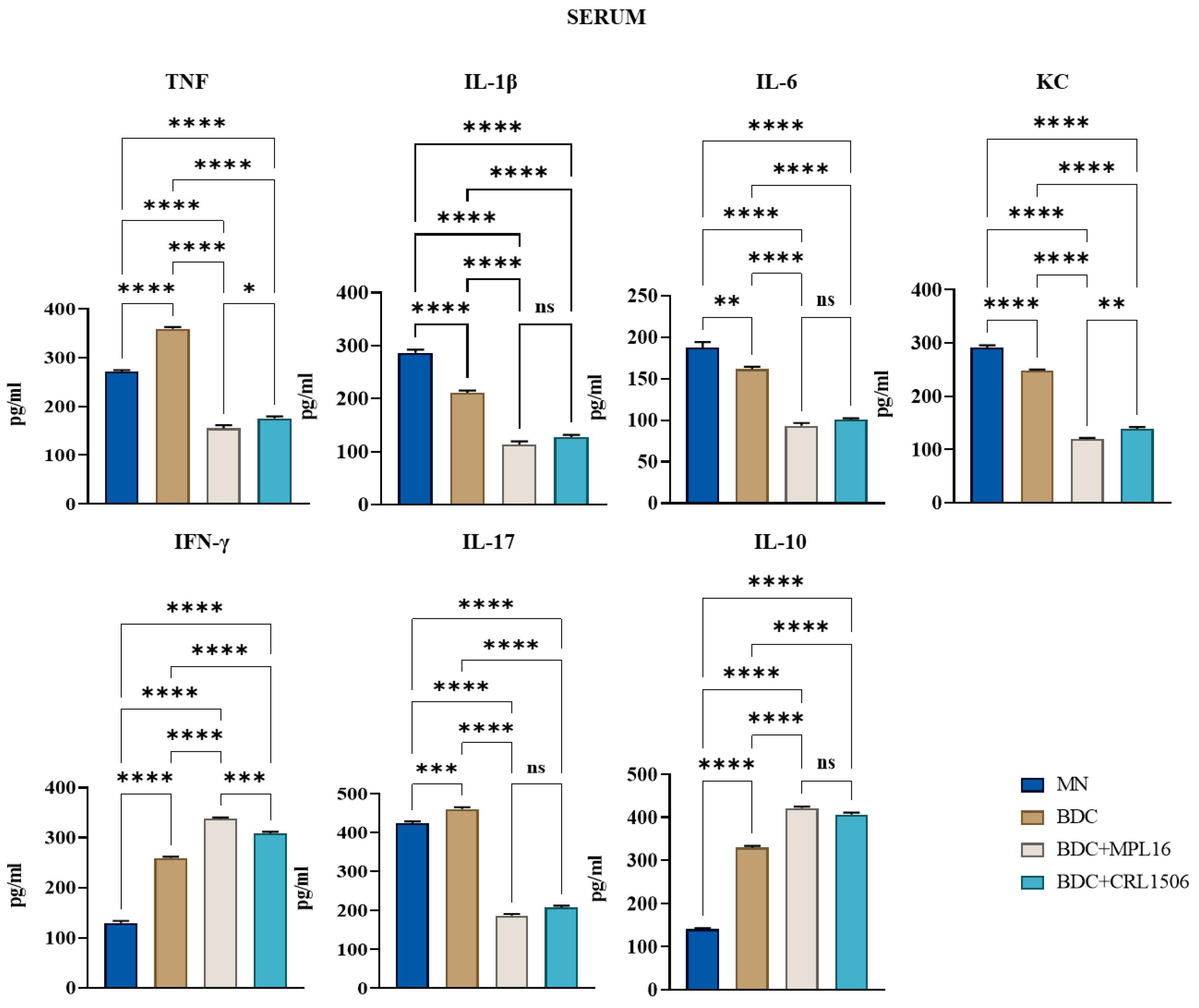
Treatment with MPL16 and CRL1506 strains significantly reduced serum levels of the inflammatory cytokines TNF, IL-1β, and IL-6. No significant differences were observed in TNF levels in intestinal fluid samples between LAB-treated and E. faecalis-infected mice. However, intestinal IL-6 levels were elevated following LAB treatment.
Additionally, the effects of LAB supplementation on IFN-γ and IL-10 levels were assessed. Treatment resulted in increased concentrations of both immunological factors in intestinal fluid and serum samples compared to infected animals. Furthermore, LAB-treated mice exhibited lower intestinal and serum levels of KC and IL-17 than those challenged with the E. faecalis strain. (Figure 4 and Figure 5).

3.4. Restoration of Small Intestine Histology During Re-Nutrition

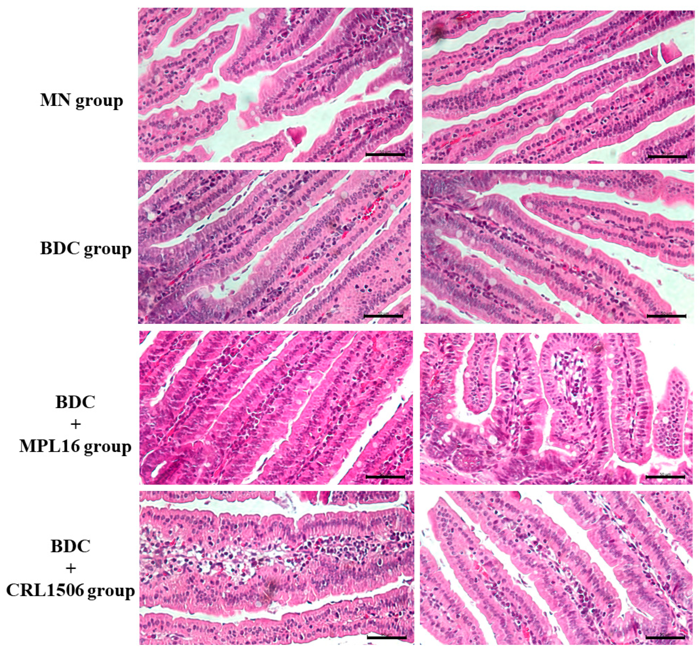
The histological structure of the small intestine was altered in the MN group, with shorter and thinner intestinal villi compared to the BDC group (Figure 6). Administration of immunobiotic strains during the renourishment period partially restored these histological features (Figure 6). Mice that received MPL16 and CRL1506 also showed a remarkable recruitment of immune cells in the lamina propria.

4. Discussion

E. faecalis has been associated with life-threatening infections in patients with chronic antibiotic use or a compromised immune status [8,9]. In this regard, it was shown that the treatment of mice with antibiotics together with the immunosuppressive drug cyclophosphamide significantly augmented the susceptibility of animals to E. faecalis bacteremia after oral challenge with the pathogen, leading to increased mortality [10]. In this work, the immunosuppression induced by protein deficiency in the diet, together with the oral treatment with gentamicin, allowed E. faecalis intestinal–invasive infection of the malnourished mice. Male mice were selected as the experimental model for probiotic supplementation in the context of enterococcal infection due to epidemiological and biological factors that reflect increased male vulnerability to undernutrition and infection. Males are more affected by undernutrition, particularly in severe cases and low-resource settings, and are generally more vulnerable to infectious diseases, likely due to differences in immune and endocrine function [39]. Clinically, vancomycin-resistant enterococci (VRE) bloodstream infections show a male predominance, suggesting sex-specific factors in disease susceptibility. These considerations justify the use of male mice to better reflect the human populations most at risk [40].
We observed that MN mice have significantly higher enterococcal loads in the intestine as well as in the liver, blood, and spleen compared to mice renourished with BCD, indicating the higher susceptibility of malnourished animals to E. faecalis intestinal–invasive infection. The results presented here, and the studies performed previously in this murine malnutrition model allow us to speculate that the reduced resistance of malnourished mice to E. faecalis infection could be associated with: (i) the dysbiosis of the intestinal microbiota, (ii) the damage of the intestinal epithelial barrier and (iii) the impairment of the mucosal and systemic immune systems.
Intestinal microbiota dysbiosis is a hallmark of malnutrition. Protein malnutrition can affect gastric acid secretion and alter bile production, which contribute to intestinal microbiota dysbiosis [41]. Although in our model of protein-malnourished mice, the changes in the intestinal microbiota were not investigated using culture-independent techniques, it was previously reported that these malnourished mice had significantly lower numbers of lactobacilli and anaerobic microorganisms and higher numbers of Gram-negative bacilli in both the small intestine and colon compared to well-nourished animals [42]. These results are in line with works describing alterations of the intestinal microbiome in mice receiving diets with protein deficiencies [43,44]. In addition, studies conducted in gnotobiotic C57BL/6 mice showed that E. faecalis can pass from the intestinal lumen to the lamina propria through internalization via IECs [3,4]. Protein malnutrition impairs the IECs’ turnover [13], and induces villous atrophy, increased intestinal permeability, and crypt hyperplasia [12,14,15,17,42]. The structural and ultrastructural alterations of the intestine have been reported previously in the model of protein-malnourished mice used in this work [42,45,46], which probably allowed E. faecalis intestinal colonization and invasion. Further studies of the gut microbiota using novel omics tools and the alteration of the intestinal epithelium in our protein malnutrition model in the context of E. faecalis infection could provide further insight into the role of these intestinal barriers in the increased susceptibility of malnourished mice to E. faecalis infection. These are interesting topics for future research.
We observed that MN mice had lower levels of TNF, IL-1β, IL-6, KC, IL-17, IFN-γ, and IL-10 in the intestine compared to BCD mice after the enterococcal infection. In addition, MN animals had lower concentrations of TNF, IL-17, IFN-γ, and IL-10 as well as higher levels of IL-1β, IL-6, and KC in blood. The reduced capacity of protein-malnourished mice to produce inflammatory factors in response to E. faecalis infection is in line with our previous studies evaluating their immune response to other pathogens, such as C. albicans [28] and S. pneumoniae [29]. Pneumococcal infection can enhance the concentrations of TNF, IL-1β, IL-6, IFN-γ, and IL-10 in the respiratory tract and blood of immunocompetent well-nourished mice, while in protein-malnourished the levels of those immune mediators are significantly lower [47]. Similarly, the intraperitoneal challenge of well-nourished and protein-malnourished mice with C. albicans increased the levels of TNF, IL-6, and IFN-γ in the peritoneal cavity as well as TNF, IL-6, IFN-γ, IL-17, and IL-10 concentrations in blood. However, we observed significantly lower values of all these cytokines in MN animals than in normal controls [28]. It was reported that children with severe malnutrition often have a diminished ability to produce acute-phase proteins and pro-inflammatory mediators like TNF, IL-1β, and IL-6 [48,49] while peritoneal macrophages from protein-deficient mice produce low levels of TNF in response to lipopolysaccharide challenge [50]. Of note, macrophages from protein-deficient mice responded to TNF stimulation with lower concentrations of IL-1β and IL-12 than cells obtained from control animals [51]. However, it was also shown that uninfected malnourished children have high levels of circulating TNF [52]. Furthermore, malnourished mice infected with the enteric pathogen Salmonella Typhimurium had higher TNF, IL-6, MCP-1, and IFN-γ levels in the livers than control-infected mice [53]. This discordance may be due to differences in malnutrition severity and the distinct inflammatory challenges. Even though increases or decreases in immunological mediators have been recorded for malnourished hosts, their response has been consistently described as being inappropriate for eliminating pathogens. In fact, the different intestinal and serum cytokine profiles of malnourished mice in response to E. faecalis infection were associated with higher bacterial loads in the intestine and internal organs and with their dissemination into blood, resembling our previous results with C. albicans and S. pneumoniae [28,29].
We have previously reported quantitative and qualitative impairments of phagocytes in our model of protein malnutrition. MN mice have lower numbers of blood neutrophils [47] and reduced phagocytic and microbicidal activities in peritoneal and respiratory macrophages [28,29]. The recruitment of neutrophils into the peritoneal cavity, as well as their peroxidase activity after the challenge with C. albicans, was reduced in protein-malnourished mice compared to normal controls [28]. They also showed a significant impairment in the recruitment of neutrophils and macrophages into the respiratory tract after the infection with S. pneumoniae [29], which was related to qualitative alterations of immune cells in blood and bone marrow [47]. In line with these findings, we observed that malnourished mice had a reduced ability to recruit immune cells to intestinal tissue after the challenge with E. faecalis compared to mice repleted with BCD. Then, the failure in the establishment of this immunological phagocytic and microbicidal barrier in the gut of protein-malnourished mice could contribute to the invasive enterococcal infection.
The production of regulatory cytokines during the course of infections is of importance for avoiding dysregulated inflammation and host damage. In this work, although protein malnourished mice had lower levels of pro-inflammatory cytokines and chemokines in the intestine, they also showed higher levels of IL-1β, IL-6, and KC in blood compared to mice in the BCD groups. Of note, the levels of the regulatory cytokine IL-10 were significantly lower in both the intestine and blood in malnourished mice than in animals repleted with BCD after the enterococcal infection. Thus, malnourished mice showed an imbalance of inflammatory mediators in the different compartments along with a notable impairment in their ability to regulate the inflammatory response. This imbalance of immune mediators in protein malnourished mice would allow the colonization and invasion of E. faecalis and the inflammatory-mediated tissue damage, making the disease more severe than in animals repleted with BCD.
We demonstrated here that BCD supplemented with L. plantarum MPL16 or L. plantarum CRL1506 was more efficient than BCD alone to improve the resistance and modulate the immune response to E. faecalis infection in repleted malnourished animals. This is in line with our previous studies showing that immunomodulatory lactobacilli can accelerate the recovery of immune defense mechanisms when used as supplements in repletion diets [28,29]. Protein-malnourished mice renourished with BCD and the MPL16 or CRL1506 strains had lower intestinal, liver, spleen, and blood loads of enterococci than mice receiving only BCD. This distinct resistance to E. faecalis infection could be associated with the different cytokine profiles found in both the intestine and serum. Previously, we showed that protein-malnourished mice treated with both BCD and BCD supplemented with the CRL431 strain enhanced the concentrations of TNF, IL-6, and IFN-γ in the peritoneal cavity, blood, liver, and spleen in response to C. albicans infection [28]. Cytokine levels were higher than the concentrations observed in malnourished controls, but only mice renourished with the immunomodulatory lactobacilli reached the values of well-nourished animals. Interestingly, the levels of IL-10 were higher in mice treated with BCD and L. casei CRL431 than in well-nourished mice. In addition, both immunomodulatory CRL431 and CRL1505 strains improved the production of TNF, IL-1β, IL-6, and IFN-γ in the respiratory tract compared to malnourished and BCD-treated mice, after the pneumococcal infection. Interestingly, IL-10 production in response to S. pneumoniae was higher in BCD and CRL341-treated malnourished mice than in controls but similar to well-nourished controls in mice receiving the BCD and the CRL1505 strain [47]. The results of this study contrast with those of previous works, since protein-malnourished mice renourished with BCD and the MPL16 or CRL1506 strains had lower TNF, IL-1β, KC, and IL-17 in the gut and blood, while IL-6 was higher in the intestine but lower in blood, compared with animals treated only with BCD. In addition, we observed improved levels of IFN-γ and IL-10 in mice of the BCD + MPL16 and BCD + CRL1506 groups than in the BCD mice. These differences in the effect of lactobacilli on cytokine profiles of malnourished hosts may be due to three combined factors: the pathogen used, the initial anatomical site of infection, and the different strains of immunomodulatory lactobacilli used.
Malnourished mice treated with BCD and the MPL16 or CRL1506 have reduced levels of inflammatory cytokines and enhanced IL-10, a balance that could be involved in the protection of inflammatory-mediated intestinal barrier damage during the enterococcal infection. The production of inflammatory mediators in response to E. faecalis is important to eliminate the pathogen. However, the inflammatory response must be tightly regulated to avoid the host’s tissues. As mentioned before, IL-10 deficiency can lead to enhanced inflammatory disease in mice infected with E. faecalis [54,55]. On the other hand, TNF can disrupt epithelial barrier function through the activation of NF-κB and MAPK pathways [56] and the modulation of zonulin-1 [57] and cytoskeletal filaments [58].
Our findings reveal that malnourished mice treated with BCD and the immunobiotic strains L. plantarum MPL16 or CRL1506 exhibit increased levels of IL-6 and IFN-γ, which help reinforce gut epithelial and immune barriers. IFN-γ production by phagocytes plays a key role in protecting against E. faecalis invasion, while IL-6 is essential for intestinal homeostasis, tight junction integrity, and epithelial cell proliferation [59,60,61]. In immunocompetent hosts, both L. plantarum strains enhanced IL-6 and IFN-γ levels, improved the recruitment of immune cells to the intestinal mucosa, and modulated IECs and antigen-presenting cell responses. These strains can upregulate IFN-β, IL-6, and TNF in IECs [25,26]. Additionally, L. plantarum CRL1506 stimulated antigen-presenting cells in Peyer’s patches, boosting IL-1β, IFN-γ, IL-12, and activation markers (MHC-II and CD80/86), which correlated with an enhanced Th1 response [25,62]. Then, it is tempting to speculate that lactobacilli exerted similar effects on IECs and phagocytes in malnourished mice treated with BCD. While our study highlights the immunomodulatory benefits of L. plantarum MPL16 and CRL1506, a limitation is that cytokine levels were assessed at a single time point after E. faecalis challenge. Future studies should investigate the kinetics of immune responses and the specific roles of IECs and antigen-presenting cells in immunobiotic-induced protection.

5. Conclusions

This work provides evidence that protein deficiency and antibiotic treatment compromise gut defenses, facilitating the intestinal colonization and invasion of E. faecalis. This underscores the need for cautious antibiotic use during malnutrition recovery, given the rise in antibiotic-resistant enterococci. Further, short-term BCD repletion alone was insufficient to restore defenses against E. faecalis infection, aligning with previous findings that immune recovery requires at least 21 days of BCD treatment [29]. However, supplementing BCD with L. plantarum MPL16 and CRL1506 accelerated immune recovery and improved resistance to infection by modulating systemic and gut cytokine profiles. These results suggest that immunobiotic lactobacilli could be integrated into nutritional rehabilitation strategies for malnourished children to enhance immune function and protect against opportunistic infections.

Supplementary Materials

The following supporting information can be downloaded at https://www.mdpi.com/article/10.3390/nu17111770/s1. Table S1: Composition of the conventional balanced diet and low-protein diet. Table S2: LAB survival data under in vitro gastrointestinal conditions. Table S3: Data on the effect of LAB on the resistance of malnourished mice to E. faecalis infection. Table S4: Data on the effect of LAB on intestinal cytokine levels after infection with E. faecalis 102. Table S5: Data on the effect of LAB on serum cytokine levels after infection with E. faecalis 102.

Author Contributions

Conceptualization, M.G.V.-P. and J.V.; Formal analysis, M.D.V. and M.G.V.-P.; Funding acquisition, H.K., M.G.V.-P., and J.V.; Investigation, M.D.V., L.P.A., M.F.M., F.R.T., R.O.M., and H.L.B.; Methodology, M.F.M.; Resources, M.G.V.-P. and J.V.; Supervision, H.K., M.G.V.-P., and J.V.; Writing—original draft, M.D.V. and L.P.A.; Writing—review and editing, H.K., M.G.V.-P., and J.V. All authors have read and agreed to the published version of the manuscript.

Funding

This study was supported by ANPCyT–FONCyT grants PICT2021-159 and PICT2019-3382 to M.G.V.-P. and by PICT-2016-424 0410 and PICT 2021-I-A-00705 to J.V. This study was also supported by a Grant-in-Aid for Scientific Research (A) (23H00354, H.K.), and Challenging Research (Exploratory, 23K18072, H.K.) from the Japan Society for the Promotion of Science (JSPS); by the Research Program on Development of Innovative Technology grants (JPJ007097) from the Project of the Bio oriented Technology Research Advancement Institution (BRAIN), by the Japan Racing Association to H.K., and by AMED grant number JP21zf0127001.

Institutional Review Board Statement

Institutional Committee for the Care and Use of Laboratory Animals at CERELA (CICUAL CERELA)CRL-CICUAL-IBT2024/3A2025-06-24.

Informed Consent Statement

Not applicable.

Data Availability Statement

The data that support the findings of this study are available from the corresponding author upon reasonable request.

Acknowledgments

M.G.V.-P. and J.V. are research members of the National Research Council of Argentina (CONICET).

Conflicts of Interest

The authors declare no conflicts of interest.

References

  1. Yuen, G.J.; Ausubel, F.M. Enterococcus infection biology: Lessons from invertebrate host models. J. Microbiol. 2014, 52, 200–210. [Google Scholar] [CrossRef] [PubMed]
  2. Lindenstrauss, A.G.; Ehrmann, M.A.; Behr, J.; Landstorfer, R.; Haller, D.; Sartor, R.B.; Vogel, R.F. Transcriptome analysis of Enterococcus faecalis toward its adaption to surviving in the mouse intestinal tract. Arch. Microbiol. 2014, 196, 423–433. [Google Scholar] [CrossRef] [PubMed]
  3. Medina-Contreras, O.; Geem, D.; Laur, O.; Williams, I.R.; Lira, S.A.; Nusrat, A.; Parkos, C.A.; Denning, T.L. CX3CR1 regulates intestinal macrophage homeostasis, bacterial translocation, and colitogenic Th17 responses in mice. J. Clin. Investig. 2011, 121, 4787–4795. [Google Scholar] [CrossRef] [PubMed]
  4. Wells, C.L.; Jechorek, R.P.; Erlandsen, S.L. Evidence for the translocation of Enterococcus faecalis across the mouse intestinal tract. J. Infect. Dis. 1990, 162, 82–90. [Google Scholar] [CrossRef]
  5. Chakraborty, R.; Lam, V.; Kommineni, S.; Stromich, J.; Hayward, M.; Kristich, C.J.; Salzman, N.H. Ceftriaxone Administration Disrupts Intestinal Homeostasis, Mediating Noninflammatory Proliferation and Dissemination of Commensal Enterococci. Infect. Immun. 2018, 86, 10–1128. [Google Scholar] [CrossRef]
  6. Archambaud, C.; Derre-Bobillot, A.; Lapaque, N.; Rigottier-Gois, L.; Serror, P. Intestinal translocation of enterococci requires a threshold level of enterococcal overgrowth in the lumen. Sci. Rep. 2019, 9, 8926. [Google Scholar] [CrossRef]
  7. Geldart, K.G.; Kommineni, S.; Forbes, M.; Hayward, M.; Dunny, G.M.; Salzman, N.H.; Kaznessis, Y.N. Engineered E. coli Nissle 1917 for the reduction of vancomycin-resistant Enterococcus in the intestinal tract. Bioeng. Transl. Med. 2018, 3, 197–208. [Google Scholar] [CrossRef]
  8. McKenney, P.T.; Yan, J.; Vaubourgeix, J.; Becattini, S.; Lampen, N.; Motzer, A.; Larson, P.J.; Dannaoui, D.; Fujisawa, S.; Xavier, J.B.; et al. Intestinal Bile Acids Induce a Morphotype Switch in Vancomycin-Resistant Enterococcus that Facilitates Intestinal Colonization. Cell Host Microbe 2019, 25, 695–705.e695. [Google Scholar] [CrossRef]
  9. Repoila, F.; Le Bohec, F.; Guerin, C.; Lacoux, C.; Tiwari, S.; Jaiswal, A.K.; Santana, M.P.; Kennedy, S.P.; Quinquis, B.; Rainteau, D.; et al. Adaptation of the gut pathobiont Enterococcus faecalis to deoxycholate and taurocholate bile acids. Sci. Rep. 2022, 12, 8485. [Google Scholar] [CrossRef]
  10. Miyazaki, S.; Fujikawa, T.; Kobayashi, I.; Matsumoto, T.; Tateda, K.; Yamaguchi, K. Development of systemic bacteraemia after oral inoculation of vancomycin-resistant enterococci in mice. J. Med. Microbiol. 2001, 50, 695–701. [Google Scholar] [CrossRef]
  11. Tuano, K.S.; Seth, N.; Chinen, J. Secondary immunodeficiencies: An overview. Ann. Allergy Asthma Immunol. 2021, 127, 617–626. [Google Scholar] [CrossRef]
  12. Ibrahim, M.K.; Zambruni, M.; Melby, C.L.; Melby, P.C. Impact of Childhood Malnutrition on Host Defense and Infection. Clin. Microbiol. Rev. 2017, 30, 919–971. [Google Scholar] [CrossRef]
  13. Liu, J.; Bolick, D.T.; Kolling, G.L.; Fu, Z.; Guerrant, R.L. Protein Malnutrition Impairs Intestinal Epithelial Cell Turnover, a Potential Mechanism of Increased Cryptosporidiosis in a Murine Model. Infect. Immun. 2016, 84, 3542–3549. [Google Scholar] [CrossRef]
  14. Lykke, M.; Hother, A.L.; Hansen, C.F.; Friis, H.; Molgaard, C.; Michaelsen, K.F.; Briend, A.; Larsen, T.; Sangild, P.T.; Thymann, T. Malnutrition induces gut atrophy and increases hepatic fat infiltration: Studies in a pig model of childhood malnutrition. Am. J. Transl. Res. 2013, 5, 543–554. [Google Scholar] [PubMed]
  15. Kasai, A.; Gama, P.; Alvares, E.P. Protein restriction inhibits gastric cell proliferation during rat postnatal growth in parallel to ghrelin changes. Nutrition 2012, 28, 707–712. [Google Scholar] [CrossRef]
  16. Eyzaguirre-Velasquez, J.; Olavarria-Ramirez, L.; Gonzalez-Arancibia, C.; Diaz-Merino, C.; Ariz, R.; Lopez, S.; Quiroz, W.; Beltran, C.J.; Bravo, J.A.; Julio-Pieper, M. Protein Malnutrition During Juvenile Age Increases Ileal and Colonic Permeability in Rats. J. Pediatr. Gastroenterol. Nutr. 2017, 64, 707–712. [Google Scholar] [CrossRef]
  17. Ling, C.; Versloot, C.J.; Arvidsson Kvissberg, M.E.; Hu, G.; Swain, N.; Horcas-Nieto, J.M.; Miraglia, E.; Thind, M.K.; Farooqui, A.; Gerding, A.; et al. Rebalancing of mitochondrial homeostasis through an NAD(+)-SIRT1 pathway preserves intestinal barrier function in severe malnutrition. EBioMedicine 2023, 96, 104809. [Google Scholar] [CrossRef]
  18. Amaral, J.F.; Foschetti, D.A.; Assis, F.A.; Menezes, J.S.; Vaz, N.M.; Faria, A.M. Immunoglobulin production is impaired in protein-deprived mice and can be restored by dietary protein supplementation. Braz. J. Med. Biol. Res. 2006, 39, 1581–1586. [Google Scholar] [CrossRef]
  19. Flo, J.; Elias, F.; Benedetti, R.; Massouh, E. Reversible effects on B and T cells of the gut-associated lymphoid tissues in rats malnourished during suckling: Impaired induction of the immune response to intra-Peyer patches immunization with cholera toxin. Clin. Immunol. Immunopathol. 1996, 80, 147–154. [Google Scholar] [CrossRef]
  20. Huus, K.E.; Hoang, T.T.; Creus-Cuadros, A.; Cirstea, M.; Vogt, S.L.; Knuff-Janzen, K.; Sansonetti, P.J.; Vonaesch, P.; Finlay, B.B. Cross-feeding between intestinal pathobionts promotes their overgrowth during undernutrition. Nat. Commun. 2021, 12, 6860. [Google Scholar] [CrossRef]
  21. Kau, A.L.; Planer, J.D.; Liu, J.; Rao, S.; Yatsunenko, T.; Trehan, I.; Manary, M.J.; Liu, T.C.; Stappenbeck, T.S.; Maleta, K.M.; et al. Functional characterization of IgA-targeted bacterial taxa from undernourished Malawian children that produce diet-dependent enteropathy. Sci. Transl. Med. 2015, 7, 276ra224. [Google Scholar] [CrossRef]
  22. Oldenburg, C.E.; Hinterwirth, A.; Ourohire, M.; Dah, C.; Ouedraogo, M.; Sie, A.; Boudo, V.; Chen, C.; Ruder, K.; Zhong, L.; et al. Gut Resistome after Antibiotics among Children with Uncomplicated Severe Acute Malnutrition: A Randomized Controlled Trial. Am. J. Trop. Med. Hyg. 2022, 107, 59–64. [Google Scholar] [CrossRef]
  23. Villena, J.; Li, C.; Vizoso-Pinto, M.G.; Sacur, J.; Ren, L.; Kitazawa, H. Lactiplantibacillus plantarum as a Potential Adjuvant and Delivery System for the Development of SARS-CoV-2 Oral Vaccines. Microorganisms 2021, 9, 683. [Google Scholar] [CrossRef]
  24. Mizuno, H.; Arce, L.; Tomotsune, K.; Albarracin, L.; Funabashi, R.; Vera, D.; Islam, M.A.; Vizoso-Pinto, M.G.; Takahashi, H.; Sasaki, Y.; et al. Lipoteichoic Acid Is Involved in the Ability of the Immunobiotic Strain Lactobacillus plantarum CRL1506 to Modulate the Intestinal Antiviral Innate Immunity Triggered by TLR3 Activation. Front. Immunol. 2020, 11, 571. [Google Scholar] [CrossRef]
  25. Villena, J.; Chiba, E.; Vizoso-Pinto, M.G.; Tomosada, Y.; Takahashi, T.; Ishizuka, T.; Aso, H.; Salva, S.; Alvarez, S.; Kitazawa, H. Immunobiotic Lactobacillus rhamnosus strains differentially modulate antiviral immune response in porcine intestinal epithelial and antigen presenting cells. BMC Microbiol. 2014, 14, 126. [Google Scholar] [CrossRef]
  26. Albarracin, L.; Garcia-Castillo, V.; Masumizu, Y.; Indo, Y.; Islam, M.A.; Suda, Y.; Garcia-Cancino, A.; Aso, H.; Takahashi, H.; Kitazawa, H.; et al. Efficient Selection of New Immunobiotic Strains With Antiviral Effects in Local and Distal Mucosal Sites by Using Porcine Intestinal Epitheliocytes. Front. Immunol. 2020, 11, 543. [Google Scholar] [CrossRef]
  27. Salva, S.; Nunez, M.; Villena, J.; Ramon, A.; Font, G.; Alvarez, S. Development of a fermented goats’ milk containing Lactobacillus rhamnosus: In vivo study of health benefits. J. Sci. Food Agric. 2011, 91, 2355–2362. [Google Scholar] [CrossRef]
  28. Villena, J.; Salva, S.; Aguero, G.; Alvarez, S. Immunomodulatory and protective effect of probiotic Lactobacillus casei against Candida albicans infection in malnourished mice. Microbiol. Immunol. 2011, 55, 434–445. [Google Scholar] [CrossRef]
  29. Villena, J.; Racedo, S.; Aguero, G.; Bru, E.; Medina, M.; Alvarez, S. Lactobacillus casei improves resistance to pneumococcal respiratory infection in malnourished mice. J. Nutr. 2005, 135, 1462–1469. [Google Scholar] [CrossRef]
  30. Salva, S.; Villena, J.; Alvarez, S. Immunomodulatory activity of Lactobacillus rhamnosus strains isolated from goat milk: Impact on intestinal and respiratory infections. Int. J. Food Microbiol. 2010, 141, 82–89. [Google Scholar] [CrossRef] [PubMed]
  31. Villena, J.; Saavedra, L.; Hebert, E.M.; Suda, Y.; Masumizu, Y.; Albarracin, L.; Clua, P.; Ikeda-Ohtsubo, W.; Kitazawa, H. Draft Genome Sequence of Lactobacillus plantarum MPL16, a Wakame-Utilizing Immunobiotic Strain Isolated from Swine Feces. Genome Announc. 2017, 5, 10–1128. [Google Scholar] [CrossRef]
  32. Charteris, W.P.; Kelly, P.M.; Morelli, L.; Collins, J.K. Development and application of an in vitro methodology to determine the transit tolerance of potentially probiotic Lactobacillus and Bifidobacterium species in the upper human gastrointestinal tract. J. Appl. Microbiol. 1998, 84, 759–768. [Google Scholar] [CrossRef]
  33. Conway, P.L.; Gorbach, S.L.; Goldin, B.R. Survival of lactic acid bacteria in the human stomach and adhesion to intestinal cells. J. Dairy Sci. 1987, 70, 1–12. [Google Scholar] [CrossRef]
  34. Vizoso Pinto, M.G.; Franz, C.M.; Schillinger, U.; Holzapfel, W.H. Lactobacillus spp. with in vitro probiotic properties from human faeces and traditional fermented products. Int. J. Food Microbiol. 2006, 109, 205–214. [Google Scholar] [CrossRef]
  35. Muller, M.F.; Sacur, J.; Brancher, J.M.; Vera, M.D.; Arce, L.; Raya-Tonetti, M.F.; Kitazawa, H.; Villena, J.; Vizoso-Pinto, M.G. An experimental chimeric hepatitis E virus vaccine elicits both local and systemic immune responses. Front. Microbiol. 2024, 15, 1512018. [Google Scholar] [CrossRef]
  36. Villena, J.; Racedo, S.; Aguero, G.; Alvarez, S. Yoghurt accelerates the recovery of defence mechanisms against Streptococcus pneumoniae in protein-malnourished mice. Br. J. Nutr. 2006, 95, 591–602. [Google Scholar] [CrossRef]
  37. Kolling, Y.; Salva, S.; Villena, J.; Alvarez, S. Are the immunomodulatory properties of Lactobacillus rhamnosus CRL1505 peptidoglycan common for all Lactobacilli during respiratory infection in malnourished mice? PLoS ONE 2018, 13, e0194034. [Google Scholar] [CrossRef]
  38. Ge, X.; Ding, C.; Zhao, W.; Xu, L.; Tian, H.; Gong, J.; Zhu, M.; Li, J.; Li, N. Antibiotics-induced depletion of mice microbiota induces changes in host serotonin biosynthesis and intestinal motility. J. Transl. Med. 2017, 15, 13. [Google Scholar] [CrossRef]
  39. Thurstans, S.; Opondo, C.; Seal, A.; Wells, J.C.; Khara, T.; Dolan, C.; Briend, A.; Myatt, M.; Garenne, M.; Mertens, A.; et al. Understanding Sex Differences in Childhood Undernutrition: A Narrative Review. Nutrients 2022, 14, 948. [Google Scholar] [CrossRef]
  40. Correa-Martinez, C.L.; Schuler, F.; Kampmeier, S. Sex differences in vancomycin-resistant enterococci bloodstream infections-a systematic review and meta-analysis. Biol. Sex Differ. 2021, 12, 36. [Google Scholar] [CrossRef] [PubMed]
  41. Zhang, L.; Voskuijl, W.; Mouzaki, M.; Groen, A.K.; Alexander, J.; Bourdon, C.; Wang, A.; Versloot, C.J.; Di Giovanni, V.; Wanders, R.J.; et al. Impaired Bile Acid Homeostasis in Children with Severe Acute Malnutrition. PLoS ONE 2016, 11, e0155143. [Google Scholar] [CrossRef] [PubMed]
  42. Allori, C.; Aguero, G.; de Ruiz Holgado, A.P.; de Nader, O.M.; Perdigon, G. Gut mucosa morphology and microflora changes in malnourished mice after renutrition with milk and administration of Lactobacillus casei. J. Food Prot. 2000, 63, 83–90. [Google Scholar] [CrossRef] [PubMed]
  43. Corsetti, G.; Romano, C.; Pasini, E.; Testa, C.; Dioguardi, F.S. Qualitative Nitrogen Malnutrition Damages Gut and Alters Microbiome in Adult Mice. A Preliminary Histopathological Study. Nutrients 2021, 13, 1089. [Google Scholar] [CrossRef] [PubMed]
  44. Mayneris-Perxachs, J.; Bolick, D.T.; Leng, J.; Medlock, G.L.; Kolling, G.L.; Papin, J.A.; Swann, J.R.; Guerrant, R.L. Protein- and zinc-deficient diets modulate the murine microbiome and metabolic phenotype. Am. J. Clin. Nutr. 2016, 104, 1253–1262. [Google Scholar] [CrossRef] [PubMed]
  45. Gauffin, C.P.; Aguero, G.; Perdigon, G. Adjuvant effects of Lactobacillus casei added to a renutrition diet in a malnourished mouse model. Biocell 2002, 26, 35–48. [Google Scholar] [CrossRef]
  46. Cano, P.G.; Aguero, G.; Perdigon, G. Immunological effects of yogurt addition to a re-nutrition diet in a malnutrition experimental model. J. Dairy Res. 2002, 69, 303–316. [Google Scholar] [CrossRef]
  47. Herrera, M.; Salva, S.; Villena, J.; Barbieri, N.; Marranzino, G.; Alvarez, S. Dietary supplementation with Lactobacilli improves emergency granulopoiesis in protein-malnourished mice and enhances respiratory innate immune response. PLoS ONE 2014, 9, e90227. [Google Scholar] [CrossRef]
  48. Doherty, J.F.; Golden, M.H.; Remick, D.G.; Griffin, G.E. Production of interleukin-6 and tumour necrosis factor-alpha in vitro is reduced in whole blood of severely malnourished children. Clin. Sci. 1994, 86, 347–351. [Google Scholar] [CrossRef]
  49. Manary, M.J.; Yarasheski, K.E.; Berger, R.; Abrams, E.T.; Hart, C.A.; Broadhead, R.L. Whole-body leucine kinetics and the acute phase response during acute infection in marasmic Malawian children. Pediatr. Res. 2004, 55, 940–946. [Google Scholar] [CrossRef]
  50. Fock, R.A.; Vinolo, M.A.; de Moura Sa Rocha, V.; de Sa Rocha, L.C.; Borelli, P. Protein-energy malnutrition decreases the expression of TLR-4/MD-2 and CD14 receptors in peritoneal macrophages and reduces the synthesis of TNF-alpha in response to lipopolysaccharide (LPS) in mice. Cytokine 2007, 40, 105–114. [Google Scholar] [CrossRef]
  51. de Oliveira, D.C.; Hastreiter, A.A.; Mello, A.S.; de Oliveira Beltran, J.S.; Oliveira Santos, E.W.; Borelli, P.; Fock, R.A. The effects of protein malnutrition on the TNF-RI and NF-kappaB expression via the TNF-alpha signaling pathway. Cytokine 2014, 69, 218–225. [Google Scholar] [CrossRef] [PubMed]
  52. Azevedo, Z.M.; Luz, R.A.; Victal, S.H.; Kurdian, B.; Fonseca, V.M.; Fitting, C.; Camara, F.P.; Haeffner-Cavaillon, N.; Cavaillon, J.M.; Gaspar Elsas, M.I.; et al. Increased production of tumor necrosis factor-alpha in whole blood cultures from children with primary malnutrition. Braz. J. Med. Biol. Res. 2005, 38, 171–183. [Google Scholar] [CrossRef] [PubMed]
  53. Brown, E.M.; Wlodarska, M.; Willing, B.P.; Vonaesch, P.; Han, J.; Reynolds, L.A.; Arrieta, M.C.; Uhrig, M.; Scholz, R.; Partida, O.; et al. Diet and specific microbial exposure trigger features of environmental enteropathy in a novel murine model. Nat. Commun. 2015, 6, 7806. [Google Scholar] [CrossRef]
  54. Hoffmann, M.; Kim, S.C.; Sartor, R.B.; Haller, D. Enterococcus faecalis strains differentially regulate Alix/AIP1 protein expression and ERK 1/2 activation in intestinal epithelial cells in the context of chronic experimental colitis. J. Proteome Res. 2009, 8, 1183–1192. [Google Scholar] [CrossRef]
  55. Steck, N.; Hoffmann, M.; Sava, I.G.; Kim, S.C.; Hahne, H.; Tonkonogy, S.L.; Mair, K.; Krueger, D.; Pruteanu, M.; Shanahan, F.; et al. Enterococcus faecalis metalloprotease compromises epithelial barrier and contributes to intestinal inflammation. Gastroenterology 2011, 141, 959–971. [Google Scholar] [CrossRef]
  56. Patrick, D.M.; Leone, A.K.; Shellenberger, J.J.; Dudowicz, K.A.; King, J.M. Proinflammatory cytokines tumor necrosis factor-alpha and interferon-gamma modulate epithelial barrier function in Madin-Darby canine kidney cells through mitogen activated protein kinase signaling. BMC Physiol. 2006, 6, 2. [Google Scholar] [CrossRef]
  57. Ma, T.Y.; Iwamoto, G.K.; Hoa, N.T.; Akotia, V.; Pedram, A.; Boivin, M.A.; Said, H.M. TNF-alpha-induced increase in intestinal epithelial tight junction permeability requires NF-kappa B activation. Am. J. Physiol. Gastrointest. Liver Physiol. 2004, 286, G367–G376. [Google Scholar] [CrossRef]
  58. Turner, J.R. Molecular basis of epithelial barrier regulation: From basic mechanisms to clinical application. Am. J. Pathol. 2006, 169, 1901–1909. [Google Scholar] [CrossRef] [PubMed]
  59. Onyeji, C.O.; Bui, K.Q.; Nicolau, D.P.; Nightingale, C.H.; Bow, L.; Quintiliani, R. Influence of adjunctive interferon-gamma on treatment of gentamicin- and vancomycin-resistant Enterococcus faecalis infection in mice. Int. J. Antimicrob. Agents 1999, 12, 301–309. [Google Scholar] [CrossRef]
  60. Kuhn, K.A.; Manieri, N.A.; Liu, T.C.; Stappenbeck, T.S. IL-6 stimulates intestinal epithelial proliferation and repair after injury. PLoS ONE 2014, 9, e114195. [Google Scholar] [CrossRef]
  61. Suzuki, T.; Yoshinaga, N.; Tanabe, S. Interleukin-6 (IL-6) regulates claudin-2 expression and tight junction permeability in intestinal epithelium. J. Biol. Chem. 2011, 286, 31263–31271. [Google Scholar] [CrossRef] [PubMed]
  62. Islam, M.A.; Albarracin, L.; Tomokiyo, M.; Valdez, J.C.; Sacur, J.; Vizoso-Pinto, M.G.; Andrade, B.G.N.; Cuadrat, R.R.C.; Kitazawa, H.; Villena, J. Immunobiotic Lactobacilli Improve Resistance of Respiratory Epithelial Cells to SARS-CoV-2 Infection. Pathogens 2021, 10, 1197. [Google Scholar] [CrossRef] [PubMed]
Figure 1. Experimental design. For 21 days, mice were fed a low-protein diet. Two control groups were included: malnourished mice without renourishment (MN group) and mice renourished with a conventional balanced diet (BDC) for 7 days without LAB supplementation (BDC group). The LAB was administered at a dose of 108 cells/mouse/day in drinking water (BDC + MPL16 and BDC + CRL1506 groups). Three days before the challenge with E. faecalis (day 26), the mice were treated with gentamicin (1.6 mg/kg) (ATB) supplemented in drinking water. On day 29, BALB/c mice were orally challenged with E. faecalis (50 µL, 108 UFC/mL). After euthanasia (day 29), samples were collected (blood, feces, intestinal lavage, liver, and spleen).
Nutrients 17 01770 g001
Figure 2. Survival of LAB in vitro gastrointestinal conditions. Effect of the resistance of L. plantarum (A) MLP16 and (B) CRL156 strains, previously suspended in MRS or 15% reconstituted skim milk, to an artificial gastrointestinal passage (simulated saliva, gastric juice, and intestinal juice solutions). The viable LAB cells were serially diluted (10-fold dilutions), plated onto MRS agar plates, and incubated at 37 °C for 48 h under anaerobic conditions. Results are presented as mean ± SD. Statistical differences between the groups were indicated as * p < 0.05.
Nutrients 17 01770 g002
Figure 3. Effect of LAB on resistance of malnourished mice to E. faecalis infection. After malnutrition, mice were renourished with a conventional balanced diet (BDC) alone (BDC group), or supplemented with LAB (BDC + MPL16 and BDC + CRL1506 groups), and malnourished mice without renourishment (MN group). The mice were subsequently challenged with a single dose of E. faecalis (50 µL, 108 UFC/mL) and euthanized 48 h post-challenge. Post-euthanasia, E. faecalis was counted in mouse samples (stool, blood, spleen, liver, and bowel lavage). Results are presented as mean ± SD. Statistical differences between the groups were indicated as follows: * p < 0.05, ** p < 0.01, *** p < 0.001, **** p < 0.000, and not significant (ns) > 0.05.
Nutrients 17 01770 g003
Figure 4. Effect of LAB on intestinal cytokine levels after infection with E. faecalis 102. After malnutrition, mice were renourished with a conventional balanced diet (BDC) alone (BDC group) or supplemented with LAB (BDC + MPL16 and BDC + CRL1506 groups), and malnourished mice without renourishment (MN group). The mice were subsequently challenged with a single dose of E. faecalis 102 (50 µL, 108 UFC/mL). Two days post-challenge with E. faecalis 102, a serum sample was collected to determine IL-6, IL-8, IL-1, TNF, IFN-γ, IL-10, and IL-17 levels. Results are presented as mean ± SD. Statistical differences between the groups were indicated as follows: * p < 0.05, ** p < 0.01, *** p < 0.001, **** p < 0.000, and not significant (ns) > 0.05.
Nutrients 17 01770 g004
Figure 5. Effect of LAB on serum cytokine levels after infection with E. faecalis 102. After malnutrition, mice were renourished with a conventional balanced diet (BDC) alone (BDC group), or supplemented with LAB (BDC + MPL16 and BDC + CRL1506 groups), and malnourished mice without renourishment (MN group). The mice were subsequently challenged with a single dose of E. faecalis 102 (50 µL, 108 UFC/mL). Two days after the challenge, IL-6, IL-8, IL-1, TNF, IFN-γ, IL-10, and IL-17 levels were determined in gut fluid. Results are presented as mean ± SD. Asterisks indicate significant differences between the indicated groups (* p < 0.05, ** p < 0.01, *** p < 0.001, **** p < 0.000, and not significant (ns) > 0.05).
Nutrients 17 01770 g005
Figure 6. Effects of a low-protein diet and renourishment with LAB on the intestinal mucosal morphology in mice after infection with E. faecalis 102. The malnourished mice without renourishment, MN group, were renourished with a conventional balanced diet (BDC) alone, BDC group, or supplemented with LAB, BDC + MPL16, and BDC + CRL1506 groups, and subsequently challenged with E. faecalis. Histological analysis of the intestine mucosal showed thinner villi in the MN group compared to the BDC group. Additionally, groups treated with LAB (MPL16 and CRL1506) exhibited increased immune cell recruitment compared with MN and BDC groups. Scale bar = 50 μm.
Nutrients 17 01770 g006
Disclaimer/Publisher’s Note: The statements, opinions and data contained in all publications are solely those of the individual author(s) and contributor(s) and not of MDPI and/or the editor(s). MDPI and/or the editor(s) disclaim responsibility for any injury to people or property resulting from any ideas, methods, instructions or products referred to in the content.