Article

Supplementing Ryegrass Ameliorates Commercial Diet-Induced Gut Microbial Dysbiosis-Associated Spleen Dysfunctions by Gut–Microbiota–Spleen Axis

1
Department of Animal Nutrition and Feed Science, College of Animal Science and Technology, Henan Agricultural University, Zhengzhou 450046, China
2
Henan Key Laboratory of Innovation and Utilization of Grassland Resources, Zhengzhou 450002, China
3
Henan Herbage Engineering Technology Research Center, Zhengzhou 450001, China
4
Department of Animal Nutrition, Sichuan Agricultural University, Chengdu 611130, China
*
Author to whom correspondence should be addressed.
Nutrients 2024, 16(5), 747; https://doi.org/10.3390/nu16050747
Submission received: 11 January 2024 / Revised: 24 February 2024 / Accepted: 29 February 2024 / Published: 5 March 2024

Abstract

:
The type and composition of food strongly affect the variation and enrichment of the gut microbiota. The gut–microbiota–spleen axis has been developed, incorporating the spleen’s function and maturation. However, how short-chain fatty-acid-producing gut microbiota can be considered to recover spleen function, particularly in spleens damaged by changed gut microbiota, is unknown in geese. Therefore, the gut microbial composition of the caecal chyme of geese was assessed by 16S rRNA microbial genes, and a Tax4Fun analysis identified the enrichment of KEGG orthologues involved in lipopolysaccharide production. The concentrations of LPS, reactive oxygen species, antioxidant/oxidant enzymes, and immunoglobulins were measured from serum samples and spleen tissues using ELISA kits. Quantitative reverse transcription PCR was employed to detect the Kelch-like-ECH-associated protein 1–Nuclear factor erythroid 2-related factor 2 (Keap1-Nrf2), B cell and T cell targeting markers, and anti-inflammatory/inflammatory cytokines from the spleen tissues of geese. The SCFAs were determined from the caecal chyme of geese by using gas chromatography. In this study, ryegrass-enriched gut microbiota such as Eggerthellaceae, Oscillospiraceae, Rikenellaceae, and Lachnospiraceae attenuated commercial diet-induced gut microbial alterations and spleen dysfunctions in geese. Ryegrass significantly improved the SCFAs (acetic, butyric, propionic, isovaleric, and valeric acids), AMPK pathway-activated Nrf2 redox signaling cascades, B cells (B220, CD19, and IgD), and T cells (CD3, CD4, CD8, and IL-2, with an exception of IL-17 and TGF-β) to activate anti-inflammatory cytokines (IL-4 and IL-10) and immunoglobulins (IgA, IgG, and IgM) in geese. In conclusion, ryegrass-improved reprogramming of the gut microbiota restored the spleen functions by attenuating LPS-induced oxidative stress and systemic inflammation through the gut–microbiota–spleen axis in geese.

1. Introduction

Food composition and its intake duration largely impact the enrichment and diversity of the intestinal microbiota [1]. On the other hand, a higher utilization of saturated fats, salts, animal protein sources, and sugars damages beneficial bacteria, leading to substantial alterations in gut microbiota richness and gut barrier dysfunctions. However, the use of plant protein sources and dietary fibers may be linked with an increased number of beneficial bacteria [2]. Dietary fat, to varying degrees, can cause oxidative stress in the gut’s epithelial cells [3]. In addition, a high-fat diet enriches lipopolysaccharide (LPS)-producing bacteria, particularly Proteobacteria and Firmicutes [4]. Surprisingly, these gut microbial alterations can be restored by high-dietary-fiber-source diets [5].
The production of oxidative stress due to altered gut microbial LPS activation may lead to systemic inflammation [2,6,7], metabolic disorders, and inflammatory diseases, including type II diabetes, sepsis, splenectomy, etc. [8,9]. The spleen is an immune organ that elicits hematological functions [10]. Animals with damaged spleens from oxidative stress [11], chronic stress [12], and LPS-induced stress [13] show compromised immune functions because the spleen is responsible for removing aged and damaged erythrocytes and bacteria, recycling iron, and producing antibodies [14,15]. The gut microbiota, upon maturing the spleen [16,17], controls its functions, such as those involved in systemic inflammatory processes [17,18,19].
Different possible modifiable factors such as alginate oligosaccharides [19], fine particulate matter [20], feed antibiotics [5], dietary fiber [21,22], immunoglobulin G [23], berry pomaces and phenolic-enriched extractives [24], astragalus polysaccharides [25], oat beta-glucan [26], and Lactobacillus acidophilus, Lactobacillus reuteri, and Lactobacillus salivarius probiotic strains [27] have been used in animal models to deal with spleen dysfunctions. In addition, Guelph-resistant (Gu-R) and single-comb (S.C.) strains have been injected in domestic chickens to treat splenomegaly [28]. Moreover, ryegrass shows an anti-inflammatory effect; however, the exact mechanisms by which ryegrass attenuates gut microbial LPS-induced systemic inflammation in spleen organs are still unknown in geese.
Geese are naturally grazing animals, and due to their special capacity to consume high-fiber feeds, pasture is recommended to improve their health and save grain feed [29]. The current poultry industry is established on cereals with lower dietary fiber contents [30], which may lead to higher growth performance and compromised digestive health. However, these improvements are dangerous for visceral growth and may cause low immunity, intestinal infections, and metabolic diseases [31]. Recently, we have investigated the useful impacts of ryegrass on regulating the gut microbiota, antioxidant, antimicrobial, and anti-inflammatory characteristics in Wanfu geese [2]. Based on previous reports that the gut–microbiota–spleen axis modulates systemic inflammation-related processes [18,19,32], we aimed to investigate the effects of ryegrass on attenuating the gut microbial alteration-associated spleen dysfunctions in geese. In this context, first, we developed a mechanistic pathway in which ryegrass, upon restoring SCFA-producing gut microbiota, activates the Nrf2 redox signaling pathway in the spleen organs of geese. Then, it was investigated that this pathway elicits B cells and regulatory T cells to stimulate anti-inflammatory cytokines and immunoglobulins, which alternatively attenuate oxidative stress-induced systemic inflammation in geese. Lastly, a correlation analysis of the gut microbiome with host markers was established to better understand the spleen functions through the gut–microbiota–spleen axis.

2. Materials and Methods

2.1. Ethical Approval Statement

The animal experiments were approved by the Research Bioethics Committee of the Henan Agricultural University.

2.2. Animals, Diets, and Management

Wanfu mixed-sex geese (n = 180, 1 day old) were sourced from the Henan Daidai Goose Agriculture and Animal Husbandry Development Co., Ltd. (Zhumadian, China). Reaching the age of 25 days, geese with similar body weights were assigned into two groups, i.e., (1) the in-house feeding group (IHF, n = 90) and (2) the artificial pasture grazing group (AGF, n = 90). Geese in each group were divided into six replicates of 15 birds, each with an ad libitum supply of feed and water. The in-house feeding group was provided with commercial diets (two stages). The artificial pasture grazing group was fed ryegrass (6:00 a.m. to 6:00 p.m.) with a commercial diet (only once a day, 7:00 p.m.). The nutrients and chemical composition of the diet offered during the trial are shown in Table S1. The trial duration was 66 days (Figure S1).

2.3. Sample Collection

At the ages of 45 d, 60 d, and 90 d, six geese were slaughtered from each group by the halal method. The average daily feed intake and live body weight gain were measured weekly. From the jugular vein of the geese, blood samples were collected in sterile blood vessels, which were then centrifuged at 4000× g for 15 min at 4 °C, and the serum samples were preserved at −80 °C for further examinations. The fresh caecal chyme was taken into sterile centrifuge tubes and stored at −80 °C. The spleen and caecal tissues were washed with phosphate-buffered saline, kept in liquid nitrogen, and then stored at −80 °C for further examination.

2.4. Determination of LPS, ROS, IgA, IgG, and IgM

The concentrations of LPS and ROS were measured from the serum samples and spleen tissues, while the values of IgA, IgG, and IgM were determined from the serum samples and spleen and caecal tissues. All of the kits were bought from Shanghai Enzyme Link Biotechnology Co., Ltd. (Shanghai, China), and the experimental protocols were carried out following the manufacturer’s directions.

2.5. Determination of Antioxidant Activity

Using ELISA kits (Shanghai Meilian Biology Technology, Shanghai, China), the levels of glutathione reductase (GSR) and heme oxygenase 1 (HO-1) in splenic tissues were assessed. Total superoxide dismutase (T-SOD, #A001-1), malondialdehyde (MDA, #A003-1), glutathione peroxidase (GSH-Px, #A005), catalase (CAT, #A007-1), and total antioxidant capacity (T-AOC, #A015-2–1) were determined from spleen tissues by applying diagnostic kits (Nanjing Jiancheng Bioengineering Institute, Nanjing, China) based on the manufacturer’s directions.

2.6. RNA Extraction and Gene Expression Analysis

Total RNA from caecal and spleen tissues was collected and reverse-transcribed into cDNA using HiScript® III RT SuperMix for qPCR (+gDNA wiper) (Vazyme, Nanjing, China). The specific primers of genes produced by Primer3Web version 4.1.0 (https://primer3.ut.ee/ (accessed on 3 March 2023)) are listed in Table S2. The RT-qPCR was carried out using ChamQ Universal SYBR qPCR Master Mix from Vazyme Biotechnology (Nanjing, China) on a C1000 Touch PCR Thermal Cycler (BIO-RAD Laboratories, Shanghai, China). It was executed under the following conditions: 40 cycles of 95 °C for 15 s and 60 °C for 30 s. The messenger ribonucleic acid (mRNA) expression of target genes relative to beta-actin was estimated using the 2−ΔΔCT approach [33].

2.7. Determination of Short-Chain Fatty Acids

The in vivo testing for the concentration of short-chain fatty acids in caecal chyme was assessed using gas chromatography (GC), as revealed by Liu et al. [34].

2.8. DNA Extraction and 16S rRNA Gene Sequencing

The microbial community genomic DNA and the bacterial 16S rRNA gene sequencing were determined using the method described by Ali et al. [2].

2.9. Illumina MiSeq Sequencing

The Illumina MiSeq technology (Illumina, San Diego, CA, USA) was utilized to pool purified amplicons and perform paired-end sequencing (2 × 300) under standard protocols provided by Majorbio Bio-Pharm Technology Co., Ltd. (Shanghai, China).

2.10. Bioinformatics Analysis of Sequencing Data

The raw 16S rRNA gene sequencing reads were demultiplexed, quality-filtered by Trimmomatic, and merged by FLASH according to the method described by Ali et al. [2]. Alpha diversity was calculated based on the OTU profiles from the MOTHUR program (version v.1.30.2, https://mothur.org/wiki/calculators/ (accessed on 10 July 2023)). For alpha diversity, the Kruskal–Wallis H test was applied to determine the Ace index, Chao index, Shannon index, Simpson index, and Sobs index among the six groups. The false discovery rate (FDR) was considered to adjust p values. NMDS-based β-diversity analysis by the Bray–Curtis dissimilarity matrix was employed to determine species composition between the samples. A stamp variance analysis was applied to analyze the taxonomic profiles of gut microbes (http://www.cloudtutu.com/#/stamp (accessed on 17 July 2023)). Tax4Fun v.0.3.1 (http://tax4fun.gobics.de/ (accessed on 20 July 2023)) was utilized for inferred metagenome profiling against canonical pathways of the Kyoto Encyclopedia of Genes and Genomes (KEGG).

2.11. Statistical Analysis

The raw data are expressed as the mean ± SD. Statistical analyses were accomplished using the SPSS 20.0 software (=D3 SPSS, Inc., 2009, Chicago, IL, USA, www.spss.com (accessed on 12 April 2023)). The difference between the two groups was found by Student’s t-test, and p < 0.05 was considered statistically significant. GraphPad Prism (version 8.3.0.) was used to make the graphs, and the Spearman correlation analysis was achieved using Majorbio (https://cloud.majorbio.com (accessed on 13 August 2023)) and OECloud Tools (https://cloud.oebiotech.cn (accessed on 17 August 2023)) to evaluate the host markers’ relationships.

3. Results

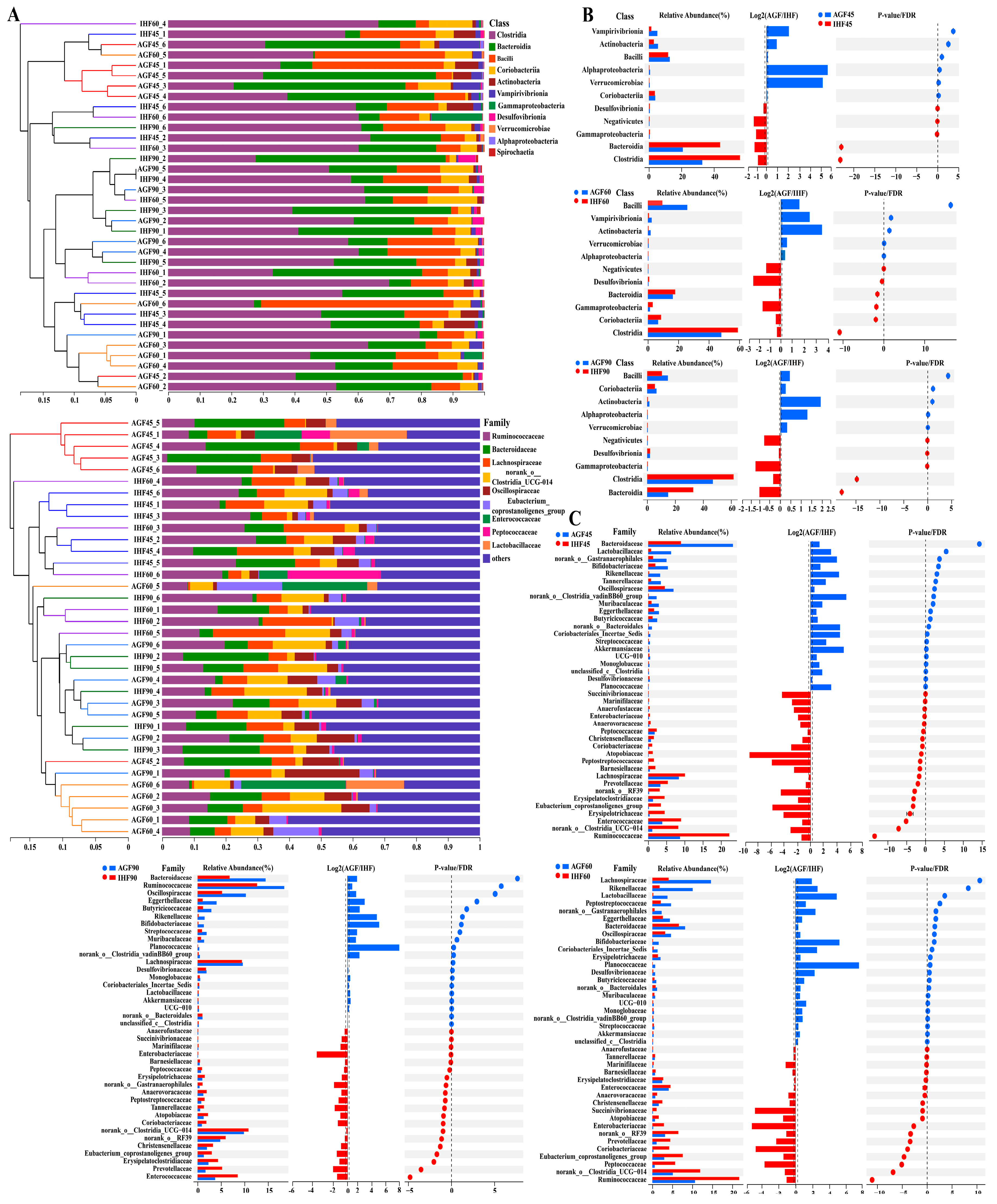
3.1. Commercial Diet Caused Gut Microbial Alterations in Geese

To study the impacts of ryegrass on the geese’s gut microbiota, we performed high-throughput amplicon sequencing of 16S rRNA microbial genes from geese in the IHF and AGF groups. To determine the diversity, richness, and evenness of the gut microbiota of geese in the IHF and AGF groups, α-diversity was determined by applying the Ace index, Chao index, Shannon index, Simpson index, and Sobs index. We noticed a considerable decline in the α-diversity of the IHF group compared to that of the AGF group (Figure S2A). The NMDS-based β-diversity analysis based on the Bray–Curtis dissimilarity matrix revealed significant modifications in caecal microbiota at 45d (Stress: 0.069, R = 0.754, p = 0.003), 60d (Stress: 0.087, R = 0.387, p = 0.003), and 90d (Stress: 0.049, R = 0.274, p = 0.059) between the IHF and AGF geese groups (Figure S2B). The microbial and bacterial taxonomies of the caecal microbiota were equated between the two groups at the class and family levels (Figure 1A). Equating the percentages of bacteria at the class level, ryegrass significantly reduced the Clostridia, Bacteroidia, Gammaproteobacteria, Desulfovibrionia, and Negativicutes compared to those of the IHF group (Figure 1A), while at the family level, the percentages of Ruminococcaceae, norank_o__RF39, Lachnospiraceae, norank_o__Clostridia_UCG-014, Eubacterium_coprostanoligenes_group, and Erysipelatoclostridiaceae were more reduced in the AGF group than in the IHF group (Figure 1A).
Next, we examined detailed alterations in the caecal microbiota at various taxonomic levels in the AGF group compared to that of the IHF group. At the class level, the AGF group had the greatest abundances of Bacilli, Coriobacteriia, Actinobacteria, Vampirivibrionia, Verrucomicrobiae, and Alphaproteobacteria relative to the IHF group (Figure 1B and Table S3). In contrast, Clostridia, Bacteroidia, Gammaproteobacteria, Desulfovibrionia, and Negativicutes were absent in the AGF group compared to those in the IHF group (Figure 1B and Table S3). The abundance of 40 bacterial species at the family level was detected to differ between the two groups, of which 20, 18, and 20 were significantly lowered after ryegrass supplementation at 45 d, 60 d, and 90 d, respectively (Figure 1C and Table S4). Interestingly, at 45 d, five species—norank_o__Clostridia_vadinBB60_group, Akkermansiaceae, norank_o__Bacteroidales, Coriobacteriales_Incertae_Sedis, and Enterococcaceae—and at 60 d and 90 d, three species—Planococcaceae, Bifidobacteriaceae, and Lactobacillaceae—were abundantly higher in the AGF group than in the IHF group (Figure 1C and Table S4).

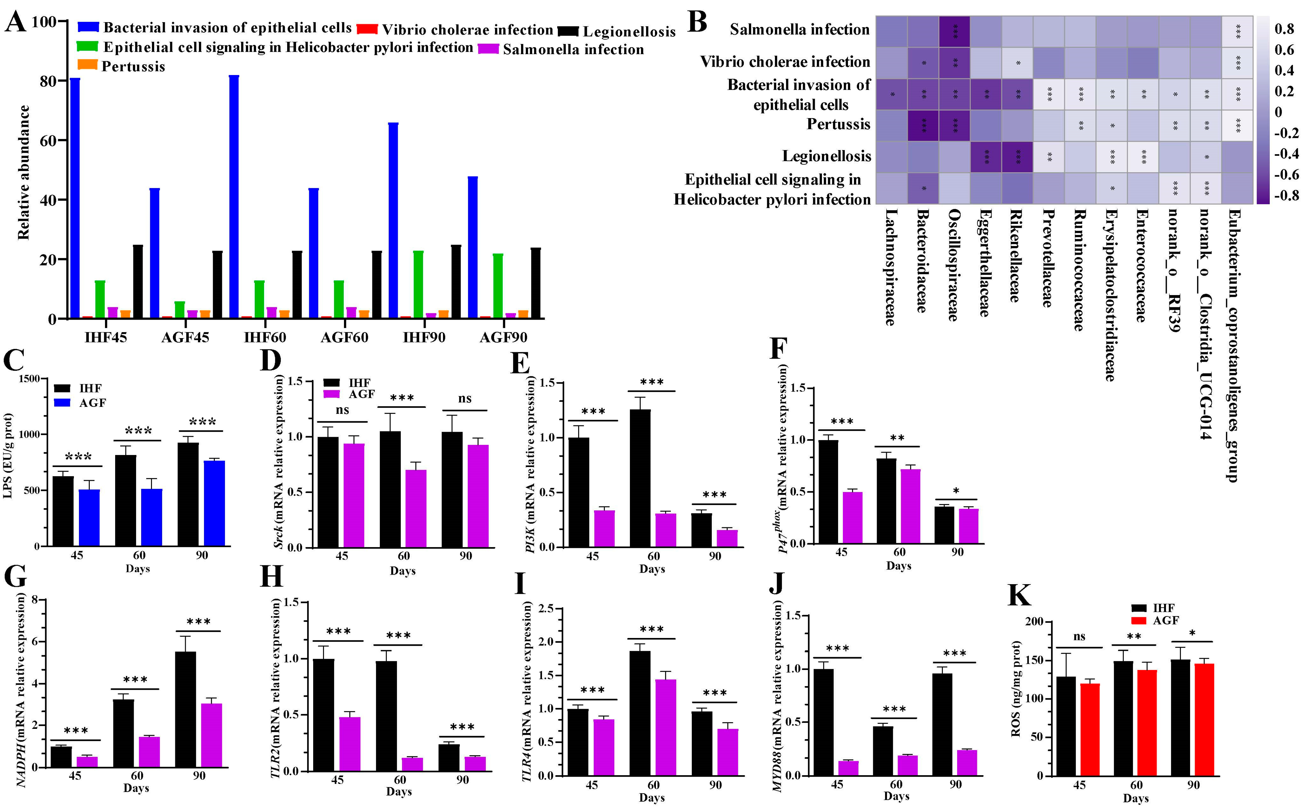
3.2. Commercial diet-Dependent Gut Microbial Alterations Are Paralleled by LPS-Induced Oxidative Stress Production

The metagenome-predicted functional modifications of caecal microbial KEGG terms were detected by applying Tax4Fun to 36 samples of geese. A total of 6, 40, and 257 KEGG terms were identified at KEGG levels 1, 2, and 3, respectively. To recognize which strains aid in LPS biosynthesis, we concentrated on KEGG terms at level 3 (Figure 2A) and gut microbiota at the family level (Figure 2B). By applying a Spearman correlation analysis between the LPS-synthesizing strains and gut microbiota, we detected that Eubacterium_coprostanoligenes_group, Norank_o__Clostridia_UCG-014, and Erysipelatoclostridiaceae were the main bacteria that were positively correlated with pertussis, the bacterial invasion of epithelial cells, Vibrio cholera infection, and Salmonella infection (Figure 2B and Table S5). Most of the highly abundant bacteria in the IHF group were involved in the bacterial invasion of epithelial cells. Interestingly, the highly prevalent bacteria in the ryegrass intake group (i.e., Oscillospiraceae and Bacteroidaceae) were involved in reducing pertussis, the bacterial invasion of epithelial cells, Vibrio cholera infection, and Salmonella infection (Figure 2B and Table S5).
Comparing the LPS-causing bacterial strains with the concentration of LPS in the caecum and spleen tissues, we hypothesized that diet-dependent LPS production could disturb spleen functions by generating oxidative stress in geese. We first confirmed this by establishing the LPS/NADPH-induced ROS mechanism on the reliance on dietary intervention. In this mechanism, the concentration of LPS measured using the ELISA kits was increased in the spleen tissues and serum samples of the IHF group (Figure 2C and Figure S3A). LPS plays a pivotal role in activating the regulatory subunit gene (p47phox) of nicotinamide adenine dinucleotide phosphate (NADPH) by augmenting proto-oncogene tyrosine-protein kinase (Srck) and phosphatidylinositol-3-kinase (PI3K). Indeed, we confirmed this by measuring the mRNA expression levels of Srck, PI3K, and p47phox that were surprisingly more increased in the spleen tissues of the IHF group than in the AGF group at 45 d, 60 d, and 90 d (Figure 2D–F). Next, we sought to identify whether the increased NADPH allows LPS to connect with Toll-like receptors (TLRs) and then promotes the myeloid differentiation primary response 88 (MyD88) pathway. Herein, first, we determined the mRNA expression levels of NADPH, TLR2, and TLR4 from the spleen tissues of geese. We found that the expression levels of NADPH, TLR2, and TLR4 were increased in ryegrass-lacking geese (Figure 2G–I). Then, we detected a higher mRNA level of MyD88 in the IHF group than in the AGF group (Figure 2J), which suggested that a gradual increase in NADPH may permit LPS to bind with MyD88. The NADPH/MyD88 activation may promote ROS production. Intriguingly, the splenic and serum ROS production was paralleled by increased NADPH and MyD88 in the IHF group compared to those of the AGF group (Figure 2K and Figure S3B).

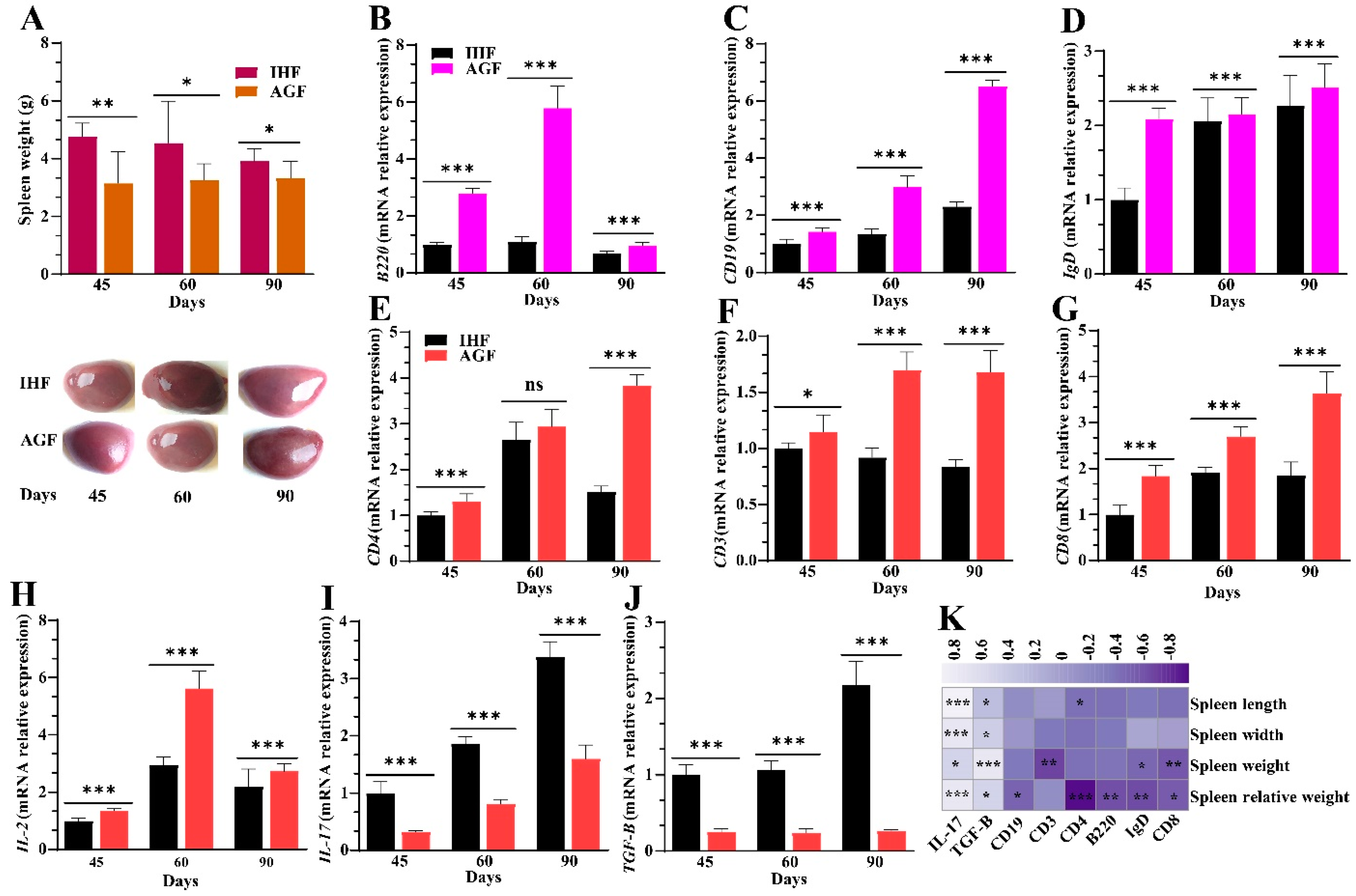
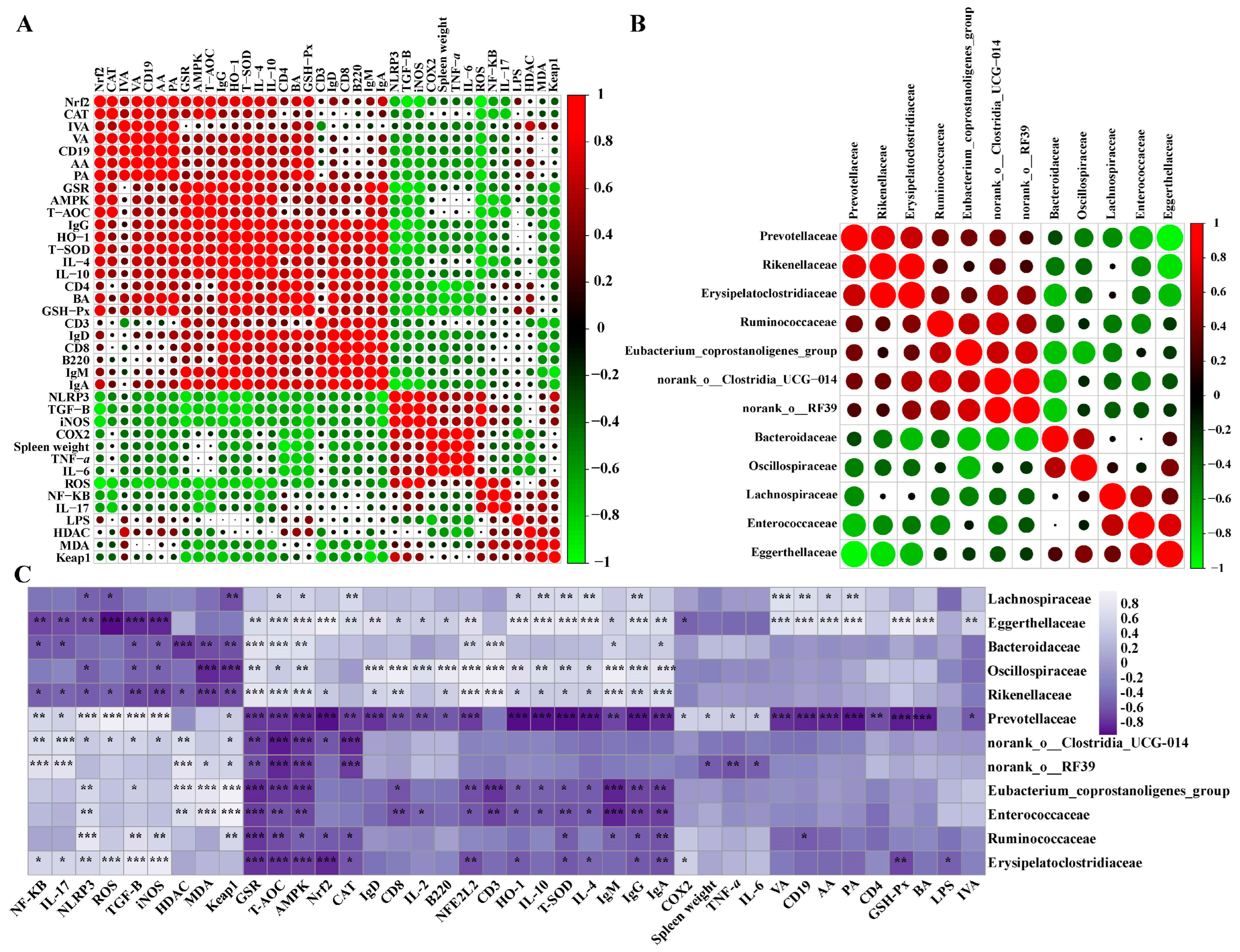
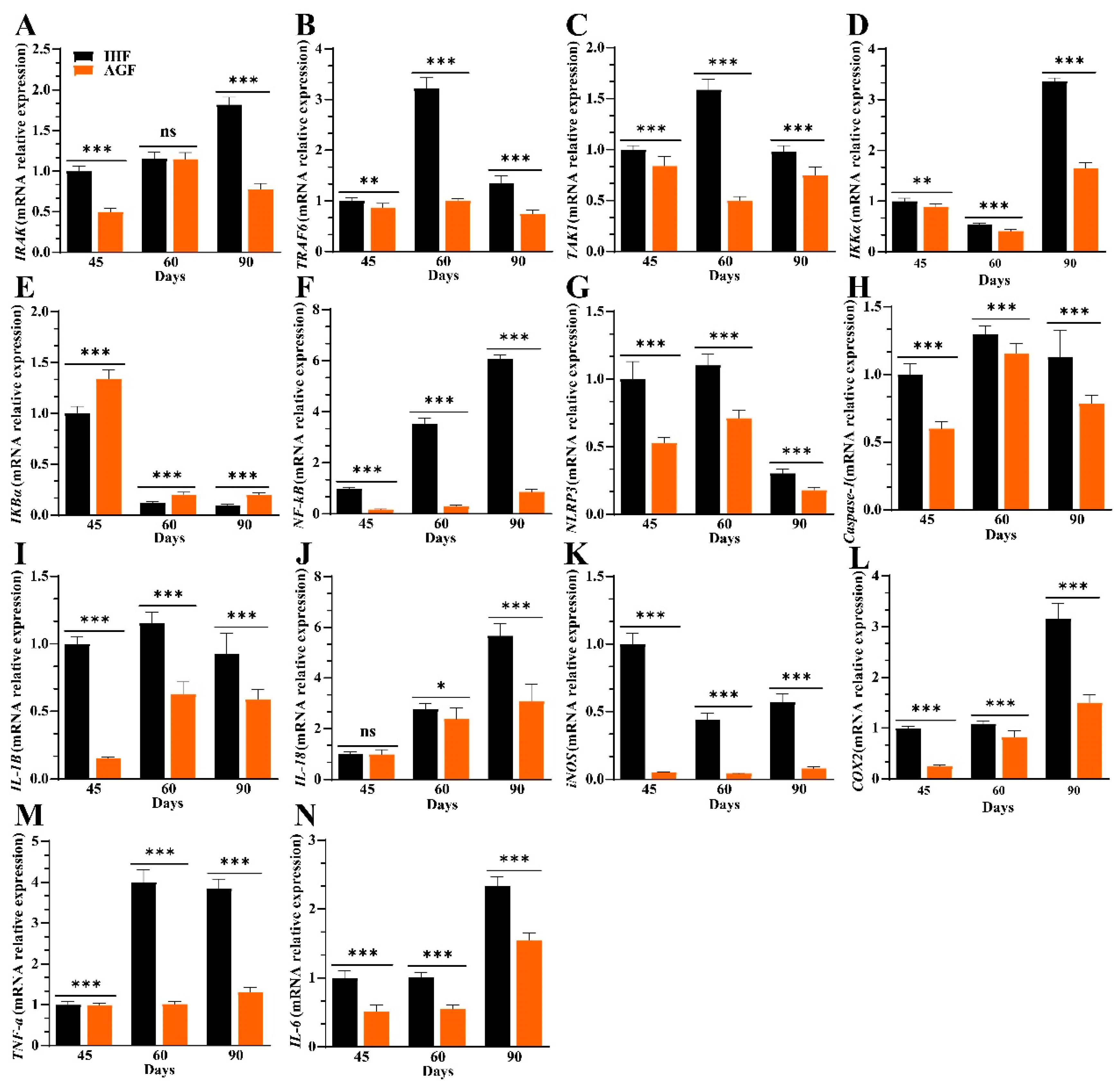
3.3. Commercial Diet-Induced Oxidative Stress Modulates Spleen Weight and Splenocyte Populations via NF-κB/NLRP3 Pathway-Induced Systemic Inflammation

Given that the oxidative stress-induced NF-κB pathway exacerbates NLR family pyrin domain containing 3 (NLRP3) activity, which influences systemic inflammation through regulating inflammatory cytokines, we hypothesized that its regulation could affect splenocyte populations. First, we developed a priming signal pathway for NLRP3 activation and hypothesized that the regulation of NF-κB could be MyD88-dependent TRAF6 activation in geese.
We detected higher mRNA expression levels of MyD88 (Figure 2J), interleukin-1 receptor (IL-1R)-associated kinase (IRAK), TNF receptor-associated factor (TRAF6), and transforming growth factor β-activated kinase 1 (TAK1) in the IHF group compared with the AGF group (Figure 3A–C). The increased mRNA expression level of TAK1 suggested that it must activate IκB kinase α (IKKα) (Figure 3D) to degrade NF-κB inhibitor IκBα (Figure 3E) and, in turn, activate NF-κB-facilitated NLRP3 in IHF geese as compared to AGF geese (Figure 3F,G). In response to NLRP3 activation, caspase-1 was increased in the IHF group which further influenced the pro-inflammatory cytokines interleukin 1 beta (IL-1β), IL-18, inducible nitric oxide synthase (iNOS), cytochrome c oxidase subunit 2 (COX2), tumor necrosis factor-alpha (TNF-α), and IL-6 in the spleen tissues of geese (Figure 3H–N).
How NLRP3-induced systemic inflammation affects splenocyte populations needs to be explored in geese. To obtain insight into splenocyte population detection, we first showed the effect of inflammation on spleen weight, length, and width. Contrary to the IHF group, ryegrass resulted in a significant reduction in spleen weight, the ratio of spleen to body weight, and spleen length and width (Figure 4A and Figure S4A–C). To investigate the splenocyte populations related to the AGF-induced decrease in spleen weight, length, and width, we then identified the mRNA expression levels of B cells (B220, CD19, and IgD) and T cells (CD3, CD4, CD8, IL-2, IL-17, and TGF-β). Of note, the mRNA expression levels of B220, cluster of differentiation 19 (CD19), immunoglobulin D (IgD), CD3, CD4, CD8, and interleukin 2 (IL-2) in the AGF group were higher than in the IHF group (Figure 4B–H). In contrast, the mRNA levels of IL-17 and transforming growth factor-β (TGF-β) were significantly decreased in the AGF group compared with the IHF group (Figure 4I,J). Positive correlations were detected between the expression levels of IL-17, TGF-β, spleen weight, spleen relative weight, and spleen length and width. On the contrary, a negative correlation was found between the expression levels of B220, CD19, IgD, CD3, CD4, CD8, IL-2, spleen weight, spleen relative weight, and spleen length and width (Figure 4K).

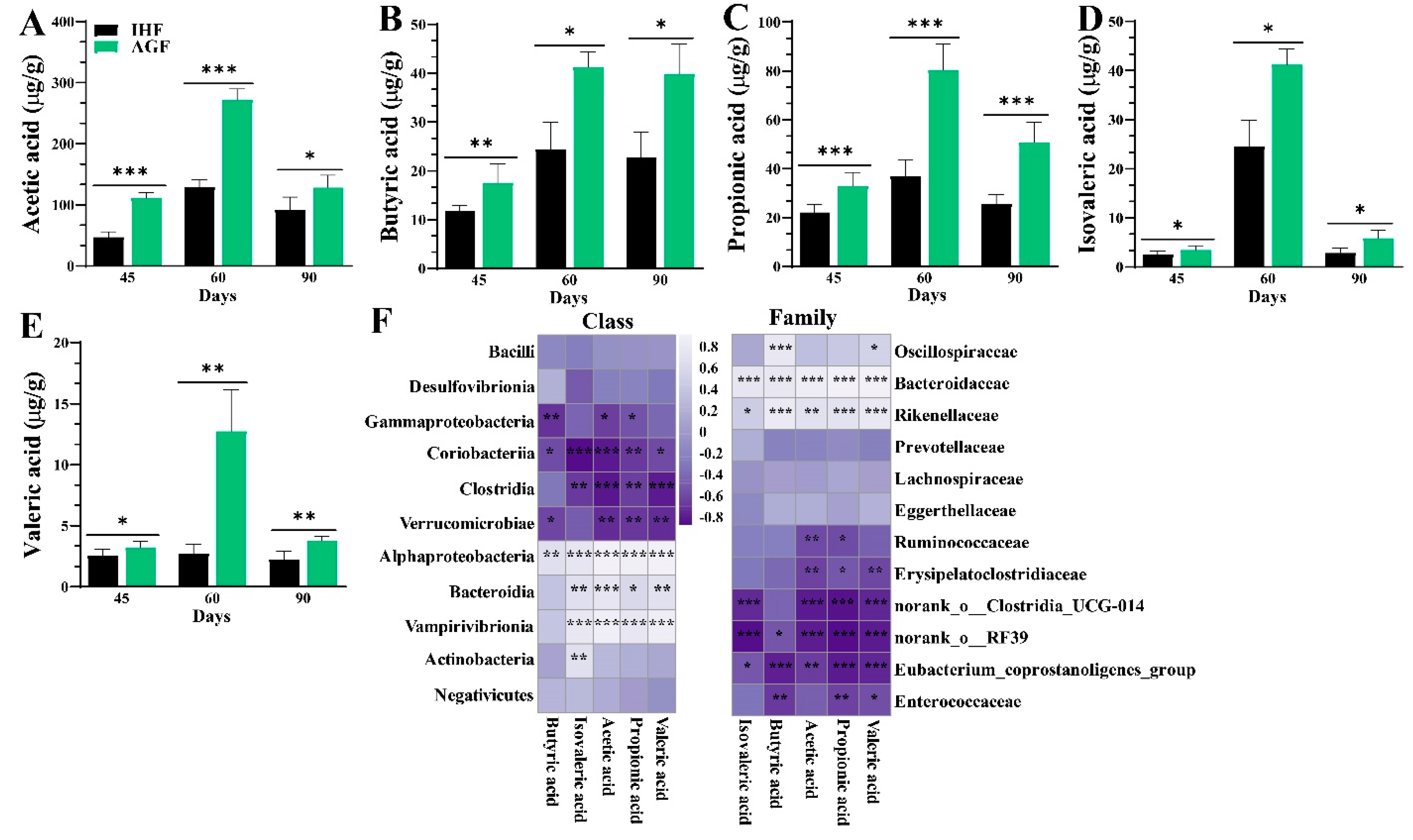
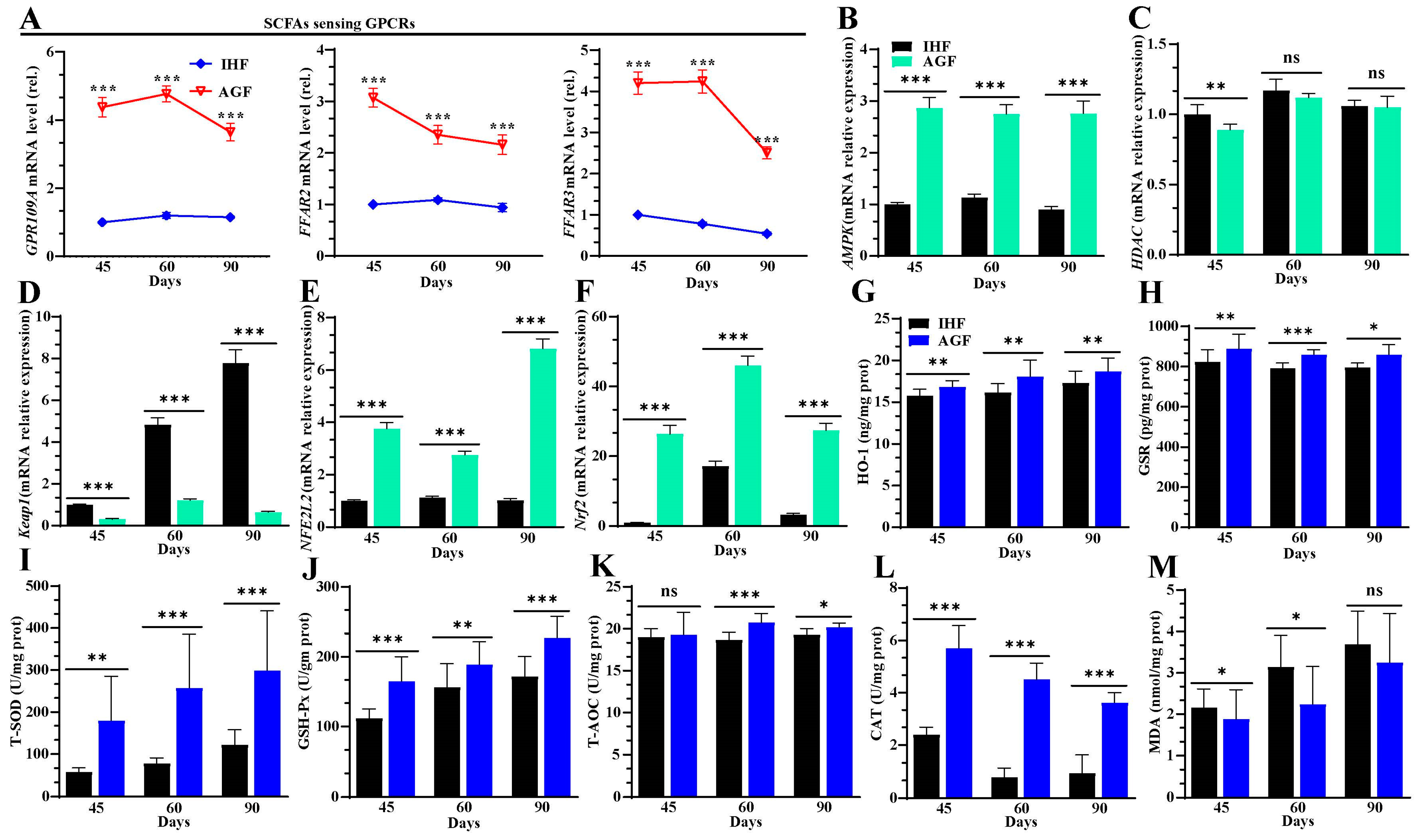
3.4. Long-Term Ryegrass Intake Causes Gut Microbial Short-Chain Fatty Acid Enrichment

The production of SCFAs by the gut microbiome plays an important role in controlling myeloid and lymphocyte cell populations to induce immune molecules that support balanced immunity [35,36]. We determined the concentrations of SCFAs through gas chromatography from the caecal chyme of geese (Figure 5A–E). The levels of butyric, propionic, and acetic acids were dominant over valeric and isovaleric acids in the AGF group compared to the IHF group. To determine the potential associations between the relative bacterial abundance and SCFA levels in caecal chyme samples, we developed a Spearman correlation analysis. There were significant positive correlations of the enrichment of Alphaproteobacteria with acetic acid (R = 0.895, p < 0.001), propionic acid (R = 0.866, p < 0.001), valeric acid (R = 0.888, p < 0.001), isovaleric acid (R = 0.743, p < 0.001), and butyric acid (R = 0.661, p < 0.001) at the class level (Figure 5F and Table S6). In contrast, a significant positive correlation of Bacteroidaceae with acetic acid (R = 0.912, p < 0.001), propionic acid (R = 0.920, p < 0.001), butyric acid (R = 0.793, p < 0.001), isovaleric acid (R = 0.764, p < 0.001), and valeric acid (R = 0.865, p < 0.001) was observed at the family level (Figure 5F and Table S7).

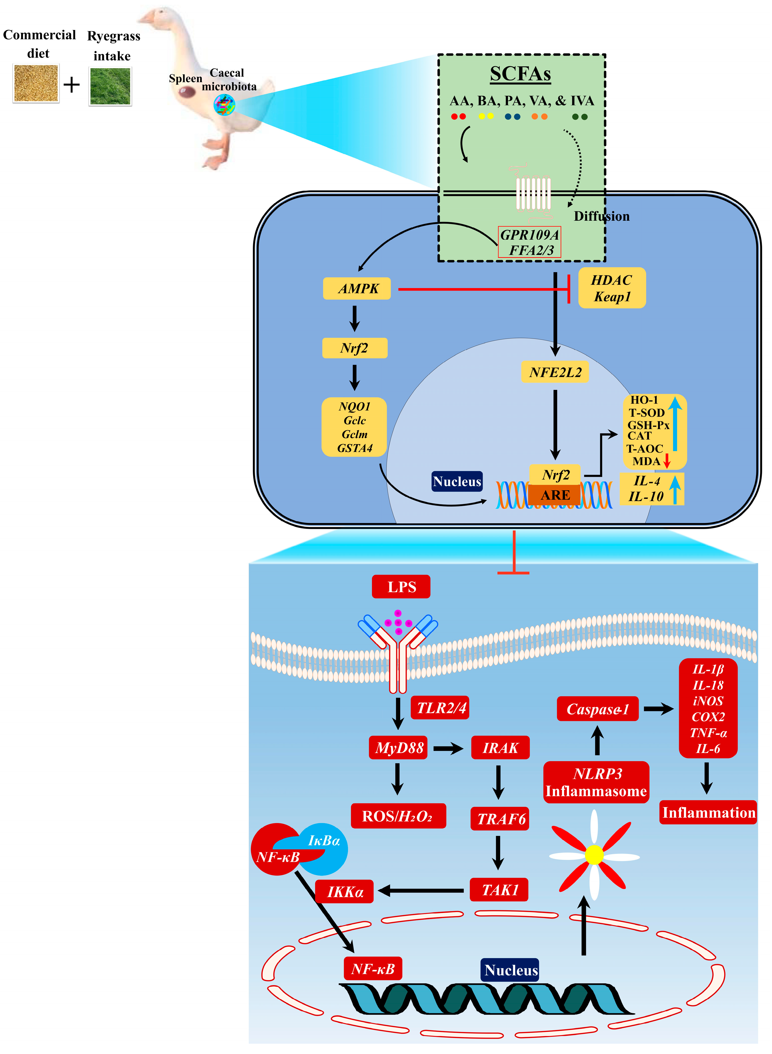
3.5. Ryegrass-Dependent SCFA Regulation Improved Keap1-Nrf2 Pathway Activation

Gut microbial SCFAs play a pivotal role in the activation of the Keap1-Nrf2 redox signaling pathway by patrolling the AMPK pathway [37]. How SCFAs activate the redox signaling pathway is not described in geese. We hypothesized that the higher expression of SCFA receptors could activate the AMPK pathway and then collectively inhibit HDAC. Upon HDAC inhibition, Keap1 dissociates from Nrf2 and then enters the activation of antioxidant-responsive element (ARE)-dependent genes. With this agreement, we first identified that the mRNA expression levels of GPR109A, FFAR2, FFAR3, and AMPK were increased (Figure 6A,B) and the expression levels of HDAC declined in the spleen tissues of AGF geese (Figure 6C). This suggests that the HDAC inhibition reduced the Nrf2 suppressor Keap1 mRNA expression (Figure 6D) and then separated Keap1/Nrf2 in augmenting the Nrf2 encoding gene NFE2L2 (Figure 6E) and Nrf2 itself (Figure 6F).
The activation of the Nrf2 pathway is considered to induce cellular redox homeostasis, and any defect in Nrf2 activity is known to change the cellular defense system. Therefore, we measured Nrf2-regulated genes and the oxidant/antioxidant enzymes controlled by the Nrf2 pathway in the IHF and AGF groups. With long-term ryegrass supplementation, the Nrf2-regulated genes NQO1, Gclc, Gclm, and GSTA4 were higher in the AGF group than those in the IHF group (Figure S5A). In addition, we detected higher protein levels of HO-1, GSR, T-SOD, GSH-Px, T-AOC, and CAT in the AGF group than in the IHF group (Figure 6G–L). To validate our results, we determined the mRNA expression levels of HMOX1 (HO-1), SOD, GSH-Px, and CAT from the spleen tissues of the AGF and IHF groups. Again, we identified that ryegrass was more involved in the upregulation of HMOX1 (HO-1), SOD, GSH-Px, and CAT in the AGF group than in the IHF group (Figure S5B–E). Furthermore, to identify whether these antioxidants control oxidative mediators that cause ROS/HDAC/Keap1 insults in the AGF and IHF groups, we determined protein levels of MDA from the spleen tissues. We identified higher expression of protein levels of MDA in the IHF group compared to that in the AGF group (Figure 6M).

3.6. Ryegrass-Dependent Keap1-Nrf2 Pathway Activation Impedes Endotoxemia, Systemic Inflammation, and Spleen Dysfunctions

To further explore the effects of ryegrass on gut microbial alterations that facilitate spleen functions at 45 d, 60 d, and 90 d, we hypothesized that gut microbial SCFAs could activate the Nrf2 pathway and mimic oxidative stress-induced systemic inflammation through the gut–microbiota–spleen axis in geese. Carsetti et al. [17] described that circulating IgM can influence the production of SIgA in the gut and thus directly involve the mechanism of a gut–spleen axis. Therefore, we measured the protein levels of IgA, IgM, and IgG with ELISA kits from the caecal tissues, followed by serum samples and spleen tissues. We identified a higher concentration of IgA, IgM, and IgG in caecal tissues, serum samples, and spleen tissues of AGF geese compared to that in IHF geese, suggesting that the circulating immunoglobulins may contribute to spleen functions by influencing the production of IgA, IgM, and IgG in spleen tissues (Figure 7A–I). Furthermore, SCFAs, particularly butyric, acetic, and propionic acids, encourage the Nrf2 redox signaling pathway [38] and regulatory T cells to release IL-10, which enters the gut–spleen axis by patrolling CD8 T cells and ameliorating severe cytotoxicity and inflammation [36,39]. To explore the role of anti-inflammatory cytokines in controlling inflammation, we measured the mRNA levels of Nrf2, IL-4, and IL-10 and the protein levels of HO-1, GSR, T-SOD, GSH-Px, T-AOC, and CAT from spleen tissues. Compared to gut microbial SCFAs, Nrf2, and antioxidant concentrations, the mRNA expression levels of IL-4 and IL-10 were higher in the spleen tissues of the AGF group than in the IHF group (Figure 7J,K).
To further prove the gut–microbiota–spleen axis, we performed a correlation analysis of host markers and gut microbial SCFAs. The results obtained from the correlation of host markers showed that acetic, butyric, propionic, valeric, and isovaleric acids were significantly positively correlated with Nrf2, antioxidant enzymes (HO-1, GSR, T-SOD, GSH-Px, T-AOC, and CAT), B cells (B220, CD19, and IgD), T cells (CD3, CD4, CD8, and IL-2), immunoglobulins (IgA, IgM, and IgG), and anti-inflammatory cytokines (IL-4 and IL-10) in the AGF group compared with the IHF group (Figure 8A). In contrast, the gut microbial SCFA-induced Nrf2 redox signaling enzymes were more negatively correlated with endotoxemia (LPS), oxidative stress (ROS), NF-κB, NLRP3, and inflammatory cytokines in the AGF group compared with the IHF group. These results indicated that the existence of dietary fiber in ryegrass is the main influencer of gut microbial metabolic modulations that, on the one hand, promote the redox signaling pathway and immune balance and, on the other hand, attenuate systemic inflammation in geese.

3.7. Long-Term Ryegrass Supplementation Impedes Splenic Dysfunctions by Maintaining the Gut Microbial Microenvironment

The intestinal microbiota plays an important role in the development of the spleen [16,17,18]. How diet affects spleen maturation by modulating gut microbiota needs to be explained in geese. For this consensus, we developed a correlation analysis among the gut microbes (Figure 8B) and with the host markers during the different growth phases of geese (Figure 8C and Figure S6). As shown in Figure 8C and Figure S6, Eggerthellaceae, Oscillospiraceae, Rikenellaceae, and Lachnospiraceae were the most abundant bacteria that existed in the caecal chyme of the AGF group that were positively correlated with SCFAs, Nrf2 redox signaling enzymes, B cells, T cells (with an exception of IL-17 and TGF-β), and anti-inflammatory mediators in spleen tissues of geese. In contrast, Enterococcaceae and Prevotellaceae were highly abundant in the IHF group compared with the AGF group, which were positively correlated with LPS, ROS, NF-κB, NLRP3, inflammatory cytokines, and spleen weight in the spleen tissues of geese (Figure 8C and Figure S6, and Table S8). In conclusion, these results describe the role of ryegrass as a high-dietary-fiber source that controls LPS-induced oxidative stress-facilitated systemic inflammation by activating the SCFAs/Nrf2 pathway in geese.

4. Discussion

Low-dietary-fiber-dependent gut microbial LPS-induced oxidative stress [2] undergoes systemic inflammation in the spleen [6] and inflammatory diseases [40]. Today, the current poultry industry is based on low-dietary-fiber-enriched feed ingredients [30], which may lead to higher growth performance with compromised intestinal infections, low immunity, and metabolic diseases [31]. In addition, the global trend of consuming a low-dietary-fiber diet has been concerned with improved autoimmunity, diabetes, cardiovascular health, and cancer [22]. Despite this, more research on geese is required to determine how dietary fiber content lowers the risk of inflammatory disorders by triggering a redox signaling pathway that is dependent on SCFAs. Thus, for the first time, the present research explores that ryegrass intake can ameliorate commercial diet-dependent gut microbial alterations, endotoxemia, oxidative stress, and inflammatory diseases.
However, fewer findings have explained the relationship of altered gut microbiota with spleen functions in humans [41], mice [18], chickens [24], and tilapia [42]. Within these abrogative findings, the mechanisms linked with the diverse association of dietary fiber intervention with the gut–microbiota–spleen axis have not been explored. Notably, we developed a ryegrass-dependent artificial pasture grazing system that concords with concomitant mechanisms to attenuate commercial diet-dependent intestinal and splenic LPS and inflammatory disorders. Subsequently, the advanced role of ryegrass is to develop a pathway-based mechanism, thereby restoring the enrichment of intestinal SCFA-generating microbes and impeding commercial diet-induced LPS-producing bacteria. These modifications improve the Nrf2 redox signaling pathway, splenocyte population (B cells and T cells), immunoglobulins, and anti-inflammatory cytokines, resulting in decreased metabolic endotoxemia and LPS-induced oxidative stress in the spleen organ of geese. The reduction in pro-inflammatory cytokines results in a decrease in chronic inflammatory disorders. In light of the aforementioned data, Spearman correlation analysis provides substantial support for the suggested mechanisms.
Only a few studies explain the character of altered gut microbiota and LPS levels in spleen dysfunctions [43,44]. In this study, we discovered altered microbiota using predicted functions based on 16S rRNA sequencing and Tax4Fun analysis that were involved in LPS-producing strains. Some of the bacteria, Ruminococcaceae, norank_o__RF39, Enterocococcaceae, norank_o__Clostridia_UCG-014, Eubacterium_coprostanoligenes_group, and Erysipelatoclostridiaceae, were strongly involved in KEGG orthologues of LPS production in the IHF group. In contrast, our findings were underscored by those of dietary capsaicin in human participants [45]. This suggests that the reduced enrichment of KEGG orthologues involved in LPS production may be due to the higher abundances of Oscillospiraceae, Eggerthellaceae, and Bacteroidaceae in the AGF group.
LPS-induced ROS production is mainly due to the stimulation of the TLR4/MyD88 pathway [46]. Several research studies have demonstrated that the stimulation of the TLR4/MyD88 pathway is influenced by a low-dietary-fiber diet and high carbohydrates, lipids, protein, and calories [2,47,48]. Contrary to these studies, we developed the LPS-induced NADPH pathway and then hypothesized that this pathway should promote ROS production in commercial diet-feeding geese (Figure 9). It is known that SrcK activates PI3K in response to LPS, and then PI3K stimulates NADPH oxidase through p47phox phosphorylation in RAW264.7 macrophages [49]. Concerning this study, we detected higher protein concentrations of LPS and mRNA expression levels of Srck, PI3K, and p47phox in the IHF group compared with those of the AGF group. P47phox is a major driving factor of NADPH oxidase that is involved in TLR-MyD88 activation [50]. Of note, we detected increased mRNA levels of NADPH, TLR2, TLR4, and MyD88 in the IHF group compared with the AGF group. According to Ichikawa et al. [46], the activated RLT4/MyD88 pathway further contributes to the production of ROS. Considering this report, we identified higher concentrations of ROS in the serum and spleen tissues of the IHF group compared with those of the AGF group.
The production of oxidative stress due to ROS insults provides an environment for the activation of the NF-κB pathway [51], which plays a vital role in the establishment of systemic inflammation through the maturation of pro-inflammatory cytokines [52,53]. Our previous work had limitations, whereby we showed that the development of inflammation by the LPS/ROS-induced NF-κB pathway was LC8- and IkB-a-dependent [2]. However, in the current study, we showed a mechanistic pathway by which the first NF-κB pathway activates NLRP3 and then allows it to induce pro-inflammatory cytokine stimulation.
In many studies, the augmentation of the NLRP3 inflammasome is NF-κB-dependent [52]. However, in our investigation, we identified that the stimulation of the NLRP3 inflammasome by NF-κB is MyD88-dependent TRAF6 regulation. Several studies using LPS as an MyD88 activator support our findings [54]. Furthermore, our investigations, somewhat modified from those of Lu et al. [55] and Sato et al. [56], demonstrated that MyD88 triggering promoted IRAK, TRAF6, and TAK1 activation, which might have collectively activated NF-κB signaling cascades and finally resulted in NLRP3 activation in the IHF group. Several studies demonstrated that the overexpression of NLRP3 results in increased caspase-1, which matures pro-inflammatory cytokines IL-1β and IL-18 [57,58] and then influences other inflammatory cytokines [59,60]. Comparing these studies with our current work, we identified higher mRNA expression levels of NLRP3, Casoase-1, and pro-inflammatory cytokines in the IHF group compared to those of the AGF group. A defensive role of the spleen against inflammation and obesity-related diseases has been reported elsewhere [61,62]. How dietary interventions impact immune responses of the spleen against inflammatory mediators is not reported clearly in geese. Herein, comparing the expression levels of inflammatory markers, we detected a parallel decrease in the expression levels of splenocyte populations (B cells and T cells) except IL-17, TGF-β, and spleen weight in the IHF group compared with those of the AGF group.
The perpetuated regulation of HDACs and NF-κB is primarily concerned with the activation of genes that are commonly expressed during inflammation [63,64]. In our previous study, we described the mechanism by which the AGF system-induced alkaline phosphatase enzyme can dephosphorylate LPS and attenuate the existence of NF-κB-facilitated chronic low-grade inflammation in the intestines of geese. In this study, we developed a pathway by which ryegrass-dependent gut microbial SCFA receptors activate the AMPK pathway and then collectively inhibit HDAC. Upon HDAC inhibition, Keap1 dissociates from Nrf2 and then enters the activation of ARE-dependent genes in the spleen tissues of the AGF group. Our findings were followed by the reports of Wang et al. [37], in which we detected higher concentrations of SCFA-producing bacteria and mRNA expressions of SCFAs receptors GPCR109A, FFAR2, FFAR3, and AMPK that attenuated the severity of HDAC in the AGF group compared to those of the IHF group. Additionally, according to the reports of Xu et al. [65] and Xu et al. [66], we detected a significant difference between the expression levels of Keap1, Nrf2, and Nrf2-regulated genes, describing the important role of ryegrass in activating the Nrf2 redox signaling pathway in geese.
It is well known that Nrf2 increases the ARE-dependent gene expression of a series of antioxidant proteins [67] and regulatory T cells to release anti-inflammatory cytokines [68], which enter the gut–spleen axis by patrolling CD8 T cells and ameliorating severe cytotoxicity and inflammation [36,39]. To further investigate the function of gut microbial SCFAs in controlling spleen functions, we performed a correlation analysis among host markers and gut microbial SCFAs (caecum vs. spleen). We described, for the first time, that the SCFAs were significantly positively correlated with Nrf2 redox signaling enzymes, T cells (except IL-17 and TFG-β), B cells, immunoglobulins, and anti-inflammatory mediators. We can say that this is our first report in which long-term ryegrass-induced gut microbial SCFAs, along with immunoglobulins, IL-4, and IL-10 expression, can collectively attenuate the LPS-induced oxidative stress and systemic inflammation in geese.
The gut microbiota controls the maturation of the spleen [16,17,18]. To determine how diet affects spleen maturation by modulating the gut microbiota, we performed a correlation analysis among the gut microbes and with the host markers during the different growth phases of geese. Herein, the most notable findings were the higher abundance of Eggerthellaceae, Oscillospiraceae, and Lachnospiraceae in the AGF group that were positively correlated with SCFAs, Nrf2 redox signaling enzymes, B cells, T cells (with the exception of IL-17 and TGF-β), and anti-inflammatory mediators in the spleen tissues of geese. A well-known SCFA-producing Oscillospiraceae is known to treat obesity-related diseases [69,70]. Similarly, the role of Lachnospiraceae as probiotics has been acknowledged in attenuating colonic dysbiosis-related metabolic syndrome and cancer [71]. In contrast, ryegrass ameliorated the higher abundances of Enterococcaceae and Prevotellaceae that were positively correlated with LPS, ROS, NF-κB, NLRP3, inflammatory cytokines, and spleen weight in the spleen tissues of IHF geese. Few studies have shown that the higher abundance of Prevotellaceae causes myocardial ischemia [8] and intestinal inflammation [72] in rats. In contrast, Enterococcaceae is known as a multidrug-resistant nosocomial pathogen that causes roughly 66,000 infections/year in the USA [73]. It primarily causes septic arthritis and meningitis in immunocompromised patients [74]. In conclusion, these results describe the role of ryegrass as a high-dietary-fiber source that attenuates systemic inflammation-producing bacteria by activating SCFA-producing bacteria in geese.

5. Conclusions

In conclusion, gut microbial SCFA production by ryegrass supplementation appears to restore gut microbial architecture, which then promotes Nrf2 redox signaling enzymes, B cells, and regulatory T cells to release anti-inflammatory cytokines and immunoglobulins in the spleens of geese. In addition, the higher abundances of these markers seemed to attenuate endotoxemia, oxidative stress, and systemic inflammation in the spleens of commercial diet-fed geese through the gut–microbiota–spleen axis.

Supplementary Materials

The following supporting information can be downloaded at: https://www.mdpi.com/article/10.3390/nu16050747/s1, Figure S1: Overview of experimental design; Figure S2: Effects of ryegrass on the alpha- and beta-diversity of the gut microbiota; Figure S3: Commercial diet-dependent gut microbial alterations are paralleled by LPS-induced oxidative stress production; Figure S4: Effect of feeding systems on spleen relative weight, spleen length, and spleen width of geese; Figure S5: Ryegrass-dependent SCFA regulation improved Nrf2-regulated genes and antioxidant-responsive element (ARE)-dependent genes; Figure S6: A Spearman correlation between the host markers and gut microbiota; Table S1: The nutrients and chemical composition of the diet (two stages) offered to geese; Table S2: Primer sequences used for quantitative real-time PCR; Table S3: Effect of ryegrass on gut microbial richness of geese at class level; Table S4: Effect of ryegrass on the gut microbial richness of geese at a family level; Table S5: Gut microbiota and KEGG orthologues involved in LPS production; Table S6: Effect of ryegrass on the relationship of gut microbiota at class level and SCFAs; Table S7: Effect of ryegrass on the relationship of gut microbiota at a family level and SCFAs; Table S8: Effect of ryegrass on the relationship of gut microbiota and host markers.

Author Contributions

Q.A., B.L., J.N., M.L. and D.L. performed studies and analyzed data. A.M., Z.W., H.S. and Y.C. supervised experiments, provided essential tools and samples, analyzed data, and edited the manuscript. S.M. significantly contributed to the discussion and interpretation of data. Y.S. conceptualized and supervised the project and analyzed data. Q.A. wrote the manuscript. All authors have read and agreed to the published version of the manuscript.

Funding

This work was supported by grants from the Modern Agroindustry Technology Research System of China (CARS-34) and the Science and Technology Innovation Team of Henan Province High Quality Forage and Animal Health (No.22IRTSTHN022).

Institutional Review Board Statement

The animal experiments were approved by the Research Bioethics Committee of the Henan Agricultural University (Animal ethics number: HNND2020041011; approved on 10 April 2020).

Informed Consent Statement

Not applicable.

Data Availability Statement

Data are contained within the article and supplementary materials.

Conflicts of Interest

The authors declare no conflicts of interest.

References

  1. Zhang, Y.; Zhou, M.; Zhou, Y.; Guan, X. Dietary components regulate chronic diseases through gut microbiota: A review. J. Sci. Food Agric. 2023, 103, 6752–6766. [Google Scholar] [CrossRef] [PubMed]
  2. Ali, Q.; Ma, S.; Farooq, U.; Niu, J.; Li, F.; Li, D.; Wang, Z.; Sun, H.; Cui, Y.; Shi, Y. Pasture intake protects against commercial diet-induced lipopolysaccharide production facilitated by gut microbiota through activating intestinal alkaline phosphatase enzyme in meat geese. Front. Immunol. 2022, 13, 1041070. [Google Scholar] [CrossRef] [PubMed]
  3. Rohr, M.W.; Narasimhulu, C.A.; Rudeski-Rohr, T.A.; Parthasarathy, S. Negative Effects of a High-Fat Diet on Intestinal Permeability: A Review. Adv. Nutr. 2020, 11, 77–91. [Google Scholar] [CrossRef] [PubMed]
  4. Cani, P.D.; Amar, J.; Iglesias, M.A.; Poggi, M.; Knauf, C.; Bastelica, D.; Neyrinck, A.M.; Fava, F.; Tuohy, K.M.; Chabo, C.; et al. Metabolic endotoxemia initiates obesity and insulin resistance. Diabetes 2007, 56, 1761–1772. [Google Scholar] [CrossRef] [PubMed]
  5. Ali, Q.; Ma, S.; La, S.; Guo, Z.; Liu, B.; Gao, Z.; Farooq, U.; Wang, Z.; Zhu, X.; Cui, Y.; et al. Microbial short-chain fatty acids: A bridge between dietary fibers and poultry gut health—A review. Anim. Biosci. 2022, 35, 1461–1478. [Google Scholar] [CrossRef] [PubMed]
  6. Seemann, S.; Zohles, F.; Lupp, A. Comprehensive comparison of three different animal models for systemic inflammation. J. Biomed. Sci. 2017, 24, 60. [Google Scholar] [CrossRef] [PubMed]
  7. Barrea, L.; Di Somma, C.; Muscogiuri, G.; Tarantino, G.; Tenore, G.C.; Orio, F.; Colao, A.; Savastano, S. Nutrition, inflammation and liver-spleen axis. Crit. Rev. Food Sci. Nutr. 2018, 58, 3141–3158. [Google Scholar] [CrossRef]
  8. Sun, L.; Jia, H.; Li, J.; Yu, M.; Yang, Y.; Tian, D.; Zhang, H.; Zou, Z. Cecal Gut Microbiota and Metabolites Might Contribute to the Severity of Acute Myocardial Ischemia by Impacting the Intestinal Permeability, Oxidative Stress, and Energy Metabolism. Front. Microbiol. 2019, 10, 1745. [Google Scholar] [CrossRef]
  9. Sahin, G.S.; Lee, H.; Engin, F. An accomplice more than a mere victim: The impact of β-cell ER stress on type 1 diabetes pathogenesis. Mol. Metab. 2021, 54, 101365. [Google Scholar] [CrossRef]
  10. Lewis, S.M.; Williams, A.; Eisenbarth, S.C. Structure and function of the immune system in the spleen. Sci. Immunol. 2019, 4, eaau6085. [Google Scholar] [CrossRef]
  11. Ishii, T.; Miyazawa, M.; Takanashi, Y.; Tanigawa, M.; Yasuda, K.; Onouchi, H.; Kawabe, N.; Mitsushita, J.; Hartman, P.S.; Ishii, N. Genetically induced oxidative stress in mice causes thrombocytosis, splenomegaly and placental angiodysplasia that leads to recurrent abortion. Redox Biol. 2014, 2, 679–685. [Google Scholar] [CrossRef] [PubMed]
  12. Zhang, K.; Sakamoto, A.; Chang, L.; Qu, Y.; Wang, S.; Pu, Y.; Tan, Y.; Wang, X.; Fujita, Y.; Ishima, T.; et al. Splenic NKG2D confers resilience versus susceptibility in mice after chronic social defeat stress: Beneficial effects of (R)-ketamine. Eur. Arch. Psychiatry Clin. Neurosci. 2021, 271, 447–456. [Google Scholar] [CrossRef] [PubMed]
  13. Zhang, J.; Ma, L.; Chang, L.; Pu, Y.; Qu, Y.; Hashimoto, K. A key role of the subdiaphragmatic vagus nerve in the depression-like phenotype and abnormal composition of gut microbiota in mice after lipopolysaccharide administration. Transl. Psychiatry 2020, 10, 186. [Google Scholar] [CrossRef] [PubMed]
  14. Weledji, E.P. Benefits and risks of splenectomy. Int. J. Surg. 2014, 12, 113–119. [Google Scholar] [CrossRef] [PubMed]
  15. Alexandre, Y.O.; Mueller, S.N. Splenic stromal niches in homeostasis and immunity. Nat. Rev. Immunol. 2023, 23, 705–719. [Google Scholar] [CrossRef]
  16. Rosado, M.M.; Aranburu, A.; Scarsella, M.; Cascioli, S.; Giorda, E.; Del Chierico, F.; Mortera, S.L.; Mortari, E.P.; Petrini, S.; Putignani, L.; et al. Spleen development is modulated by neonatal gut microbiota. Immunol. Lett. 2018, 199, 1–15. [Google Scholar] [CrossRef]
  17. Carsetti, R.; Di Sabatino, A.; Rosado, M.M.; Cascioli, S.; Piano Mortari, E.; Milito, C.; Grimsholm, O.; Aranburu, A.; Giorda, E.; Tinozzi, F.P.; et al. Lack of Gut Secretory Immunoglobulin A in Memory B-Cell Dysfunction-Associated Disorders: A Possible Gut-Spleen Axis. Front. Immunol. 2019, 10, 2937. [Google Scholar] [CrossRef]
  18. Zhang, Y.; Xie, B.; Chen, X.; Zhang, J.; Yuan, S. A key role of gut microbiota-vagus nerve/spleen axis in sleep deprivation-mediated aggravation of systemic inflammation after LPS administration. Life Sci. 2021, 265, 118736. [Google Scholar] [CrossRef]
  19. Fang, H.; Feng, X.; Xu, T.; Zhong, R.; Lu, D.; Zhang, H.; Shen, W.; Zhao, Y.; Chen, L.; Wang, J. Gut-Spleen Axis: Microbiota via Vascular and Immune Pathways Improve Busulfan-Induced Spleen Disruption. mSphere 2023, 8, e0058122. [Google Scholar] [CrossRef]
  20. He, Z.; Zhang, H.; Song, Y.; Yang, Z.; Cai, Z. Exposure to ambient fine particulate matter impedes the function of spleen in the mouse metabolism of high-fat diet. J. Hazard. Mater. 2022, 423, 127129. [Google Scholar] [CrossRef]
  21. Fang, T.; Wu, X.; Cao, W.; Jia, G.; Zhao, H.; Chen, X.; Wu, C.; Tang, J.; Wang, J.; Liu, G. Effects of dietary fiber on the antioxidant capacity, immune status, and antioxidant-relative signaling molecular gene expression in rat organs. RSC Adv. 2017, 7, 19611–19620. [Google Scholar] [CrossRef]
  22. Schäfer, A.L.; Eichhorst, A.; Hentze, C.; Kraemer, A.N.; Amend, A.; Sprenger, D.T.L.; Fluhr, C.; Finzel, S.; Daniel, C.; Salzer, U.; et al. Low Dietary Fiber Intake Links Development of Obesity and Lupus Pathogenesis. Front. Immunol. 2021, 12, 696810. [Google Scholar] [CrossRef]
  23. Zhang, Q.; Xiang, L.; Zaman, M.H.; Dong, W.; He, G.; Deng, G.M. Predominant Role of Immunoglobulin G in the Pathogenesis of Splenomegaly in Murine Lupus. Front. Immunol. 2019, 10, 3020. [Google Scholar] [CrossRef]
  24. Das, Q.; Islam, M.R.; Lepp, D.; Tang, J.; Yin, X.; Mats, L.; Liu, H.; Ross, K.; Kennes, Y.M.; Yacini, H.; et al. Gut Microbiota, Blood Metabolites, and Spleen Immunity in Broiler Chickens Fed Berry Pomaces and Phenolic-Enriched Extractives. Front. Vet. Sci. 2020, 7, 150. [Google Scholar] [CrossRef] [PubMed]
  25. Li, Y.; Lei, X.; Yin, Z.; Guo, W.; Wu, S.; Yang, X. Transgenerational effects of paternal dietary Astragalus polysaccharides on spleen immunity of broilers. Int. J. Biol. Macromol. 2018, 115, 90–97. [Google Scholar] [CrossRef]
  26. Błaszczyk, K.; Wilczak, J.; Harasym, J.; Gudej, S.; Suchecka, D.; Królikowski, T.; Lange, E.; Gromadzka-Ostrowska, J. Impact of low and high molecular weight oat beta-glucan on oxidative stress and antioxidant defense in spleen of rats with LPS induced enteritis. Food Hydrocoll. 2015, 51, 272–280. [Google Scholar] [CrossRef]
  27. Brisbin, J.T.; Gong, J.; Parvizi, P.; Sharif, S. Effects of lactobacilli on cytokine expression by chicken spleen and cecal tonsil cells. Clin. Vaccine Immunol. CVI 2010, 17, 1337–1343. [Google Scholar] [CrossRef] [PubMed]
  28. Morris, J.R.; Jerome, F.N.; Reinhart, B.S. Degree of Splenomegaly in Domestic Chickens Selected for Resistance and Susceptibility to Marek’s Disease. Poult. Sci. 1969, 48, 2097–2102. [Google Scholar] [CrossRef] [PubMed]
  29. Yin, H.-c.; Huang, J. Effects of soybean meal replacement with fermented alfalfa meal on the growth performance, serum antioxidant functions, digestive enzyme activities, and cecal microflora of geese. J. Integr. Agric. 2016, 15, 2077–2086. [Google Scholar] [CrossRef]
  30. Mourão, J.L.; Pinheiro, V.M.; Prates, J.A.; Bessa, R.J.; Ferreira, L.M.; Fontes, C.M.; Ponte, P.I. Effect of dietary dehydrated pasture and citrus pulp on the performance and meat quality of broiler chickens. Poult. Sci. 2008, 87, 733–743. [Google Scholar] [CrossRef]
  31. Huyghebaert, G.; Ducatelle, R.; Van Immerseel, F. An update on alternatives to antimicrobial growth promoters for broilers. Vet. J. 2011, 187, 182–188. [Google Scholar] [CrossRef]
  32. Wan, X.; Eguchi, A.; Sakamoto, A.; Fujita, Y.; Yang, Y.; Qu, Y.; Hatano, M.; Mori, C.; Hashimoto, K. Impact of broad-spectrum antibiotics on the gut-microbiota-spleen-brain axis. Brain Behav. Immun.—Health 2023, 27, 100573. [Google Scholar] [CrossRef]
  33. Livak, K.J.; Schmittgen, T.D. Analysis of relative gene expression data using real-time quantitative PCR and the 2(-Delta Delta C(T)) Method. Methods 2001, 25, 402–408. [Google Scholar] [CrossRef]
  34. Liu, B.; Cui, Y.; Ali, Q.; Zhu, X.; Li, D.; Ma, S.; Wang, Z.; Wang, C.; Shi, Y. Gut Microbiota Modulate Rabbit Meat Quality in Response to Dietary Fiber. Front. Nutr. 2022, 9, 849429. [Google Scholar] [CrossRef]
  35. Lu, Y.; Zhang, Y.; Zhao, X.; Shang, C.; Xiang, M.; Li, L.; Cui, X. Microbiota-derived short-chain fatty acids: Implications for cardiovascular and metabolic disease. Front. Cardiovasc. Med. 2022, 9, 900381. [Google Scholar] [CrossRef] [PubMed]
  36. Lee, R.; Yoon, B.I.; Hunter, C.A.; Kwon, H.M.; Sung, H.W.; Park, J. Short chain fatty acids facilitate protective immunity by macrophages and T cells during acute fowl adenovirus-4 infection. Sci. Rep. 2023, 13, 17999. [Google Scholar] [CrossRef] [PubMed]
  37. Wang, B.; Zhu, X.; Kim, Y.; Li, J.; Huang, S.; Saleem, S.; Li, R.C.; Xu, Y.; Dore, S.; Cao, W. Histone deacetylase inhibition activates transcription factor Nrf2 and protects against cerebral ischemic damage. Free Radic. Biol. Med. 2012, 52, 928–936. [Google Scholar] [CrossRef] [PubMed]
  38. González-Bosch, C.; Boorman, E.; Zunszain, P.A.; Mann, G.E. Short-chain fatty acids as modulators of redox signaling in health and disease. Redox Biol. 2021, 47, 102165. [Google Scholar] [CrossRef] [PubMed]
  39. Bachem, A.; Makhlouf, C.; Binger, K.J.; de Souza, D.P.; Tull, D.; Hochheiser, K.; Whitney, P.G.; Fernandez-Ruiz, D.; Dähling, S.; Kastenmüller, W.; et al. Microbiota-Derived Short-Chain Fatty Acids Promote the Memory Potential of Antigen-Activated CD8(+) T Cells. Immunity 2019, 51, 285–297.e5. [Google Scholar] [CrossRef]
  40. Sun, X.; Cui, Y.; Su, Y.; Gao, Z.; Diao, X.; Li, J.; Zhu, X.; Li, D.; Li, Z.; Wang, C.; et al. Dietary Fiber Ameliorates Lipopolysaccharide-Induced Intestinal Barrier Function Damage in Piglets by Modulation of Intestinal Microbiome. mSystems 2021, 6, e01374-20. [Google Scholar] [CrossRef]
  41. Zhu, H.; Liu, Y.; Li, S.; Jin, Y.; Zhao, L.; Zhao, F.; Feng, J.; Yan, W.; Wei, Y. Altered gut microbiota after traumatic splenectomy is associated with endotoxemia. Emerg. Microbes Infect. 2018, 7, 197. [Google Scholar] [CrossRef] [PubMed]
  42. Ofek, T.; Lalzar, M.; Izhaki, I.; Halpern, M. Intestine and spleen microbiota composition in healthy and diseased tilapia. Anim. Microbiome 2022, 4, 50. [Google Scholar] [CrossRef] [PubMed]
  43. Hernandez, A.; Patil, N.K.; Stothers, C.L.; Luan, L.; McBride, M.A.; Owen, A.M.; Burelbach, K.R.; Williams, D.L.; Sherwood, E.R.; Bohannon, J.K. Immunobiology and application of toll-like receptor 4 agonists to augment host resistance to infection. Pharmacol. Res. 2019, 150, 104502. [Google Scholar] [CrossRef] [PubMed]
  44. Xu, D.; Zhang, Y.; Xie, B.; Yao, H.; Yuan, Y.; Yuan, S.; Zhang, J. The spleen mediates chronic sleep restriction-mediated enhancement of LPS-induced neuroinflammation, cognitive deficits, and anxiety-like behavior. Aging 2020, 12, 15446–15461. [Google Scholar] [CrossRef] [PubMed]
  45. Kang, C.; Zhang, Y.; Zhu, X.; Liu, K.; Wang, X.; Chen, M.; Wang, J.; Chen, H.; Hui, S.; Huang, L.; et al. Healthy Subjects Differentially Respond to Dietary Capsaicin Correlating with Specific Gut Enterotypes. J. Clin. Endocrinol. Metab. 2016, 101, 4681–4689. [Google Scholar] [CrossRef]
  46. Ichikawa, S.; Miyake, M.; Fujii, R.; Konishi, Y. MyD88 associated ROS generation is crucial for Lactobacillus induced IL-12 production in macrophage. PLoS ONE 2012, 7, e35880. [Google Scholar] [CrossRef] [PubMed]
  47. Moustafa, A. Effect of Omega-3 or Omega-6 Dietary Supplementation on Testicular Steroidogenesis, Adipokine Network, Cytokines, and Oxidative Stress in Adult Male Rats. Oxidative Med. Cell. Longev. 2021, 2021, 5570331. [Google Scholar] [CrossRef]
  48. Ali, Q.; Ma, S.; Liu, B.; Mustafa, A.; Wang, Z.; Sun, H.; Cui, Y.; Li, D.; Shi, Y. Artificial Pasture Grazing System Attenuates Lipopolysaccharide-Induced Gut Barrier Dysfunction, Liver Inflammation, and Metabolic Syndrome by Activating ALP-Dependent Keap1-Nrf2 Pathway. Animals 2023, 13, 3574. [Google Scholar] [CrossRef]
  49. Check, J.; Byrd, C.L.; Menio, J.; Rippe, R.A.; Hines, I.N.; Wheeler, M.D. Src kinase participates in LPS-induced activation of NADPH oxidase. Mol. Immunol. 2010, 47, 756–762. [Google Scholar] [CrossRef]
  50. Kong, X.; Thimmulappa, R.; Kombairaju, P.; Biswal, S. NADPH oxidase-dependent reactive oxygen species mediate amplified TLR4 signaling and sepsis-induced mortality in Nrf2-deficient mice. J. Immunol. 2010, 185, 569–577. [Google Scholar] [CrossRef]
  51. Nakajima, S.; Kitamura, M. Bidirectional regulation of NF-κB by reactive oxygen species: A role of unfolded protein response. Free Radic. Biol. Med. 2013, 65, 162–174. [Google Scholar] [CrossRef] [PubMed]
  52. Liu, T.; Zhang, L.; Joo, D.; Sun, S.C. NF-κB signaling in inflammation. Signal Transduct. Target. Ther. 2017, 2, 17023. [Google Scholar] [CrossRef] [PubMed]
  53. Okorji, U.P.; Velagapudi, R.; El-Bakoush, A.; Fiebich, B.L.; Olajide, O.A. Antimalarial Drug Artemether Inhibits Neuroinflammation in BV2 Microglia Through Nrf2-Dependent Mechanisms. Mol. Neurobiol. 2016, 53, 6426–6443. [Google Scholar] [CrossRef] [PubMed]
  54. Jangula, A.; Murphy, E.J. Lipopolysaccharide-induced blood brain barrier permeability is enhanced by alpha-synuclein expression. Neurosci. Lett. 2013, 551, 23–27. [Google Scholar] [CrossRef]
  55. Lu, Y.C.; Yeh, W.C.; Ohashi, P.S. LPS/TLR4 signal transduction pathway. Cytokine 2008, 42, 145–151. [Google Scholar] [CrossRef]
  56. Sato, S.; Sanjo, H.; Takeda, K.; Ninomiya-Tsuji, J.; Yamamoto, M.; Kawai, T.; Matsumoto, K.; Takeuchi, O.; Akira, S. Essential function for the kinase TAK1 in innate and adaptive immune responses. Nat. Immunol. 2005, 6, 1087–1095. [Google Scholar] [CrossRef] [PubMed]
  57. Kelley, N.; Jeltema, D.; Duan, Y.; He, Y. The NLRP3 Inflammasome: An Overview of Mechanisms of Activation and Regulation. Int. J. Mol. Sci. 2019, 20, 3328. [Google Scholar] [CrossRef] [PubMed]
  58. Lindblad, A.; Johansson, C.; Persson, K.; Demirel, I. The role of caspase-1, caspase-4 and NLRP3 in regulating the host cell response evoked by uropathogenic Escherichia coli. Sci. Rep. 2022, 12, 2005. [Google Scholar] [CrossRef]
  59. Guo, H.; Callaway, J.B.; Ting, J.P. Inflammasomes: Mechanism of action, role in disease, and therapeutics. Nat. Med. 2015, 21, 677–687. [Google Scholar] [CrossRef]
  60. Zhang, R.; Han, S.; Zhang, Z.; Zhang, W.; Yang, J.; Wan, Z.; Qin, L. Cereal Fiber Ameliorates High-Fat/Cholesterol-Diet-Induced Atherosclerosis by Modulating the NLRP3 Inflammasome Pathway in ApoE−/− Mice. J. Agric. Food Chem. 2018, 66, 4827–4834. [Google Scholar] [CrossRef]
  61. Inoue, M.; Gotoh, K.; Seike, M.; Masaki, T.; Honda, K.; Kakuma, T.; Yoshimatsu, H. Role of the spleen in the development of steatohepatitis in high-fat-diet-induced obese rats. Exp. Biol. Med. 2012, 237, 461–470. [Google Scholar] [CrossRef]
  62. Wu, L.; Parekh, V.V.; Hsiao, J.; Kitamura, D.; Van Kaer, L. Spleen supports a pool of innate-like B cells in white adipose tissue that protects against obesity-associated insulin resistance. Proc. Natl. Acad. Sci. USA 2014, 111, E4638–E4647. [Google Scholar] [CrossRef] [PubMed]
  63. Neurath, M.F.; Becker, C.; Barbulescu, K. Role of NF-kappaB in immune and inflammatory responses in the gut. Gut 1998, 43, 856–860. [Google Scholar] [CrossRef] [PubMed]
  64. Kulthinee, S.; Yano, N.; Zhuang, S.; Wang, L.; Zhao, T.C. Critical Functions of Histone Deacetylases (HDACs) in Modulating Inflammation Associated with Cardiovascular Diseases. Pathophysiol. Off. J. Int. Soc. Pathophysiol. 2022, 29, 471–485. [Google Scholar] [CrossRef] [PubMed]
  65. Xu, W.; Luo, Y.; Yin, J.; Huang, M.; Luo, F. Targeting AMPK signaling by polyphenols: A novel strategy for tackling aging. Food Funct. 2023, 14, 56–73. [Google Scholar] [CrossRef] [PubMed]
  66. Xu, Y.; Song, D.; Su, Y.; Chen, J.; Wu, L.; Lian, H.; Hai, N.; Li, J.; Jiang, J.; Zhao, J.; et al. Pharmacology-based molecular docking of 4-methylcatechol and its role in RANKL-mediated ROS/Keap1/Nrf2 signalling axis and osteoclastogenesis. Biomed. Pharmacother. 2023, 159, 114101. [Google Scholar] [CrossRef] [PubMed]
  67. Magesh, S.; Chen, Y.; Hu, L. Small molecule modulators of Keap1-Nrf2-ARE pathway as potential preventive and therapeutic agents. Med. Res. Rev. 2012, 32, 687–726. [Google Scholar] [CrossRef]
  68. Noel, S.; Martina, M.N.; Bandapalle, S.; Racusen, L.C.; Potteti, H.R.; Hamad, A.R.; Reddy, S.P.; Rabb, H. T Lymphocyte-Specific Activation of Nrf2 Protects from AKI. J. Am. Soc. Nephrol. 2015, 26, 2989–3000. [Google Scholar] [CrossRef]
  69. Konikoff, T.; Gophna, U. Oscillospira: A Central, Enigmatic Component of the Human Gut Microbiota. Trends Microbiol. 2016, 24, 523–524. [Google Scholar] [CrossRef]
  70. Yang, J.; Li, Y.; Wen, Z.; Liu, W.; Meng, L.; Huang, H. Oscillospira—A candidate for the next-generation probiotics. Gut Microbes 2021, 13, 1987783. [Google Scholar] [CrossRef]
  71. Wu, M.-R.; Chou, T.; Huang, C.-Y.; Hsiao, J.-K. A potential probiotic—Lachnospiraceae NK4A136 group: Evidence from the restoration of the dietary pattern from a high-fat diet. Res. Sq. 2020. [Google Scholar] [CrossRef]
  72. Chen, J.H.; Zeng, L.Y.; Zhao, Y.F.; Tang, H.X.; Lei, H.; Wan, Y.F.; Deng, Y.Q.; Liu, K.X. Causal effects of gut microbiota on sepsis: A two-sample Mendelian randomization study. Front. Microbiol. 2023, 14, 1167416. [Google Scholar] [CrossRef] [PubMed]
  73. Munita, J.M.; Arias, C.A. Mechanisms of Antibiotic Resistance. Microbiol. Spectr. 2016, 4, 2. [Google Scholar] [CrossRef] [PubMed]
  74. Agudelo Higuita, N.I.; Huycke, M.M. Enterococcal Disease, Epidemiology, and Implications for Treatment. In Enterococci: From Commensals to Leading Causes of Drug Resistant Infection; Gilmore, M.S., Clewell, D.B., Ike, Y., Shankar, N., Eds.; Massachusetts Eye and Ear Infirmary: Boston, MA, USA, 2014. [Google Scholar]
Figure 1. Effects of ryegrass on the gut microbial composition of geese. (A) Bar chart for the bacterial community composition at class and family levels. (B) Differences in bacterial abundance between the two groups at the class level (statistical results are described in Table S3). (C) Differences in bacterial abundance between the two groups at the family level (statistical results are explained in Table S4).
Nutrients 16 00747 g001
Figure 2. Commercial diet-dependent gut microbial alterations are paralleled by LPS-induced oxidative stress production. (A) Relative abundance of KEEG orthologues involved in LPS biosynthesizing-related functions (statistical results are described in Table S5). (B) Relative contributions of various species to the gut microbiome’s overall LPS-encoding ability. (C) LPS concentration in spleen tissues. (D) Srck mRNA relative expression in spleen tissues. (E) PI3k mRNA relative expression in spleen tissues. (F) P47phox mRNA relative expression in spleen tissues. (G) NADPH mRNA relative expression in spleen tissues. (H) TLR2 mRNA relative expression in spleen tissues. (I) TLR4 mRNA relative expression in spleen tissues. (J) MyD88 mRNA relative expression in spleen tissues. (K) ROS concentration in spleen tissues. In-house feeding system (IHF) and artificial pasture grazing system (AGF). Data with * p < 0.05, ** p < 0.01, and *** p < 0.001 are considered significant; ns: not significant (Student’s t-test, p < 0.05).
Nutrients 16 00747 g002
Figure 3. Commercial diet-induced oxidative stress causes NF-κB/NLRP3 pathway-induced systemic inflammation. (A) IRAK mRNA relative expression in spleen tissues. (B) TRAF6 mRNA relative expression in spleen tissues. (C) TAK1 mRNA relative expression in spleen tissues. (D) IKKα mRNA relative expression in spleen tissues. (E) IKBα mRNA relative expression in spleen tissues. (F) NF-κB mRNA relative expression in spleen tissues. (G) NLRP3 mRNA relative expression in spleen tissues. (H) Caspase-1 mRNA relative expression in spleen tissues. (I) IL-1β mRNA relative expression in spleen tissues. (J) IL-18 mRNA relative expression in spleen tissues. (K) iNOS mRNA relative expression in spleen tissues. (L) COX2 mRNA relative expression in spleen tissues. (M) TNF-α mRNA relative expression in spleen tissues. (N) IL-6 mRNA relative expression in spleen tissues. In-house feeding system (IHF) and artificial pasture grazing system (AGF). Data with * p < 0.05, ** p < 0.01, and *** p < 0.001 were considered significant; ns: not significant (Student’s t-test, p < 0.05).
Nutrients 16 00747 g003
Figure 4. Commercial diet-induced systemic inflammation increases spleen weight and modulates splenocyte populations. (A) Spleen weight. (B) B220 mRNA relative expression in spleen tissues. (C) CD19 mRNA relative expression in spleen tissues. (D) IgD mRNA relative expression in spleen tissues. (E) CD4 mRNA relative expression in spleen tissues. (F) CD3 mRNA relative expression in spleen tissues. (G) CD8 mRNA relative expression in spleen tissues. (H) IL-2 mRNA relative expression in spleen tissues. (I) IL-17 mRNA relative expression in spleen tissues. (J) TGF-β mRNA relative expression in spleen tissues. (K) Spearman correlation analysis between spleen weight, spleen relative weight, spleen length, spleen width, and splenocyte populations. Purple squares indicate a negative correlation, and white squares indicate a positive correlation. A deeper color describes a stronger correlation. In-house feeding system (IHF) and artificial pasture grazing system (AGF). Data with * p < 0.05, ** p < 0.01, and *** p < 0.001 are considered significant; ns: not significant (Student’s t-test, p < 0.05).
Nutrients 16 00747 g004
Figure 5. Effect of long-term ryegrass intake on gut microbial short-chain fatty acids. (AE) Measurement of acetic acid, butyric acid, propionic acid, valeric acid, and isovaleric acid from caecal chyme. (F) Spearman correlation analysis of SCFAs with bacterial abundance at the class and family levels (statistical results are described in Tables S6 and S7). Purple squares indicate a negative correlation, and white squares indicate a positive correlation. A deeper color describes a stronger correlation. In-house feeding system (IHF) and artificial pasture grazing system (AGF). Data with * p < 0.05, ** p < 0.01, and *** p < 0.001 are considered significant (Student’s t-test, p < 0.05).
Nutrients 16 00747 g005
Figure 6. Ryegrass-dependent SCFA regulation improved Keap1-Nrf2 pathway activation. (A) mRNA relative expression of GPCRs (GPR109A, FFAR2, and FFAR3) in spleen tissues. (B) AMPK mRNA relative expression in spleen tissues. (C) HDAC mRNA relative expression in spleen tissues. (D) Keap1 mRNA relative expression in spleen tissues. (E) NFE2L2 mRNA relative expression in spleen tissues. (F) Nrf2 mRNA relative expression in spleen tissues. (GM) HO-1, GSR, T-SOD, GSH-Px, T-AOC, CAT, and MDA protein levels were measured from spleen tissues using ELISA kits. In-house feeding system (IHF) and artificial pasture grazing system (AGF). Data with * p < 0.05, ** p < 0.01, and *** p < 0.001 are considered significant; ns: not significant (Student’s t-test, p < 0.05).
Nutrients 16 00747 g006
Figure 7. Effects of ryegrass on immunoglobulins and anti-inflammatory cytokine production. (AC) IgM, IgA, and IgG protein levels measurement from caecal tissues. (DF) IgM, IgA, and IgG protein levels measurement from serum samples. (GI) IgM, IgA, and IgG protein levels measurement from spleen tissues. (J) IL-4 mRNA relative expression in spleen tissues. (K) IL-10 mRNA relative expression in spleen tissues. In-house feeding system (I,H,F) and artificial pasture grazing system (A,G,F). Data with ** p < 0.01, and *** p < 0.001 are considered significant (Student’s t-test, p < 0.05).
Nutrients 16 00747 g007
Figure 8. Effect of ryegrass supplementation on ameliorating splenic dysfunctions by maintaining gut microbial microenvironment. (A) A Spearman correlation analysis among host markers. (B) A Spearman correlation analysis among highly abundant gut microbiota at the family level. (C) A Spearman correlation analysis of host markers and gut microbiota at the family level (statistical results are described in Table S8). Data with * p < 0.05, ** p < 0.01, and *** p < 0.001 are considered significant (Student’s t-test, p < 0.05).
Nutrients 16 00747 g008
Figure 9. Graphical illustration of the interaction between the gut microbial SCFA-induced Nrf2 redox signaling pathway and LPS/ROS facilitated systemic inflammatory cytokines. Ryegrass upregulates SCFAs and SCFA-producing bacteria, which enhances the GPCR-induced AMPK pathway. This pathway causes the dissociation of Keap1 and Nrf2, dependent on HDAC inhibition, and then regulates the Nrf2 signaling pathway. Next, the increased concentrations of redox signaling Nrf2 pathway-regulated enzymes such as HO-1, T-SOD, GSH-Px, CAT, and T-AOC with an exception of MDA and anti-inflammatory cytokines (IL-4 and IL-10) collectively ameliorate LPS-induced ROS-mediated NF-κB/NLRP3 inflammasome formation. The downstream regulation of NF-κB/NLRP3 inflammasome formation resulted in reduced systemic inflammation in the spleen tissues of AGF geese. AA, acetic acid; BA, butyric acid; PA, propionic acid; VA, valeric acid; IVA, isovaleric acid.
Nutrients 16 00747 g009
Disclaimer/Publisher’s Note: The statements, opinions and data contained in all publications are solely those of the individual author(s) and contributor(s) and not of MDPI and/or the editor(s). MDPI and/or the editor(s) disclaim responsibility for any injury to people or property resulting from any ideas, methods, instructions or products referred to in the content.

