
Structures, Sources, Identification/Quantification Methods, Health Benefits, Bioaccessibility, and Products of Isorhamnetin Glycosides as Phytonutrients
 , and
, and
Abstract
1. Introduction
2. Structure of IGs
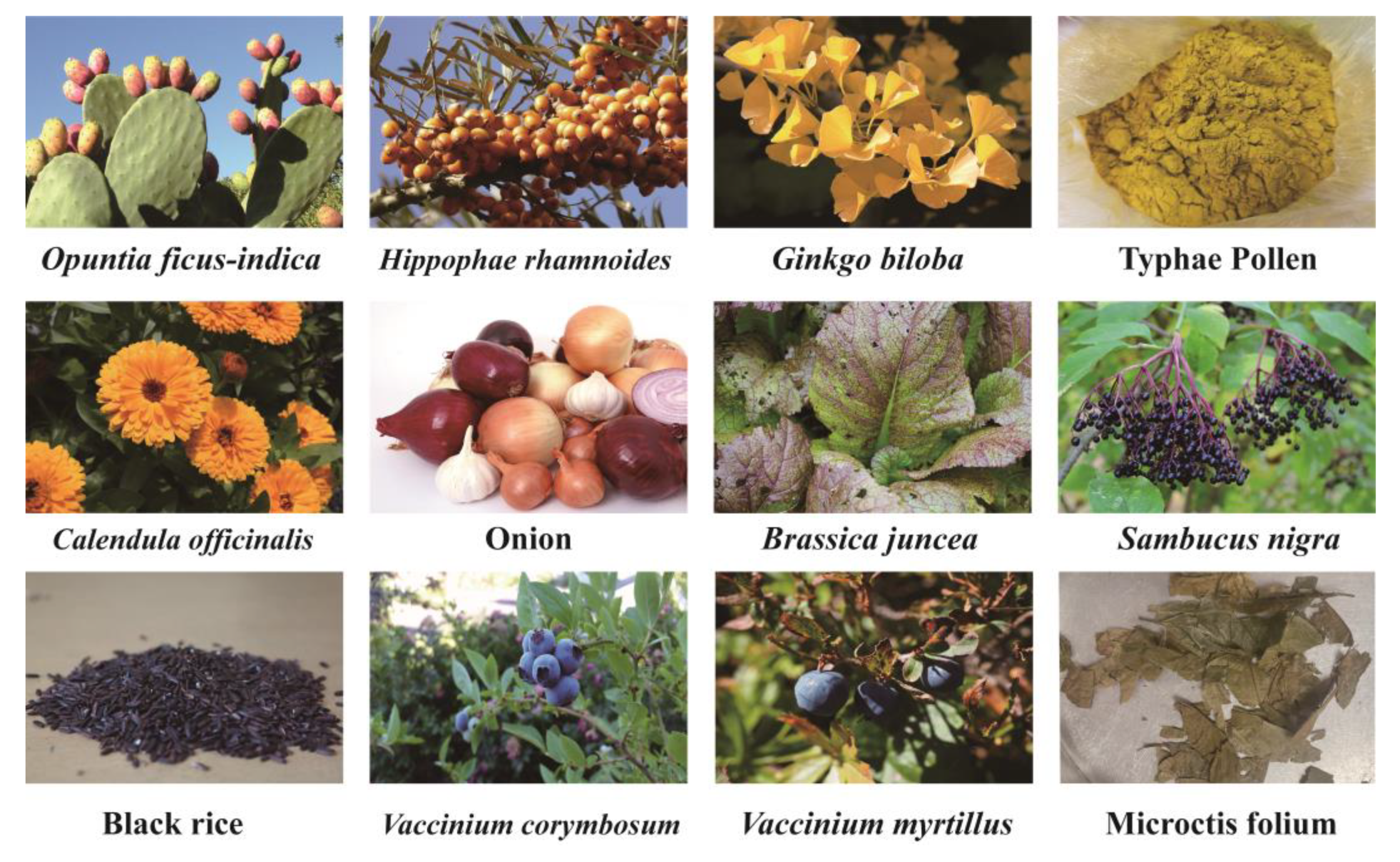
3. Sources of IGs
3.1. Opuntia ficus-indica
3.2. Hippophae rhamnoides
3.3. Ginkgo biloba
3.4. Pollen Typhae
3.5. Calendula officinalis
3.6. Other Sources
4. IG Identification and Quantification Methods
4.1. Spectral Techniques and Mass Spectrometry
4.1.1. UV
4.1.2. IR
4.1.3. NMR
4.1.4. MS
4.2. Chromatographic Techniques
4.2.1. TLC
4.2.2. HPLC and UPLC
4.2.3. HSCCC
5. The Health-Promoting Effects of IGs
5.1. Antioxidant Activity

| Isorhamnetin Glycosides | Study Model | Method/Assay | Conclusion | Ref. |
|---|---|---|---|---|
| Isorhamnetin-3-O-glucoside (4), Narcissin (24) | / | DPPH, ONOO- | Showed potent antioxidant activity, with IC50 values of 11.76 and 9.01 μM in DPPH assay, and 3.34 and 2.56 μM in the ONOO- assay. | [11] |
| Brassicin (1) | / | DPPH, ABTS | Showed radical-scavenging activity of DPPH radical and peroxynitrite, with IC50 values of 13.3 and 2.07 μM. | [13] |
| Brassicin (1) | / | DPPH, peroxynitrite | Showed radical-scavenging activity of DPPH radical and peroxynitrite, with IC50 values of 13.3 and 2.07μM. | [122] |
| Narcissin (24); isorhamnetin, 3,4′-diglucoside (17) | LPS-induced Raw264.7 mouse macrophage cells | NO | Had an inhibitory effect on the production of NO induced by LPS. | [137] |
| Isorhamnetin-3-O-glucoside (4), 3-O-galactoside (8) | β-carotene- linoleic acid | DPPH, ABTS, CUPRAC | Act as free radical scavengers and chain-breaking antioxidants of DPPH, with IC50 values of 4.84 and 4.51 μM. | [127] |
| Isorhamnetin 3-O-galactoside (8) | DPPH | Showed high antioxidant activity compared to Trolox (standard antioxidant compound). | [128] | |
| Typhaneoside (45); isorhamnetin-3-O-neohesperidoside (15) | HUVECs treated with LPS | NO, MDA, SOD | Reduced levels of MDA, increased SOD activity and NO bioactivity. | [124] |
| Isorhamnetin 3-O-robinobioside (22) | K562 cell line induced by H2O2 | CAA | Inhibited oxidation (IC50 = 0.225 mg/mL) and genotoxicity (by 80.55% at 1000 μg/mL). | [131] |
| Isorhamnetin 3-O-robinoside (22); isorhamnetin 3-O-(2″,6″-O-α- dirhamnosyl)-β-galactoside (37) | / | DPPH | Effectively scavenged DPPH radicals, with IC50 values of 3.8 and 4.3 μM. | [123] |
| Isorhamnetin-3-O-glucoside (4) | / | DPPH, ABTS, FRAP | Highly correlated with DPPH, ABTS, and FRAP (r = 0.672, r = 0.660, r = 0.943, respectively). | [130] |
| Astragaloside (13), narcissin (24) | / | ABTS | Possessed antioxidant capacity, with IC50 values of 33.43 and 40.97 μg/mL. | [118] |
| Narcissin (24); isorhamnetin 3-O-glucoside (4) | / | DPPH | Showed pronounced antioxidant activity, with IC50 values of 165.62 and 177.91 μg/mL. | [65] |
| Narcissin (24); isorhamnetin-3-O-rutinoside-7-O-glucoside (47) | / | DPPH, ABTS | Showed obvious antioxidant activity. | [125] |
| Narcissin (24) | HepG2 cells | CAA | Showed significant in vitro antioxidant activity, with CAA value significantly correlated with narcissin (24) (R2 = 0.998). | [136] |
| IGs | H2O2-induced intestinal epithelial cells | ORAC | Able to counteract protein oxidation. | [132] |
| Isorhamnetin 3-O-neohesperidoside (15) | Hydroxyl radical-induced DNA damage pKS plasmid | MDA, DNA-strand scission assay | Transcriptions of several genes related to the antioxidant system (HMOX2 and TXNL) were upregulated. | [133] |
| Isorhamnetin 3-O-neohesperidoside (15) | / | ABTS, xanthine/xanthine oxidase | Was a potent inhibitor of xanthine oxidase (IC50 = 48.75 μg/mL) and superoxide anion scavengers (IC50 = 30 μg/mL). | [126] |
| Isorhamnetin 3-O-galactoside (8) | / | ABTS | Showed ABTS radical-scavenging activity (IC50 = 6 ± 0 μM). | [129] |
| Narcissin (24); isorhamnetin 3-O-glucoside (4) | Whole blood, neutrophils, or monocytes | ROS | Demonstrated potent inhibition of ROS production. | [134] |
5.2. Anti-Inflammatory Activity
5.3. Anti-Cancer Activity
5.4. Hepatoprotective Ability
5.5. Antidiabetic Activity
5.6. Anti-Obesity Activity
5.7. Antithrombotic Activity
5.8. Toxic Effects
6. Bioaccessibility of IGs
7. Marketed Products Related to IGs
7.1. Food and Functional Food Products Using Opuntia ficus-indica
7.2. Food and Functional Food Products of Hippophae rhamnoides
8. Conclusions and Prospects
Author Contributions
Funding
Institutional Review Board Statement
Informed Consent Statement
Data Availability Statement
Acknowledgments
Conflicts of Interest
Abbreviations
| 5-HMF | 5-hydroxymethylfurfural |
| ABTS | 2,2′-azino-bis(3-ethylbenzothiazoline-6-sulfonate) |
| ADH | alcohol dehydrogenase |
| AGEs | advanced glycation end products |
| ALDH | aldehyde dehydrogenase |
| ALT | alanine aminotransferase |
| Ara | l-arabinose |
| AST | aspartate aminotransferase |
| ATM | ataxia telangiectasia mutated gene |
| ATR | ATM and Rad3-related gene |
| BB | bee read |
| CCl4 | carbon tetrachloride |
| Chk1 | checkpoint kinase1 |
| Chk2 | checkpoint kinase2 |
| CAA | cellular antioxidant activity assay |
| COX-2 | cyclooxygenase-2 |
| CUPRAC | CUPric reducing antioxidant capacity |
| DAD | diode array detection |
| DPPH | 2,2-diphenyl-1-picrylhydrazil |
| DPP-IV | dipeptidyl peptidase-IV |
| DW | dry weight |
| ERK | extracellular regulated kinases |
| ESI | electrospray ionization |
| FRAP | ferric reducing antioxidant power |
| Gal | d-galactose |
| Glc | d-glucose |
| Glccur | d-glucuronic |
| HFD | high-fat diet |
| HHP | high hydrostatic pressure |
| HMGB1 | high-mobility-group protein 1 |
| HO-1 | heme oxygenase-1 |
| HPLC | high-performance liquid chromatography |
| HSCCC | high-speed counter-current chromatography |
| HSCs | hepatic stellate cells |
| IGs | isorhamnetin glycosides |
| IL-6 | interleukin-6 |
| IL-1β | interleukin-1β |
| iNOS | inducible nitric oxide synthase |
| IR | infrared spectroscopy |
| JNK | c-Jun N-terminal kinase |
| LPS | lipopolysaccharide |
| MAPK | mitogen-activated protein kinase |
| MDA | malondialdehyde |
| MEOS | microsomal ethanol oxidizing system |
| MS | mass spectrometry |
| NF-κB | nuclear factor kappa-B |
| NMR | nuclear magnetic resonance |
| Nrf2 | nuclear factor E2-related factor 2 |
| OFI-E | opuntia ficus-indica extract |
| ONOO(-) | peroxynitrite |
| ORAC | oxygen radical absorbance capacity |
| PAI-1 | plasminogen activator inhibitor type 1 |
| PBMC | human peripheral blood mononuclear cells |
| PSC | peroxyl radical-scavenging capacity |
| PTP1B | protein tyrosine phosphatase 1B |
| Rha | l-rhamnose |
| ROS | reactive oxygen species |
| Xyl | D-xylose |
| TGF-β | transforming growth factor-β |
| TLC | thin-layer chromatography |
| TNF-α | tumor necrosis factor |
| tPA | tissue-type plasminogen activator |
| UPLC | ultra-performance liquid chromatography |
| UV | ultraviolet radiation |
| α-SMA | alpha-smooth muscle actin |
References
- Monjotin, N.; Amiot, M.; Fleurentin, J.; Morel, J.; Raynal, S. Clinical Evidence of the Benefits of Phytonutrients in Human Healthcare. Nutrients 2022, 14, 1712. [Google Scholar] [CrossRef] [PubMed]
- Valente, I.; Cabrita, A.; Malushi, N.; Oliveira, H.; Papa, L.; Rodrigues, J.; Fonseca, A.; Maia, M. Unravelling the phytonutrients and antioxidant properties of European Vicia faba L. seeds. Food Res. Int. 2019, 116, 888–896. [Google Scholar] [CrossRef] [PubMed]
- Saraei, R.; Marofi, F.; Naimi, A.; Talebi, M.; Ghaebi, M.; Javan, N.; Salimi, O.; Hassanzadeh, A. Leukemia therapy by flavonoids: Future and involved mechanisms. J. Cell. Physiol. 2018, 234, 8203–8220. [Google Scholar] [CrossRef] [PubMed]
- Roche, A.; Ross, E.; Walsh, N.; O’Donnell, K.; Williams, A.; Klapp, M.; Fullard, N.; Edelstein, S. Representative literature on the phytonutrients category: Phenolic acids. Crit. Rev. Food Sci. Nutr. 2017, 57, 1089–1096. [Google Scholar] [CrossRef] [PubMed]
- Zhou, D.; Bai, Z.S.; Guo, T.T.; Li, J.Y.; Li, Y.W.; Hou, Y.; Chen, G.; Li, N. Dietary flavonoids and human top-ranked diseases: The perspective of in vivo bioactivity and bioavailability. Trends Food Sci. Technol. 2022, 120, 374–386. [Google Scholar] [CrossRef]
- Ross, J.A.; Kasum, C.M. Dietary flavonoids: Bioavailability, metabolic effects, and safety. Annu. Rev. Nutr. 2002, 22, 19–34. [Google Scholar] [CrossRef]
- Tao, H.; Li, L.; He, Y.; Zhang, X.; Zhao, Y.; Wang, Q.; Hong, G. Flavonoids in vegetables: Improvement of dietary flavonoids by metabolic engineering to promote health. Crit. Rev. Food Sci. Nutr. 2022. [Google Scholar] [CrossRef]
- Marilena, A.R.; César, R.-R.; Janet, G.-U.; Eduardo, C.C.E.; Sergio, S.-S. Bioaccessibility, Intestinal Permeability and Plasma Stability of Isorhamnetin Glycosides from Opuntia ficus-indica (L.). Int. J. Mol. Sci. 2017, 18, 1816. [Google Scholar]
- Wang, L.; Fan, X.; Jian, Y.; Dong, M.; Yang, Q.; Meng, D.; Fu, Y. A sensitive and selective multiple reaction monitoring mass spectrometry method for simultaneous quantification of flavonol glycoside, terpene lactones, and biflavonoids in Ginkgo biloba leaves. J. Pharm. Biomed. Anal. 2019, 170, 335–340. [Google Scholar] [CrossRef]
- Ma, X.; Laaksonen, O.; Zheng, J.; Yang, W.; Trépanier, M.; Kallio, H.; Yang, B. Flavonol glycosides in berries of two major subspecies of sea buckthorn (Hippophaë rhamnoides L.) and influence of growth sites. Food Chem. 2016, 200, 189–198. [Google Scholar] [CrossRef]
- Hyun, S.; Jung, Y.; Chung, H.; Jung, H.; Choi, J. Isorhamnetin glycosides with free radical and ONOO-scavenging activities from the stamens of Nelumbo nucifera. Arch. Pharmacal. Res. 2006, 29, 287–292. [Google Scholar] [CrossRef] [PubMed]
- Abdel Motaal, A.; Salem, H.; Almaghaslah, D.; Alsayari, A.; Bin Muhsinah, A.; Alfaifi, M.; Elbehairi, S.; Shati, A.; El-Askary, H. Flavonol Glycosides: In Vitro Inhibition of DPPIV, Aldose Reductase and Combating Oxidative Stress are Potential Mechanisms for Mediating the Antidiabetic Activity of Cleome droserifolia. Molecules 2020, 25, 5864. [Google Scholar] [CrossRef] [PubMed]
- Cho, J.; Song, N.; Nam, T.; Shrestha, S.; Park, H.; Lyu, H.; Kim, D.; Lee, G.; Woo, Y.; Jeong, T.; et al. Flavonoids from the grains of C1/R-S transgenic rice, the transgenic Oryza sativa spp. japonica, and their radical scavenging activities. J. Agric. Food Chem. 2013, 61, 10354–10359. [Google Scholar] [CrossRef] [PubMed]
- Ku, S.K.; Han, M.S.; Bae, J.S. Down-regulation of endothelial protein C receptor shedding by persicarin and isorhamnetin-3-O-galactoside. Thromb. Res. 2013, 132, e58–e63. [Google Scholar] [CrossRef]
- Antunes-Ricardo, M.; Hernández-Reyes, A.; Uscanga-Palomeque, A.C.; Rodríguez-Padilla, C.; Martínez-Torres, A.C.; Gutiérrez-Uribe, J.A. Isorhamnetin glycoside isolated from Opuntia ficus-indica (L.) MilI induces apoptosis in human colon cancer cells through mitochondrial damage. Chem. Biol. Interact. 2019, 310, 108734. [Google Scholar] [CrossRef]
- Kim, D.W.; Cho, H.I.; Kim, K.M.; Kim, S.J.; Choi, J.S.; Kim, Y.S.; Lee, S.M. Isorhamnetin-3-O-galactoside Protects against CCl4-Induced Hepatic Injury in Mice. Biomol. Ther. 2012, 20, 406–412. [Google Scholar] [CrossRef]
- Hussain, H.; Green, I.; Abbas, G.; Adekenov, S.; Hussain, W.; Ali, I. Protein tyrosine phosphatase 1B (PTP1B) inhibitors as potential anti-diabetes agents: Patent review (2015–2018). Expert Opin. Ther. Pat. 2019, 29, 689–702. [Google Scholar] [CrossRef] [PubMed]
- Barba, F.J.; Garcia, C.; Fessard, A.; Munekata, P.E.S.; Lorenzo, J.M.; Aboudia, A.; Ouadia, A.; Remize, F. Opuntia Ficus Indica Edible Parts: A Food and Nutritional Security Perspective. Food Rev. Int. 2022, 38, 930–952. [Google Scholar] [CrossRef]
- Wang, K.; Xu, Z.; Liao, X. Bioactive compounds, health benefits and functional food products of sea buckthorn: A review. Crit. Rev. Food Sci. Nutr. 2022, 62, 6761–6782. [Google Scholar] [CrossRef]
- Ciesarova, Z.; Murkovic, M.; Cejpek, K.; Kreps, F.; Tobolkova, B.; Koplik, R.; Belajova, E.; Kukurova, K.; Dasko, L.; Panovska, Z.; et al. Why is sea buckthorn (Hippophae rhamnoides L.) so exceptional? A review. Food Res. Int. 2020, 133, 109170. [Google Scholar] [CrossRef]
- Yang, M.C.; Choi, S.Z.; Lee, S.O.; Chung, A.K.; Nam, J.H.; Lee, K.H.; Lee, K.R. Flavonoid constituents and their antioxidant activity of Laportea bulbifera Weddell. Saengyak Hakhoechi 2003, 34, 18–24. [Google Scholar]
- Jia, Z.; Zhu, G.; Wang, J. Flavonoid constituents of the seeds of Nitraria tangutorum Bolor. Lanzhou Daxue Xuebao Ziran Kexueban 1991, 27, 102. [Google Scholar]
- Park, Y.-K.; Lee, C.Y. Identification of Isorhamnetin 4’-Glucoside in Onions. J. Agric. Food Chem. 1996, 44, 34. [Google Scholar] [CrossRef]
- Fattorusso, E.; Iorizzi, M.; Lanzotti, V.; Taglialatela-Scafati, O. Chemical composition of shallot (Allium ascalonicum Hort.). J. Agric. Food Chem. 2002, 50, 5686–5690. [Google Scholar] [CrossRef]
- Kassem, M.E.S.; Afifi, M.S.; Marzouk, M.M.; Mostafa, M.A. Two new flavonol glycosides and biological activities of Diplotaxis harra (Forssk.) Boiss. Nat. Prod. Res. 2013, 27, 2272–2280. [Google Scholar] [CrossRef] [PubMed]
- Aquino, R.; Behar, I.; D’Agostino, M.; De Simone, F.; Schettino, O.; Pizza, C. Phytochemical investigation on Mercurialis annua. Biochem. Syst. Ecol. 1987, 15, 667. [Google Scholar] [CrossRef]
- Bechlem, H.; Mencherini, T.; Bouheroum, M.; Benayache, S.; Cotugno, R.; Braca, A.; De Tommasi, N. New Constituents from Gymnocarpos decander. Planta Med. 2017, 83, 1200–1206. [Google Scholar] [CrossRef]
- Batyuk, V.S.; Vasil’chenko, E.A.; Kovaleva, S.N. Flavonoids of Solidago virgaurea L. and S. canadensis L. and their pharmacological properties. Rastit. Resur. 1988, 24, 92. [Google Scholar]
- Litvinenko, V.I.; Bubenchikova, V.N. Phytochemical study of Centaurea cyanus. Chem. Nat. Compd. 1988, 792, 672–674. [Google Scholar] [CrossRef]
- Oksuz, S.; Putun, E. Flavonoids of Centaurea kotschyi var. kotschyi. Doga: Kim. Ser. 1987, 11, 66–71. [Google Scholar]
- Singh, K.N.; Pandey, V.B. Isorhamnetin 7-glucoside from Cnicus wallichi. Phytochemistry 1986, 25, 2683. [Google Scholar] [CrossRef]
- Butayarov, A.V.; Batirov, E.K.; Tadzhibaev, M.M.; Melibaev, S.; Malikov, V.M. Flavonoids from aerial parts of Russowia sogdiana. Chem. Nat. Compd. 1993, 29, 807–808. [Google Scholar] [CrossRef]
- Abdala, L.R. Flavonoids of the aerial parts from Tagetes lucida (Asteraceae). Biochem. Syst. Ecol. 1999, 27, 753–754. [Google Scholar] [CrossRef]
- He, A.; Wang, M. Flavonoids from stringy stonecrop (Sedum sarmentosum). Zhongcaoyao 1997, 28, 517–522. [Google Scholar]
- Grace, M.H.; Mohamed, T.K.; Khattab, A.M. Flavonoids of Carduncellus eriocephalus. Egypt. J. Pharm. Sci. 1999, 39, 409–416. [Google Scholar]
- Zhang, Z.-x.; Zhang, J.; Luo, J.-q.; Zhang, T.-h. Chemical constituents of the seeds of Atriplex centralasiatica. Shenyang Yaoke Daxue Xuebao 2008, 25, 708–710. [Google Scholar]
- Awaad, A.S.; Grace, M.H. Flavonoids and pharmacological activity of Vernonia galamensis ssp. galamensis var. petitiana (A. Rich) M. Gilbert. Egypt. J. Pharm. Sci. 2001, 40, 117–128. [Google Scholar]
- Krzeminski, K.; Krzeminska, K. Flavonoid heterosides in the herb of Raphanus raphanistrum L. Herba Pol. 1977, 23, 291. [Google Scholar]
- Zhang, S.; Shi, J.; Sun, Z.; Hu, C. Studies on chemical constituents from Caragana intermedia. Zhongyaocai 2006, 29, 19–21. [Google Scholar]
- Paskhov, D.; Marichkova, L. Flavonoids of Astragalus centralpinus and their effect on the smooth muscle of the gastrointestinal tract. Probl. Farm. 1983, 11, 36–42. [Google Scholar]
- Christensen, L.P.; Kaack, K.; Frette, X.C. Selection of elderberry (Sambucus nigra L.) genotypes best suited for the preparation of elderflower extracts rich in flavonoids and phenolic acids. Eur. Food Res. Technol. 2008, 227, 293–305. [Google Scholar] [CrossRef]
- Vidal-Ollivier, E.; Elias, R.; Faure, F.; Babadjamian, A.; Crespin, F.; Balansard, G.; Boudon, G. Flavonol glycosides from Calendula officinalis flowers. Planta Med. 1989, 55, 73. [Google Scholar] [CrossRef]
- Merfort, I.; Wendisch, D. Flavonoid glucuronides from the flowers of Arnica montana. Planta Med. 1988, 54, 247. [Google Scholar] [CrossRef] [PubMed]
- Kim, S.Y.; Park, J.Y.; Park, P.S.; Bang, S.H.; Lee, K.M.; Lee, Y.R.; Jang, Y.H.; Kim, M.J.; Chun, W.; Heo, M.Y.; et al. Flavonoid glycosides as acetylcholinesterase inhibitors from the whole plants of Persicaria thunbergii. Nat. Prod. Sci. 2014, 20, 191–195. [Google Scholar]
- Mezache, N.; Derbre, S.; Akkal, S.; Laouer, H.; Seraphin, D.; Richomme, P. Fast counter current chromatography of n-butanolic fraction from Senecio giganteus (Asteraceae). Nat. Prod. Commun. 2009, 4, 1357–1362. [Google Scholar] [CrossRef]
- Granica, S.; Czerwinska, M.E.; Zyzynska-Granica, B.; Kiss, A.K. Antioxidant and anti-inflammatory flavonol glucuronides from Polygonum aviculare L. Fitoterapia 2013, 91, 180–188. [Google Scholar] [CrossRef]
- Cheng, W.; Sui, C.; Yuan, J.; Zhang, H. Flavonoids of argun groundsel (Senecio argunensis). Zhongcaoyao 1999, 30, 727–729. [Google Scholar]
- Zaki, A.A.; Xu, X.; Wang, Y.; Shie, P.-H.; Qiu, L. A new anti-inflammatory flavonoid glycoside from Tetraena aegyptia. Nat. Prod. Res. 2021, 35, 1985–1990. [Google Scholar] [CrossRef] [PubMed]
- Cheng, J.; Wu, J.; Azi, g.; Li, R.; Lin, G. Chemical constituents of Aertaihuanqi (Astragalus altaicus). Zhongcaoyao 1994, 25, 563. [Google Scholar]
- Saleh, N.A.M.; Mansour, R.M.A.; Markham, K.R. An acylated isorhamnetin glycoside from Aerva javanica. Phytochemistry 1990, 29, 1344. [Google Scholar] [CrossRef]
- Jia, S.; Liu, Y.; Ma, C.; Yang, S.; Zhou, H.; Zhao, D.; Liu, D.; Li, S. Flavonoid constituents of the pollen of Typha angustfolia L. (Puhuang). Yaoxue Xuebao 1986, 21, 441. [Google Scholar]
- Olszewska, M.A.; Kwapisz, A. Metabolite profiling and antioxidant activity of Prunus padus L. flowers and leaves. Nat. Prod. Res. 2011, 25, 1115–1131. [Google Scholar] [CrossRef] [PubMed]
- Wang, S.; Shi, P.; Qu, L.; Ruan, J.; Yang, S.; Yu, H.; Zhang, Y.; Wang, T. Bioactive constituents obtained from the seeds of Lepidium apetalum willd. Molecules 2017, 22, 540. [Google Scholar] [CrossRef] [PubMed]
- Hoerhammer, L.; Wagner, H.; Kraemer, H.; Farkas, L. Isorhamnetin glycosides. II. Isolation and composition of new glycosides from Brassica napus and Sinapis arvensis. Chem. Ber. 1967, 100, 2301. [Google Scholar] [CrossRef]
- Halim, A.F.; Saad, H.-E.A.; Hashish, N.E. Flavonol glycosides from Nitraria retusa. Phytochemistry 1995, 40, 349. [Google Scholar] [CrossRef] [PubMed]
- Tang, Y.; Lou, F.; Wang, J.; Li, Y.; Zhuang, S. Coumaroyl flavonol glycosides from the leaves of Ginkgo biloba. Phytochemistry 2001, 58, 1251–1256. [Google Scholar] [CrossRef]
- Wang, D.-M.; Pu, W.-J.; Wang, Y.-H.; Zhang, Y.-J.; Wang, S.-S. A new isorhamnetin glycoside and other phenolic compounds from Callianthemum taipaicum. Molecules 2012, 17, 4595–4603. [Google Scholar] [CrossRef]
- Schoensiegel, I.; Egger, K. Flavonol glycosides in the petals of Narcissus pseudonarcissus. Z. Naturforsch. B 1969, 24, 1215. [Google Scholar] [CrossRef]
- Zhang, Z.-l.; Cai, M.-t.; Zuo, Y.-m.; Wang, Y.-y. Studies on chemical constituents in fruits of Trillium tschonoskii Maxim. Shizhen Guoyi Guoyao 2014, 25, 541–543. [Google Scholar] [CrossRef]
- Yoshitama, K.; Shida, Y.; Oyamada, T.; Takasaki, N.; Yahara, S. "Studies of the flavonoids of the genus Trillium". 3. Flavonol glycosides in the leaves of Trillium apetalon Makino and T. kamtschaticum Pallas. J. Plant Res. 1997, 110, 443–448. [Google Scholar] [CrossRef]
- Rodriguez, E.; Shen, M.C.; Mabry, T.J.; Dominguez, X.A. Isorhamnetin 3-0-glucoside 7-0-arabinoside from Eschscholzia mexicana. Phytochemistry 1973, 12, 2069. [Google Scholar] [CrossRef]
- Li, H.; Kim, U.H.; Yoon, J.H.; Ji, H.S.; Park, H.M.; Park, H.Y.; Jeong, T.S. Suppression of Hyperglycemia and Hepatic Steatosis by Black-Soybean-Leaf Extract via Enhanced Adiponectin-Receptor Signaling and AMPK Activation. J. Agric. Food Chem. 2019, 67, 90–101. [Google Scholar] [CrossRef] [PubMed]
- Kijima, H.; Ide, T.; Otsuka, H.; Takeda, Y. Alangiflavoside, a new flavonol glycoside from the leaves of Alangium premnifolium. J. Nat. Prod. 1995, 58, 1753. [Google Scholar] [CrossRef]
- Yasukawa, K.; Sekine, H.; Takido, M. Studies of the constituents of genus Lysimachia. Part 4. Two flavonol glycosides from Lysimachia fortunei. Phytochemistry 1989, 28, 2215. [Google Scholar] [CrossRef]
- Liu, H.; Mou, Y.; Zhao, J.; Wang, J.; Zhou, L.; Wang, M.; Wang, D.; Han, J.; Yu, Z.; Yang, F. Flavonoids from Halostachys caspica and their antimicrobial and antioxidant activities. Molecules 2010, 15, 7933–7945. [Google Scholar] [CrossRef]
- Kaouadji, M.; Doucoure, A.; Mariotte, A.M.; Chulia, A.J.; Thomasson, F. Flavonol triglycosides from Blackstonia perfoliata. Phytochemistry 1990, 29, 1283. [Google Scholar] [CrossRef]
- El-Sayed, N.H.; Abu Dooh, A.M.; El-Khrisy, E.A.M.; Mabry, T.J. Flavonoids of Cassia italica. Phytochemistry 1992, 31, 2187. [Google Scholar] [CrossRef]
- Trineeva, O.V.; Perova, I.B.; Slivkin, A.I.; Eller, K.I. Study the composition of flavonoids fruits of sea buckthorn. Sorbtsionnye Khromatogr. Protsessy 2017, 17, 87–93. [Google Scholar]
- Skalski, B.; Lis, B.; Pecio, L.; Kontek, B.; Olas, B.; Zuchowski, J.; Stochmal, A. Isorhamnetin and its new derivatives isolated from sea buckthorn berries prevent H(2)O(2)/Fe—Induced oxidative stress and changes in hemostasis. Food Chem. Toxicol. 2019, 125, 614–620. [Google Scholar] [CrossRef]
- Rodríguez-Rodríguez, C.; Torres, N.; Gutiérrez-Uribe, J.; Noriega, L.; Torre-Villalvazo, I.; Leal-Díaz, A.; Antunes-Ricardo, M.; Márquez-Mota, C.; Ordaz, G.; Chavez-Santoscoy, R.; et al. The effect of isorhamnetin glycosides extracted from Opuntia ficus-indica in a mouse model of diet induced obesity. Food Funct. 2015, 6, 805–815. [Google Scholar] [CrossRef]
- Yeddes, N.; Chérif, J.; Guyot, S.; Sotin, H.; Ayadi, M. Comparative Study of Antioxidant Power, Polyphenols, Flavonoids and Betacyanins of the Peel and Pulp of Three Tunisian Opuntia Forms. Antioxidants 2013, 2, 37–51. [Google Scholar] [CrossRef]
- FAO. Cactus (Opuntia spp.) as Forage; Food and Agriculture Organization: Rome, Italy, 2001. [Google Scholar]
- Santos-Zea, L.; Gutierrez-Uribe, J.A.; Serna-Saldivar, S.O. Comparative analyses of total phenols, antioxidant activity, and flavonol glycoside profile of cladode flours from different varieties of Opuntia spp. J. Agric. Food Chem. 2011, 59, 7054–7061. [Google Scholar] [CrossRef] [PubMed]
- Albergamo, A.; Potortí, A.; Di Bella, G.; Amor, N.; Lo Vecchio, G.; Nava, V.; Rando, R.; Ben Mansour, H.; Lo Turco, V. Chemical Characterization of Different Products from the Tunisian Opuntia ficus-indica (L.) Mill. Foods 2022, 11, 155. [Google Scholar] [CrossRef] [PubMed]
- Pundir, S.; Garg, P.; Dviwedi, A.; Ali, A.; Kapoor, V.; Kapoor, D.; Kulshrestha, S.; Lal, U.; Negi, P. Ethnomedicinal uses, phytochemistry and dermatological effects of Hippophae rhamnoides L.: A review. J. Ethnopharmacol. 2021, 266, 113434. [Google Scholar] [CrossRef] [PubMed]
- Olas, B.; Skalski, B. Preparations from Various Organs of Sea Buckthorn (Elaeagnus rhamnoides (L.) A. Nelson) as Important Regulators of Hemostasis and Their Role in the Treatment and Prevention of Cardiovascular Diseases. Nutrients 2022, 14, 991. [Google Scholar] [CrossRef]
- Pop, R.; Socaciu, C.; Pintea, A.; Buzoianu, A.; Sanders, M.; Gruppen, H.; Vincken, J. UHPLC/PDA-ESI/MS analysis of the main berry and leaf flavonol glycosides from different Carpathian Hippophaë rhamnoides L. varieties. Phytochem. Anal. PCA 2013, 24, 484–492. [Google Scholar] [CrossRef]
- Tkacz, K.; Wojdyło, A.; Turkiewicz, I.; Ferreres, F.; Moreno, D.; Nowicka, P. UPLC-PDA-Q/TOF-MS profiling of phenolic and carotenoid compounds and their influence on anticholinergic potential for AChE and BuChE inhibition and on-line antioxidant activity of selected Hippophaë rhamnoides L. cultivars. Food Chem. 2020, 309, 125766. [Google Scholar] [CrossRef]
- Fang, J.; Wang, Z.; Wang, P.; Wang, M. Extraction, structure and bioactivities of the polysaccharides from Ginkgo biloba: A review. Int. J. Biol. Macromol. 2020, 162, 1897–1905. [Google Scholar] [CrossRef]
- Gray, D.; Messer, D.; Porter, A.; Hefner, B.; Logan, D.; Harris, R.; Clark, A.; Algaier, J.; Overstreet, J.; Smith, C. Analysis of flavonol aglycones and terpenelactones in Ginkgo biloba extract: A comparison of high-performance thin-layer chromatography and column high-performance liquid chromatography. J. AOAC Int. 2007, 90, 1203–1209. [Google Scholar] [CrossRef]
- Mahadevan, S.; Park, Y. Multifaceted Therapeutic Benefits of Ginkgo biloba L.: Chemistry, Efficacy, Safety, and Uses. J. Food Sci. 2010, 73, R14–R19. [Google Scholar] [CrossRef]
- Eisvand, F.; Razavi, B.; Hosseinzadeh, H. The effects of Ginkgo biloba on metabolic syndrome: A review. Phytother. Res. PTR 2020, 34, 1798–1811. [Google Scholar] [CrossRef] [PubMed]
- Gao, M.; Ge, Z.; Deng, R.; Bao, B.; Yao, W.; Cao, Y.; Shan, M.; Cheng, F.; Yan, H.; Chen, P.; et al. Evaluation of VEGF mediated pro-angiogenic and hemostatic effects and chemical marker investigation for Typhae Pollen and its processed product. J. Ethnopharmacol. 2021, 268, 113591. [Google Scholar] [CrossRef] [PubMed]
- National Health Commission of the People’s Republic of China. Notice of the Ministry of Health on Further Standardizing the Management of Health Food Raw Materials. 2002. Available online: http://www.nhc.gov.cn/wjw/gfxwj/201304/e33435ce0d894051b15490aa3219cdc4.shtml (accessed on 15 September 2022).
- Zeng, G.; Wu, Z.; Cao, W.; Wang, Y.; Deng, X.; Zhou, Y. Identification of anti-nociceptive constituents from the pollen of Typha angustifolia L. using effect-directed fractionation. Nat. Prod. Res. 2020, 34, 1041–1045. [Google Scholar] [CrossRef]
- Cao, S.; Ni, B.; Feng, L.; Yin, X.; Dou, H.; Fu, J.; Lin, L.; Ni, J. Simultaneous Determination of Typhaneoside and Isorhamnetin-3-O-Neohesperidoside in Rats After Oral Administration of Pollen Typhae Extract by UPLC-MS/MS. J. Chromatogr. Sci. 2015, 53, 866–871. [Google Scholar] [CrossRef]
- Zhao, J.; Zhang, C.; Xu, D.; Huang, G.; Xu, Y.; Wang, Z.; Fang, S.; Chen, Y.; Gu, Y. The antiatherogenic effects of components isolated from pollen typhae. Thromb. Res. 1990, 57, 957–966. [Google Scholar] [CrossRef] [PubMed]
- Wang, X.; Li, J.; Yang, X.; Gao, X.; Wang, H.; Chang, Y. A rapid and efficient extraction method based on industrial MCM-41-miniaturized matrix solid-phase dispersion extraction with response surface methodology for simultaneous quantification of six flavonoids in Pollen typhae by ultra-high-performance liquid chromatography. J. Sep. Sci. 2019, 42, 2426–2434. [Google Scholar] [CrossRef] [PubMed]
- Miguel, M.; Barros, L.; Pereira, C.; Calhelha, R.; Garcia, P.; Castro, M.; Santos-Buelga, C.; Ferreira, I. Chemical characterization and bioactive properties of two aromatic plants: Calendula officinalis L. (flowers) and Mentha cervina L. (leaves). Food Funct. 2016, 7, 2223–2232. [Google Scholar] [CrossRef]
- Dinda, M.; Dasgupta, U.; Singh, N.; Bhattacharyya, D.; Karmakar, P. PI3K-mediated proliferation of fibroblasts by Calendula officinalis tincture: Implication in wound healing. Phytother. Res. PTR 2015, 29, 607–616. [Google Scholar] [CrossRef]
- Olennikov, D.N.; Kashchenko, N.I. Componential Profile and Amylase Inhibiting Activity of Phenolic Compounds from Calendula officinalis L. Leaves. Sci. World J. 2014, 2014, 654193. [Google Scholar] [CrossRef]
- Olennikov, D.N.; Kashchenko, N.I. New Isorhamnetin Glycosides and other Phenolic Compounds from Calendula officinalis. Chem. Nat. Compd. 2013, 49, 833–840. [Google Scholar] [CrossRef]
- Bezákova, L.; Masterová, I.; Paulíková, I.; Psenák, M. Inhibitory activity of isorhamnetin glycosides from Calendula officinalis L. on the activity of lipoxygenase. Pharmazie 1996, 51, 126–127. [Google Scholar] [PubMed]
- Sriseadka, T.; Wongpornchai, S.; Rayanakorn, M. Quantification of Flavonoids in Black Rice by Liquid Chromatography-Negative Electrospray Ionization Tandem Mass Spectrometry. J. Agric. Food Chem. 2012, 60, 11723–11732. [Google Scholar] [CrossRef]
- Yokozawa, T.; Kim, H.; Cho, E.; Choi, J.; Chung, H. Antioxidant effects of isorhamnetin 3,7-di-O-beta-d-glucopyranoside isolated from mustard leaf (Brassica juncea) in rats with streptozotocin-induced diabetes. J. Agric. Food Chem. 2002, 50, 5490–5495. [Google Scholar] [CrossRef] [PubMed]
- Mikulic-Petkovsek, M.; Slatnar, A.; Stampar, F.; Veberic, R. HPLC-MSn identification and quantification of flavonol glycosides in 28 wild and cultivated berry species. Food Chem. 2012, 135, 2138–2146. [Google Scholar] [CrossRef] [PubMed]
- Pbpa, B.; Mtba, D.; Ga, C.; Htg, A.; Hg, C. Phenolics profiling by HPLC-DAD-ESI-MS n aided by principal component analysis to classify Rabbiteye and Highbush blueberries. Food Chem. 2020, 340, 127958. [Google Scholar]
- Chen, Y.G.; Li, P.; Li, P.; Yan, R.; Zhang, X.Q.; Wang, Y.; Zhang, X.T.; Ye, W.C.; Zhang, Q.W. α-Glucosidase Inhibitory Effect and Simultaneous Quantification of Three Major Flavonoid Glycosides in Microctis folium. Molecules 2013, 18, 4221–4232. [Google Scholar] [CrossRef]
- Jiang, J.Y.; Yang-Xue, L.I.; Su-Mei, L.I.; Ai-Li, X.U. Simultaneous Determination of Six Flavonoids in Extract of Microctis Folium by HPLC. Chin. J. Exp. Tradit. Med. Formulae 2016, 36, 589–593. [Google Scholar]
- Tedesco, I.; Carbone, V.; Spagnuolo, C.; Minasi, P.; Russo, G.L. Identification and Quantification of Flavonoids from Two Southern Italian Cultivars of Allium cepa L., Tropea (Red Onion) and Montoro (Copper Onion), and Their Capacity to Protect Human Erythrocytes from Oxidative Stress. J. Agric. Food Chem. 2015, 63, 5229–5238. [Google Scholar] [CrossRef]
- Price, K.R.; Rhodes, M.J.C. Analysis of the Major Flavonol Glycosides Present in Four Varieties of Onion (Allium cepa) and Changes in Composition Resulting from Autolysis. J. Sci. Food Agric. 1997, 74, 331–339. [Google Scholar] [CrossRef]
- Aalar, H.G. Elderberry (Sambucus nigra L.). In Nonvitamin Nonmineral Nutritional Supplements; Seyed, M.N., Ana, S.S., Eds.; Academic Press: Cambridge, MA, USA, 2019; pp. 211–215. [Google Scholar]
- Bhattacharya, S.; Christensen, K.B.; Olsen, L.C.B.; Christensen, L.P.; Grevsen, K.; FæRgeman, N.J.; Kristiansen, K.; Young, J.F.; Oksbjerg, N. Bioactive Components from Flowers of Sambucus nigra L. Increase Glucose Uptake in Primary Porcine Myotube Cultures and Reduce Fat Accumulation in Caenorhabditis elegans. J. Agric. Food Chem. 2013, 61, 11033–11040. [Google Scholar] [CrossRef]
- Mabry, T.J.; Markham, K.R.; Thomas, M.B. The Ultraviolet Spectra of Isoflavones, Flavanones and Dihydroflavonols. In The Systematic Identification of Flavonoids; Mabry, T.J., Markham, K.R., Thomas, M.B., Eds.; Springer: Berlin/Heidelberg, Germany, 1970; pp. 165–226. [Google Scholar]
- Mabry, T.J.; Markham, K.R.; Thomas, M.B. The Systematic Identification of Flavonoids; Springer: Berlin/Heidelberg, Germany, 1970. [Google Scholar]
- Karl, C.; Pedersen, P.A.; Schwarz, C. A new flavonoacetylglucoside from Salix viminalis. Phytochemistry 1977, 16, 1117. [Google Scholar] [CrossRef]
- Markham, K.R. 6—Flavones, Flavonols and their Glycosides. In Methods in Plant Biochemistry; Harborne, J.B., Ed.; Academic Press: Cambridge, MA, USA, 1989; Volume 1, pp. 197–235. [Google Scholar]
- Schieber, A.; Keller, P.; Streker, P.; Klaiber, I.; Carle, R. Detection of isorhamnetin glycosides in extracts of apples (Malus domestica cv. "Brettacher") by HPLC-PDA and HPLC-APCI-MS/MS. Phytochem. Anal. 2002, 13, 87–94. [Google Scholar] [CrossRef] [PubMed]
- Mata, A.; Ferreira, J.; Semedo, C.; Serra, T.; Duarte, C.; Bronze, M. Contribution to the characterization of Opuntia spp. juices by LC-DAD-ESI-MS/MS. Food Chem. 2016, 210, 558–565. [Google Scholar] [CrossRef] [PubMed]
- Frison-Norrie, S.; Sporns, P. Identification and quantification of flavonol glycosides in almond seedcoats using MALDI-TOF MS. J. Agric. Food Chem. 2002, 50, 2782–2787. [Google Scholar] [CrossRef]
- Bartnik, M.; Glowniak, K.; Gromek, A. TLC and HPLC analysis of the flavonoid glycosides in the aerial parts of Peucedanum tauricum Bieb. JPC-J. Planar Chromatogr. Mod. 2007, 20, 127–130. [Google Scholar] [CrossRef]
- Slimestad, R.; Andersen, Ø.M.; Francis, G.W.; Marston, A.; Hostettmann, K. Syringetin 3-O-(6″-acetyl)-β-glucopyranoside and other flavonols from needles of norway spruce, Picea abies. Phytochemistry 1995, 40, 1537–1542. [Google Scholar] [CrossRef]
- Lee, J.; Mitchell, A.E. Quercetin and isorhamnetin glycosides in onion (Allium cepa L.): Varietal comparison, physical distribution, coproduct evaluation, and long-term storage stability. J. Agric. Food Chem. 2011, 59, 857–863. [Google Scholar] [CrossRef]
- Arimboor, R.; Arumughan, C. HPLC-DAD-MS/MS profiling of antioxidant flavonoid glycosides in sea buckthorn (Hippophae rhamnoides L.) seeds. Int. J. Food Sci. Nutr. 2012, 63, 730–738. [Google Scholar] [CrossRef]
- Rodriguez-Aller, M.; Gurny, R.; Veuthey, J.; Guillarme, D. Coupling ultra high-pressure liquid chromatography with mass spectrometry: Constraints and possible applications. J. Chromatogr. A 2013, 1292, 2–18. [Google Scholar] [CrossRef]
- Tian, Y.; Liimatainen, J.; Alanne, A.; Lindstedt, A.; Liu, P.; Sinkkonen, J.; Kallio, H.; Yang, B. Phenolic compounds extracted by acidic aqueous ethanol from berries and leaves of different berry plants. Food Chem. 2017, 220, 266–281. [Google Scholar] [CrossRef]
- Zhang, S.; Cui, Y.; Li, L.; Li, Y.; Zhou, P.; Luo, L.; Sun, B. Preparative HSCCC isolation of phloroglucinolysis products from grape seed polymeric proanthocyanidins as new powerful antioxidants. Food Chem. 2015, 188, 422–429. [Google Scholar] [CrossRef] [PubMed]
- Yang, C.; Yang, Y.; Aisa, H.A.; Xin, X.; Ma, H.; Yili, A.; Zhao, Y. Bioassay-guided isolation of antioxidants from Astragalus altaicus by combination of chromatographic techniques. J. Sep. Sci. 2012, 35, 977–983. [Google Scholar] [CrossRef] [PubMed]
- Lei, W.; Wei, X.; Ju-Wu, H.; Zhen, G.; Jian-Guo, X.; Chuan-Ling, S.; Young-Soo, B.; Gang, X. Purification of Four Flavonoid Glycosides from Lotus (Nelumbo nucifera Gaertn) plumule by Macroporous Resin Combined with HSCCC. J. Chromatogr. Sci. 2018, 56, 108–114. [Google Scholar]
- Quispe, C.; Viveros-Valdez, E.; Yarleque, J.A.; Arones, M.R.; Paniagua, J.C.; Schmeda-Hirschmann, G. High speed centrifugal countercurrent chromatography (hsccc) isolation and identification by lc-ms n analysis of the polar phenolics from Vasconcellea quercifolia. J. Chil. Chem. Soc. 2017, 58, 1830–1835. [Google Scholar] [CrossRef]
- Speisky, H.; Shahidi, F.; Costa de Camargo, A.; Fuentes, J. Revisiting the Oxidation of Flavonoids: Loss, Conservation or Enhancement of Their Antioxidant Properties. Antioxidants 2022, 11, 133. [Google Scholar] [CrossRef] [PubMed]
- Choi, J.; Jung, M.; Park, H.; Chung, H.; Kang, S. Further isolation of peroxynitrite and 1,1-diphenyl-2-picrylhydrazyl radical scavenging isorhamnetin 7-O-glucoside from the leaves of Brassica juncea L. Arch. Pharmacal Res. 2002, 25, 625–627. [Google Scholar] [CrossRef] [PubMed]
- Hawas, U.; Abou El-Kassem, L.; Shaher, F.; Al-Farawati, R. In vitro inhibition of Hepatitis C virus protease and antioxidant by flavonoid glycosides from the Saudi costal plant. Nat. Prod. Res. 2019, 33, 3364–3371. [Google Scholar] [CrossRef]
- Chen, P.; Cao, Y.; Bao, B.; Zhang, L.; Ding, A. Antioxidant capacity of Typha angustifolia extracts and two active flavonoids. Pharm. Biol. 2017, 55, 1283–1288. [Google Scholar] [CrossRef]
- Qi, J.; Gui, X.; Chen, G. Research on active component extraction of nitraria sibirica pall. fruit and their antioxidant activity. Mod. Chin. Med. 2013, 15, 827–831. [Google Scholar] [CrossRef]
- Bouhlel, I.; Limem, I.; Skandrani, I.; Nefatti, A.; Ghedira, K.; Dijoux-Franca, M.; Leila, C. Assessment of isorhamnetin 3-O-neohesperidoside from Acacia salicina: Protective effects toward oxidation damage and genotoxicity induced by aflatoxin B1 and nifuroxazide. J. Appl. Toxicol. JAT 2010, 30, 551–558. [Google Scholar] [CrossRef]
- Demirkiran, O.; Sabudak, T.; Ozturk, M.; Topcu, G. Antioxidant and tyrosinase inhibitory activities of flavonoids from Trifolium nigrescens Subsp. petrisavi. J. Agric. Food Chem. 2013, 61, 12598–12603. [Google Scholar] [CrossRef] [PubMed]
- Hassan, R.; Tawfik, W.; Abou-Setta, L. The flavonoid constitunts of Leucaena leucocephala. Growing in Egypt, and their biological activity. Afr. J. Tradit. Complement. Altern. Med. AJTCAM 2014, 11, 67–72. [Google Scholar]
- Yuca, H.; Özbek, H.; Demirezer, L.; Kasil, H.G.; Güvenalp, Z. trans-Tiliroside: A potent α-glucosidase inhibitor from the leaves of Elaeagnus angustifolia L. Phytochemistry 2021, 188, 112795. [Google Scholar] [CrossRef]
- Li, Y.; Guo, S.; Zhu, Y.; Yan, H.; Qian, D.W.; Wang, H.Q.; Yu, J.Q.; Duan, J.A. Flowers of Astragalus membranaceus var. mongholicus as a Novel High Potential By-Product: Phytochemical Characterization and Antioxidant Activity. Molecules 2019, 24, 434. [Google Scholar] [CrossRef] [PubMed]
- Boubaker, J.; Ben Sghaier, M.; Skandrani, I.; Ghedira, K.; Chekir-Ghedira, L. Isorhamnetin 3-O-robinobioside from Nitraria retusa leaves enhance antioxidant and antigenotoxic activity in human chronic myelogenous leukemia cell line K562. BMC Complement. Altern. Med. 2012, 12, 135. [Google Scholar] [CrossRef] [PubMed]
- Abdallah, H.; Esmat, A. Antioxidant and anti-inflammatory activities of the major phenolics from Zygophyllum simplex L. J. Ethnopharmacol. 2017, 205, 51–56. [Google Scholar] [CrossRef]
- Bouhlel, I.; Skandrani, I.; Nefatti, A.; Valenti, K.; Ghedira, K.; Mariotte, A.M.; Hininger-Favier, I.; Laporte, F.; Dijoux-Franca, M.G.; Chekir-Ghedira, L. Antigenotoxic and antioxidant activities of isorhamnetin 3-O neohesperidoside from Acacia salicina. Drug Chem. Toxicol. 2009, 32, 258–267. [Google Scholar] [CrossRef]
- Yeskaliyeva, B.; Mesaik, M.; Abbaskhan, A.; Kulsoom, A.; Burasheva, G.; Abilov, Z.; Choudhary, M.; Atta-ur-Rahman. Bioactive flavonoids and saponins from Climacoptera obtusifolia. Phytochemistry 2006, 67, 2392–2397. [Google Scholar] [CrossRef]
- Matias, A.; Nunes, S.; Poejo, J.; Mecha, E.; Serra, A.; Madeira, P.; Bronze, M.; Duarte, C. Antioxidant and anti-inflammatory activity of a flavonoid-rich concentrate recovered from Opuntia ficus-indica juice. Food Funct. 2014, 5, 3269–3280. [Google Scholar] [CrossRef]
- Guo, R.; Guo, X.; Li, T.; Fu, X.; Liu, R. Comparative assessment of phytochemical profiles, antioxidant and antiproliferative activities of Sea buckthorn (Hippophaë rhamnoides L.) berries. Food Chem. 2017, 221, 997–1003. [Google Scholar] [CrossRef]
- Kang, Y.J.; Kim, H.Y.; Lee, C.; Park, S.Y. Nitric oxide inhibitory constituents from fruits of Opuntia humifusa. Nat. Prod. Sci. 2014, 20, 211–215. [Google Scholar]
- Andersson, U.; Yang, H.; Harris, H. Extracellular HMGB1 as a therapeutic target in inflammatory diseases? Expert Opin. Ther. Targets 2018, 22, 263–277. [Google Scholar] [CrossRef] [PubMed]
- Kim, T.; Ku, S.; Bae, J. Anti-inflammatory activities of isorhamnetin-3-O-galactoside against HMGB1-induced inflammatory responses in both HUVECs and CLP-induced septic mice. J. Cell. Biochem. 2013, 114, 336–345. [Google Scholar] [CrossRef] [PubMed]
- Yong, H.; Koh, M.; Moon, A. The p38 MAPK inhibitors for the treatment of inflammatory diseases and cancer. Expert Opin. Investig. Drugs 2009, 18, 1893–1905. [Google Scholar] [CrossRef]
- Park, J.; Kim, S.; Lee, H.; Kim, S.; Kwon, Y.; Chun, W. Isorhamnetin-3-O-Glucuronide Suppresses JNK and p38 Activation and Increases Heme-Oxygenase-1 in Lipopolysaccharide-Challenged RAW264.7 Cells. Drug Dev. Res. 2016, 77, 143–151. [Google Scholar] [CrossRef]
- Ahmed, A.F.; Wen, Z.-H.; Bakheit, A.H.; Basudan, O.A.; Ghabbour, H.A.; Al-Ahmari, A.; Feng, C.-W. A Major Diplotaxis harra-Derived Bioflavonoid Glycoside as a Protective Agent against Chemically Induced Neurotoxicity and Parkinson’s Models; In Silico Target Prediction; and Biphasic HPTLC-Based Quantification. Plants 2022, 11, 648. [Google Scholar] [CrossRef]
- Fu, Y.; Jia, Y.; Sun, Y.; Liu, X.; Yi, J.; Cai, S. Dietary Flavonoids Alleviate Inflammation and Vascular Endothelial Barrier Dysfunction Induced by Advanced Glycation End Products In Vitro. Nutrients 2022, 14, 1026. [Google Scholar] [CrossRef]
- Jin, H.; Ko, H.; Chowdhury, M.; Lee, D.; Woo, E. A new indole glycoside from the seeds of Raphanus sativus. Arch. Pharmacal Res. 2016, 39, 755–761. [Google Scholar] [CrossRef]
- Osman, S.; El Kashak, W.; Wink, M.; El Raey, M. New Isorhamnetin Derivatives from Salsola imbricata Forssk. Leaves with Distinct Anti-inflammatory Activity. Pharmacogn. Mag. 2016, 12, S47–S51. [Google Scholar] [CrossRef]
- Ameur, A.S.; Negab, I.; Zouzou, F.; Legseir, B. Anti-inflammatory and antispasmodic activities of isorhamnetin glycosides isolated from Opuntia ficus-indica (L.) mill. Flowers. Res. J. Pharm. Biol. Chem. Sci. 2016, 7, 432–437. [Google Scholar]
- Antunes-Ricardo, M.; Gutiérrez-Uribe, J.; Martínez-Vitela, C.; Serna-Saldívar, S. Topical anti-inflammatory effects of isorhamnetin glycosides isolated from Opuntia ficus-indica. BioMed Res. Int. 2015, 2015, 847320. [Google Scholar] [CrossRef] [PubMed]
- Ren, Q.-C.; Li, X.-H.; Li, Q.-Y.; Yang, H.-L.; Wang, H.-L.; Zhang, H.; Zhao, L.; Jiang-Yong, S.-L.; Meng, X.-L.; Zhang, Y.; et al. Total flavonoids from sea buckthorn ameliorates lipopolysaccharide/cig arette smoke-induced airway inflammation. Phytother. Res. PTR 2019, 33, 2102–2117. [Google Scholar] [CrossRef] [PubMed]
- Raffa, D.; Maggio, B.; Raimondi, M.; Plescia, F.; Daidone, G. Recent discoveries of anticancer flavonoids. Eur. J. Med. Chem. 2017, 142, 213–228. [Google Scholar] [CrossRef] [PubMed]
- Mohammed, M.; El-Sharkawy, E.R.; Matloub, A.A. Cytotoxic flavonoids from Diplotaxis harra (Forssk.) Boiss. growing in Sinai. J. Med. Plant Res. 2013, 520, 5099–5103. [Google Scholar]
- Tofighi, Z.; Asgharian, P.; Goodarzi, S.; Hadjiakhoondi, A.; Ostad, S.N.; Yassa, N. Potent cytotoxic flavonoids from Iranian Secur. Securidaca. Med. Chem. Res. 2014, 23, 1718–1724. [Google Scholar] [CrossRef]
- Tundis, R.; Loizzo, M.; Bonesi, M.; Menichini, F.; Statti, G.; Menichini, F. In vitro cytotoxic activity of Salsola oppositifolia Desf. (Amaranthaceae) in a panel of tumour cell lines. Z. Fur Naturforschung. C J. Biosci. 2008, 63, 347–354. [Google Scholar] [CrossRef]
- Liu, Y.; Xiao, Z.Y.; Liu, P.; Huang, J.; Algradi, A.M.; Pan, J.; Guan, W.; Zhou, Y.Y.; Yang, B.Y.; Kuang, H.X. New flavonoids from the aerial part of Bupleurum chinense DC. Fitoterapia 2020, 147, 104739. [Google Scholar] [CrossRef]
- Pfeffer, C.; Singh, A. Apoptosis: A Target for Anticancer Therapy. Int. J. Mol. Sci. 2018, 19, 448. [Google Scholar] [CrossRef]
- Yao, N.; Li, Y.; Lei, Y.; Hu, N.; Chen, W.; Yao, Z.; Yu, M.; Liu, J.; Ye, W.; Zhang, D. A piperazidine derivative of 23-hydroxy betulinic acid induces a mitochondria-derived ROS burst to trigger apoptotic cell death in hepatocellular carcinoma cells. J. Exp. Clin. Cancer Res. CR 2016, 35, 192. [Google Scholar] [CrossRef]
- Rademaker, G.; Boumahd, Y.; Peiffer, R.; Anania, S.; Wissocq, T.; Liégeois, M.; Luis, G.; Sounni, N.; Agirman, F.; Maloujahmoum, N.; et al. Myoferlin targeting triggers mitophagy and primes ferroptosis in pancreatic cancer cells. Redox Biol. 2022, 53, 102324. [Google Scholar] [CrossRef]
- Mendes, S.; Sá, R.; Magalhães, M.; Marques, F.; Sousa, M.; Silva, E. The Role of ROS as a Double-Edged Sword in (In)Fertility: The Impact of Cancer Treatment. Cancers 2022, 14, 1585. [Google Scholar] [CrossRef] [PubMed]
- Wu, Q.; Kroon, P.A.; Shao, H.; Needs, P.W.; Yang, X. Differential Effects of Quercetin and Two of Its Derivatives, Isorhamnetin and Isorhamnetin-3-glucuronide, in Inhibiting the Proliferation of Human Breast-Cancer MCF-7 Cells. J. Agric. Food Chem. 2018, 66, 7181–7189. [Google Scholar] [CrossRef]
- Antunes-Ricardo, M.; Guardado-Félix, D.; Rocha-Pizaña, M.; Garza-Martínez, J.; Acevedo-Pacheco, L.; Gutiérrez-Uribe, J.; Villela-Castrejón, J.; López-Pacheco, F.; Serna-Saldívar, S. Opuntia ficus-indica Extract and Isorhamnetin-3-O-Glucosyl-Rhamnoside Diminish Tumor Growth of Colon Cancer Cells Xenografted in Immune-Suppressed Mice through the Activation of Apoptosis Intrinsic Pathway. Plant Foods Hum. Nutr. 2021, 76, 434–441. [Google Scholar] [CrossRef] [PubMed]
- Antunes-Ricardo, M.; Moreno-García, B.; Gutiérrez-Uribe, J.; Aráiz-Hernández, D.; Alvarez, M.; Serna-Saldivar, S. Induction of apoptosis in colon cancer cells treated with isorhamnetin glycosides from Opuntia ficus-indica pads. Plant Foods Hum. Nutr. 2014, 69, 331–336. [Google Scholar] [CrossRef]
- Jihed, B.; Bhouri, W.; Ben Sghaier, M.; Bouhlel, I.; Kriffi, M.; Skandrani, I.; Dijoux, F.; Ghedira, K.; Chekir-Ghedira, L. Flavonoids products from Nitraria retusa leaves promote lymphoblastoid cells apoptosis. Nutr. Cancer 2012, 64, 1095–1102. [Google Scholar] [CrossRef] [PubMed]
- Serra, A.T.; Poejo, J.; Matias, A.A.; Bronze, M.R.; Duarte, C. Evaluation of Opuntia spp. derived products as antiproliferative agents in human colon cancer cell line (HT29). Food Res. Int. 2013, 54, 892–901. [Google Scholar] [CrossRef]
- Hoang Anh, N.; Tam, K.; Tuan, N.; Thien, D.; Quan, T.; Tam, N.; Bao, N.; Do, T.; Nga, N.; Thuy, T.; et al. Chemical constituents of Oldenlandia pinifolia and their antiproliferative activities. Nat. Prod. Res. 2019, 33, 796–802. [Google Scholar] [CrossRef] [PubMed]
- Boubaker, J.; Bhouri, W.; Ben Sghaier, M.; Ghedira, K.; Dijoux Franca, M.G.; Chekir-Ghedira, L. Ethyl acetate extract and its major constituent, isorhamnetin 3-O-rutinoside, from Nitraria retusa leaves, promote apoptosis of human myelogenous erythroleukaemia cells. Cell Prolif. 2011, 44, 453–461. [Google Scholar] [CrossRef] [PubMed]
- Kong, C.; Kim, Y.; Kim, M.; Park, J.; Kim, J.; Kim, S.; Lee, B.; Nam, T.; Seo, Y. Flavonoid glycosides isolated from Salicornia herbacea inhibit matrix metalloproteinase in HT1080 cells. Toxicol. Vitr. Int. J. Publ. Assoc. BIBRA 2008, 22, 1742–1748. [Google Scholar] [CrossRef]
- Nasri, I.; Chawech, R.; Girardi, C.; Mas, E.; Ferrand, A.; Vergnolle, N.; Fabre, N.; Mezghani-Jarraya, R.; Racaud-Sultan, C. Anti-inflammatory and anticancer effects of flavonol glycosides from Diplotaxis harra through GSK3beta regulation in intestinal cells. Pharm. Biol. 2017, 55, 124–131. [Google Scholar] [CrossRef]
- Highton, A.; Schuster, I.; Degli-Esposti, M.; Altfeld, M. The role of natural killer cells in liver inflammation. Semin. Immunopathol. 2021, 43, 519–533. [Google Scholar] [CrossRef]
- Lücke, J.; Sabihi, M.; Zhang, T.; Bauditz, L.; Shiri, A.; Giannou, A.; Huber, S. The good and the bad about separation anxiety: Roles of IL-22 and IL-22BP in liver pathologies. Semin. Immunopathol. 2021, 43, 591–607. [Google Scholar] [CrossRef]
- Sayed, A.M.; Hassanein, E.; Hassan, S.; Hussein, O.E.; Mahmoud, A.M. Flavonoids-mediated SIRT1 signaling activation in hepatic disorders. Life Sci. 2020, 259, 118173. [Google Scholar] [CrossRef] [PubMed]
- Igarashi, K.; Mikami, T.; Takahashi, Y.; Sato, H. Comparison of the preventive activity of isorhamnetin glycosides from atsumi-kabu (red turnip, Brassica, campestris L.) leaves on carbon tetrachloride-induced liver injury in mice. Biosci. Biotechnol. Biochem. 2008, 72, 856–860. [Google Scholar] [CrossRef] [PubMed]
- Galati, E.; Mondello, M.; Lauriano, E.; Taviano, M.; Galluzzo, M.; Miceli, N. Opuntia ficus indica (L.) Mill. fruit juice protects liver from carbon tetrachloride-induced injury. Phytother. Res. PTR 2005, 19, 796–800. [Google Scholar] [CrossRef] [PubMed]
- Maheshwari, D.; Yogendra Kumar, M.; Verma, S.; Singh, V.; Singh, S. Antioxidant and hepatoprotective activities of phenolic rich fraction of Seabuckthorn (Hippophae rhamnoides L.) leaves. Food Chem. Toxicol. Int. J. Publ. Br. Ind. Biol. Res. Assoc. 2011, 49, 2422–2428. [Google Scholar] [CrossRef] [PubMed]
- Kuang, H.; Tang, Z.; Wang, X.; Yang, B.; Wang, Z.; Wang, Q. Chemical constituents from Sambucus williamsii Hance fruits and hepatoprotective effects in mouse hepatocytes. Nat. Prod. Res. 2018, 32, 2008–2016. [Google Scholar] [CrossRef]
- Ben Saad, A.; Dalel, B.; Rjeibi, I.; Smida, A.; Ncib, S.; Zouari, N.; Zourgui, L. Phytochemical, antioxidant and protective effect of cactus cladodes extract against lithium-induced liver injury in rats. Pharm. Biol. 2017, 55, 516–525. [Google Scholar] [CrossRef] [PubMed]
- Guo, Z.; Cheng, J.; Zheng, L.; Xu, W.; Xie, Y. Mechanochemical-Assisted Extraction and Hepatoprotective Activity Research of Flavonoids from Sea Buckthorn (Hippophaë rhamnoides L.) Pomaces. Molecules 2021, 26, 7615. [Google Scholar] [CrossRef]
- Zhang, G.; Liu, Y.; Liu, P. Active Components from Sea Buckthorn (Hippophae rhamnoides L.) Regulate Hepatic Stellate Cell Activation and Liver Fibrogenesis. J. Agric. Food Chem. 2018, 66, 12257–12264. [Google Scholar] [CrossRef]
- Hur, J.M.; Park, S.H.; Choi, J.W.; Park, J.C. Effects of extract and isorhamnetin glycoside from Brassica juncea on hepatic alcohol-metabolizing enzyme system in rats. Nat. Prod. Sci. 2012, 18, 190–194. [Google Scholar]
- Ran, B.; Guo, C.; Li, W.; Li, W.; Wang, Q.; Qian, J.; Li, H. Sea buckthorn (Hippophae rhamnoides L.) fermentation liquid protects against alcoholic liver disease linked to regulation of liver metabolome and the abundance of gut microbiota. J. Sci. Food Agric. 2021, 101, 2846–2854. [Google Scholar] [CrossRef]
- Tanveer, A.; Akram, K.; Farooq, U.; Hayat, Z.; Shafi, A. Management of diabetic complications through fruit flavonoids as a natural remedy. Crit. Rev. Food Sci. Nutr. 2017, 57, 1411–1422. [Google Scholar] [CrossRef] [PubMed]
- Nan, X.; Jia, W.; Zhang, Y.; Wang, H.; Lin, Z.; Chen, S. An on-line detection system for screening small molecule inhibitors of α-Amylase and α-Glucosidase in Prunus mume. J. Chromatogr. A 2022, 1663, 462754. [Google Scholar] [CrossRef] [PubMed]
- Tundis, R.; Loizzo, M.R.; Statti, G.A.; Menichini, F. Inhibitory effects on the digestive enzyme alpha-amylase of three Salsola species (Chenopodiaceae) in vitro. Pharm. Die 2007, 62, 473–475. [Google Scholar]
- Pham, A.T.; Malterud, K.E.; Paulsen, B.S.; Diallo, D.; Wangensteen, H. alpha-Glucosidase inhibition, 15-lipoxygenase inhibition, and brine shrimp toxicity of extracts and isolated compounds from Terminalia macroptera leaves. Pharm. Biol. 2014, 52, 1166–1169. [Google Scholar] [CrossRef]
- Gómez-Maqueo, A.; García-Cayuela, T.; Fernández-López, R.; Welti-Chanes, J.; Cano, M. Inhibitory potential of prickly pears and their isolated bioactives against digestive enzymes linked to type 2 diabetes and inflammatory response. J. Sci. Food Agric. 2019, 99, 6380–6391. [Google Scholar] [CrossRef]
- Siegień, J.; Buchholz, T.; Popowski, D.; Granica, S.; Osińska, E.; Melzig, M.; Czerwińska, M. Pancreatic lipase and α-amylase inhibitory activity of extracts from selected plant materials after gastrointestinal digestion in vitro. Food Chem. 2021, 355, 129414. [Google Scholar] [CrossRef] [PubMed]
- Fujimura, Y.; Watanabe, M.; Morikawa-Ichinose, T.; Fujino, K.; Yamamoto, M.; Nishioka, S.; Inoue, C.; Ogawa, F.; Yonekura, M.; Nakasone, A.; et al. Metabolic Profiling for Evaluating the Dipeptidyl Peptidase-IV Inhibitory Potency of Diverse Green Tea Cultivars and Determining Bioactivity-Related Ingredients and Combinations. J. Agric. Food Chem. 2022, 70, 6455–6466. [Google Scholar] [CrossRef] [PubMed]
- Gao, F.; Fu, Y.; Yi, J.; Gao, A.; Jia, Y.; Cai, S. Effects of Different Dietary Flavonoids on Dipeptidyl Peptidase-IV Activity and Expression: Insights into Structure-Activity Relationship. J. Agric. Food Chem. 2020, 68, 12141–12151. [Google Scholar] [CrossRef]
- Cai, J.; Zhao, L.; Tao, W. Potent protein tyrosine phosphatase 1B (PTP1B) inhibiting constituents from Anoectochilus chapaensis and molecular docking studies. Pharm. Biol. 2015, 53, 1030–1034. [Google Scholar] [CrossRef] [PubMed]
- Kousaxidis, A.; Petrou, A.; Lavrentaki, V.; Fesatidou, M.; Nicolaou, I.; Geronikaki, A. Aldose reductase and protein tyrosine phosphatase 1B inhibitors as a promising therapeutic approach for diabetes mellitus. Eur. J. Med. Chem. 2020, 207, 112742. [Google Scholar] [CrossRef] [PubMed]
- Lee, Y.; Lee, S.; Lee, H.; Kim, B.; Ohuchi, K.; Shin, K. Inhibitory effects of isorhamnetin-3-O-beta-d-glucoside from Salicornia herbacea on rat lens aldose reductase and sorbitol accumulation in streptozotocin-induced diabetic rat tissues. Biol. Pharm. Bull. 2005, 28, 916–918. [Google Scholar] [CrossRef] [PubMed]
- Lim, S.; Jung, Y.; Hyun, S.; Lee, Y.; Choi, J. Rat lens aldose reductase inhibitory constituents of Nelumbo nucifera stamens. Phytother. Res. PTR 2006, 20, 825–830. [Google Scholar] [CrossRef] [PubMed]
- Gil-Cardoso, K.; Ginés, I.; Pinent, M.; Ardévol, A.; Blay, M.; Terra, X. Effects of flavonoids on intestinal inflammation, barrier integrity and changes in gut microbiota during diet-induced obesity. Nutr. Res. Rev. 2016, 29, 234–248. [Google Scholar] [CrossRef]
- Kwon, E.; Lee, J.; Kim, Y.; Do, A.; Choi, J.; Cho, S.; Jung, U.; Lee, M.; Park, Y.; Choi, M. Seabuckthorn Leaves Extract and Flavonoid Glycosides Extract from Seabuckthorn Leaves Ameliorates Adiposity, Hepatic Steatosis, Insulin Resistance, and Inflammation in Diet-Induced Obesity. Nutrients 2017, 9, 569. [Google Scholar] [CrossRef]
- Yang, Z.; Wen, X.; Li, Y.; Matsuzaki, K.; Kitanaka, S. Inhibitory effects of the constituents of Hippophae rhamnoides on 3T3-L1 cell differentiation and nitric oxide production in RAW264.7 cells. Chem. Pharm. Bull. 2013, 61, 279–285. [Google Scholar] [CrossRef]
- Kong, C.S.; Seo, Y. Antiadipogenic activity of isohamnetin 3-O-beta-d-glucopyranoside from Salicornia herbacea. Immunopharmacol. Immunotoxicol. 2012, 34, 907–911. [Google Scholar] [CrossRef]
- Aboura, I.; Nani, A.; Belarbi, M.; Murtaza, B.; Fluckiger, A.; Dumont, A.; Benammar, C.; Tounsi, M.; Ghiringhelli, F.; Rialland, M.; et al. Protective effects of polyphenol-rich infusions from carob (Ceratonia siliqua) leaves and cladodes of Opuntia ficus-indica against inflammation associated with diet-induced obesity and DSS-induced colitis in Swiss mice. Biomed. Pharmacother. Biomed. Pharmacother. 2017, 96, 1022–1035. [Google Scholar] [CrossRef]
- Yang, X.; Wang, Q.; Pang, Z.; Pan, M.; Zhang, W. Flavonoid-enriched extract from Hippophae rhamnoides seed reduces high fat diet induced obesity, hypertriglyceridemia, and hepatic triglyceride accumulation in C57BL/6 mice. Pharm. Biol. 2017, 55, 1207–1214. [Google Scholar] [CrossRef]
- Mulati, A.; Ma, S.; Zhang, H.; Ren, B.; Zhao, B.; Wang, L.; Liu, X.; Zhao, T.; Kamanova, S.; Sair, A.; et al. Sea-Buckthorn Flavonoids Alleviate High-Fat and High-Fructose Diet-Induced Cognitive Impairment by Inhibiting Insulin Resistance and Neuroinflammation. J. Agric. Food Chem. 2020, 68, 5835–5846. [Google Scholar] [CrossRef]
- Furie, B.; Furie, B.C. Mechanisms of thrombus formation. N. Engl. J. Med. 2008, 359, 938–949. [Google Scholar] [CrossRef]
- Furie, B.; Furie, B.C. In vivo thrombus formation. J. Thromb. Haemost. JTH 2007, 5 (Suppl. S1), 12–17. [Google Scholar] [CrossRef]
- Mega, J.L.; Simon, T. Pharmacology of antithrombotic drugs: An assessment of oral antiplatelet and anticoagulant treatments. Lancet 2015, 386, 281–291. [Google Scholar] [CrossRef]
- Ku, S.K.; Kim, T.H.; Lee, S.; Kim, S.M.; Bae, J.S. Antithrombotic and profibrinolytic activities of isorhamnetin-3-O-galactoside and hyperoside. Food Chem. Toxicol. 2013, 53, 197–204. [Google Scholar] [CrossRef]
- Stochmal, A.; Rolnik, A.; Skalski, B.; Zuchowski, J.; Olas, B. Antiplatelet and Anticoagulant Activity of Isorhamnetin and Its Deriva tives Isolated from Sea Buckthorn Berries, Measured in Whole Blood. Molecules 2022, 27, 4429. [Google Scholar] [CrossRef]
- Galati, G.; O’Brien, P.J. Potential toxicity of flavonoids and other dietary phenolics: Significance for their chemopreventive and anticancer properties. Free Radic. Biol. Med. 2004, 37, 287–303. [Google Scholar] [CrossRef] [PubMed]
- Othman, Z.A.; Zakaria, Z.; Suleiman, J.B.; Che Jalil, N.A.; Wan Ghazali, W.S.; Mohamed, M. Bee bread attenuates the progression of atherosclerosis by activating Nrf2/Keap1 and modulating TNF-alpha/NF-kappabeta-associated mast cell migration and a mitochondrial-dependent apoptotic pathway in the obese rat model. Food Funct. 2022, 13, 8119–8130. [Google Scholar] [CrossRef] [PubMed]
- Sobral, F.; Calhelha, R.C.; Barros, L.; Duenas, M.; Tomas, A.; Santos-Buelga, C.; Vilas-Boas, M.; Ferreira, I.C. Flavonoid Composition and Antitumor Activity of Bee Bread Collected in Northeast Portugal. Molecules 2017, 22, 248. [Google Scholar] [CrossRef] [PubMed]
- Budzianowska, A.; Toton, E.; Romaniuk-Drapala, A.; Kikowska, M.; Budzianowski, J. Cytotoxic Effect of Phenylethanoid Glycosides Isolated from Plantago lanceolata L. Life 2023, 13, 556. [Google Scholar] [CrossRef] [PubMed]
- Naksuriya, O.; Daowtak, K.; Tima, S.; Okonogi, S.; Mueller, M.; Toegel, S.; Khonkarn, R. Hydrolyzed Flavonoids from Cyrtosperma johnstonii with Superior Antioxidant, Antiproliferative, and Anti-Inflammatory Potential for Cancer Prevention. Molecules 2022, 27, 3226. [Google Scholar] [CrossRef] [PubMed]
- Peanparkdee, M.; Borompichaichartkul, C.; Iwamoto, S. Bioaccessibility and antioxidant activity of phenolic acids, flavonoids, and anthocyanins of encapsulated Thai rice bran extracts during in vitro gastrointestinal digestion. Food Chem. 2021, 361, 130161. [Google Scholar] [CrossRef] [PubMed]
- Mrudulakumari Vasudevan, U.; Lee, E.Y. Flavonoids, terpenoids, and polyketide antibiotics: Role of glycosylat ion and biocatalytic tactics in engineering glycosylation. Biotechnol. Adv. 2020, 41, 107550. [Google Scholar] [CrossRef] [PubMed]
- Manach, C.; Williamson, G.; Morand, C.; Scalbert, A.; Remesy, C. Bioavailability and bioefficacy of polyphenols in humans. I. Review of 97 bioavailability studies. Am. J. Clin. Nutr. 2005, 81, 230S–242S. [Google Scholar] [CrossRef]
- Al-Ishaq, R.K.; Liskova, A.; Kubatka, P.; Busselberg, D. Enzymatic Metabolism of Flavonoids by Gut Microbiota and Its Impact on Gastrointestinal Cancer. Cancers 2021, 13, 3934. [Google Scholar] [CrossRef] [PubMed]
- Yang, B.; Liu, H.L.; Yang, J.L.; Gupta, V.K.; Jiang, Y.M. New insights on bioactivities and biosynthesis of flavonoid glycosides. Trends Food Sci. Technol. 2018, 79, 116–124. [Google Scholar] [CrossRef]
- Mandalari, G.; Tomaino, A.; Rich, G.T.; Curto, R.L.; Arcoraci, T.; Martorana, M.; Bisignano, C.; Saija, A.; Parker, M.L.; Waldron, K.W.; et al. Polyphenol and nutrient release from skin of almonds during simulated human digestion—ScienceDirect. Food Chem. 2010, 122, 1083–1088. [Google Scholar] [CrossRef]
- Gómez-Maqueo, A.; Antunes-Ricardo, M.; Welti-Chanes, J.; Cano, M.P. Digestive Stability and Bioaccessibility of Antioxidants in Prickly Pear Fruits from the Canary Islands: Healthy Foods and Ingredients. Antioxidants 2020, 9, 164. [Google Scholar] [CrossRef]
- De Santiago, E.; Gill, C.I.; Carafa, I.; Tuohy, K.M.; De Peña, M.P.; Cid, C. Digestion and Colonic Fermentation of Raw and Cooked Opuntia ficus-indica Cladodes Impacts Bioaccessibility and Bioactivity. J. Agric. Food Chem. 2019, 67, 2490–2499. [Google Scholar] [CrossRef]
- Kim, H.Y.; Lee, J.M.; Yokozawa, T.; Sakata, K.; Lee, S. Protective activity of flavonoid and flavonoid glycosides against gluc ose-mediated protein damage. Food Chem. 2010, 126, 892–895. [Google Scholar] [CrossRef]
- Yu, L.; Chen, C.; Wang, L.-F.; Kuang, X.; Liu, K.; Zhang, H.; Du, J.-R. Neuroprotective effect of kaempferol glycosides against brain injury a nd neuroinflammation by inhibiting the activation of NF-κB and STAT3 i n transient focal stroke. PLoS ONE 2013, 8, e55839. [Google Scholar] [CrossRef]
- Michael, H.N.; Salib, J.Y.; Eskander, E.F. Bioactivity of Diosmetin Glycosides Isolated from the Epicarp of Date Fruits, Phoenix dactylifera, on the Biochemical Profile of Alloxan Diabetic Male Rats. Phytother. Res. 2012, 27, 699–704. [Google Scholar] [CrossRef]
- Makino, T.; Kanemaru, M.; Okuyama, S.; Shimizu, R.; Tanaka, H.; Mizukami, H. Anti-allergic effects of enzymatically modified isoquercitrin (α-oligo glucosyl quercetin 3-O-glucoside), quercetin 3-O-glucoside, α-oligoglu cosyl rutin, and quercetin, when administered orally to mice. J. Nat. Med. 2013, 67, 881–886. [Google Scholar] [CrossRef] [PubMed]
- Xiao, J. Dietary flavonoid aglycones and their glycosides: Which show better bi ological significance? Crit. Rev. Food Sci. Nutr. 2015, 57, 1874–1905. [Google Scholar] [CrossRef]
- Zeng, H.; Xue, P.; Su, S.; Huang, X.; Shang, E.; Guo, J.; Qian, D.; Tang, Y.; Duan, J. Comparative Pharmacokinetics of three major bioactive components in rats after oral administration of Typhae Pollen-Trogopterus Feces drug pair before and after compatibility. Daru J. Fac. Pharm. Tehran Univ. Med. Sci. 2016, 24, 2. [Google Scholar] [CrossRef]
- Lehtonen, H.M.; Lehtinen, O.; Suomela, J.P.; Viitanen, M.; Kallio, H. Flavonol glycosides of sea buckthorn (Hippophae rhamnoides ssp. sinensis) and lingonberry (Vaccinium vitis-idaea) are bioavailable in humans and monoglucuronidated for excretion. J. Agric. Food Chem. 2010, 58, 620–627. [Google Scholar] [CrossRef] [PubMed]
- Aziz, A.A.; Edwards, C.A.; Lean, M.E.; Crozier, A. Absorption and excretion of conjugated flavonols, including quercetin-4’-O-beta-glucoside and isorhamnetin-4’-O-beta-glucoside by human volunteers after the consumption of onions. Free Radic. Res. 1998, 29, 257–269. [Google Scholar] [CrossRef]
- Boyle, S.P.; Dobson, V.L.; Duthie, S.J.; Kyle, J.A.; Collins, A.R. Absorption and DNA protective effects of flavonoid glycosides from an onion meal. Eur. J. Nutr. 2000, 39, 213–223. [Google Scholar] [CrossRef]
- Baell, J.B.; Holloway, G.A. New Substructure Filters for Removal of Pan Assay Interference Compounds (PAINS) from Screening Libraries and for Their Exclusion in Bioassays. J. Med. Chem. 2010, 53, 2719–2740. [Google Scholar] [CrossRef]
- de Matos, A.M.; Blazquez-Sanchez, M.T.; Sousa, C.; Oliveira, M.C.; de Almeida, R.F.M.; Rauter, A.P. C-Glucosylation as a tool for the prevention of PAINS-induced membrane dipole potential alterations. Sci. Rep. 2021, 11, 4443. [Google Scholar] [CrossRef]
- Ditu, L.-M.; Grigore, M.E.; Camen-Comanescu, P.; Holban, A.M. Introduction in Nutraceutical and Medicinal Foods; Elsevier: Amsterdam, The Netherlands, 2018. [Google Scholar]
- de Albuquerque, J.; Escalona-Buendía, H.; de Souza Aquino, J.; da Silva Vasconcelos, M. Nopal beverage (Opuntia ficus-indica) as a non-traditional food: Sensory properties, expectations, experiences, and emotions of low-income and food-insecure Brazilian potential consumers. Food Res. Int. 2022, 152, 110910. [Google Scholar] [CrossRef]
- Gómez-López, I.; Lobo-Rodrigo, G.; Portillo, M.; Cano, M. Characterization, stability, and bioaccessibility of Betalain and phenolic compounds from Opuntia stricta var. Dillenii fruits and products of their industrialization. Foods 2021, 10, 1593. [Google Scholar] [CrossRef] [PubMed]
- Kang, J.; Shin, J.; Koh, E.; Ryu, H.; Kim, H.; Lee, S. Opuntia ficus-indica seed attenuates hepatic steatosis and promotes M2 macrophage polarization in high-fat diet-fed mice. Nutr. Res. 2016, 36, 369–379. [Google Scholar] [CrossRef] [PubMed]
- Corona-Cervantes, K.; Parra-Carriedo, A.; Hernández-Quiroz, F.; Martínez-Castro, N.; Vélez-Ixta, J.; Guajardo-López, D.; García-Mena, J.; Hernández-Guerrero, C. Physical and dietary intervention with Opuntia ficus-indica (Nopal) in women with obesity improves health condition through gut microbiota adjustment. Nutrients 2022, 14, 1008. [Google Scholar] [CrossRef]
- Amaya-Cruz, D.; Pérez-Ramírez, I.; Delgado-García, J.; Mondragón-Jacobo, C.; Dector-Espinoza, A.; Reynoso-Camacho, R. An integral profile of bioactive compounds and functional properties of prickly pear (Opuntia ficus indica L.) peel with different tonalities. Food Chem. 2019, 278, 568–578. [Google Scholar] [CrossRef]
- Silva, M.; Albuquerque, T.; Pereira, P.; Ramalho, R.; Vicente, F.; Oliveira, M.; Costa, H. Opuntia ficus-indica (L.) Mill.: A Multi-Benefit Potential to Be Exploited. Molecules 2021, 26, 951. [Google Scholar] [CrossRef]
- Bellafiore, M.; Pintaudi, A.; Thomas, E.; Tesoriere, L.; Bianco, A.; Cataldo, A.; Cerasola, D.; Traina, M.; Livrea, M.; Palma, A. Redox and autonomic responses to acute exercise-post recovery following Opuntia ficus-indica juice intake in physically active women. J. Int. Soc. Sport. Nutr. 2021, 18, 43. [Google Scholar] [CrossRef] [PubMed]
- Ennouri, M.; Khemakhem, B.; Ben Hassen, H.; Ammar, I.; Belghith, K.; Attia, H. Purification and characterization of an amylase from Opuntiaficus-indica seeds. J. Sci. Food Agric. 2013, 93, 61–66. [Google Scholar] [CrossRef] [PubMed]
- Navarrete-Bolaños, J.; Fato-Aldeco, E.; Gutiérrez-Moreno, K.; Botello-Álvarez, J.; Jiménez-Islas, H.; Rico-Martínez, R. A strategy to design efficient fermentation processes for traditional beverages production: Prickly pear wine. J. Food Sci. 2013, 78, M1560–M1568. [Google Scholar] [CrossRef]
- Oniszczuk, A.; Wójtowicz, A.; Oniszczuk, T.; Matwijczuk, A.; Dib, A.; Markut-Miotła, A.E. Opuntia Fruits as Food Enriching Ingredient, the First Step towards New Functional Food Products. Molecules 2020, 25, 916. [Google Scholar] [CrossRef]
- Chougui, N.; Djerroud, N.; Naraoui, F.; Hadjal, S.; Aliane, K.; Zeroual, B.; Larbat, R. Physicochemical properties and storage stability of margarine containing Opuntia ficus-indica peel extract as antioxidant. Food Chem. 2015, 173, 382–390. [Google Scholar] [CrossRef]
- Koshak, A.; Algandaby, M.; Mujallid, M.; Abdel-Naim, A.; Alhakamy, N.; Fahmy, U.; Alfarsi, A.; Badr-Eldin, S.; Neamatallah, T.; Nasrullah, M.; et al. Wound Healing Activity of Opuntia ficus-indica Fixed Oil Formulated in a Self-Nanoemulsifying Formulation. Int. J. Nanomed. 2021, 16, 3889–3905. [Google Scholar] [CrossRef]
- Moussa-Ayoub, T.; Youssef, K.; El-Samahy, S.; Kroh, L.; Rohn, S. Flavonol profile of cactus fruits (Opuntia ficus-indica) enriched cereal-based extrudates: Authenticity and impact of extrusion. Food Res. Int. 2015, 78, 442–447. [Google Scholar] [CrossRef] [PubMed]
- Anchondo-Trejo, C.; Loya-Carrasco, J.; Galicia-García, T.; Estrada-Moreno, I.; Mendoza-Duarte, M.; Castellanos-Gallo, L.; Márquez-Meléndez, R.; Portillo-Arroyo, B.; Soto-Figueroa, C. Development of a Third Generation Snack of Rice Starch Enriched with Nopal Flour (Opuntia ficus indica). Molecules 2020, 26, 54. [Google Scholar] [CrossRef]
- Parafati, L.; Restuccia, C.; Palmeri, R.; Fallico, B.; Arena, E. Characterization of Prickly Pear Peel Flour as a Bioactive and Functional Ingredient in Bread Preparation. Foods 2020, 9, 1189. [Google Scholar] [CrossRef] [PubMed]
- Namir, M.; Elzahar, K.; Ramadan, M.F.; Allaf, K. Cactus pear peel snacks prepared by instant pressure drop texturing: Effect of process variables on bioactive compounds and functional properties. J. Food Meas. Charact. 2017, 11, 388–400. [Google Scholar] [CrossRef]
- Moussa-Ayoub, T.; Jaeger, H.; Youssef, K.; Knorr, D.; El-Samahy, S.; Kroh, L.; Rohn, S. Technological characteristics and selected bioactive compounds of Opuntia dillenii cactus fruit juice following the impact of pulsed electric field pre-treatment. Food Chem. 2016, 210, 249–261. [Google Scholar] [CrossRef]
- Ma, X.; Moilanen, J.; Laaksonen, O.; Yang, W.; Tenhu, E.; Yang, B. Phenolic compounds and antioxidant activities of tea-type infusions processed from sea buckthorn (Hippophaë rhamnoides) leaves. Food Chem. 2019, 272, 1–11. [Google Scholar] [CrossRef]
- Beveridge, T.; Harrison, J.; Drover, J. Processing effects on the composition of sea buckthorn juice from Hippophae rhamnoides L. Cv. Indian Summer. J. Agric. Food Chem. 2002, 50, 113–116. [Google Scholar] [CrossRef]
- Tulsawani, R. Ninety day repeated gavage administration of Hipphophae rhamnoides extract in rats. Food Chem. Toxicol. Int. J. Publ. Br. Ind. Biol. Res. Assoc. 2010, 48, 2483–2489. [Google Scholar] [CrossRef]
- Arimboor, R.; Venugopalan, V.; Sarinkumar, K.; Arumughan, C.; Sawhney, R.C. Integrated processing of fresh Indian sea buckthorn (Hippophae rhamnoides) berries and chemical evaluation of products. J. Sci. Food Agric. 2006, 86, 2345–2353. [Google Scholar] [CrossRef]
- Périno-Issartier, S.; Zill-e-Huma; Abert-Vian, M.; Chemat, F. Solvent free microwave-assisted extraction of antioxidants from Sea Buckthorn (Hippophae rhamnoides) food by-products. Food Bioprocess Technol. 2011, 4, 1020–1028. [Google Scholar] [CrossRef]
- Afrin, S.; Giampieri, F.; Gasparrini, M.; Forbes-Hernández, T.; Cianciosi, D.; Reboredo-Rodriguez, P.; Zhang, J.; Manna, P.; Daglia, M.; Atanasov, A.; et al. Dietary phytochemicals in colorectal cancer prevention and treatment: A focus on the molecular mechanisms involved. Biotechnol. Adv. 2020, 38, 107322. [Google Scholar] [CrossRef]
- Ma, Q.; Wei, R.; Shang, D.; Sang, Z.; Dong, J. Structurally diverse flavonolignans with immunosuppressive and neuroprotective activities from the fruits of Hippophae rhamnoides L. J. Agric. Food Chem. 2020, 68, 6564–6575. [Google Scholar] [CrossRef] [PubMed]
- Khémiri, I.; Bitri, L. Effectiveness of Opuntia ficus indica L. Seed oil in the protection and the healing of experimentally induced gastric mucosa ulcer. Oxidative Med. Cell. Longev. 2019, 2019, 1568720. [Google Scholar] [CrossRef] [PubMed]


| No. | Name | Trivial Name | Source | Ref. |
|---|---|---|---|---|
| Monoglycosides | ||||
| 1 | Isorhamnetin-7-O-β-d-glucoside | Brassicin | Centaurea cyanus Centaurea kotschyi var. kotschyi Cnicus wallichi Russowia Sogdiana Tagetes lucida (Asteraceae) Sedum sarmentosum Bunge Nitraria tangutorum Bolor | [29] [30] [31] [32] [33] [34] [22] |
| 2 | Isorhamnetin-7-O-α-l-rhamnoside | Carduncellus eriocephalus Nitraria tangutorum Bolor Atriplex centralasiatica Laportea bulbifera Wedd. V. galamensis ssp. galamensis var. petitiana (A. Rich) M. Gilbert Raphanus raphanistrum L. Caragana intermedia | [35] [22] [36] [21] [37] [38] [39] | |
| 3 | Isorhamnetin-3-O-α-l-rhamnoside | Laportea bulbifera Wedd. | [21] | |
| 4 | Isorhamnentin-3-O-β-d-glucoside | Astragalus centralpinus Solidago canadensis L. Hippophae rhamnoids Sambucus nigra L. Calendula officinalis | [40] [28] [20] [41] [42] | |
| 5 | Isorhamnetin-3-O-β-d-glucuronide | Arnica montana Persicaria thunbergii Senecio giganteus Polygonum aviculare L. Senecio argunensis Turcz. | [43] [44] [45] [46] [47] | |
| 6 | Isorhamnetin-3-O-β-d-(2-acetyl-glucuronide) | Polygonum aviculare L. | [46] | |
| 7 | Isorhamnetin-3-O-β-d (6-acetyl-glucoside) | Solidago canadensis L. | [28] | |
| 8 | Isorhamnetin-3-O-β-d-galactoside | Senecio argunensis Turcz. | [47] | |
| 9 | Isorhamnetin-4′-O-β-d glucoside | Allium cepa L. | [23] | |
| Diglycosides | ||||
| 10 | Isorhamnetin-3-O-[2‴-O-acetyl−β-d-xyloside-(1→6)-β-d-glucoside] | Gymnocarpos decander | [27] | |
| 11 | Isorhamnetin-3-O-[2‴,3‴-O-isopropylidene-α-l-rhamnoside]—(1→6)-β-d-glucoside | Tetraena aegyptia | [48] | |
| 12 | Isorhamnetin-7-O-α-l-rhamnoside-(1→2)-β-d-glucoside | Isorhamnetin-7-O-β-neohesperidoside | Cleome droserifolia | [12] |
| 13 | Isorhamnetin-7-O-β-d-glucoside-(1→6)-β-d-glucoside | Astragaloside or Isorhamnetin-7-O-gentiobioside | Astragalus altaicus | [49] |
| 14 | Isorhamnetin-3-O-β-(4‴-p-coumaroyl-α-rhamnosy]—(1→6)-galactoside) | Aerva javanica | [50] | |
| 15 | Isorhamnetin-3-O-α-l-rhamnoside-(1→2)-β-d-glucoside | Isorhamnetin-3-O-β-neohesperidoside | Hippophae rhamnoids Typha augustifolia L. Calendula officinalis | [20] [51] [42] |
| 16 | Isorhamnetin-3-O-β-d-xylosidel-(1→2)-β-d-galactoside | Prunus padus L. | [52] | |
| 17 | Isorhamnetin-3,4′-O-β-d-diglucoside | Allium ascalonicum Lepidium apetalum willd | [24] [53] | |
| 18 | Isorhamnetin-3,7-O-β-d-diglucoside | Sedum sarmentosum Bunge Carduncellus eriocephalus | [34] [35] | |
| 19 | Isorhamnetin-3,7-O-α-l-dirhamnoside | Laportea bulbifera Wedd. | [21] | |
| 20 | Isorhamnetin-3-O-β-d-glucoside-7-O-α-l-rhamnoside | Brassidine | Sinapis arvensis Atriplex centralasiatica Hippophae rhamnoids | [54] [36] [20] |
| 21 | Isorhamnetin-3-O-β-d-glucoside-4′-O-β-d-xyloside | Diplotaxis harra (Forssk.) Boiss | [26] | |
| 22 | Isorhamnetin-3-O-α-l-rhamnoside-(1→6)-β-d-galactoside | Isorhamnetin-3-O-robinobioside | Nitraria retusa | [55] |
| 23 | Isorhamnetin-3-O-α-rhamnoside-(1→2)-rhamnoside | Laportea bulbifera Wedd. | [21] | |
| 24 | Isorhamnetin-3-O-α-l-rhamnoside-(1→6)-β-d-glucoside | Narcissin Isorhamnetin-3-O-rutinoside | V. galamensis ssp. galamensis var. petitiana (A. Rich) M. Gilbert opuntia ficus-indica Hippophae rhamnoids Ginkgo biloba Sambucus nigra L. Calendula officinalis | [37] [18] [20] [9,56] [41] [42] |
| 25 | Isorhamnetin-3-O-β-d-apioide (1→2)-β-d-galactoside | V. galamensis ssp. galamensis var. petitiana (A. Rich) M. Gilbert | [37] | |
| 26 | Isorhamnetin-3-O-α-l-arabinoside-7-O-β-d-glucoside | Callianthemum taipaicum Narcissus pseudonarcissus | [57] [58] | |
| 27 | Isorhamnetin-3-O-β-d- (6‴-p-coumaroyl-α-glucoside-(1→2)-rhamnoside) | Ginkgo biloba | [56] | |
| 28 | Isorhamnetin-3-O-β-d-glucoside-(1→2)-α-l-rhamnoside | Ginkgo biloba | [56] | |
| 29 | Isorhamnetin-3-O-[2‴-O-acetyl−α-l-arabinoside-(1→6)-β-d-galactoside] | Trillium tschonoskii Maxim. Trillium apetalon Makino. and T. kamtschaticum Pallas. | [59] [60] | |
| 30 | Isorhamnetin-3-O−α-l-arabinoside-(1→6)-β-d-galactoside | Trillium apetalon Makino. and T. kamtschaticum Pallas. | [60] | |
| 31 | Isorhamnetin-3-O-α-(4″-acetyl-rhamnoside)-7-O-α-rhamnoside | Cleome droserifolia | [12] | |
| 32 | Isorhamnetin-3-O-β-d-glucoside-7-O-α-l-arabinoside | Eschscholtzia mexicana Greene | [61] | |
| 33 | Isorhamnetin-3-O-α-l-rhamnoside(1→2)]-β-d-galactoside | Glycine max (L.) Merr. | [62] | |
| 34 | Isorhamnetin-3-O-β-glucoside-7-O-α-(3″′-isovaleryl)-rhamnoside | Lepidium apetalum | [53] | |
| Triglycosides | ||||
| 35 | Isorhamnetin-3-O-α-l-rhamnoside-(1→6)-β-d-glucoside-4′-O-β-d-glucoside | Isorhamnetin-3-rutinoside-4′-glucoside | Mercurialis annua | [26] |
| 36 | Isorhamnetin-3-O-(2G-β-d-apiofuranosyl) [2‴-O-acetyl−β-d-xyloside-(1→6)-β-d-glucoside] | Gymnocarpos decander | [27] | |
| 37 | Isorhamnetin-3-O-(2″,6″-O-α-l-dirhamnoside)-β-d-galactoside | Alangium premnifolium Lysimachia fortunei | [63] [64] | |
| 38 | Isorhamnetin-3-O-(4Rham-β-d-galactosyl)-α-l-rhamnoside-(1→6)-β-d-galactoside] | Isorhamnetin-3-O-4Rham-galactosyl-robinobioside | Nitraria retusa | [55,65] |
| 39 | Isorhamnetin-3-O-α-l-rhamnoside-(1→2)-β-d-galactoside-7-O-β-d-glucoside | Blackstonia perfoliata | [66] | |
| 40 | Isorhamnetin-3-O-α-l-rhamnoside-(1→6)-β-d-glucoside-7-O-α-l-rhamnoside | Isorhamnetin-3-rutinoside-7-rhamnoside | Cassia italica Hippophae rhamnoides | [67] [68] |
| 41 | Isorhamnetin-3-O-β-glucoside-(1→2)-β-d-glucoside-7-β-d-glucoside | Brassicoside or Isorhamnetin-3-O-sophoroside-7-O-β-d-glucoside | Brassica napus | [54] |
| 42 | Isorhamnetin-3-O-β-d-xyloside-(1→3Rham)-α-l-rhamnoside-(1→6)-β-d-galactoside | Isorhamnetin 3-xylosyl-robinobioside | Nitraria retusa | [55] |
| 43 | Isorhamnetin-3-O-β-glucoside-(1→2)-β-d-glucoside-7-O-α-l-rhamnoside | Isorhamnetin-3-O-sophoroside-7-O-rhamnoside | Hippophae rhamnoids | [20] |
| 44 | Isorhamnetin-3-O-[(6-O-E-sinapoyl)-β-d-glucoside-(1 → 2)]-β-d-glucoside-7-O-α-l-rhamnoside | Hippophae rhamnoids | [20] | |
| 45 | Isorhamnetin-3-O-(2G-α-l-rhamnoside)-α-l-rhamnoside-(1→6)-β-d-glucoside | Typhaneoside | Typha augustifolia L. Calendula officinalis | [51] [42] |
| 46 | Isorhamnetin-3-O-(2G-β-d-glucoside)-α-l-rhamnoside-(1→6)-β-d-glucoside | Boldo Folium | [69] | |
| 47 | Isorhammetin-3-O-α-l-rhamnoside-(1→6)-β-d-glucoside-7-O-β-d-glucoside | Isorhammetin-3-rutinoside-7-glucoside | Hippophae rhamnoids Mercurialis annua | [20] [26] |
| 48 | Isorhamnetin-3-O-β-d-glucoside-7-O-β-d-glucoside-(1→6)-β-d-glucoside | Isorhamnetin-3-O-glucoside-7-O-gentiobioside | Lepidium apetalum willd | [53] |
| Tetraglycosides | ||||
| 49 | Isorhamnetin-3-O-[2G-α-l-rhamnoside-(1→6)-β-d-glucoside]-α-l-rhamnoside-(1→6)-β-d-glucoside | Boldo Folium | [69] | |
| Product Type | Ingredients | Brand | Country |
|---|---|---|---|
| Tender nopalitos | Cladode | La Costena | Mexico |
| Sauce | Cladode, fruit | Marie Sharp’s, Navajo Mike’s | Belize, United States |
| Beer | Whole plant | Michelob Ultra | United States |
| Juice | Fruit | Dynamic Health, Maxx Herb | United States |
| Drink | Cladode | Yunseonae Cactus, San Pellegrino | Korea, Italian |
| Cocktail syrup | Fruit | The Prickly Pear Pantry | United States |
| Water | Fruit, whole plant | Pricklee, True Nopal | United States |
| Tea | Fruit | Snapple | United States |
| Tea bags | Cladode and fruit, fruit | Only Natural, Loyd | United States, Poland |
| Sugar | Cladode, fruit | HealthForce SuperFoods, Arizona Gifts | United States |
| Capsules | Cladode | Swanson, Solaray, Natural Home Cures, Tadin, Carlyle | United States |
| Tablets | Whole plant | Planetary Herbals | United States |
| Pills | Whole plant | Flyby | United States |
| Meal | Seed | Nuestra NS Salud | United States |
| Powder | Cladode | BareOrganics | United States |
| Liquid supplements | Fruit | Nochtli SuperiorFruit | United States |
| Drops | Whole plant | Natural Home Cures | United States |
| Campanelle pasta | Cladode | Merkin Vineyards | United States |
| Product | Ingredients | Brand | Country |
|---|---|---|---|
| Juice | Flesh, juice, skin, pulp, seed oil | Genesis Today, Dynamic Health, Tongrentang, Vitba | United States, United States, China, Russia |
| Oil | Seed, berry | SeabuckWonders, SIBU, PipingRock, Swanson | United States, United States, United States, United States |
| Pure | Berry | SIBU | United States |
| Powder | Berry | LOOV | Estonia |
| Tea | Leave, berry | OBH, Far East echipam, Apotheke, Xiuzheng, Wanmei | Lithuania, Korea, Germany, China, China |
| Capsules | Seed, berry | Terezia | Czech Republic |
Disclaimer/Publisher’s Note: The statements, opinions and data contained in all publications are solely those of the individual author(s) and contributor(s) and not of MDPI and/or the editor(s). MDPI and/or the editor(s) disclaim responsibility for any injury to people or property resulting from any ideas, methods, instructions or products referred to in the content. |
© 2023 by the authors. Licensee MDPI, Basel, Switzerland. This article is an open access article distributed under the terms and conditions of the Creative Commons Attribution (CC BY) license (https://creativecommons.org/licenses/by/4.0/).
Share and Cite
Wang, H.; Chen, L.; Yang, B.; Du, J.; Chen, L.; Li, Y.; Guo, F. Structures, Sources, Identification/Quantification Methods, Health Benefits, Bioaccessibility, and Products of Isorhamnetin Glycosides as Phytonutrients. Nutrients 2023, 15, 1947. https://doi.org/10.3390/nu15081947
Wang H, Chen L, Yang B, Du J, Chen L, Li Y, Guo F. Structures, Sources, Identification/Quantification Methods, Health Benefits, Bioaccessibility, and Products of Isorhamnetin Glycosides as Phytonutrients. Nutrients. 2023; 15(8):1947. https://doi.org/10.3390/nu15081947
Chicago/Turabian StyleWang, Hong, Lijia Chen, Binrui Yang, Jun Du, Liang Chen, Yiming Li, and Fujiang Guo. 2023. "Structures, Sources, Identification/Quantification Methods, Health Benefits, Bioaccessibility, and Products of Isorhamnetin Glycosides as Phytonutrients" Nutrients 15, no. 8: 1947. https://doi.org/10.3390/nu15081947
APA StyleWang, H., Chen, L., Yang, B., Du, J., Chen, L., Li, Y., & Guo, F. (2023). Structures, Sources, Identification/Quantification Methods, Health Benefits, Bioaccessibility, and Products of Isorhamnetin Glycosides as Phytonutrients. Nutrients, 15(8), 1947. https://doi.org/10.3390/nu15081947