Abstract
Non-alcoholic steatohepatitis (NASH) can cause liver cirrhosis and hepatocellular carcinoma (HCC), with cases increasing worldwide. To reduce the incidence of liver cirrhosis and HCC, NASH is targeted for the development of treatments, along with viral hepatitis and alcoholic hepatitis. Lactoferrin (LF) has antioxidant, anti-cancer, and anti-inflammatory activities. However, whether LF affects NASH and fibrosis remains unelucidated. We aimed to clarify the chemopreventive effect of LF on NASH progression. We used a NASH model with metabolic syndrome established using connexin 32 (Cx32) dominant negative transgenic (Cx32ΔTg) rats. Cx32ΔTg rats (7 weeks old) were fed a high-fat diet and intraperitoneally injected with dimethylnitrosamine (DMN). Rats were divided into three groups for LF treatment at 0, 100, or 500 mg/kg/day for 17 weeks. Lactoferrin significantly protected steatosis and lobular inflammation in Cx32ΔTg rat livers and attenuated bridging fibrosis or liver cirrhosis induced by DMN. By quantitative RT–PCR, LF significantly down-regulated inflammatory (Tnf-α, Il-6, Il-18, and Il-1β) and fibrosis-related (Tgf-β1, Timp2, and Col1a1) cytokine mRNAs. Phosphorylated nuclear factor (NF)-κB protein decreased in response to LF, while phosphorylated JNK protein was unaffected. These results indicate that LF might act as a chemopreventive agent to prevent hepatic injury, inflammation, and fibrosis in NASH via NF-κB inactivation.
1. Introduction
The development of non-alcoholic fatty liver disease (NAFLD) is associated with obesity and disorders of lipid metabolism in patients with metabolic syndrome. With a global increase in recent years of the obese population, the number of cases of NAFLD has also increased [1,2]. A global meta-analysis describes the prevalence of NAFLD worldwide, which is approximately 25%, with the highest rates of 31% and 32% occurring in South America and the Middle East, respectively [3]. The concept of NAFLD is a broad spectrum of disease, ranging from simple steatosis without inflammation to non-alcoholic steatohepatitis (NASH) with chronic progressive inflammation and fibrosis. Continuous inflammation produces abundant inflammatory cytokines and accumulates reactive oxygen species (ROS) in the liver, leading to fibrosis. Once fibrosis develops to bridge cirrhosis, they are irreversible and can develop into hepatocellular carcinoma (HCC), as occurs in various chronic liver diseases, such as alcohol-induced injury and viral hepatitis [1,4].
Connexin (Cx) is a component of gap junctions, which exist between cells and is responsible for the transfer of small molecules less than 1 kDa, such as second messengers, ions, and cell metabolites [5,6]. This cellular interaction is called gap junctional intercellular communication (GJIC) and contributes to maintain tissue homeostasis and control cell growth and differentiation [7,8]. Within the liver, Cx32 exists as a major gap junction protein of hepatocytes [9,10]. In particular, decreased expression of Cx32 is followed by the continued progression of chronic liver diseases, such as liver cirrhosis and HCC [11].
We previously assessed the function of Cx32 in liver diseases via the establishment of Cx32 dominant negative transgenic (Cx32ΔTg) rats with a dominant negative mutant of Cx32 controlled by an albumin (Alb) promoter [12]. Cx32ΔTg rats showed greatly decreased Cx32 expression localized at the membrane and depressed GJIC capacity in their hepatocytes, as well as a high susceptibility to chemical-induced hepatocarcinogenesis compared to wild-type (Wt) rats [13,14]. Cx32 is involved in not only carcinogenesis but also NASH. There was no difference in susceptibilities to hepatotoxicity and hepatocarcinogenesis in the Cx32ΔTg as compared to Wt rats in basal diet feeding without any chemical treatment [14]. However, dysfunction of Cx32 in Cx32ΔTg rats exacerbated hepatocyte injury, steatohepatitis, and fibrosis due to increased ROS levels in the NASH induced by the methionine-choline deficient diet (MCDD) [10]. The acceleration of NASH development by Cx32 inactivation was also observed in another model that was induced by a combination of a high-fat diet (HFD) plus dimethylnitrosamine (DMN) in Cx32ΔTg rats [15]. Comparing the two models, nuclear factor (NF)-κB was commonly activated with the up-regulation of inflammatory cytokines, such as tumor necrosis factor (TNF)-α and transforming growth factor (TGF)-β1 in NASH induced by Cx32 dysfunction. Therefore, NF-κB is one of the key contributors to the progression of NASH.
Lactoferrin (LF) is an 80-kDa iron-binding glycoprotein found in all exocrine fluids, including tears, sweat, and saliva, and is especially abundant in milk. It was first isolated and purified in 1960 and was involved in the promotion of iron absorption and lipid metabolism. LF has various physiological functions, including anti-bacterial, anti-fungal, anti-viral, anti-oxidant, anti-cancer, anti-inflammatory effects, which have been reported. We focused on LF as a suppressor of inflammation. A previous study indicated that bovine LF inhibited chronic inflammation in the lungs in a mouse cystic fibrosis model. Tanaka et al. reported that bovine LF improved colitis in a dextran sulfate sodium-induced colitis model in rats and mice due to a reduction in the inflammation level by LF correlated with a decrease in proinflammatory cytokines, such as TNF-α, IL-1β, and interleukin (IL)-6 [16,17]. With regard to the liver, decreased IL-1β by LF leads to the inhibition of carbon tetrachloride-induced hepatitis in a rat model [18]. Another report suggests that LF reduced the expression of TGF-β1, IL-1β, and TNF-α and suppressed liver fibrosis in a rat systemic lupus erythematosus model [19]. The anti-tumor abilities of LF have also been described in various cancer cell lines, such as those of the breast [20], stomach [21], head, and neck [22]. A randomized placebo-controlled clinical trial indicated that the growth of colorectal adenomatous polyp was significantly retarded by intake of 3 g LF without any adverse events related to the intervention [23]. However, the effects of LF on hepatotoxicity, as well as fibrosis and carcinogenesis on NASH, have not been clearly established as yet.
In this study, we aimed to determine the chemopreventive effect of dietary LF on NASH development and hepatocarcinogenesis using a Cx32ΔTg–HFD–DMN NASH model.
2. Materials and Methods
2.1. Chemicals
A HFD (HFD-60) was bought from Oriental BioService, Inc. (Kyoto, Japan). DMN was supplied by Tokyo Kasei Kogyo Co. Ltd. (Tokyo, Japan). Bovine LF was provided by the Dairy Techno Inc. (Tokyo, Japan).
2.2. Development and Screening of Transgenic Rats
Cx32ΔTg rats were bred and screened as previously described [12]. Rats were housed in cages containing hardwood chips under specific pathogen-free conditions at 22 ± 2 °C and 50% humidity using a 12 h light/12 h dark cycle. Rats ate food and tap water that were available ad libitum. Protocols for animal experiments were approved by the Institutional Animal Care and Use Committee of Nagoya City University School of Medical Sciences (no. 19-025, approved on 24 September 2019).
2.3. Animal Treatments and Biochemical Analysis
A total of 48 male Cx32ΔTg rats (7 weeks old) ate a HFD for 17 weeks. After 5 weeks, DMN was injected intraperitoneally six times once every 2 weeks. DMN was used at 15 mg/kg (injections 1 and 2), 10 mg/kg (injections 3 and 4), and 5 mg/kg (injections 5 and 6). Rats were randomly divided into three groups (n = 16 each). One group of rats received tap water (Control), and the other two groups of rats continuously received either 100 or 500 mg/kg/day LF (LF100 or LF500) in drinking water for 17 weeks. During animal experiments, one rat in the control group unexpectedly died at week 10. Therefore, we analyzed 47 rats in total (Control: 15 rats, LF100: 16 rats, and LF500: 16 rats) once the experiment was completed. All rats were sacrificed under deep anesthesia, and samples of blood were taken from the abdominal aorta. Total adipose tissues around spermatic ducts were weighed to assess visceral fat.
The serum levels of Alb, total protein, alkaline phosphatase, aspartate aminotransferase (AST), alanine aminotransferase, high-density lipoprotein cholesterol, low-density lipoprotein cholesterol (LDL-C), total cholesterol, and glucose were assessed and measured at the DIMS Institute of Medical Science, Inc. (Aichi, Japan).
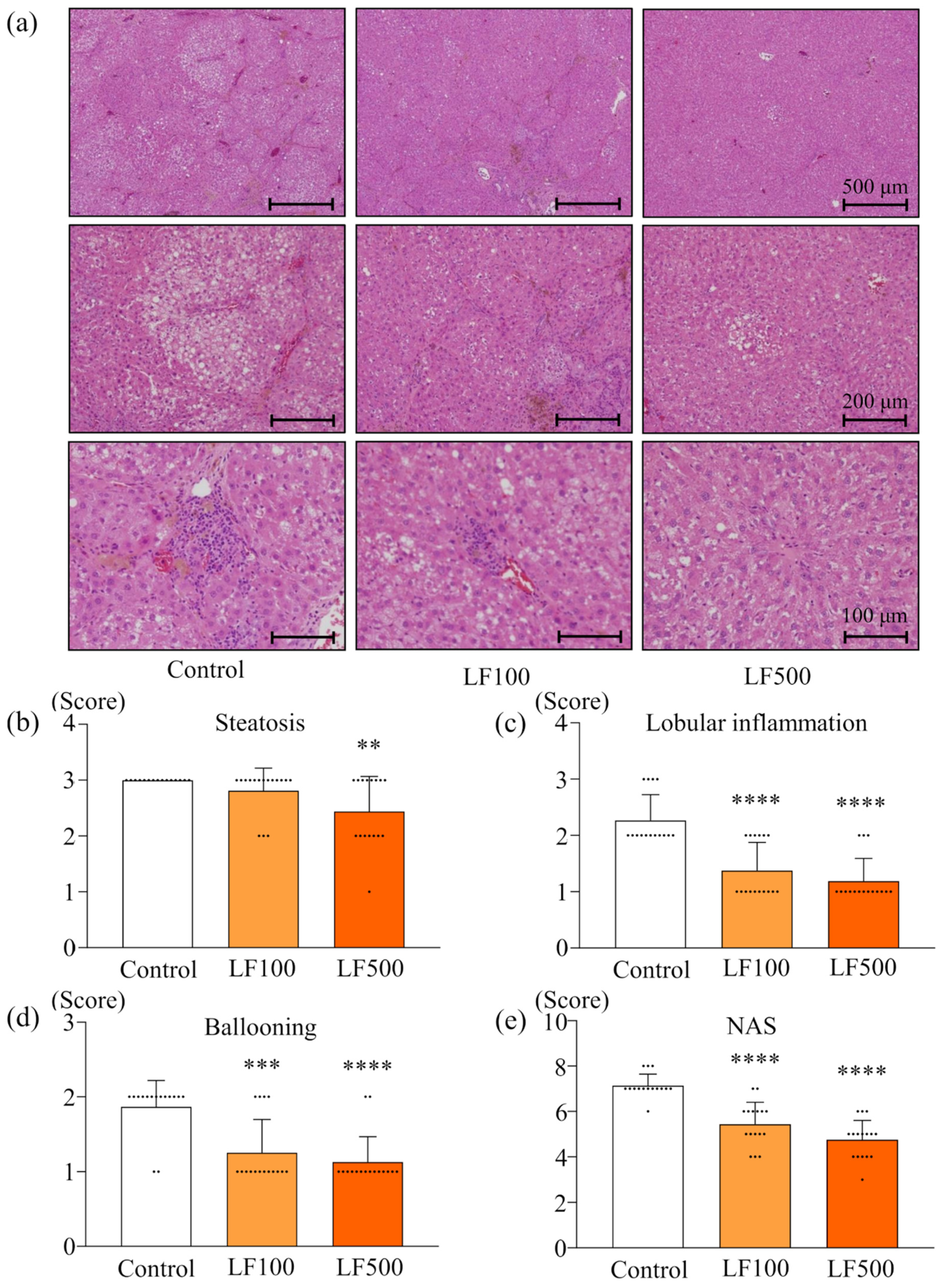
2.4. Histology of NASH
Rat livers were surgically excised and sliced into 3–4 mm thick sections. After fixing with 10% buffered formalin, sections were embedded in paraffin for histological evaluation (thickness 2–3 μm). Histological sections were stained with Azan or hematoxylin and eosin (H&E), as well as immunohistochemically stained with antibody against α-smooth muscle actin (α-SMA; Dako, Tokyo, Japan). Steatohepatitis and fibrosis were evaluated using a non-alcoholic fatty liver disease activity score (NAS), as previously described in detail [10,24]. The NAS and fibrosis scores were evaluated by three very experienced pathologists (A.N.-I., M.K., and S.Takahashi).
2.5. Evaluation of Preneoplastic Foci in the Liver
The glutathione S-transferase placental form (GST-P) was immunohistochemically stained, as previously described [25]. Averages of GST-P–positive foci that were >80 μm in diameter in the entire liver section were evaluated using an image analyzer (Keyence, Osaka, Japan).
2.6. Western Blotting
Protein samples were extracted from frozen liver tissues using radioimmunoprecipitation buffer (Thermo Fisher Scientific, Rockford, IL, USA) with added protease and phosphatase inhibitors (Thermo Fisher Scientific). The protein concentration of samples was quantified by a Bradford assay. Protein samples (30 μg per lane) were separated in 12% polyacrylamide gels and transferred onto nitrocellulose membranes (Hybond-ECL; GE Healthcare UK Ltd., Buckinghamshire, UK). Membranes were probed with primary antibodies against: Cdc42, IκB-α, NF-κB, phosphorylated (p) NF-κB (Ser536), Mkk4, pMkk4 (Thr261), Jnk, pJnk (Thr183/Tyr185) (Cell Signaling Technology, Danvers, MA, USA), and β-actin (Sigma-Aldrich, St. Louis, MI, USA). Anti–β-actin was used at a 1:5000 dilution, and all other antibodies were used at 1:1000. ImageJ software, ver.1.52 (National Cancer Institute, Bethesda, MD, USA), was used to quantify bands from blots.
2.7. Quantitative Reverse Transcription PCR
RNA samples were extracted, and quantitative reverse transcription (qRT)–PCR was performed, as previously described [15]. Phenol–chloroform was used to isolate total RNA from liver tissue (Isogen, Nippon Gene Co., Ltd., Tokyo, Japan) and then converted to cDNA with Moloney murine leukemia virus reverse transcriptase (Takara, Otsu, Japan). Quantitative reverse transcription was performed using an AriaMx Real-Time PCR system (g8830a, Agilent, Santa Clara, CA, USA). The sequences of primers used in this study were provided in a previous study [15].
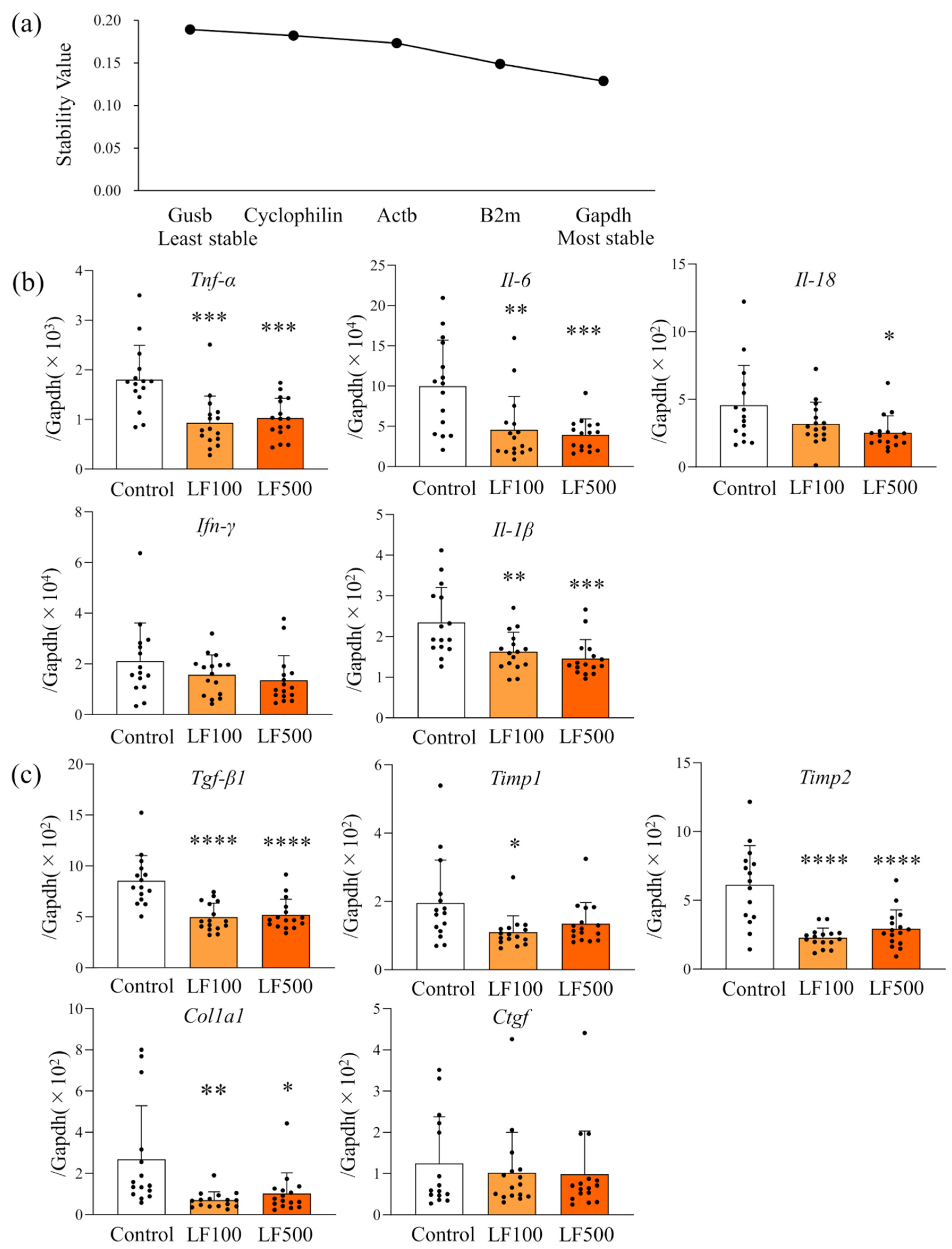
2.8. Selection of a Candidate Reference Gene
In order to select a reference gene that is stably expressed and has low variability in the present experiment system, the stability of the five candidate housekeeping genes (Table 1) was validated using NormFinder (MOMA, Aarhus, Denmark). The relative quantification for qRT-PCR was performed by standard curve method.
Table 1.
Sequence of primers for housekeeping genes tested with quantitative reverse transcription PCR.
2.9. Statistical Analysis
Data are presented as the mean ± standard deviation (SD), and one-way ANOVA and Tukey multiple comparison tests were used to compare differences between groups using the software package, Graph Pad Prism 8 (GraphPad Software, Inc., La Jolla, CA, USA). p < 0.05 was considered significant.
3. Results
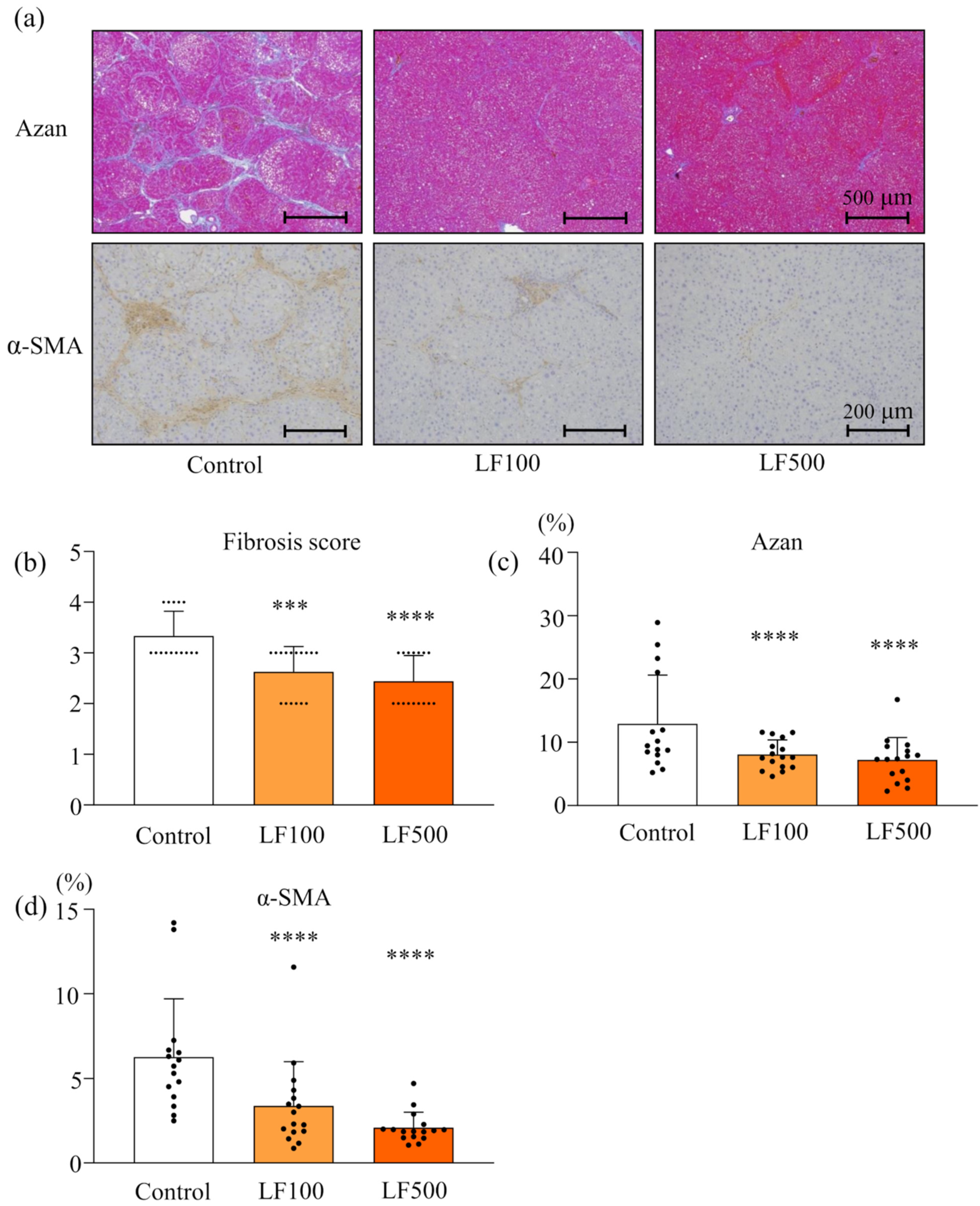
3.1. LF Prevents Steatohepatitis and Fibrosis in Cx32ΔTg Rats
We initially investigated the safety and chemopreventive effect of LF on NASH in a Cx32ΔTg–HFD–DMN rat NASH model. The dosage of LF in previous clinical studies was to reflect the selection of dosages in the present study [23]. A significant difference in body weights between control and LF-treated groups was not found. A dose-dependent change in organ weights was not noted, although liver weights were significantly increased in the LF100 compared to control group (Table 2).
Table 2.
Body and various organ weights in connexin 32 dominant negative transgenic rats fed a high-fat diet and dimethylnitrosamine with or without lactoferrin (100 or 500 mg/kg/day) at week 17.
Histological observation by H&E staining also indicated that LF did not induce any changes in kidneys. In the liver, treatments of a HFD and DMN induced diffuse deposits of fat droplets with hepatocellular ballooning and neutrophil infiltration in the lobule (Figure 1a). Lactoferrin treatment significantly reduced fat deposition, lobular inflammation, and ballooning injury of hepatocytes in a dose-dependent manner (Figure 1a–d and Table S1), resulting in decreased NAS (Figure 1e and Table S1). Bridging fibrosis and activated hepatic stellate cells (HSC) were visualized by Azan staining and α-SMA immunohistochemical staining, respectively, in a Cx32ΔTg–HFD–DMN rat NASH model (Figure 2a). The histological fibrosis score, percentages of the Azan-positive area (collagen), and percentages of the α-SMA-positive area (activated HSCs) were significantly reduced by LF in the NASH model (Figure 2a–d and Table S1).
Figure 1.
Preventive effect of lactoferrin on nonalcoholic steatohepatitis in rats. Connexin 32 dominant negative transgenic (Cx32ΔTg) rats were fed a high-fat diet (HFD), given an intraperitoneal injection of dimethylnitrosamine (DMN), and treated with lactoferrin (LF) for 17 weeks. (a) Representative histological findings of hematoxylin and eosin (H&E) stains in liver sections taken from Control, LF 100 mg/kg/day (LF100) or LF 500 mg/kg/day (LF500) rat groups. (b–e) Histopathological analysis of non-alcoholic steatohepatitis (NASH) was evaluated by severity scores for (b) steatosis, (c) lobular inflammation, (d) hepatocellular ballooning, and (e) a non-alcoholic fatty liver disease activity score (NAS). Data is shown as the mean ± SD, n = 15–16 per group, ** p < 0.01, *** p < 0.001, **** p < 0.0001 compared to the Control group.
Figure 2.
Attenuation effect of lactoferrin on fibrosis in rat nonalcoholic steatohepatitis. Connexin 32 dominant negative transgenic (Cx32ΔTg) rats were fed a high-fat diet (HFD), given an intraperitoneal injection of dimethylnitrosamine (DMN), and treated with lactoferrin (LF) for 17 weeks. (a) Azan staining (upper panels) and α-smooth muscle actin (α-SMA; lower panels) immunohistochemical stains of liver sections from Control, LF 100 mg/kg/day (LF100), or LF 500 mg/kg/day (LF500) rat groups. (b) Azan staining was used to evaluate the fibrosis score and (c) percentage of fibrosis area. (d) α-SMA–positive area. Data is shown as the mean ± SD, n = 15–16 per group, *** p < 0.001, **** p < 0.0001 compared to the Control group.
Biochemical analysis of serum indicated that the level of AST, T-chol, and LDL-C in the LF–treated groups was lower than that in the control group and the level of glucose of the LF100 was significantly higher than that of the control group. However, there was no dose-dependent change in serum hepatic enzymes, proteins, glucose, or lipids (Table 3). These results indicated that LF administration prevented the development of steatohepatitis and fibrosis without any adverse effects observed in a rat NASH model.
Table 3.
Hepatic enzyme serum levels in connexin 32 dominant negative transgenic rats fed a high-fat diet and dimethylnitrosamine with or without lactoferrin (100 or 500 mg/kg/day) at week 17.
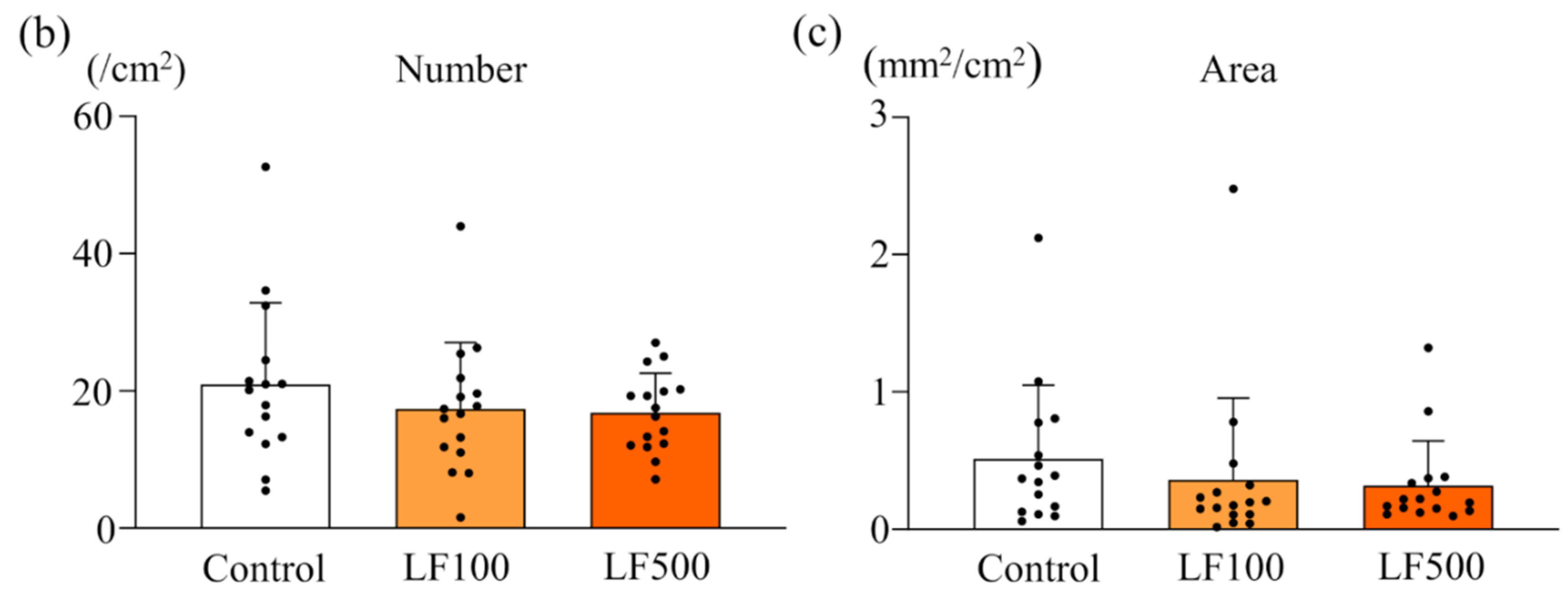
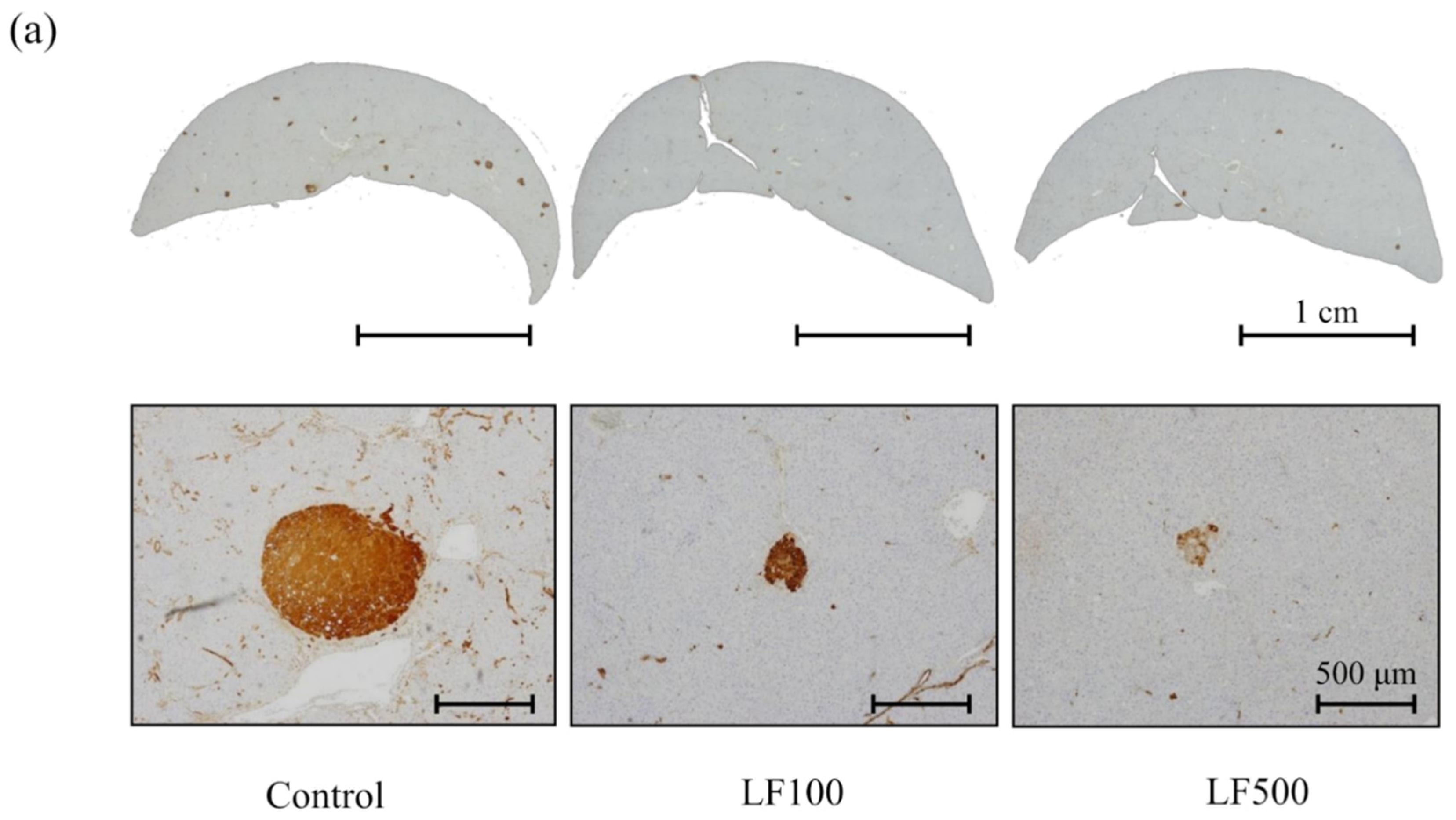
3.2. LF Tends to Decrease the Induction of Preneoplastic Lesions in Cx32ΔTg Rats
To explore the effect of LF on carcinogenic potential during the development of NASH, the formation of preneoplastic hepatic foci, namely GST-P–positive foci, was quantitated by immunohostochemistry. A combination of HFD and DMN treatment increased both the number and area of GST-P–positive foci in Cx32ΔTg rats, although the carcinogenic potential was weaker than that induced by MCDD plus diethylnitrosamine (DEN; Figure 3a–c) [10,15]. In contrast, both the number and area of GST-P–positive lesions tended to be decreased by LF intake (Figure 3a–c and Table S1). In accordance with these results, LF may have the potential to reduce hepatocarcinogenesis in NASH.

Figure 3.
Effect of lactoferrin on hepatocarcinogenesis in rat nonalcoholic steatohepatitis. Connexin 32 dominant negative transgenic (Cx32ΔTg) rats were fed a high-fat diet (HFD), given an intraperitoneal injection of dimethylnitrosamine (DMN), and treated with lactoferrin (LF) for 17 weeks. (a) Liver sections showing representative foci positive for glutathione S-transferase placental form (GST-P) from Control, LF 100 mg/kg/day (LF100), or LF 500 mg/kg/day (LF500) rat groups. (b) The number and (c) area of GST-P–positive hepatic foci. Data is shown as the mean ± SD, n = 15–16 per group.
3.3. LF Down-Regulates mRNA Expression of Inflammatory Cytokines in Cx32ΔTg Rats
Previous studies, including ours, strongly indicated that expression of inflammatory cytokines associated with inflammation (Tnf-α, Il-6, Il-18, Ifn-γ, and Il-1β) and fibrosis (Tgf-β1, Timp1, Timp2, Col1a1, and Ctgf) correlated with histological NASH activity in human and rodent models [15,26,27,28]. Thus, we further quantitated their mRNA expression level using qRT–PCR. NormFinder analysis revealed that the stability value of Gapdh was the smallest among examined candidate housekeeping genes (Figure 4a). Therefore, we concluded Gapdh as the most stable gene and used it as a reference. As shown in Figure 4b, the inflammatory cytokines, Il-6, Tnf-α, Il-18, and Il-1β, were significantly down-regulated by LF compared with the control, and a dose-dependency was observed with Il-6, Il-18, and Il-1β. While not significant, Ifn-γ mRNA expression also tended to be decreased by LF (Figure 4b and Table S2). The mRNA expression of Tgf-β1, Col1a1, Timp1, Timp2, and Ctgf as fibrosis-related cytokines was also measured; Timp2, Col1a1, and Tgf-β1 were significantly down-regulated by LF (Figure 4c and Table S2). These results suggests that down-regulation of inflammatory cytokines by LF was involved in the attenuation of steatohepatitis and hepatic fibrosis in a Cx32ΔTg–HFD–DMN rat NASH model.
Figure 4.
Down-regulation of inflammatory cytokines by lactoferrin in rat nonalcoholic steatohepatitis. Connexin 32 dominant negative transgenic (Cx32ΔTg) rats were fed a high-fat diet (HFD), given an intraperitoneal injection of dimethylnitrosamine (DMN), and treated with lactoferrin (LF) for 17 weeks. (a) NormFinder stability values for candidate housekeeping genes (Gapdh, B2m, Actb, Ppia, and Gusb). (b,c) mRNA levels of (b) pro-inflammatory cytokines (Tnf-α, Il-6, Il-18, Ifn-γ, and Il-1β) and (c) pro-fibrotic cytokines (Tgf-β1, Timp1, Timp2, Col1a1, and Ctgf) in Control, LF 100 mg/kg/day (LF100), or LF 500 mg/kg/day (LF500) rat groups were measured using quantitative reverse transcription (RT)–PCR. Data is shown as the mean ± SD, n = 15–16 per group, * p < 0.05, ** p < 0.01, *** p < 0.001, **** p < 0.0001 compared to the Control group.
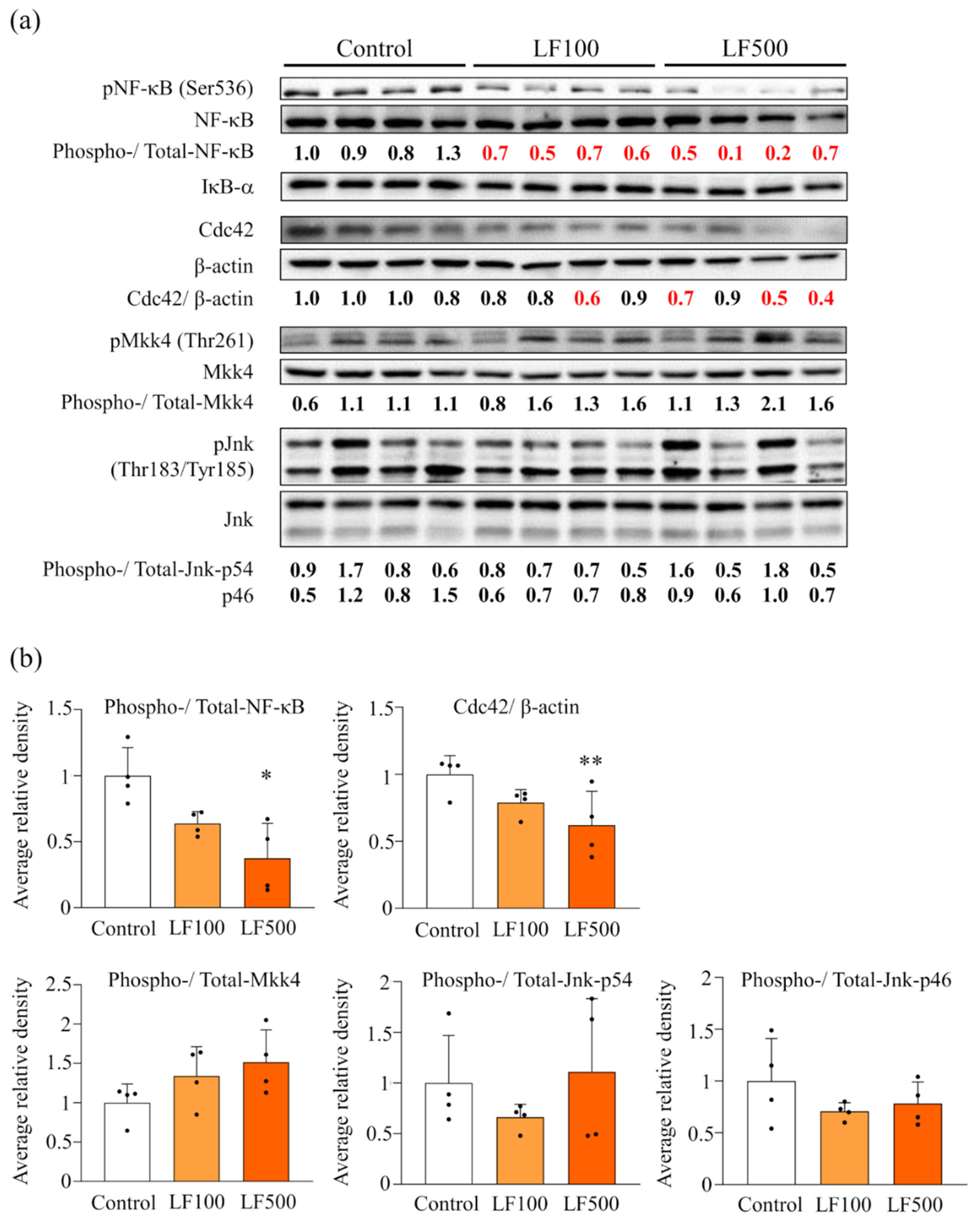
3.4. LF Administration Reduces NF-κB Signaling in Cx32ΔTg Rats
Previous studies showed that NF-κB and JNK/SAPK signaling were switched on in a rat NASH model mediated by MCDD or HFD and DMN combined [10,15]. Therefore, we investigated how such signal transduction was altered by the administration of LF. Western blotting showed that elevated pNF-κB protein expression in NASH was significantly decreased by the administration of LF in a dose-dependent manner. In contrast, phosphorylated Mkk4 and Jnk, which belong to JNK/SAPK signaling, were not affected by LF, even though their upstream protein, Cdc42, was significantly reduced in LF–treated groups (Figure 5). Such results indicate that the inactivation of NF-κB, but not JNK/SAPK signaling, is involved in the preventive effect of LF against NASH development in rats.
Figure 5.
Down-regulation of inflammatory cytokines and deactivation of NF-κB and JNK signaling after the administration of lactoferrin in nonalcoholic steatohepatitis induced in Cx32 dominant negative transgenic rats. Connexin 32 dominant negative transgenic (Cx32ΔTg) rats were fed a high-fat diet (HFD), given an intraperitoneal injection of dimethylnitrosamine (DMN), and treated with lactoferrin (LF) for 17 weeks. (a) Protein levels of nuclear factor (NF)-κB-related (NF-κB, phosphorylated (p)NF-κB, IκB-α) and SAPK/JNK (Cdc42, Mkk4, pMkk4, Jnk, and pJnk) signaling proteins in Control, LF 100 mg/kg/day (LF100), or LF 500 mg/kg/day (LF500) rat groups were assessed by western blotting. Each lane represents a protein sample from an individual rat. Phospho, phosphorylated. (b) Data is shown as the mean ± SD. * p < 0.05, ** p < 0.01 compared to the Control group.
4. Discussion
In this study, we examined the chemopreventive effect of LF on NASH in a Cx32ΔTg–HFD–DMN rat NASH model. NASH is an internationally prevalent chronic liver disease that shows fatty accumulations in the liver, ballooning, and inflamed hepatocyte. Non-alcoholic fatty liver disease often occurs in adults as a complication of lifestyle-related diseases, although it has also been found in children and is increasing all over the world regardless of a country’s industrialization level [3,29]. Eventually, continuous inflammation leads to fibrosis and progresses to cirrhosis and HCC. It is known that nearly 20% of NASH cases progress to liver cirrhosis and HCC, with or without liver cirrhosis [30,31]. Therefore, NASH is one of the most crucial targets for deterring liver cirrhosis and HCC.
Currently, the main treatments for NASH include an improvement in life-style, represented by diet and exercise therapies. In addition, existing drugs for other diseases might also be effective for countering NASH. For instance, for drug repositioning, insulin sensitizers, such as pioglitazone, improved hepatocyte injury and fibrosis in a randomized, placebo-controlled trial [32]. Ratziu et al. reported that rosiglitazone decreased liver steatosis, but an improvement in hepatocyte injury and fibrosis was not found [33]. Vitamin E [34,35] and drugs for hypercholesterolemia [36] have also been used as drug therapies for NASH but have not been established as standard treatments due to their less potent medicinal effects and possible side effects after long-term use [33,37,38]. Consequently, daily intervention with functional supplements, along with lifestyle modification, is considered essential in preventing NASH progression. Considering the pathogenesis of NASH, we hypothesized that the anti-inflammatory effects of LF would prevent this disease. Previous studies proposed that LF expression in the liver was decreased in high-fructose, high-fat, or MCD-induced mouse NAFLD models [39]. In contrast, LF ameliorated HFD-induced hepatic steatosis and elevated the triglyceride level in mouse models [40,41,42]. In addition, the levels of hepatic triglycerides and visceral fat were decreased by LF and were positively correlated in ICR mice [43], indicating that LF has the potential to reduce fat accumulation in the liver.
In the present study, for the first time, the effect of LF on histological features of NASH, including steatosis, hepatocyte injury, and inflammation, was quantitated using scoring systems originally designed for humans [24,44]. Lactoferrin significantly improved steatosis in the liver but did not affect visceral fat weight in the rat NASH model. These findings indicated that LF might protect hepatic steatosis by moderating fatty acid metabolism in the liver or in adipose tissue. The balance of lipid metabolism in a whole body may determine the effect on the lipid environment in each organ. Hepatocyte injury and inflammation in NASH were also decreased in LF–treated groups. It is well-known that the inflammation-associated cytokines, Il-6, Tnf-α, and Il-1β, were up-regulated and involved in the evocation of chronic inflammation in the colon and liver [15,17]. As already demonstrated in the colon, down-regulation of these cytokines by LF was also induced in NASH in the present study. Altogether, LF may prevent steatohepatitis via a decrease of inflammatory cytokines.
Persistent chronic inflammation in NASH leads to increased fibrosis; similar to other chronic hepatitis diseases, the irreversible alteration of liver structure due to progressive fibrosis eventually leads to cirrhosis [45]. Therefore, fibrosis is one of the most important prognostic factors for patients with NASH. In accordance with previous studies, LF has the potential to suppress liver fibrosis induced by thioacetamide [46,47]; however, the effect on fibrosis during NASH has not yet been established. This is due to the fact that it is not easy to induce fibrosis with NASH over a short time period in an animal model. As shown in Figure 3a, advanced fibrosis when bridging between lobules, or a lobule and portal vein, was induced by LF in a Cx32ΔTg–HFD–DMN rat NASH model. Lactoferrin significantly decreased not only steatohepatitis but also the histological score and area of fibrosis in the model. The numbers of hepatic stellate cells with an active phenotype were increased in NASH and decreased in the livers of LF–treated rats. These novel findings indicate that LF prevents the liver fibrosis of NASH via the inactivation of HSC.
To date, the anti-tumor effect of LF on HCC was described in previous studies using a DEN-induced HCC model in rats or mice [48,49], but effects of LF on NASH-related hepatocarcinogenesis have not been established. The glutathione S-transferase placental form is a well-known marker for preneoplastic lesions in rat liver. Therefore, carcinogenic potentials in the liver can be measured by GST-P immunohistochemistry in the early phase of hepatocarcinogenesis [25]. The number and area of GST-P-positive foci in the liver tended to decrease in LF-treated groups, although a significant difference was not found (Figure 4). We previously induced GST-P-positive foci in a NASH model using DEN [10] or DMN [15]. Both the number and area of GST-P-positive foci induced by DMN were decreased compared to those induced by DEN, which might influence the lack of significant difference by LF in this study. The chemopreventive effect of LF on NASH-related hepatocarcinogenesis should be investigated in a future study.
Nuclear factor-κB signaling plays central roles in inflammation and fibrosis during NASH progression. Especially in regard to fibrosis, activation of NF-κB stimulates parenchymal cells, including Kupffer cells [50] and enhanced TGF-β1 signaling that is essential as a profibrogenic mediator [51]. Transforming growth factor-β1 signaling modulates HSC as an active phenotype [27,52]. However, other reports indicated that TGF-β1 induced NF-κB activation [53]. In the present study, LF treatment decreased activated HSC and prevented fibrosis in a rat NASH model. Furthermore, NF-κB activation and TGF-β1 up-regulation in the model were attenuated by LF. However, JNK, which is also an important signaling pathway for fibrosis, was not altered by LF administration. These results suggest that LF protected the development of fibrosis by inhibiting NF-κB and TGF-β1 signaling.
5. Conclusions
This study demonstrated that LF prevents steatohepatitis and fibrosis without any adverse effects in a Cx32ΔTg–HFD–DMN rat NASH model. Therefore, LF may be a potential preventive or therapeutic application for this disease.
Supplementary Materials
The following supporting information can be downloaded at: https://www.mdpi.com/article/10.3390/nu14010042/s1, Table S1: Histopathology of NASH, fibrosis, and hepatocarcinogenesis in connexin 32 dominant negative transgenic rats fed a high-fat diet and dimethylnitrosamine with or without lactoferrin (100 or 500 mg/kg/day) at week 17; Table S2: mRNA level of inflammatory cytokines using quantitative reverse transcription PCR.
Author Contributions
Conceptualization and methodology: A.N.-I. and S.T. (Satoru Takahashi); validation, formal analysis, and investigation: Y.A., A.N.-I., K.X., M.K., H.K., Y.N., S.I., Y.M., S.T. (Shuji Takiguchi) and S.T. (Satoru Takahashi); data curation, writing of original draft preparation: Y.A. and A.N.-I.; writing by reviewing and editing: A.N.-I. and S.T. (Satoru Takahashi); resources: H.T., M.T. and S.T. (Satoru Takahashi); project administration, funding acquisition, visualization, and supervision: A.N.-I. and S.T. (Satoru Takahashi). All authors have read and agreed to the published version of the manuscript.
Funding
This work was supported by JSPS KAKENHI Grant Number 26460492 and 19K07509 to A.N-I.
Institutional Review Board Statement
The study was conducted according to the guidelines of the Declaration of Helsinki and approved by the Animal Care and Use Committee (ethic code: no. 19-025, approved on 24 September 2019) at Nagoya City University Graduate School of Medical Sciences.
Informed Consent Statement
Not applicable.
Data Availability Statement
The data presented in this study are available on request from the corresponding author.
Acknowledgments
The authors are sincerely grateful to Koji Kato and Junko Takekawa for excellent technical assistance with preparing tissue sections and immunohistochemical staining.
Conflicts of Interest
The authors declare that they have no conflict of interest.
Abbreviations
The following abbreviations are used in this manuscript:
| Alb | Albumin |
| AST | Aspartate aminotransferase |
| Bex1 | Brain expressed, X-linked 1 |
| Cx | Connexin |
| Cx32ΔTg | Cx32 dominant negative transgenic |
| DEN | Diethylnitrosamine |
| DMN | Dimethylnitrosamine |
| GST-P | Glutathione S-transferase placental form |
| HCC | Hepatocellular carcinoma |
| HFD | High-fat diet |
| HSC | Hepatic stellate cell |
| IL | Interleukin |
| LDL-C | Low-density lipoprotein cholesterol |
| LF | Lactoferrin |
| MCDD | Methionine choline-deficient diet |
| NAFLD | Non-alcoholic fatty liver disease |
| NAS | Non-alcoholic fatty liver disease activity score |
| NASH | Non-alcoholic steatohepatitis |
| NF-κB | Nuclear factor-κB |
| ROS | Reactive oxygen species |
| TGF | Transforming growth factor |
| TNF | Tumor necrosis factor |
| Wt | Wild-type |
References
- Chalasani, N.; Younossi, Z.; Lavine, J.E.; Diehl, A.M.; Brunt, E.M.; Cusi, K.; Charlton, M.; Sanyal, A.J. The diagnosis and management of non-alcoholic fatty liver disease: Practice guideline by the American Gastroenterological Association, American Association for the Study of Liver Diseases, and American College of Gastroenterology. Gastroenterology 2012, 142, 1592–1609. [Google Scholar] [CrossRef] [PubMed] [Green Version]
- Younossi, Z.M. Non-alcoholic fatty liver disease—A global public health perspective. J. Hepatol. 2019, 70, 531–544. [Google Scholar] [CrossRef] [PubMed] [Green Version]
- Younossi, Z.; Anstee, Q.M.; Marietti, M.; Hardy, T.; Henry, L.; Eslam, M.; George, J.; Bugianesi, E. Global burden of NAFLD and NASH: Trends, predictions, risk factors and prevention. Nat. Rev. Gastroenterol. Hepatol. 2018, 15, 11–20. [Google Scholar] [CrossRef]
- Anstee, Q.M.; Targher, G.; Day, C.P. Progression of NAFLD to diabetes mellitus, cardiovascular disease or cirrhosis. Nat. Rev. Gastroenterol. Hepatol. 2013, 10, 330–344. [Google Scholar] [CrossRef] [PubMed]
- Evans, W.H.; Martin, P.E. Gap junctions: Structure and function (Review). Mol. Membr. Biol. 2002, 19, 121–136. [Google Scholar] [CrossRef]
- Loewenstein, W.R. Junctional intercellular communication: The cell-to-cell membrane channel. Physiol. Rev. 1981, 61, 829–913. [Google Scholar] [CrossRef]
- Yamasaki, H. Gap junctional intercellular communication and carcinogenesis. Carcinogenesis 1990, 11, 1051–1058. [Google Scholar] [CrossRef] [Green Version]
- Trosko, J.E.; Chang, C.C. Role of stem cells and gap junctional intercellular communication in human carcinogenesis. Radiat. Res. 2001, 155, 175–180. [Google Scholar] [CrossRef]
- Paul, D.L. Molecular cloning of cDNA for rat liver gap junction protein. J. Cell Biol. 1986, 103, 123–134. [Google Scholar] [CrossRef] [Green Version]
- Sagawa, H.; Naiki-Ito, A.; Kato, H.; Naiki, T.; Yamashita, Y.; Suzuki, S.; Sato, S.; Shiomi, K.; Kato, A.; Kuno, T.; et al. Connexin 32 and luteolin play protective roles in non-alcoholic steatohepatitis development and its related hepatocarcinogenesis in rats. Carcinogenesis 2015, 36, 1539–1549. [Google Scholar] [CrossRef]
- Nakashima, Y.; Ono, T.; Yamanoi, A.; El-Assal, O.N.; Kohno, H.; Nagasue, N. Expression of gap junction protein connexin32 in chronic hepatitis, liver cirrhosis, and hepatocellular carcinoma. J. Gastroenterol. 2004, 39, 763–768. [Google Scholar] [CrossRef]
- Asamoto, M.; Hokaiwado, N.; Murasaki, T.; Shirai, T. Connexin 32 dominant-negative mutant transgenic rats are resistant to hepatic damage by chemicals. Hepatology 2004, 40, 205–210. [Google Scholar] [CrossRef] [PubMed]
- Hokaiwado, N.; Asamoto, M.; Ogawa, K.; Shirai, T. Transgenic disruption of gap junctional intercellular communication enhances early but not late stage hepatocarcinogenesis in the rat. Toxicol. Pathol. 2005, 33, 695–701. [Google Scholar] [CrossRef] [PubMed]
- Hokaiwado, N.; Asamoto, M.; Futakuchi, M.; Ogawa, K.; Takahashi, S.; Shirai, T. Both early and late stages of hepatocarcinogenesis are enhanced in Cx32 dominant negative mutant transgenic rats with disrupted gap junctional intercellular communication. J. Membr. Biol. 2007, 218, 101–106. [Google Scholar] [CrossRef] [PubMed]
- Naiki-Ito, A.; Kato, H.; Naiki, T.; Yeewa, R.; Aoyama, Y.; Nagayasu, Y.; Suzuki, S.; Inaguma, S.; Takahashi, S. A novel model of non-alcoholic steatohepatitis with fibrosis and carcinogenesis in connexin 32 dominant-negative transgenic rats. Arch. Toxicol. 2020, 94, 4085–4097. [Google Scholar] [CrossRef] [PubMed]
- Tanaka, H.; Gunasekaran, S.; Saleh, D.M.; Alexander, W.T.; Alexander, D.B.; Ohara, H.; Tsuda, H. Effects of oral bovine lactoferrin on a mouse model of inflammation associated colon cancer. Biochem. Cell Biol. 2021, 99, 159–165. [Google Scholar] [CrossRef]
- Togawa, J.; Nagase, H.; Tanaka, K.; Inamori, M.; Nakajima, A.; Ueno, N.; Saito, T.; Sekihara, H. Oral administration of lactoferrin reduces colitis in rats via modulation of the immune system and correction of cytokine imbalance. J. Gastroenterol. Hepatol. 2002, 17, 1291–1298. [Google Scholar] [CrossRef]
- Farid, A.S.; El Shemy, M.A.; Nafie, E.; Hegazy, A.M.; Abdelhiee, E.Y. Anti-inflammatory, anti-oxidant and hepatoprotective effects of lactoferrin in rats. Drug Chem. Toxicol. 2021, 44, 286–293. [Google Scholar] [CrossRef]
- Chen, H.A.; Chiu, C.C.; Huang, C.Y.; Chen, L.J.; Tsai, C.C.; Hsu, T.C.; Tzang, B.S. Lactoferrin Increases Antioxidant Activities and Ameliorates Hepatic Fibrosis in Lupus-Prone Mice Fed with a High-Cholesterol Diet. J. Med. Food 2016, 19, 670–677. [Google Scholar] [CrossRef]
- Duarte, D.C.; Nicolau, A.; Teixeira, J.A.; Rodrigues, L.R. The effect of bovine milk lactoferrin on human breast cancer cell lines. J. Dairy Sci. 2011, 94, 66–76. [Google Scholar] [CrossRef] [Green Version]
- Xu, X.X.; Jiang, H.R.; Li, H.B.; Zhang, T.N.; Zhou, Q.; Liu, N. Apoptosis of stomach cancer cell SGC-7901 and regulation of Akt signaling way induced by bovine lactoferrin. J. Dairy Sci. 2010, 93, 2344–2350. [Google Scholar] [CrossRef] [PubMed]
- Xiao, Y.; Monitto, C.L.; Minhas, K.M.; Sidransky, D. Lactoferrin down-regulates G1 cyclin-dependent kinases during growth arrest of head and neck cancer cells. Clin. Cancer Res. 2004, 10, 8683–8686. [Google Scholar] [CrossRef] [PubMed] [Green Version]
- Kozu, T.; Iinuma, G.; Ohashi, Y.; Saito, Y.; Akasu, T.; Saito, D.; Alexander, D.B.; Iigo, M.; Kakizoe, T.; Tsuda, H. Effect of orally administered bovine lactoferrin on the growth of adenomatous colorectal polyps in a randomized, placebo-controlled clinical trial. Cancer Prev. Res. 2009, 2, 975–983. [Google Scholar] [CrossRef] [PubMed] [Green Version]
- Kleiner, D.E.; Brunt, E.M.; Van Natta, M.; Behling, C.; Contos, M.J.; Cummings, O.W.; Ferrell, L.D.; Liu, Y.C.; Torbenson, M.S.; Unalp-Arida, A.; et al. Design and validation of a histological scoring system for nonalcoholic fatty liver disease. Hepatology 2005, 41, 1313–1321. [Google Scholar] [CrossRef]
- Naiki-Ito, A.; Kato, H.; Asamoto, M.; Naiki, T.; Shirai, T. Age-dependent carcinogenic susceptibility in rat liver is related to potential of gap junctional intercellular communication. Toxicol. Pathol. 2012, 40, 715–721. [Google Scholar] [CrossRef] [Green Version]
- Dela Pena, A.; Leclercq, I.; Field, J.; George, J.; Jones, B.; Farrell, G. NF-kappaB activation, rather than TNF, mediates hepatic inflammation in a murine dietary model of steatohepatitis. Gastroenterology 2005, 129, 1663–1674. [Google Scholar] [CrossRef]
- Seki, E.; De Minicis, S.; Osterreicher, C.H.; Kluwe, J.; Osawa, Y.; Brenner, D.A.; Schwabe, R.F. TLR4 enhances TGF-beta signaling and hepatic fibrosis. Nat. Med. 2007, 13, 1324–1332. [Google Scholar] [CrossRef]
- Wieckowska, A.; Papouchado, B.G.; Li, Z.; Lopez, R.; Zein, N.N.; Feldstein, A.E. Increased hepatic and circulating interleukin-6 levels in human nonalcoholic steatohepatitis. Am. J. Gastroenterol. 2008, 103, 1372–1379. [Google Scholar] [CrossRef]
- Nobili, V.; Alisi, A.; Valenti, L.; Miele, L.; Feldstein, A.E.; Alkhouri, N. NAFLD in children: New genes, new diagnostic modalities and new drugs. Nat. Rev. Gastroenterol. Hepatol. 2019, 16, 517–530. [Google Scholar] [CrossRef]
- Bullock, R.E.; Zaitoun, A.M.; Aithal, G.P.; Ryder, S.D.; Beckingham, I.J.; Lobo, D.N. Association of non-alcoholic steatohepatitis without significant fibrosis with hepatocellular carcinoma. J. Hepatol. 2004, 41, 685–686. [Google Scholar] [CrossRef]
- Marrero, J.A.; Fontana, R.J.; Su, G.L.; Conjeevaram, H.S.; Emick, D.M.; Lok, A.S. NAFLD may be a common underlying liver disease in patients with hepatocellular carcinoma in the United States. Hepatology 2002, 36, 1349–1354. [Google Scholar] [CrossRef]
- Aithal, G.P.; Thomas, J.A.; Kaye, P.V.; Lawson, A.; Ryder, S.D.; Spendlove, I.; Austin, A.S.; Freeman, J.G.; Morgan, L.; Webber, J. Randomized, placebo-controlled trial of pioglitazone in nondiabetic subjects with nonalcoholic steatohepatitis. Gastroenterology 2008, 135, 1176–1184. [Google Scholar] [CrossRef] [PubMed] [Green Version]
- Ratziu, V.; Giral, P.; Jacqueminet, S.; Charlotte, F.; Hartemann-Heurtier, A.; Serfaty, L.; Podevin, P.; Lacorte, J.M.; Bernhardt, C.; Bruckert, E.; et al. Rosiglitazone for nonalcoholic steatohepatitis: One-year results of the randomized placebo-controlled Fatty Liver Improvement with Rosiglitazone Therapy (FLIRT) Trial. Gastroenterology 2008, 135, 100–110. [Google Scholar] [CrossRef] [PubMed]
- Sanyal, A.J.; Chalasani, N.; Kowdley, K.V.; McCullough, A.; Diehl, A.M.; Bass, N.M.; Neuschwander-Tetri, B.A.; Lavine, J.E.; Tonascia, J.; Unalp, A.; et al. Pioglitazone, vitamin E, or placebo for nonalcoholic steatohepatitis. N. Engl. J. Med. 2010, 362, 1675–1685. [Google Scholar] [CrossRef] [PubMed] [Green Version]
- Harrison, S.A.; Torgerson, S.; Hayashi, P.; Ward, J.; Schenker, S. Vitamin E and vitamin C treatment improves fibrosis in patients with nonalcoholic steatohepatitis. Am. J. Gastroenterol. 2003, 98, 2485–2490. [Google Scholar] [CrossRef] [PubMed]
- Athyros, V.G.; Tziomalos, K.; Gossios, T.D.; Griva, T.; Anagnostis, P.; Kargiotis, K.; Pagourelias, E.D.; Theocharidou, E.; Karagiannis, A.; Mikhailidis, D.P. Safety and efficacy of long-term statin treatment for cardiovascular events in patients with coronary heart disease and abnormal liver tests in the Greek Atorvastatin and Coronary Heart Disease Evaluation (GREACE) Study: A post-hoc analysis. Lancet 2010, 376, 1916–1922. [Google Scholar] [CrossRef]
- Nissen, S.E.; Wolski, K. Effect of rosiglitazone on the risk of myocardial infarction and death from cardiovascular causes. N. Engl. J. Med. 2007, 356, 2457–2471. [Google Scholar] [CrossRef] [Green Version]
- Filozof, C.; Goldstein, B.J.; Williams, R.N.; Sanyal, A. Non-Alcoholic Steatohepatitis: Limited Available Treatment Options but Promising Drugs in Development and Recent Progress Towards a Regulatory Approval Pathway. Drugs 2015, 75, 1373–1392. [Google Scholar] [CrossRef] [Green Version]
- Lee, S.; Son, B.; Jeon, J.; Park, G.; Kim, H.; Kang, H.; Youn, H.; Jo, S.; Song, J.Y.; Youn, B. Decreased Hepatic Lactotransferrin Induces Hepatic Steatosis in Chronic Non-Alcoholic Fatty Liver Disease Model. Cell Physiol. Biochem. 2018, 47, 2233–2249. [Google Scholar] [CrossRef]
- Min, Q.Q.; Qin, L.Q.; Sun, Z.Z.; Zuo, W.T.; Zhao, L.; Xu, J.Y. Effects of Metformin Combined with Lactoferrin on Lipid Accumulation and Metabolism in Mice Fed with High-Fat Diet. Nutrients 2018, 10, 1628. [Google Scholar] [CrossRef] [Green Version]
- Xiong, L.; Ren, F.; Lv, J.; Zhang, H.; Guo, H. Lactoferrin attenuates high-fat diet-induced hepatic steatosis and lipid metabolic dysfunctions by suppressing hepatic lipogenesis and down-regulating inflammation in C57BL/6J mice. Food Funct. 2018, 9, 4328–4339. [Google Scholar] [CrossRef] [PubMed]
- Li, Y.C.; Hsieh, C.C. Lactoferrin dampens high-fructose corn syrup-induced hepatic manifestations of the metabolic syndrome in a murine model. PLoS ONE 2014, 9, e97341. [Google Scholar] [CrossRef] [PubMed] [Green Version]
- Morishita, S.; Ono, T.; Fujisaki, C.; Ishihara, Y.; Murakoshi, M.; Kato, H.; Hosokawa, M.; Miyashita, K.; Sugiyama, K.; Nishino, H. Bovine lactoferrin reduces visceral fat and liver triglycerides in ICR mice. J. Oleo Sci. 2013, 62, 97–103. [Google Scholar] [CrossRef] [PubMed] [Green Version]
- Brunt, E.M.; Janney, C.G.; Di Bisceglie, A.M.; Neuschwander-Tetri, B.A.; Bacon, B.R. Nonalcoholic steatohepatitis: A proposal for grading and staging the histological lesions. Am. J. Gastroenterol. 1999, 94, 2467–2474. [Google Scholar] [CrossRef]
- Tsuchida, T.; Friedman, S.L. Mechanisms of hepatic stellate cell activation. Nat. Rev. Gastroenterol. Hepatol. 2017, 14, 397–411. [Google Scholar] [CrossRef]
- Rizk, F.H.; Sarhan, N.I.; Soliman, N.A.; Ibrahim, M.A.A.; Abd-Elsalam, M.; Abd-Elsalam, S. Heat shock protein 47 as indispensible participant in liver fibrosis: Possible protective effect of lactoferrin. IUBMB Life 2018, 70, 795–805. [Google Scholar] [CrossRef] [Green Version]
- Hessin, A.; Hegazy, R.; Hassan, A.; Yassin, N.; Kenawy, S. Lactoferrin Enhanced Apoptosis and Protected Against Thioacetamide-Induced Liver Fibrosis in Rats. Open Access Maced. J. Med. Sci. 2015, 3, 195–201. [Google Scholar] [CrossRef] [Green Version]
- Hegazy, R.R.; Mansour, D.F.; Salama, A.A.; Abdel-Rahman, R.F.; Hassan, A.M. Regulation of PKB/Akt-pathway in the chemopreventive effect of lactoferrin against diethylnitrosamine-induced hepatocarcinogenesis in rats. Pharmacol. Rep. 2019, 71, 879–891. [Google Scholar] [CrossRef]
- Mohammed, M.M.; Ramadan, G.; Zoheiry, M.K.; El-Beih, N.M. Antihepatocarcinogenic activity of whey protein concentrate and lactoferrin in diethylnitrosamine-treated male albino mice. Environ. Toxicol. 2019, 34, 1025–1033. [Google Scholar] [CrossRef]
- Sun, B.; Karin, M. NF-kappaB signaling, liver disease and hepatoprotective agents. Oncogene 2008, 27, 6228–6244. [Google Scholar] [CrossRef] [Green Version]
- Yu, Y.; Liu, Y.; An, W.; Song, J.; Zhang, Y.; Zhao, X. STING-mediated inflammation in Kupffer cells contributes to progression of nonalcoholic steatohepatitis. J. Clin. Investig. 2019, 129, 546–555. [Google Scholar] [CrossRef] [PubMed]
- Okina, Y.; Sato-Matsubara, M.; Matsubara, T.; Daikoku, A.; Longato, L.; Rombouts, K.; Thanh Thuy, L.T.; Ichikawa, H.; Minamiyama, Y.; Kadota, M.; et al. TGF-beta-driven reduction of cytoglobin leads to oxidative DNA damage in stellate cells during non-alcoholic steatohepatitis. J. Hepatol. 2020, 73, 882–895. [Google Scholar] [CrossRef] [PubMed]
- Liu, C.; Yuan, X.; Tao, L.; Cheng, Z.; Dai, X.; Sheng, X.; Xue, D. Xia-yu-xue decoction (XYXD) reduces carbon tetrachloride (CCl4)-induced liver fibrosis through inhibition hepatic stellate cell activation by targeting NF-kappaB and TGF-beta1 signaling pathways. BMC Complement. Altern. Med. 2015, 15, 201. [Google Scholar] [CrossRef] [PubMed] [Green Version]
Publisher’s Note: MDPI stays neutral with regard to jurisdictional claims in published maps and institutional affiliations. |
© 2021 by the authors. Licensee MDPI, Basel, Switzerland. This article is an open access article distributed under the terms and conditions of the Creative Commons Attribution (CC BY) license (https://creativecommons.org/licenses/by/4.0/).