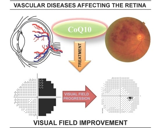
The Use of Vitamins and Coenzyme Q10 for the Treatment of Vascular Occlusion Diseases Affecting the Retina

 , and
, and 
Abstract
1. Introduction
2. Materials and Methods
3. Results
4. Discussion
5. Conclusions
Supplementary Materials
Author Contributions
Funding
Acknowledgments
Conflicts of Interest
References
- Hayreh, S.S. Ischemic optic neuropathy. Prog. Retin. Eye Res. 2009, 28, 34–62. [Google Scholar] [CrossRef] [PubMed]
- Buono, L.M.; Foroozan, R.; Sergott, R.C.; Savino, P.J. Nonarteritic anterior ischemic optic neuropathy. Curr. Opin. Ophthalmol. 2002, 13, 357–361. [Google Scholar] [CrossRef] [PubMed]
- Varma, D.D.; Cugati, S.; Lee, A.W.; Chen, C.S. A review of central retinal artery occlusion: Clinical presentation and management. Eye 2013, 27, 688–697. [Google Scholar] [CrossRef]
- Limaye, K.; Wall, M.; Uwaydat, S.; Ali, S.; Shaban, A.; Al Kasab, S.; Adams, H., Jr. Is Management of Central Retinal Artery Occlusion the Next Frontier in Cerebrovascular Diseases? J. Stroke Cerebrovasc. Dis. 2018, 27, 2781–2791. [Google Scholar] [CrossRef]
- Hayreh, S.S. The 1994 Von Sallman Lecture. The optic nerve head circulation in health and disease. Exp. Eye Res. 1995, 61, 259–272. [Google Scholar] [CrossRef]
- Goodwin, D. Homonymous hemianopia: Challenges and solutions. Clin. Ophthalmol. 2014, 8, 1919–1927. [Google Scholar] [CrossRef]
- Eggenberger, E.R.; Pula, J.H. Neuro-ophthalmology in Medicine. In Aminoff’s Neurology and General Medicine, 5th ed.; Aminoff, M.J., Josephson, S.A., Eds.; Elsevier Inc.: Amsterdam, The Netherlands, 2014; pp. 479–502. ISBN 978-012-407-710-2. [Google Scholar]
- Hayreh, S.S.; Zimmerman, M.B.; Podhajsky, P.; Alward, W.L.M. Nocturnal arterial hypotension and its role in optic nerve head and ocular ischemic disorders. Am. J. Ophthalmol. 1994, 117, 603–624. [Google Scholar] [CrossRef]
- Hayreh, S.S. Acute ischemic disorders of the optic nerve: Pathogenesis, clinical manifestations and management. Ophthalmol. Clin. N. Am. 1996, 9, 407–442. [Google Scholar]
- Hayreh, S.S. Non-arteritic anterior ischemic optic neuropathy versus cerebral ischemic stroke. Graefes Arch. Clin. Exp. Ophthalmol. 2012, 250, 1255–1260. [Google Scholar] [CrossRef][Green Version]
- Pula, J.H.; Yuen, C.A. Eyes and strike: The visual aspects of cerebrovascular disease. Stroke Vasc. Neurol. 2017, 2, 210–220. [Google Scholar] [CrossRef] [PubMed]
- Hayreh, S.S. Blood flow in the optic nerve head and factors that may influence it. Prog. Retin. Eye Res. 2001, 20, 595–624. [Google Scholar] [CrossRef]
- Pambakian, A.; Currie, J.; Kennard, C. Rehabilitation strategies for patients with homonymous visual field defects. J. Neuroophthalmol. 2005, 25, 136–142. [Google Scholar] [CrossRef] [PubMed]
- Brown, N.A.; Bron, A.J.; Harding, J.J.; Dewar, H.M. Nutrition supplements and the eye. Eye (Lond.) 1998, 12, 127–133. [Google Scholar] [CrossRef] [PubMed]
- Demmig-Adams, B.; Adams, R.B. Eye Nutrition in Context: Mechanisms, Implementation, and Future Directions. Nutrients 2013, 5, 2483–2501. [Google Scholar] [CrossRef] [PubMed]
- Salama, M.; Yuan, T.F.; Machado, S.; Murillo-Rodríguez, E.; Vega, J.A.; Menéndez-González, M.; Nardi, A.E.; Arias-Carrión, O. Co-enzyme Q10 to treat neurological disorders: Basic mechanisms, clinical outcomes, and future research direction. CNS Neurol. Disord. Drug Targets 2013, 12, 641–664. [Google Scholar] [CrossRef]
- Zhang, X.; Biswas, L.; Tohari, A.M.; Reilly, J.; Tiano, L.; Shu, X. Coenzyme Q10 as a therapeutic candidate for treating inherited photoreceptor degeneration. Neural Regen. Res. 2017, 12, 1979–1981. [Google Scholar] [CrossRef]
- Zhang, X.; Tohari, A.M.; Marcheggiani, F.; Zhou, X.; Reilly, J.; Tiano, L.; Shu, X. Therapeutic Potential of Co-enzyme Q10 in Retinal Diseases. Curr. Med. Chem. 2017, 24, 4329–4339. [Google Scholar] [CrossRef]
- Martucci, A.; Nucci, C. Evidence on neuroprotective properties of coenzyme Q10 in the treatment of glaucoma. Neural Regen. Res. 2019, 14, 197–200. [Google Scholar] [CrossRef]
- Yang, X.; Zhang, Y.; Xu, H.; Luo, X.; Yu, J.; Liu, J.; Chang, R.C. Neuroprotection of Coenzyme Q10 in Neurodegenerative Diseases. Curr. Top. Med. Chem. 2016, 16, 856–866. [Google Scholar] [CrossRef]
- Somayajulu, M.; McCarthy, S.; Hung, M.; Sikorska, M.; Borowy-Borowski, H.; Pandey, S. Role of mitochondria in neuronal cell death induced by oxidative stress; neuroprotection by coenzyme Q10. Neurobiol. Dis. 2005, 18, 618–697. [Google Scholar] [CrossRef]
- Fernández-Vega, B.; González-Iglesias, H.; Vega, J.A.; Nicieza, J.; Fernández-Vega, A. Coenzyme Q10 treatment improved visual field after homonymous quadrantanopia caused by occipital lobe infarction. Am. J. Ophthalmol. Case Rep. 2018, 13, 70–75. [Google Scholar] [CrossRef]
- Prem Senthil, N.; Khadka, J.; Gilhotra, J.S.; Simon, S.; Fenwick, E.K.; Lamoureux, E.; Pesudovs, K. Understanding quality of life impact in people with retinal vein occlusion: A qualitative inquiry. Clin. Exp. Optom. 2019, 102, 406–411. [Google Scholar] [CrossRef]
- Kerkhoff, G. Restorative and compensatory therapy approaches in cerebral blindness—A review. Restor. Neurol. Neurosci. 1999, 15, 255–271. [Google Scholar] [PubMed]
- Augsburger, J.J.; Magargal, L.E. Visual prognosis following treatment of acute central retinal artery obstruction. Br. J. Ophthalmol. 1980, 64, 913–917. [Google Scholar] [CrossRef] [PubMed]
- Miller, N.J.; Arnold, A.C. Current concepts in the diagnosis, pathogenesis and management of nonarteritic anterior ischaemic optic neuropathy. Eye (Lond.) 2015, 29, 65–79. [Google Scholar] [CrossRef] [PubMed]
- Pambakian, A.L.; Kennard, C. Can visual function be restored in patients with homonymous hemianopia? Br. J. Ophthalmol. 1997, 81, 324–328. [Google Scholar] [CrossRef] [PubMed]
- Scherer, R.W.; Feldon, S.E.; Levin, L.; Langenberg, P.; Katz, J.; Keyl, P.M.; Wilson, P.D.; Kelman, S.E.; Dickersin, K. Ischemic Optic Neuropathy Decompression Trial Research Group. Visual fields at follow-up in the Ischemic Optic Neuropathy Decompression Trial: Evaluation of change in pattern defect and severity over time. Ophthalmology 2008, 115, 1809–1817. [Google Scholar] [CrossRef] [PubMed]
- Frolov, A.; Feuerstein, J.; Subramanian, P.S. Homonymous Hemianopia and Vision. Restoration Therapy. Neurol. Clin. 2017, 35, 29–43. [Google Scholar] [CrossRef]
- Mirshahi, A.; Feltgen, N.; Hansen, L.L.; Hattenbach, L.O. Retinal vascular occlusions: An interdisciplinary challenge. Dtsch. Arztebl. Int. 2008, 105, 474–479. [Google Scholar] [CrossRef]
- Han, L.; Law-Gibson, D.; Reding, M. Key neurological impairments influence function-related group outcomes after stroke. Stroke 2002, 33, 1920–1924. [Google Scholar] [CrossRef][Green Version]
- Kedar, S.; Ghate, D.; Corbett, J.J. Visual fields in neuro-ophthalmology. Indian J. Ophthalmol. 2011, 59, 103–109. [Google Scholar] [CrossRef] [PubMed]
- Litarru, G.P.; Tiano, L. Bioenergetic and antioxidant properties of coenzyme Q10: Recent developments. Mol. Biotechnol. 2007, 37, 31–37. [Google Scholar] [CrossRef] [PubMed]
- Lee, D.; Kim, K.Y.; Shim, M.S.; Kim, S.Y.; Ellisman, M.H.; Weinreb, R.N.; Ju, W.K. Coenzyme Q10 ameliorates oxidative stress and prevents mitochondrial alteration in ischemic retinal injury. Apoptosis 2014, 19, 603–614. [Google Scholar] [CrossRef] [PubMed]
- Lu, C.J.; Guo, Y.Z.; Zhang, Y.; Yang, L.; Chang, Y.; Zhang, J.W.; Jing, L.; Zhang, J.Z. Coenzyme Q10 ameliorates cerebral ischemia reperfusion injury in hyperglycemic rats. Pathol. Res. Pract. 2017, 213, 1191–1199. [Google Scholar] [CrossRef]
- Beal, M.F. Therapeutic effects of coenzyme Q10 in neurodegenerative diseases. Methods Enzymol. 2004, 382, 473–487. [Google Scholar] [CrossRef] [PubMed]
- Molyneux, A.; Florkowski, C.M.; George, P.M.; Pilbrow, A.P.; Frampton, C.M.; Lever, M.; Richards, A.M. Coenzyme Q10: An independent predictor of mortality in chronic heart failure. J. Am. Coll. Cardiol. 2008, 52, 1435–1441. [Google Scholar] [CrossRef]
- Nucci, C.; Tartaglione, R.; Cerulli, A.; Mancino, R.; Spanò, A.; Cavaliere, F.; Rombolà, L.; Bagetta, G.; Corasaniti, M.T.; Morrone, L.A. Retinal damage caused by high intraocular pressure-induced transient ischemia is prevented by coenzyme Q10 in rat. Int. Rev. Neurobiol. 2007, 82, 397–406. [Google Scholar] [CrossRef]
- Russo, R.; Cavaliere, F.; Rombolà, L.; Gliozzi, M.; Cerulli, A.; Nucci, C.; Fazzi, E.; Bagetta, G.; Corasaniti, M.T.; Morrone, L.A. Rational basis for the development of coenzyme Q10 as a neurotherapeutic agent for retinal protection. Prog. Brain Res. 2008, 173, 575–582. [Google Scholar] [CrossRef]
- Hernández-Camacho, J.D.; Bernier, M.; López-Lluch, G.; Navas, P. Coenzyme Q10 Supplementation in Aging and Disease. Front. Physiol. 2018, 9, 44. [Google Scholar] [CrossRef]


| Study Population (n) | Age at Diagnosis (mean ± SD) | Current Age (mean ± SD) | Age Range | Gender (Female/Male) |
|---|---|---|---|---|
| NAION (18) | 61 ± 8 | 61 ± 7 | 50–81 | 8(44%)/10 |
| Stroke (10) | 56 ± 21 | 63 ± 18 | 19–71 | 4(40%)/6 |
| RAO (7) | 65 ± 11 | 65 ± 11 | 48–83 | 2(29%)/5 |
| OC (13) | 47 ± 23 | 51 ± 22 | 15–85 | 8(61%)/5 |
| Population-averaged (48) | 57 ± 16 | 60 ± 15 | 15–85 | 22(46%)/26 |
| Case | Disease | Follow-Up Months | Eye | Initial VFI (%) | Final VFI (%) | Initial MD (dB) | Final MD (dB) | VFI Progression Rate (%/year) 1 |
|---|---|---|---|---|---|---|---|---|
| NAION01 | NAION (with secondary optic atrophy) | 91 2 | Right | 96 | 100 | −3.91 | 1.03 | −0.2 ± 0.2 |
| Left | 49 | 67 | −17.91 | −10.68 | +2.0 ± 1.9 | |||
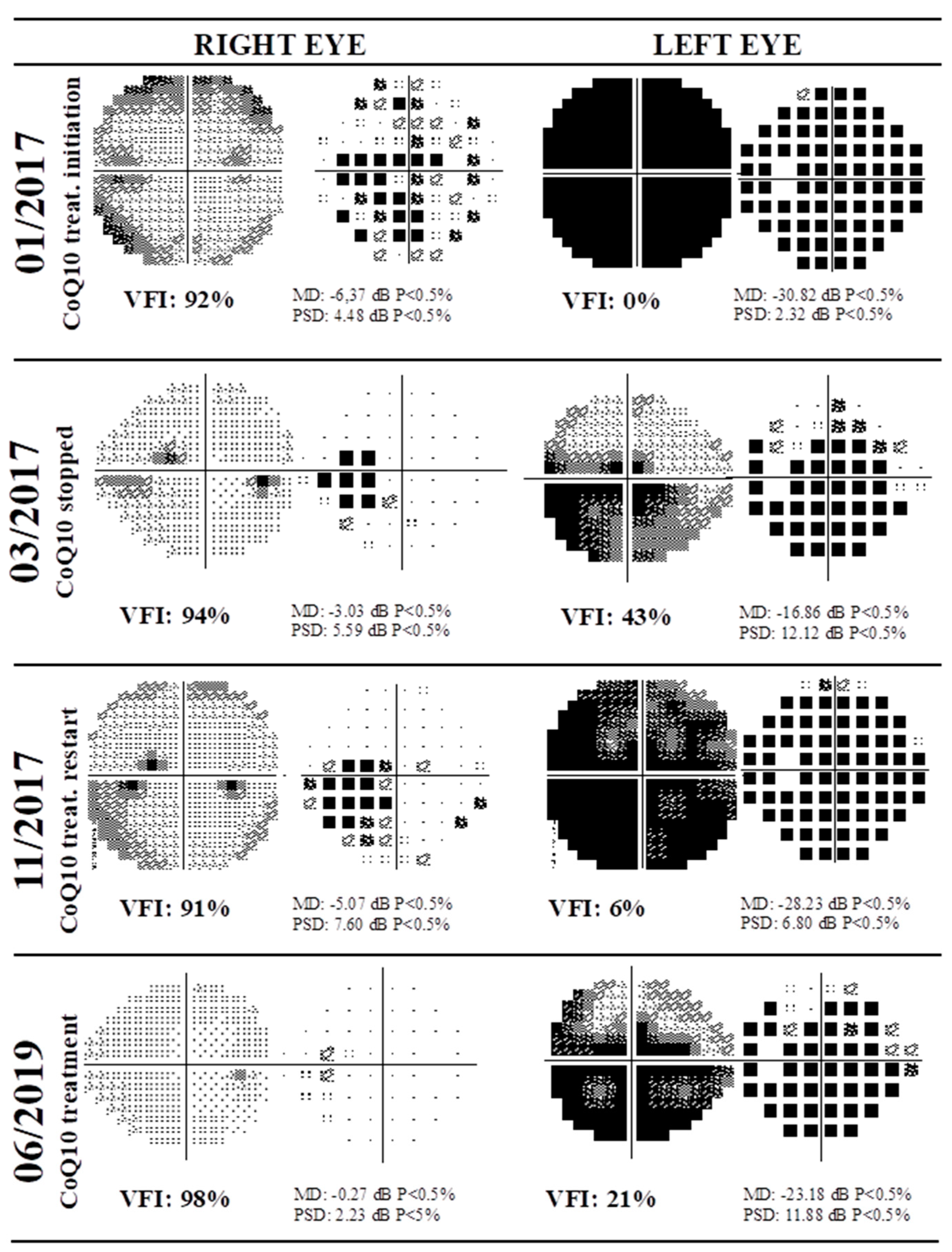
| NAION02 | NAION | 29 3 | Right | 92 | 98 | −6.37 | −0.27 | +2.6 ± 2.6 |
| Left | 0 | 21 | −30.82 | −23.18 | +5.0 ± 14.8 | |||
| NAION03 | NAION (with homonymous hemianopia) | 37 2 | Right | 97 | 98 | −2.4 | −1.81 | +1.1 ± 8.2 |
| Left | 81 | 88 | −6.79 | −2.35 | +2.4 ± 8.6 | |||
| NAION04 | NAION | 5 2 | Right | 97 | 99 | −3.72 | −1.52 | +1.0 ± 1.1 |
| Left | 14 | 35 | −27.39 | −21.64 | +58.1 ± 10.5 | |||
| NAION05 | NAION | 10 2 | Right | 88 | 93 | −5.7 | −3.0 | +2.8 ± 5.0 |
| Left | 36 | 45 | −19.22 | −15.67 | +11.8 ± 17.6 | |||
| NAION06 | NAION (with inferior hemianopia) | 64 2 | Right | 63 | 88 | −16.43 | −6.62 | +5.9 ± 2.6 |
| Left | 97 | 100 | −1.24 | 0.67 | −0.1 ± 0.3 | |||
| NAION07 | NAION | 31 2 | Right | 52 | 67 | −17.88 | −12.38 | +6.0 ± 14.4 |
| Left | 98 | 99 | −1.48 | −0.63 | +0.60 ± 0.6 | |||
| NAION08 | NAION | 14 2 | Right | 46 | 79 | −16.91 | −6.68 | +19.7 ± 13.9 |
| Left | 93 | 99 | −4.07 | −0.13 | +2.1 ± 2.6 | |||
| NAION09 | NAION | 106 4 | Right | 20 | 55 | −23.96 | −15.03 | +2.3 ± 2.1 |
| Left | 98 | 99 | −2.38 | −0.02 | +0.0 ± 0.1 | |||
| NAION10 | NAION | 76 2 | Right | 99 | 99 | −1.33 | −0.28 | 0.0 ± 0.3 |
| Left | 46 | 46 | −17.9 | −17.47 | +0.7 ± 4.0 | |||
| NAION11 | NAION | 12 2 | Right | 97 | 99 | −0.34 | 0.39 | +0.7 ± 1.1 |
| Left | 14 | 27 | −27.15 | −23.73 | +24.5 ± 6.7 | |||
| NAION12 | NAION | 42 2 | Right | 78 | 94 | −7.76 | −2.15 | +1.4 ± 6.8 |
| Left | 17 | 25 | −28.56 | −26.27 | +2.0 ± 4.1 | |||
| NAION13 | NAION | 41 2 | Right | 98 | 98 | −0.53 | 1.69 | +0.0 ± 0.0 |
| Left | 56 | 85 | −13.3 | −5.26 | +23.2 ± 14.5 | |||
| NAION14 | NAION | 18 2 | Right | 4 | 20 | −29.64 | −25.05 | +14.2 ± 10.98 |
| Left | 100 | 97 | −2.26 | −1.61 | +0.1 ± 6.3 | |||
| NAION15 | NAION | 89 2 | Right | 67 | 80 | −11.05 | −5.96 | +0.7 ± 1.2 |
| Left | 100 | 99 | −0.77 | −0.39 | −0.1 ± 0.2 | |||
| NAION16 | NAION | 5 2 | Right | 98 | 99 | −0.27 | 1.3 | +0.50 ± 0.1 |
| Left | 78 | 91 | −10.03 | −5.02 | +8.0 ± 6.6 | |||
| NAION17 | NAION | 22 2 | Right | 89 | 95 | −8.69 | −5.45 | +1.6 ± 0.3 |
| Left | 3 | 73 | −30.68 | −10.16 | +26.6 ± 16.6 | |||
| NAION18 | NAION | 102 2 | Right | 52 | 62 | −17.03 | −12.1 | −0.7 ± 2.0 |
| Left | 100 | 99 | −2.11 | −1.34 | +0.2 ± 0.6 |
| Case | Disease | Follow-up Months | Eye | Initial VFI (%) | Final VFI (%) | Initial MD (dB) | Final MD (dB) | VFI Progression Rate (%/year) 1 |
|---|---|---|---|---|---|---|---|---|
| Stroke01 | Inferior homonymous hemianopia following stroke | 203 2 | Right | 55 | 73 | −16.8 | −11.43 | +0.6 ± 0.9 |
| Left | 56 | 78 | −16.31 | −9.0 | +0.8 ± 0.8 | |||
| Stroke02 | Left homonymous hemianopia following right retrochiasmal lesion | 6 3 | Right | 60 | 73 | −14.01 | −10.47 | +22.2 ± 51.1 |
| Left | 71 | 66 | −8.65 | −9.31 | −10.5 ± 5.1 | |||
| Stroke03 | Right superior homonymous hemianopia following left inferior retrochiasmal lesion | 19 4 | Right | 84 | 84 | −7.72 | −7.61 | −0.1 ± 10.1 |
| Left | 78 | 80 | −10.01 | −10.54 | +0.7 ± 18.1 | |||
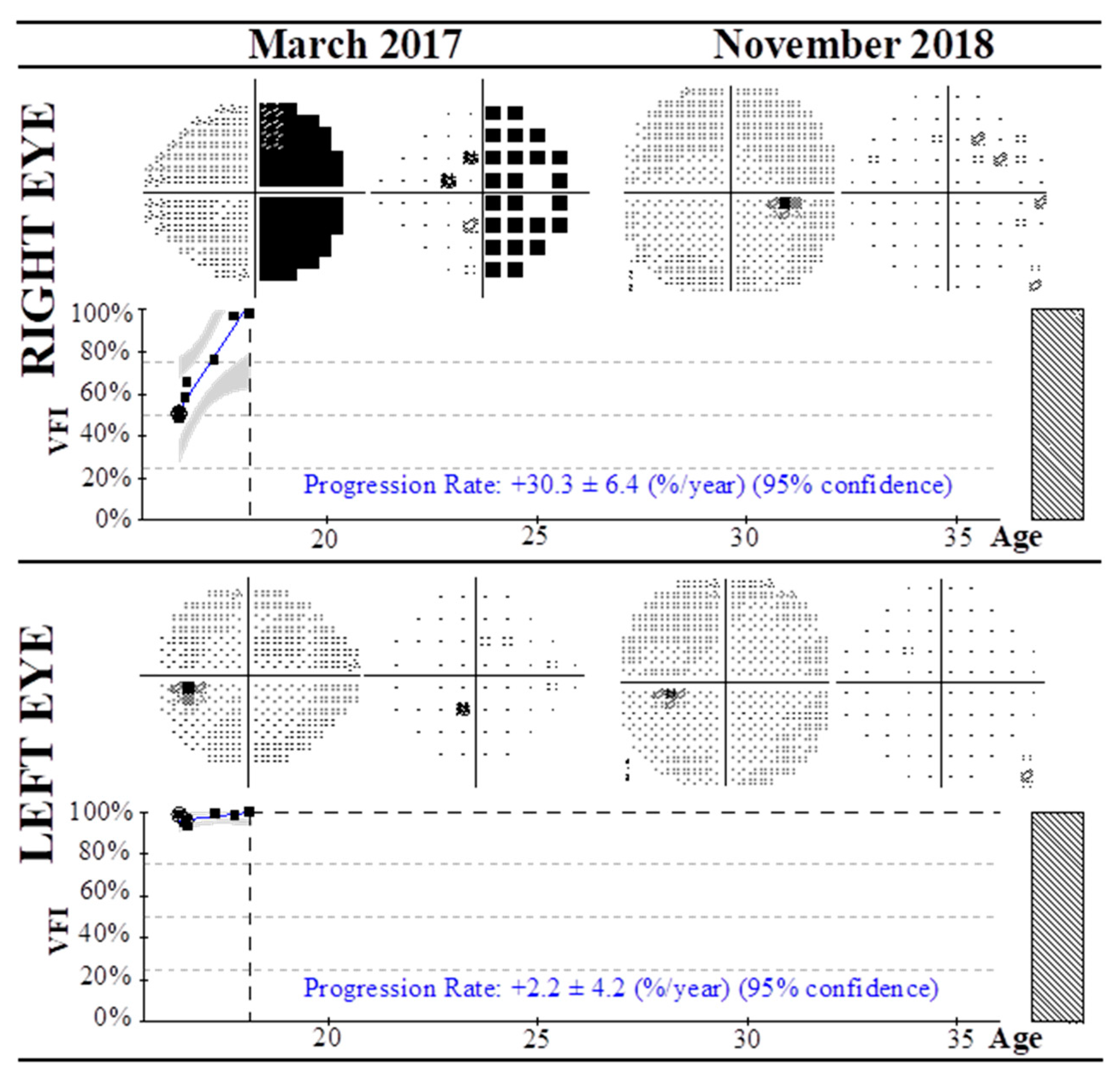
| Stroke04 | Right hemianopia (unknown origin) | 20 5 | Right | 51 | 99 | −17.21 | −1.18 | +30.3 ± 6.4 |
| Left | 99 | 100 | −2.37 | −0.58 | +2.2 ± 4.2 | |||
| Stroke05 | Right inferior homonymous quadrantanopia (left superior retrochiasmal lesion) | 13 3 | Right | 77 | 81 | −11.21 | −9.94 | +2.6 ± 15.7 |
| Left | 62 | 90 | −14.01 | −4.28 | +20.5 ± 14.4 | |||
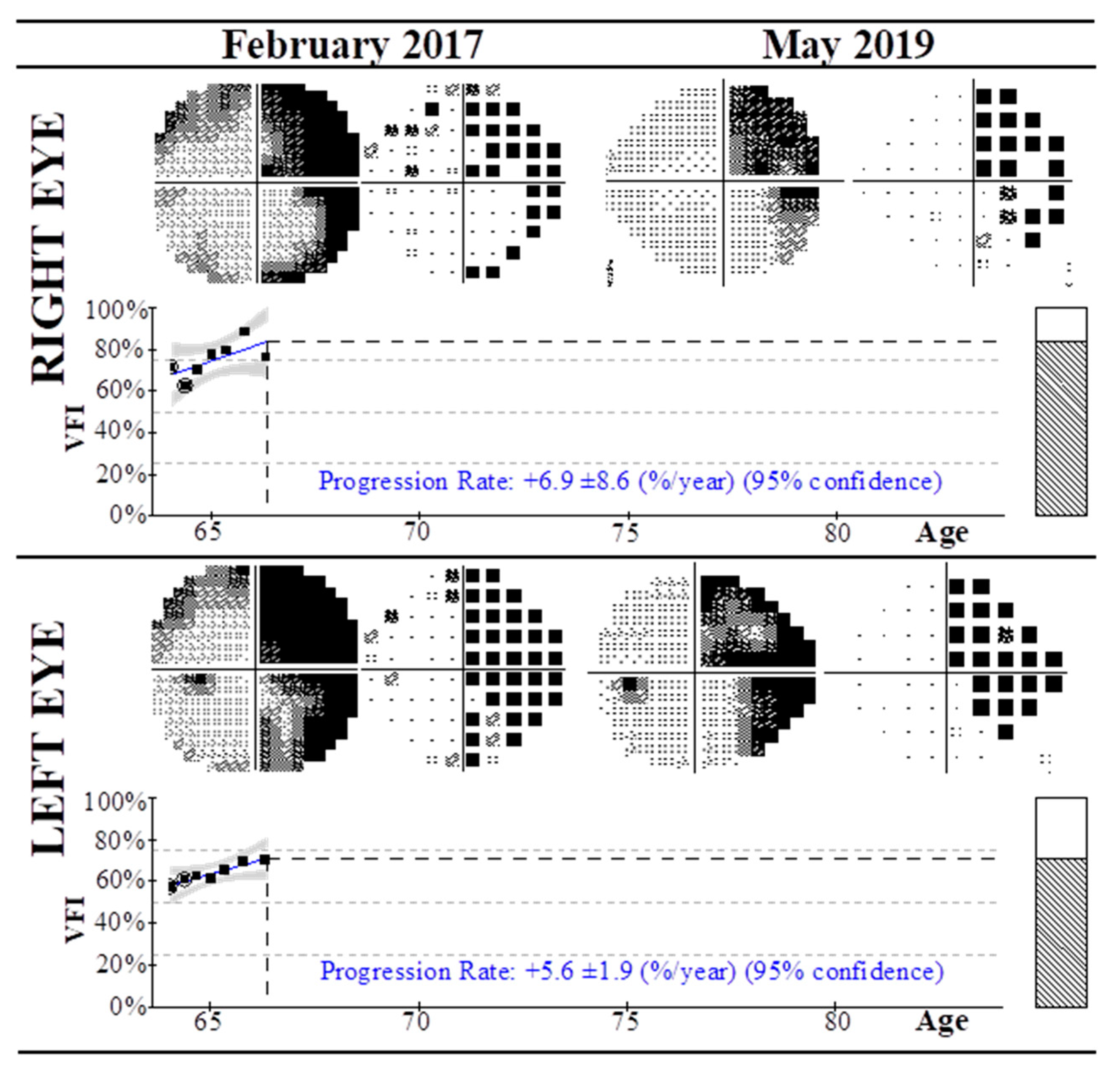
| Stroke06 | Right homonymous incomplete hemianopia (left occipital lobe stroke) | 27 3 | Right | 72 | 76 | −12.57 | −6.92 | +6.9 ± 8.6 |
| Left | 58 | 71 | −16.09 | −12.77 | +5.6 ± 1.9 | |||
| Stroke07 | Stroke (cerebrovascular ictus) with peripheral alteration | 22 6 | Right | 96 | 94 | −3.34 | −2.53 | +3.3 ± 3.8 |
| Left | 90 | 88 | −5.97 | −4.31 | +4.9 ± 4.6 | |||
| Stroke08 | Left inferior homonymous quadrantanopia (right superior retrochiasmal lesion) | 90 7 | Right | 88 | 91 | −5.62 | −1.55 | +2.3 ± 3.1 |
| Left | 89 | 90 | −5.36 | −5.24 | +3.1 ± 3.8 | |||
| Stroke09 | Right homonymous hemianopia (left retrochiasmal lesion) | 12 3 | Right | 56 | 65 | −14.34 | −10.2 | +7.7 ± 5.2 |
| Left | 50 | 57 | −16.84 | −15.31 | +3.8 ± 23.8 | |||
| Stroke10 | Left homonymous hemianopia (right retrochiasmal lesion) | 12 3 | Right | 27 | 48 | −22.53 | −18.96 | +33.3 ± 10.7 |
| Left | 26 | 35 | −21.28 | −17.62 | +16.0 ± 4.5 |
| Case | Disease | Follow-Up Months | Eye | Initial VFI (%) | Final VFI (%) | Initial MD (dB) | Final MD (dB) | VFI Progression Rate (%/year) 1 |
|---|---|---|---|---|---|---|---|---|
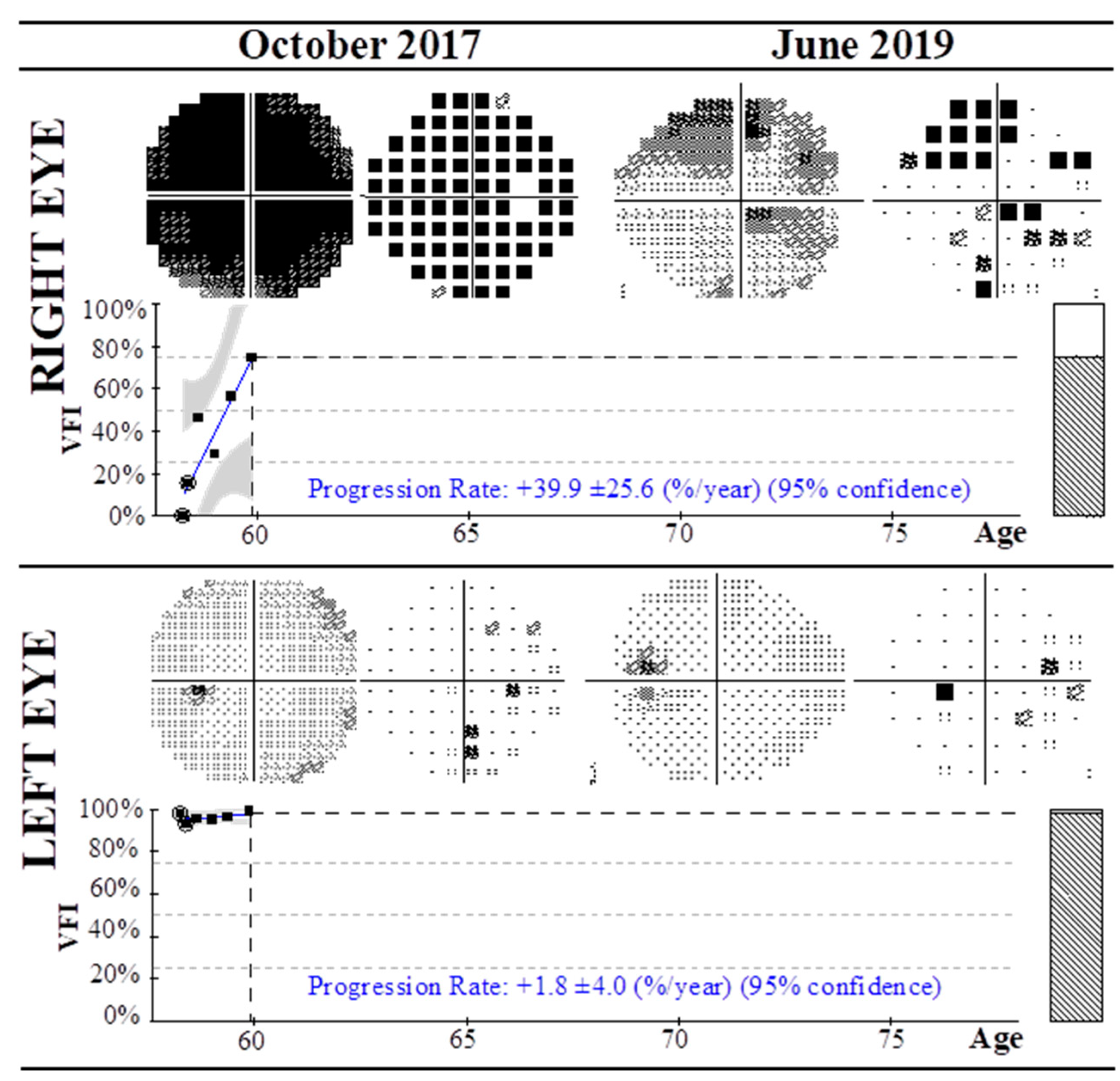
| RAO01 | CRAO | 20 2 | Right | 0 | 75 | −31.24 | −10.19 | +39.9 ± 25.6 |
| Left | 98 | 99 | −1.27 | 0.94 | +1.8 ± 4.0 | |||
| RAO02 | CRAO | 9 2 | Right | 0 | 46 | −31.88 | −17.3 | +49.4 ± 42.9 |
| Left | 96 | 94 | −3.15 | −2.19 | +1.0 ± 10.1 | |||
| RAO03 | RAO (temporal inferior with nasal superior quadrantanopia) | 4 2 | Right | 100 | 96 | 0.9 | −2.37 | −2.0 ± 1.9 |
| Left | 48 | 78 | −18.55 | −8.06 | +27.5 ± 12.7 | |||
| RAO04 | RAO (temporal inferior with optic nerve atrophy) | 7 2 | Right | 98 | 100 | −0.57 | 0.7 | +1.0 ± 0.8 |
| Left | 62 | 78 | −10.79 | −7.04 | +12.2.5 ± 10.6 | |||
| RAO05 | RAO (temporal inferior with superior hemianopia) | 15 2 | Right | 47 | 87 | −15.45 | −5.71 | +13.1 ± 16.9 |
| Left | 98 | 99 | −3.17 | −1.99 | +0.2 ± 0.2 | |||
| RAO06 | RAO (temporal superior) | 5 2 | Right | 56 | 61 | −14.44 | −11.09 | +8.9 ± 3.5 |
| Left | 98 | 98 | −2.27 | −1.54 | +0.0 ± 0.0 | |||
| RAO07 | RAO (temporal superior) | 53 2 | Right | 95 | 98 | −2.5 | −0.39 | +0.2 ± 1.1 |
| Left | 51 | 61 | −18.3 | −12.68 | +3.0 ± 5.3 |
| Case | Disease | Follow-Up Months | Eye | Initial VFI (%) | Final VFI (%) | Initial MD (dB) | Final MD (dB) | VFI Progression Rate (%/year) 1 |
|---|---|---|---|---|---|---|---|---|
| OC01 | Optic neuritis | 92 2 | Right | 87 | 97 | −9.54 | −2.61 | +0.9 ± 0.5 |
| Left | 71 | 94 | −9.89 | −3.19 | −2.4 ± 0.6 | |||
| OC02 | Optic neuritis causing optic nerve atrophy | 97 2 | Right | 99 | 98 | −1.44 | −2.22 | −0.4 ± 0.4 |
| Left | 70 | 91 | −11.12 | −7.06 | +0.8 ± 3.5 | |||
| OC03 | Bilateral optic neuritis | 16 2 | Right | 48 | 92 | −20.89 | −5.65 | +24.2 ± 28.2 |
| Left | 22 | 93 | −27.32 | −3.94 | +61.7 ± 32.9 | |||
| OC04 | Optic nerve atrophy (post-meningitis) | 63 2 | Right | 0 | 11 | −31.02 | −25.79 | +76.2 ± 12.3 |
| Left | 2 | 91 | −30.22 | −2.4 | +257.0 ± 42.30 | |||
| OC05 | Optic nerve atrophy (intracranial hypertension) | 110 2 | Right | 89 | 85 | −0.87 | −7.12 | +1.3 ± 3.8 |
| Left | 89 | 88 | 1.15 | −6.72 | +0.6 ± 2.4 | |||
| OC06 | Optic nerve atrophy (drug toxicity, etambutol) | 22 2 | Right | 86 | 99 | −4.58 | −0.28 | +4.2 ± 2.4 |
| Left | 87 | 99 | −5.67 | −0.1 | +7.1 ± 3.0 | |||
| OC07 | Neuroretinitis | 26 2 | Right | 70 | 88 | −11.47 | −2.31 | +6.8 ± 5.4 |
| Left | 100 | 100 | 4.06 | 4.18 | +0.1 ± 0.4 | |||
| OC08 | Tapetoretinal dystrophy (with superior hemianopia) | 34 2 | Right | 31 | 40 | −26.17 | −20.8 | +2.2 ± 2.9 |
| Left | 22 | 48 | −27.64 | −18.42 | +2.5 ± 3.9 | |||
| OC09 | Retinitis pigmentosa | 53 3 | Right | 12 | 49 | −27.92 | −19.46 | +0.0 ± 3.7 |
| Left | 19 | 49 | −26.88 | −19.85 | +0.4 ± 2.7 | |||
| OC10 | Unknown | 14 2 | Right | 5 | 80 | −28.75 | −7.54 | +15.5 ± 4.4 |
| Left | 11 | 88 | −27.76 | −8.5 | +10.0 ± 4.9 | |||
| OC11 | Unknown (optic nerve injury) | 68 4 | Right | 95 | 98 | −5.13 | −1.9 | +4.1 ± 24.6 |
| Left | 90 | 99 | −6.4 | −0.34 | +4.6 ± 22.0 | |||
| OC12 | Cone dystrophy | 103 5 | Right | 66 | 70 | −6.99 | −9.96 | +0.1 ± 1.7 |
| Left | 71 | 70 | −7.39 | −10.0 | −0.6 ± 1.1 | |||
| OC13 | Retinal vascular occlusion | 89 6 | Right | 99 | 99 | −0.22 | −0.51 | +0.6 ± 1.7 |
| Left | 31 | 58 | −24.44 | −17.84 | +5.2 ± 2.9 |
| p-Values (Wilcoxon Matched-Pairs Test) | ||
|---|---|---|
| Group | VFI (Initial vs. Final) (%) | MD (Initial vs. Final) (db) |
| NAION | < 0.0001 | < 0.0001 |
| Stroke | 0.0008 | < 0.0001 |
| RAO | 0.0081 | 0.0023 |
| OC | < 0.0001 | 0.0005 |
© 2020 by the authors. Licensee MDPI, Basel, Switzerland. This article is an open access article distributed under the terms and conditions of the Creative Commons Attribution (CC BY) license (http://creativecommons.org/licenses/by/4.0/).
Share and Cite
Fernández-Vega, B.; Nicieza, J.; Álvarez-Barrios, A.; Álvarez, L.; García, M.; Fernández-Vega, C.; Vega, J.A.; González-Iglesias, H. The Use of Vitamins and Coenzyme Q10 for the Treatment of Vascular Occlusion Diseases Affecting the Retina. Nutrients 2020, 12, 723. https://doi.org/10.3390/nu12030723
Fernández-Vega B, Nicieza J, Álvarez-Barrios A, Álvarez L, García M, Fernández-Vega C, Vega JA, González-Iglesias H. The Use of Vitamins and Coenzyme Q10 for the Treatment of Vascular Occlusion Diseases Affecting the Retina. Nutrients. 2020; 12(3):723. https://doi.org/10.3390/nu12030723
Chicago/Turabian StyleFernández-Vega, Beatriz, Javier Nicieza, Ana Álvarez-Barrios, Lydia Álvarez, Montserrat García, Carlos Fernández-Vega, José A. Vega, and Héctor González-Iglesias. 2020. "The Use of Vitamins and Coenzyme Q10 for the Treatment of Vascular Occlusion Diseases Affecting the Retina" Nutrients 12, no. 3: 723. https://doi.org/10.3390/nu12030723
APA StyleFernández-Vega, B., Nicieza, J., Álvarez-Barrios, A., Álvarez, L., García, M., Fernández-Vega, C., Vega, J. A., & González-Iglesias, H. (2020). The Use of Vitamins and Coenzyme Q10 for the Treatment of Vascular Occlusion Diseases Affecting the Retina. Nutrients, 12(3), 723. https://doi.org/10.3390/nu12030723