Article

Multiple Sclerosis Patients Show Lower Bioavailable 25(OH)D and 1,25(OH)2D, but No Difference in Ratio of 25(OH)D/24,25(OH)2D and FGF23 Concentrations

by
Mariska C Vlot
1,2,†,
Laura Boekel
1,†,
Jolijn Kragt
3,
Joep Killestein
4,
Barbara M. van Amerongen
5,
Robert de Jonge
1,
Martin den Heijer
2 and
Annemieke C. Heijboer
1,6,*
1
Department of Clinical Chemistry, Endocrine Laboratory, Amsterdam University Medical Center, 1081 HV Amsterdam, The Netherlands
2
Department of Internal Medicine, Amsterdam UMC, Amsterdam University medical Center, 1081 HV Amsterdam, The Netherlands
3
Department of Neurology, Reinier de Graaf Gasthuis, 2625 AD Delft, The Netherlands
4
Department of Neurology, Amsterdam Neuroscience, MS Center Amsterdam, Amsterdam University Medical Center, 1081 HV, Amsterdam, The Netherlands
5
Department of Molecular Cell Biology and Immunology, Amsterdam University Medical Center, Vrije Universiteit Amsterdam, 1081 HV Amsterdam, The Netherlands
6
Department of Clinical Chemistry, Endocrine Laboratory, Amsterdam University Medical Center, 1105 AZ Amsterdam, The Netherlands
*
Author to whom correspondence should be addressed.
Both authors contributed equally.
Nutrients 2019, 11(11), 2774; https://doi.org/10.3390/nu11112774
Submission received: 10 October 2019 / Revised: 31 October 2019 / Accepted: 13 November 2019 / Published: 15 November 2019
(This article belongs to the Special Issue Calcium, Vitamin D and Health)

Abstract

:
Vitamin D (VitD) insufficiency is common in multiple sclerosis (MS). VitD has possible anti-inflammatory effects on the immune system. The ratio between VitD metabolites in MS patients and the severity of the disease are suggested to be related. However, the exact effect of the bone-derived hormone fibroblast-growth-factor-23 (FGF23) and VitD binding protein (VDBP) on this ratio is not fully elucidated yet. Therefore, the aim is to study differences in total, free, and bioavailable VD metabolites and FGF23 between MS patients and healthy controls (HCs). FGF23, vitD (25(OH)D), active vitD (1,25(OH)2D), inactive 24,25(OH)D, and VDBP were measured in 91 MS patients and 92 HCs. Bioavailable and free concentrations were calculated. No difference in FGF23 (p = 0.65) and 25(OH)D/24.25(OH)2D ratio (p = 0.21) between MS patients and HCs was observed. Bioavailable 25(OH)D and bioavailable 1.25(OH)2D were lower (p < 0.01), while VDBP concentrations were higher in MS patients (p = 0.02) compared with HCs, specifically in male MS patients (p = 0.01). In conclusion, FGF23 and 25(OH)D/24.25(OH)2D did not differ between MS patients and HCs, yet bioavailable VitD concentrations are of potential clinical relevance in MS patients. The possible immunomodulating role of VDBP and gender-related differences in the VD-FGF23 axis in MS need further study.

1. Introduction

Multiple sclerosis (MS) is a chronic, progressive disease of the central nervous system characterized by an inflammatory, demyelinating, and neurodegenerative process, which can result in varying levels of disability. Consequently, MS has a negative effect on the daily life of patients, resulting from fatigue, muscle weakness, and imbalance to immobility, which all negatively affect bone health [1,2,3,4]. Earlier studies showed that decreased bone mineral density (BMD) is prevalent already shortly after clinical onset even in physically active patients with MS [2,4,5,6]. Lower BMD combined with impaired mobility can result in an increased fracture risk in MS patients and accompanying disability and economic burden [7]. The specific aetiology of MS is still unknown, but strong evidence exists regarding viral, genetic, and immunological causes of the disease [8,9].
Vitamin D is thought to play a role in the pathogenesis of MS, as it is known that the prevalence of MS increases with latitude, which in turn is associated with lower serum concentrations of vitamin D (25(OH)D) [10,11]. Vitamin D possibly modulates T-lymphocyte subset differentiation and, therefore, a lower concentration is thought to lead to an increased risk of MS [12]. In line with this, associations between lower serum concentrations 25(OH)D and an increased risk of MS are shown in several studies [13,14,15]. As a result, vitamin D supplementation is often advised to MS patients, although contra-dictionary effects regarding the beneficial effects of increased 25(OH)D concentrations on, for example, the recurrence rate of relapses, deterioration of number of brain laesions, and improvement of disability are described [16,17,18,19,20]. Because of the potential role of vitamin D in MS, other vitamin D metabolites have been studied in MS patients. 25(OH)D needs to be metabolized to the biologically active 1.25(OH)2D, and both metabolites are predominantly bound to their carrier vitamin D binding protein (VDBP). Previous research showed a higher plasma VDBP concentration in MS patients compared with healthy controls (HCs) [21]. VDBP is known to play an important role in the intracellular actin scavenging system by removing actin derived from damaged tissue and also promotes inflammation [22,23,24,25,26]. Polymorphisms of both VDBP and also of the vitamin D receptor (VDR) can result in a changed equilibrium between active and inactive vitamin D metabolites [24,27,28]. Interestingly, lower concentrations of the vitamin D metabolite 24.25(OH)2D were associated with a higher grade of disability based on the Expanded Disability Status Scale (EDSS) score in MS patients [29]. Moreover, the ratio of 25(OH)D/24.25(OH)2D was strongly inversely associated with brain parenchymal function [29].
A key player in vitamin D metabolism is the bone-derived hormone fibroblast growth factor 23 (FGF23), as it inhibits the enzyme 1-alpha hydroxylase and stimulates the enzyme 24-hydroxylase, resulting in the conversion of 25(OH)D into 24.25(OH)2D instead of into 1.25(OH)2D. We thus hypothesize that plasma FGF23 concentrations differ between MS patients and healthy controls.
The primary aim of this study is to further elucidate the vitamin D-FGF23 axis by measuring multiple D metabolites and FGF23 using accurate state-of-the art analytical methods in a well-defined cohort of MS patients and healthy controls. The second aim of our study is to assess bone turnover markers (BTMs) in our cohort MS patients and healthy controls and to study the possible associations of BTMs with vitamin D metabolites, as vitamin D deficiency is associated with increased bone turnover and lower bone mineral density [30,31,32]. Lastly, it is known that MS affects more women than men and that women have higher VDBP concentrations compared with men [13,21,33,34,35,36]. Therefore, possible gender-related differences of vitamin D metabolites and BTMs between MS patients and healthy controls will be studied.

2. Materials and Methods

2.1. Subjects and Study Protocol

The subjects and study protocol were described earlier by Kragt et al. [13]. Patients were eligible to participate in the study if they provided informed consent and were between 18 and 75 years of age. Three subtypes of MS were identified: relapsing-remitting MS (RRMS), secondary progressive MS (SPMS), and primary progressive MS (PPMS). Patients with all subtypes of MS were eligible to participate. In addition, patients with clinically isolated syndrome (CIS) were also included, which refers to a single episode of symptoms that are suggestive for MS. Patients were recruited between July and September 2003, to ensure that all blood samples were drawn during summer season. Patients were asked to bring a healthy control with them to the outpatient clinic if possible, preferably their partner in order to match the patients based on age and environmental factors. If a healthy control was lacking, hospital personnel volunteered to participate as controls. Patients were excluded if they were diagnosed with osteomalacia, hyperparathyroidism, hyperthyroidism, or hypercortisolism, or if they had been receiving glucocorticoid treatment in the previous three weeks (daily oral treatment or intravenous methylprednisolone treatment), anti-epileptic drugs, or vitamin D supplementation of more than 200 IU/day. The controls were excluded if they had MS or had a first-degree family member with MS. In addition, study participants were excluded in the case of suspicion of concomitant bone disease based on their laboratory results. All subjects gave their informed consent for inclusion before they participated in the study. The study was conducted in accordance with the Declaration of Helsinki, and the protocol was approved by the Medical Ethical Committee of the Amsterdam UMC, location VU University Medical Center (code 2003.029).

2.2. Measurements

2.2.1. General

The functional disability of MS patients was evaluated using the Expanded Disability Status Scale (EDSS) [37]. EDSS quantifies disability in MS patients, which ranges from 0 to 10, with higher scores representing higher levels of disability. Blood samples were collected before 00:00 and after an overnight fast. Samples were collected in 2003 and stored (both as serum and EDTA plasma in several aliquots) in minus 80 degrees Celsius. Analyses were performed in 2011 (parathyroid hormone (PTH), alkaline phosphatase (ALP), albumin, calcium, phosphate, estimated glomerular filtration rate (eGFR)) and in 2017 (25(OH)D, 24.25(OH)2D, 1.25(OH)2D, VDBP, FGF23, c-terminal telopeptide (CTX), osteocalcin, procollagen type 1 N-terminal propeptide (P1NP)) in the various aliquots.

2.2.2. Vitamin D Metabolites

In the current study, 25(OH)D and 24.25(OH)2D were measured in serum using a dedicated isotope dilution liquid chromatography-tandem mass spectrometry (ID-LC-MS/MS) [38]. Total serum concentrations of 25(OH)D were calculated by the sum of 25(OH)D2 and 25(OH)D3. For 25(OH)D2, the lower limit of quantitation (LLOQ) was 0.36 nmol/L and the inter-assay coefficient of variation (CV) was 6%. For 25(OH)D3, LLOQ was 1.19 nmol/L and the inter-assay CV was 6%. For 24.25(OH)2D, LLOQ was 0.12 nmol/L and the inter-assay CV was 5% [37,39]. A two-dimensional isotope dilution ultra-pressure liquid chromatography tandem mass spectrometry (2D ID-UPLC-MS/MS) was used to measure serum 1.25(OH)2D with an LLOQ of 3.4 pmol/L and inter-assay CV of 11% [40]. It is of note that, in the earlier study of Kragt et al., a radioimmunoassay was used for the measurement of 25(OH)D and 1.25(OH)2D [13], but the concentrations shown in the current study are obtained using an LC-MS/MS method. VDBP was measured using a polyclonal ELISA (Immundiagnostik AG) with a LLOQ of 2.2 µg/L and an inter-assay CV of <13%.

2.2.3. Free and Bioavailable Vitamin D Metabolites

Vitamin D metabolites bound to protein are not biologically active, whereas the unbound hormones are. Free 25(OH)D was calculated using equations and affinity constants according to Malmstroem et al. [41] and free 1.25(OH)2D was calculated using equations and affinity constants according to Bikle et al. [42]. Bioavailable 25(OH)D and 1.25(OH)2D were calculated using equations adapted from Vermeulen et al. [43] and the supplement of Powe et al. [44].

2.2.4. Bone Turnover Markers (BTMs)

FGF23 was measured in EDTA plasma using a c-terminal immunoassay (Immutopics) with an LLOQ of 20 RU/mL and inter-assay CV of <10% [45]. CTX and P1NP were measured in EDTA plasma using an immunoassay (Cobas, Roche Diagnostics, Almere, The Netherlands), with an LLOQ of 10 ng/L and inter-assay CV of <6.5% for CTX and an LLOQ of 5 µg/L and inter-assay CV of <8% for P1NP, respectively. Osteocalcin was measured in EDTA plasma using an immunometric-assay (Biosource, Nivelles, Belgium) with an LLOQ of 0.4 nmol/L and inter-assay CV of 8–15%.

2.2.5. Other Measurements

PTH was measured in EDTA plasma using an immunoassay (Architect, Abbott Diagnosher, Chicago, IL, USA) with an LLOQ 0.5 pmol/L and inter-assay CV of <9%. ALP, eGFR, calcium, phosphate, and albumin were measured in heparin plasma using the chemistry module of the Cobas (Roche Diagnostics).

2.3. Statistical Analysis
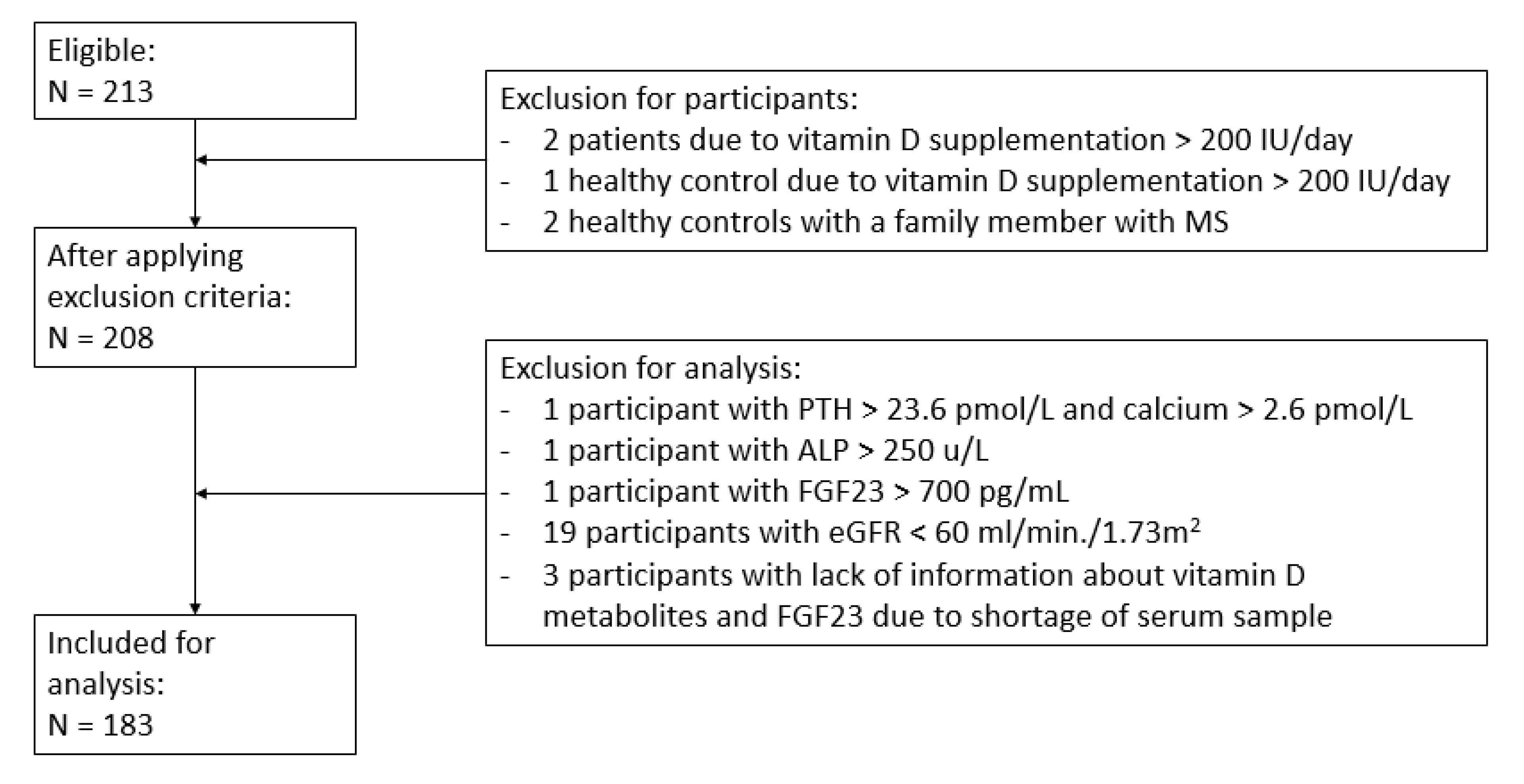
Baseline characteristics between MS patients and healthy controls were compared using a Student’s t-test or chi-square test. The Mann–Whitney U test was used in the case of non-parametric variables. To detect differences between patients and controls in biochemical indices of the vitamin D metabolites and BTMs, the Mann–Whitney U test was used as well. In addition, separate analyses for men and women were performed. Lastly, correlations between serum vitamin D metabolites, BTMs, and EDSS were calculated using Spearman correlations owing to non-parametric variables. All statistical analyses were carried out using SPSS software, version 23.0.

3. Results

3.1. General

A total of 213 participants were eligible to be included in this study. After applying the in- and exclusion criteria, a total of 30 participants were excluded; see Figure 1. The final study population that was included in the analyses consisted of 183 participants based on 91 MS patients and 92 healthy controls. Table 1 summarizes the baseline characteristics of all participants. In total, 61% of the MS patients were female. Relapsing remitting MS was the most predominant subtype of MS (57%).

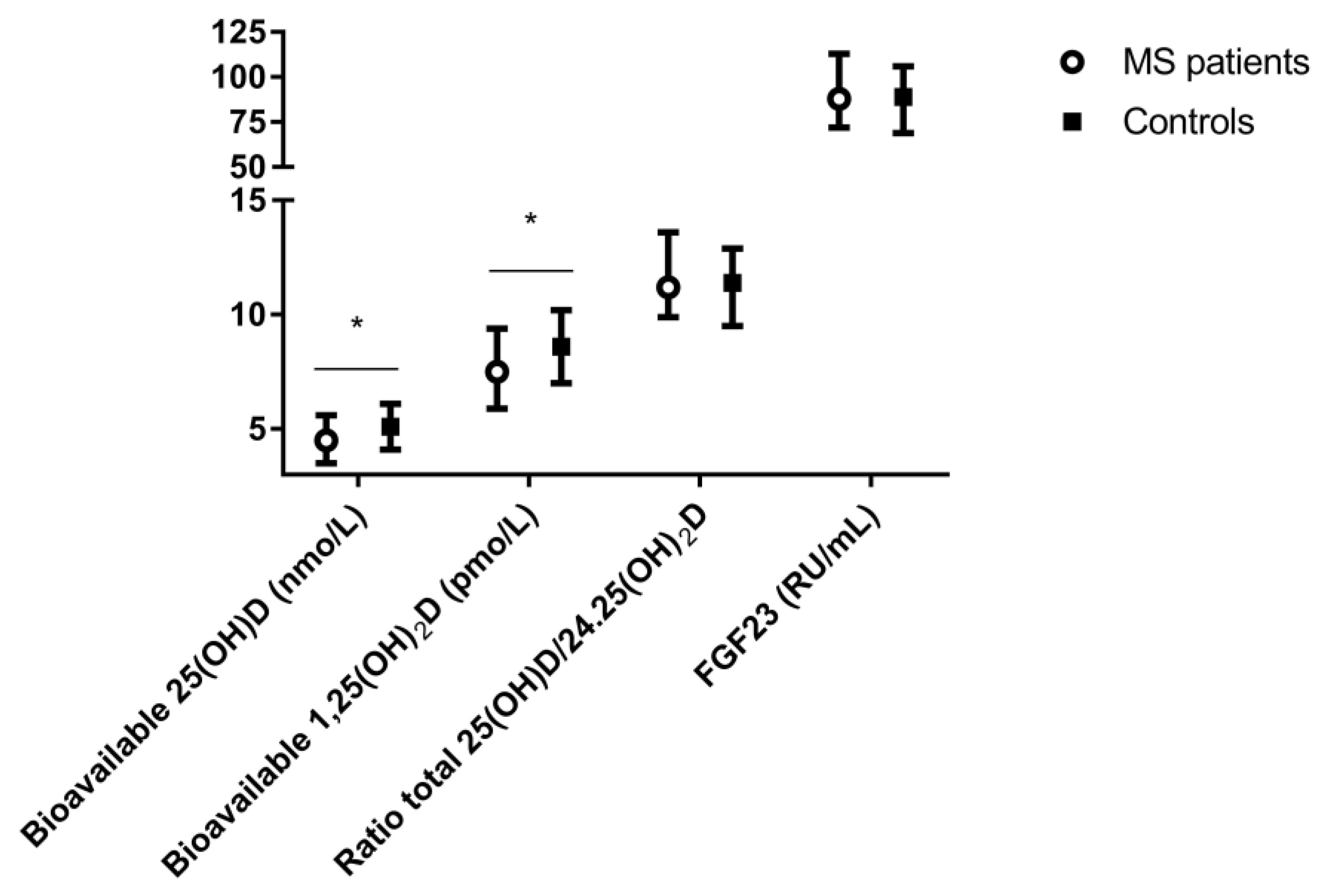
3.2. Vitamin D Metabolites, FGF23, and Bone Turnover Markers

Biochemical indices of vitamin D metabolism and bone turnover of the study population are displayed in Table 1. Again, it is of note that, in contrast to the earlier study of Kragt et al., current 25(OH)D and 1.25(OH)2D measurements are reported based on the ID-LC-MS/MS measurements [13]. Overall, no significant difference between total serum concentrations of 25(OH)D (p = 0.06) was found between MS patients and the healthy controls. MS patients had significant lower serum concentrations of free, albumin bound, and bioavailable 25(OH)D and 1.25(OH)2D compared with healthy controls (p < 0.01). In addition, MS patients had higher concentrations of phosphate and VDBP compared with controls, whereas no significant differences in BTMs were found. Plasma FGF23 concentrations did not differ between MS patient and the healthy controls (p = 0.65) (Figure 2).
Table 2 shows the significant results of separate analyses for men and women. In contrast to male patients, female MS patients had lower serum concentrations of total 25(OH)D, 25(OH)D3, 24.25(OH)2D, free 25(OH)D, and free 1.25(OH)2D compared with healthy female controls (p = 0.04). Male MS patients had higher serum concentrations of VDBP compared with male controls (p = 0.01). No other significant differences were found between male MS patients and healthy male controls. Regarding BTMs, no significant differences were found between males and females (data not shown). Lastly, EDSS scores did not differ between male and female MS patients (p = 0.77, data not shown).

3.3. Associations

3.3.1. Associations between Vitamin D Metabolites, FGF23, Bone Turnover Markers, and EDSS in MS Patients

In MS patients, all vitamin D metabolites correlated strongly with each other (all r > 0.77; p < 0.01, data not shown). EDSS showed negative correlations with bioavailable 1.25(OH)2D, bioavailable 25(OH)D, and 24.25(OH)2D (r −0.30, p < 0.01; r −0.30, p < 0.01; and r −0.23, p = 0.03, respectively). No correlations between EDSS and FGF23 or BTMs in MS patients were found (data not shown).
As shown in Table 3, FGF23 correlated positively with serum 25(OH)D and serum 24.25(OH)2D (r 0.22, p = 0.04 and r 0.22, p = 0.04, respectively), and CTX correlated negatively with serum 24.25(OH)2D and 25(OH)D (r −0.31, p < 0.01 and r −0.23, p = 0.03, respectively) in MS patients. Phosphate showed a positive correlation with osteocalcin (r 0.22, p 0.04). Negative correlations for osteocalcin with 1.25(OH)2D, and P1NP with 24.25(OH)2D, were observed in MS patients only (r −0.24, p = 0.02 and r −0.25, p = 0.02, respectively), as were positive correlations of the ratio between total serum 25(OH)D and 24.25(OH)2D with P1NP and CTX (r 0.27, p = 0.01 and r 0.31, p < 0.01, respectively). In addition, in MS patients, ALP showed a positive correlation with osteocalcin, CTX, and P1NP (r 0.36, p < 0.01; r 0.44, p < 0.01; and r 0.43, p < 0.01, respectively); P1NP showed a positive correlation with osteocalcin and CTX (r 0.75, p < 0.01 and r 0.74, p < 0.01, respectively); and CTX correlated positively with osteocalcin (r 0.67, p < 0.01).

3.3.2. Associations Based on Gender of MS Patients

Looking at gender differences, in female MS patients, the same correlations were found as described above, except for the correlations of FGF23 with serum 25(OH)D and 24.25(OH)2D (r 0.12, p = 0.37 and r 0.10, p = 0.44, respectively). In male MS patients, however, FGF23 correlated positively with total (r 0.50, p < 0.01), free (r 0.43, p = 0.02), and bioavailable 25(OH)D (r 0.43, p = 0.02); free (r 0.43, p 0.02) and bioavailable 1.25(OH)2D (r 0.43, p 0.02); and 24.25(OH)2D (r 0.46, p = 0.01), respectively. Furthermore, EDSS showed negative correlations with CTX (r −0.53, p < 0.01), P1NP (r −0.48, p < 0.01), and osteocalcin (r −0.40, p 0.03) in male MS patients only.

3.3.3. Associations in Healthy Controls

Lastly, in healthy controls, similar correlations of osteocalcin, CTX, and P1NP were found (data not shown). FGF23 correlated significantly with serum 1.25(OH)2D (r −0.35, p < 0.01; data not shown), and CTX correlated significantly with serum 25(OH)D (r −0.24, p = 0.05; data not shown). In addition, PTH showed a positive correlation with ALP (r 0.21, p 0.04; data not shown) and phosphate showed a negative correlation with P1NP (r −0.27, p 0.03; data not shown) in the healthy control group.

4. Discussion

This study examined differences in total, free, and bioavailable vitamin D, FGF23 and bone turnover markers in patients with MS compared with healthy controls and possible gender differences. Although positive correlations between FGF23 and total 25(OH)D and 24.25(OH)2D in MS patients were seen, no differences in plasma FGF23 concentrations between MS patients and healthy controls were observed. No differences between serum concentrations of BTMs in MS patients and healthy controls were found. Yet, we did observe a negative correlation between CTX and total 25(OH)D and 24.25(OH)2D. EDSS showed negative correlations with bioavailable 1.25(OH)2D, bioavailable 25(OH)D, and 24.25(OH)2D. We found gender differences in vitamin D metabolism: serum concentrations of total 25(OH)D, 25(OH)D3, 24.25(OH)2D, free 25(OH)D, and free 1.25(OH)2D were lower in female MS patients compared with female healthy controls. Serum concentrations of VDBP were higher in male MS patients compared with healthy male controls.
The main aim of this study was to further elucidate the vitamin D-FGF23 axis by measuring multiple D metabolites and FGF23 using accurate state-of-the art analytical methods in a well-defined cohort of MS patients versus healthy controls. We confirmed the finding of an earlier study that the serum 24.25(OH)2D concentration is negatively correlated with EDSS [29]. Although we found a significant positive correlation between FGF23 and 25(OH)D and 24.25(OH)2D, we did not observe a difference in FGF23 concentration between MS patients and healthy controls. A previous study described comparable plasma concentrations of FGF23 in MS patients compared with healthy controls as well [46], although two other studies found higher serum concentrations of FGF23 in MS patients compared with healthy controls [47,48]. However, the latter study was performed in patients with RRMS only and these differences were found during autumn (September–November) and winter time, respectively. Therefore, seasonal effects or the use of different FGF23 assays measuring either intact or c-term FGF23 could have resulted in these different findings. We found a higher phosphate concentration in MS patients; nevertheless, no differences in PTH or eGFR compared with healthy controls were found. Lastly, no correlations between FGF23 and 1.25(OH)2D or EDSS were found in MS patients, which is in line with two other recent studies [46,48]. Summarized, these findings do not support our hypothesis that FGF23 differs between MS patients and healthy controls.
Regarding the various vitamin D metabolites, we found similar concentrations of 25(OH)D and 1.25(OH)2D between MS patients and healthy controls, as some showed earlier [13,49,50], whereas others found lower serum concentrations of 25(OH)D or 1.25(OH)2D in MS patients compared with healthy controls, respectively [15,51,52,53,54,55,56,57]. These differences between studies might be caused by differences in sample size, analysis of both male and female participants in different seasons of the year, use of different assays to measure vitamin D and its metabolites, and possible VDBP polymorphisms [24,27,28]. In addition, a number of studies did report a relationship between lower serum concentrations of 1,25(OH)2D and an increased risk of MS [51,52]. Furthermore, our study showed that MS patients were using supplementation of vitamin D more often compared with their healthy controls. However, the maximal supplementation dosage was <200 IU of vitamin D per day, of which no clinical relevant effect on the vitamin D metabolites is expected, as a higher supplementation dosage is normally advised [18,19,29]. Moreover, controls had similar baseline concentrations of 25(OH)D.
As suggested in recent studies, bioavailable 25(OH)D might be preferred above total 25(OH)D as a marker for vitamin D status, and might thus be a better marker of mineral metabolism [58,59,60,61,62]. The sum of the albumin bound fraction of 25(OH)D plus the freely circulating 25(OH)D results in the bioavailable 25(OH)D, which can be calculated similarly for bio-available 1.25(OH)2D, respectively [58,60]. In the circulation, VDBP and albumin bind over 99% of the 25(OH)D and 1.25(OH)2D [60], whereas the binding affinity of vitamin D to albumin is much lower than the binding affinity to VDBP [63]. We found lower serum concentrations of both free and bioavailable 25(OH)D and 1.25(OH)2D, respectively, in MS patients compared with healthy controls, in contrast to comparable serum concentrations of total 25(OH)D and 1.25(OH)2D between MS patients and healthy controls. In contrast to our findings, Behrens et al. found no difference in free and bioavailable 25(OH)D between MS patients and controls, yet 1.25(OH)2D was not measured in this study [64]. However, their study included patients with CIS only (not yet diagnosed with MS), used non-parametrical statistical tests, and a de-seasonalized concentration of vitamin D was calculated afterwards. Our study measured summer concentrations of vitamin D only and included all subtypes of MS, which may explain the different findings.
The second aim of our study was to assess BTMs in MS patients versus healthy controls and to study the possible associations of BTMs with vitamin D metabolites, as vitamin D deficiency is associated with increased bone turnover [32]. Our study did not show any differences between BTMs and FGF23 in MS patients and their healthy controls. This finding is in line with earlier studies, where similar serum concentrations of CTX, P1NP, or osteocalcin between MS patients and healthy controls were found, and similar concentrations of cross-linked N terminal telopeptide type 1 collagen (NTX) and bone ALP in newly diagnosed MS patients and healthy controls were found [18,32,65,66,67]. The current study shows that, despite differences in vitamin D metabolites, bone turnover in MS patients seems to not be affected compared with healthy controls.
Lastly, possible gender-related differences of vitamin D metabolites and BTMs between MS patients and healthy controls were studied; as known, MS affects more women than men and, in general, women have higher VDBP concentrations compared with men [13,21,33,34,35,36]. Indeed, our study showed more female MS patients, of which the largest part was post-menopausal. Further analyses based on pre- or postmenopausal status was not possible because of small groups. Our study showed lower total and free 25(OH)D, free 1.25(OH)2D, and 24.25(OH)2D only in female MS patients compared with healthy female controls, but no difference in this respect between male MS patients compared with healthy men. No differences between FGF23 in male and female MS patients were found. Interestingly, in our study, VDBP was higher in MS patients compared with controls, but after further analysis, this difference was found in male MS patients only. An earlier study found that serum VDBP concentrations were higher in MS patients compared with healthy controls as well; however, this study included both female and males [21]. In contrast, other studies showed no difference in VDBP concentrations between MS patients and controls [64,68]. The differences between VDBP concentrations in the various studies could be the result of variation between numbers of male and female patients and controls included in these studies or by using either a polyclonal or monoclonal assay. It is known that VDBP can affect inflammatory processes [22,23,25,26,61,69,70]. As we found higher VDBP in male MS patients, in the presence of a similar EDSS score as in female MS patients, this suggests a possible modulating effect of VDBP in male MS patients. It was shown before that higher concentrations of VDBP restrict the uptake of free vitamin D metabolites and reduce anti-inflammatory responses of immune cells [71,72,73,74]. Moreover, VDBP can act as a chemotactic cofactor, which enhances chemotaxis of neutrophils and macrophages by complement factor C5a [23]. These macrophages are known to contribute to laesion formation and axonal damage [22,26]. Previous experimental studies showed that T-lymphocytes, glia cells, and neurons express 1-α-hydroxlyase and VDR, which enables them to convert 25(OH)D to 1.25(OH) [75,76,77]. Normally, in the central nervous system, focal inflammation is initiated by auto-reactive T-helper cells type 1 (Th 1) and T-helper cells type 17 (Th 17) [78]. The activated vitamin D is thought to induce anti-inflammatory effects in glia cells and neurons and affects vitamin D responsive genes in T-helper cells [76,79,80,81,82], thereby inhibiting pro-inflammatory T-helper cell activity (Th1 and Th 17) and promoting anti-inflammatory T-helper cell activity (Th2 and regulatory T-cells) [51,83]. However, whether vitamin D affects the T-cell response also depends on concentrations of VDBP [84,85]. On the basis of these studies, the elevated concentrations of VDBP found in our study strengthen the pro-inflammatory role of VDBP in enhancing neuronal damage in MS.
Lastly, EDSS scores were comparable between male and female MS patients, which is consistent with previous research [86,87]. In female MS patients, however, EDSS showed negative correlations with free and bioavailable 1.25(OH)2D; free, bioavailable, and total 25(OH)D; and 24.25(OH)2D, which is in line with earlier studies, which reported a negative correlation between EDSS and 1.25(OH)2D or 25(OH)D, respectively [46,88]. In contrast, in male MS patients, negative correlations between EDSS and CTX, P1NP, and osteocalcin were observed, which differs from previous studies, which found no correlations between EDSS and BTMs or only a positive correlation between EDSS and CTX [67,89].
The strengths of this study are the relative large study population and the broad spectrum of vitamin D metabolites, FGF23, and BTMs that was measured. Accurate and well-standardized LC-MS/MS assays were used to measure vitamin D metabolites. In addition, to minimize seasonal changes known to affect vitamin D metabolism [48], only blood samples drawn during summertime were used. There are also some possible limitations of this study. First, owing to the cross-sectional study design, the described differences between MS patients and controls in serum concentrations of vitamin D metabolites are not necessarily causal. In addition, 97% of our study population is Caucasian, which reduces the generalizability of the results, as it is known that vitamin D metabolism differs between races [53]. Moreover, our study was not primarily designed to study gender difference, so the differences we found in this respect should be studied further in other cohorts. No data on fractures were available. Lastly, dual energy X-ray absorptiometry (DXA) scans were not available to compare vitamin D metabolites and bone turnover markers with BMD.

5. Conclusions

In conclusion, this study provides additional knowledge of vitamin D-FGF 23 axis and bone turnover markers in MS patients compared with healthy controls, as well as gender-related differences. Similar serum concentrations of total 25(OH)D and 1.25(OH)2D in MS patients and healthy controls were found. Furthermore, no differences in plasma FGF23 concentrations and other bone turnover markers between MS patients and healthy controls were observed. The ratio total 25(OH)D/ 24.25(OH)2D did not differ between MS patients and healthy controls. However, this study suggested a relevant gender difference as serum concentrations of total 25(OH)D, 24.25(OH)2D, free 25(OH)D, and free 1.25(OH)2D were lower in female MS patients compared with female healthy controls, while serum concentrations of VDBP were higher in male MS patients compared with male controls. This study thus shows that only a total 25(OH)D measurement probably does not reflect all changes in vitamin D metabolism in MS patients. The exact role of VDBP and its polymorphisms in MS needs further studies. Finally, given the high incidence of reduced bone mineral density and the still partially unknown mechanisms that affect bone turnover in MS patients [66], further studies should be performed to evaluate the relationship between change in the vitamin D-FGF23 axis, BMD, and fracture risk in both male and female MS patients.

Author Contributions

Authors’ role: Study design: M.C.V., L.B., J.K. (Jolijn Kragt), M.d.H., and A.C.H.; Data collection: M.C.V., L.B., and J.K. (Jolijn Kragt); Data analysis: M.C.V. and L.B.; Drafting manuscript: M.C.V. and L.B.; Revising manuscript content: M.C.V., L.B., J.K. (Jolijn Kragt), J.K. (Joep Killestein), B.M.v.A., R.d.J., M.d.H., and A.C.H.; Approving final version of manuscript: A.C.H. A.C.H. takes responsibility for the integrity of the data analysis.

Funding

This research received no external funding.

Conflicts of Interest

The authors declare no conflict of interest.

References

  1. Gupta, S.; Ahsan, I.; Mahfooz, N.; Abdelhamid, N.; Ramanathan, M.; Weinstock-Guttman, B. Osteoporosis and Multiple Sclerosis: Risk Factors, Pathophysiology, and Therapeutic Interventions. CNS Drugs 2014, 28, 731–742. [Google Scholar] [CrossRef]
  2. Holmøy, T.; Kampman, M.T.; Smolders, J. Vitamin D in multiple sclerosis: Implications for assessment and treatment. Expert Rev. Neurother. 2012, 12, 1101–1112. [Google Scholar] [CrossRef]
  3. Huang, Z.; Qi, Y.; Du, S.; Chen, G.; Yan, W. BMI levels with MS Bone mineral density levels in adults with multiple sclerosis: A meta-analysis. Int. J. Neurosci. 2015, 125, 904–912. [Google Scholar] [CrossRef]
  4. Nieves, J.; Cosman, F.; Herbert, J.; Shen, V.; Lindsay, R. High prevalence of vitamin D deficiency and reduced bone mass in multiple sclerosis. Neurology 1994, 44, 1687. [Google Scholar] [CrossRef] [PubMed]
  5. Moen, S.M.; Celius, E.G.; Sandvik, L.; Nordsletten, L.; Eriksen, E.F.; Holmøy, T. Low bone mass in newly diagnosed multiple sclerosis and clinically isolated syndrome. Neurology 2011, 77, 151–157. [Google Scholar] [CrossRef] [PubMed]
  6. Steffensen, L.H.; Mellgren, S.I.; Kampman, M.T. Predictors and prevalence of low bone mineral density in fully ambulatory persons with multiple sclerosis. J. Neurol. 2010, 257, 410–418. [Google Scholar] [CrossRef] [PubMed]
  7. Dobson, R.; Ramagopalan, S.; Giovannoni, G.; Bazelier, M.T.; De Vries, F. Risk of fractures in patients with multiple sclerosis: A population-based cohort study. Neurology 2012, 79, 1934–1935. [Google Scholar] [CrossRef] [PubMed]
  8. Thompson, A.J.; Baranzini, S.E.; Geurts, J.; Hemmer, B.; Ciccarelli, O. Multiple sclerosis. Lancet 2018, 391, 1622–1636. [Google Scholar] [CrossRef]
  9. Tobore, T.O. Towards a Comprehensive Etiopathogenetic and Pathophysiological Theory of Multiple Sclerosis. Int. J. Neurosci. 2019, 1–41. [Google Scholar] [CrossRef]
  10. Acheson, E.D.; Bachrach, C.A.; Wright, F.M. Some comments on the relationship of the distribution of multiple sclerosis to latitude, solar radiation, and other variables. Acta Psychiatr. Scand. 1960, 35, 132–147. [Google Scholar] [CrossRef]
  11. Kurtzke, J.F. Geography in multiple sclerosis. J. Neurol. 1977, 215, 1–26. [Google Scholar] [CrossRef] [PubMed]
  12. Hemmer, B.; Kerschensteiner, M.; Korn, T. Role of the innate and adaptive immune responses in the course of multiple sclerosis. Lancet Neurol. 2015, 14, 406–419. [Google Scholar] [CrossRef]
  13. Kragt, J.; Van Amerongen, B.; Killestein, J.; Dijkstra, C.; Uitdehaag, B.; Polman, C.; Lips, P. Higher levels of 25-hydroxyvitamin D are associated with a lower incidence of multiple sclerosis only in women. Mult. Scler. J. 2009, 15, 9–15. [Google Scholar] [CrossRef] [PubMed]
  14. Munger, K.L.; Levin, L.I.; Hollis, B.W.; Howard, N.S.; Ascherio, A. Serum 25-Hydroxyvitamin D Levels and Risk of Multiple Sclerosis. JAMA 2006, 296, 2832–2838. [Google Scholar] [CrossRef]
  15. Soilu-Hänninen, M.; Airas, L.; Mononen, I.; Heikkilä, A.; Viljanen, M.; Hänninen, A. 25-Hydroxyvitamin D levels in serum at the onset of multiple sclerosis. Mult. Scler. J. 2005, 11, 266–271. [Google Scholar] [CrossRef]
  16. Berezowska, M.; Coe, S.; Dawes, H. Effectiveness of Vitamin D Supplementation in the Management of Multiple Sclerosis: A Systematic Review. Int. J. Mol. Sci. 2019, 20, 1301. [Google Scholar] [CrossRef]
  17. Fitzgerald, K.C.; Munger, K.L.; Köchert, K.; Arnason, B.G.W.; Comi, G.; Cook, S.; Goodin, D.S.; Filippi, M.; Hartung, H.-P.; Jeffery, D.R.; et al. Association of Vitamin D Levels With Multiple Sclerosis Activity and Progression in Patients Receiving Interferon Beta-1b. JAMA Neurol. 2015, 72, 1458–1465. [Google Scholar] [CrossRef]
  18. Holmøy, T.; Lindstrøm, J.C.; Eriksen, E.F.; Steffensen, L.H.; Kampman, M.T. High dose vitamin D supplementation does not affect biochemical bone markers in multiple sclerosis—A randomized controlled trial. BMC Neurol. 2017, 17, 67. [Google Scholar] [CrossRef]
  19. Jagannath, V.A.; Filippini, G.; Di Pietrantonj, C.; Asokan, G.V.; Robak, E.W.; Whamond, L.; Robinson, S.A. Vitamin D for the management of multiple sclerosis. Cochrane Database Syst. Rev. 2018, 9, CD008422. [Google Scholar] [CrossRef]
  20. Sintzel, M.B.; Rametta, M.; Reder, A.T. Vitamin D and Multiple Sclerosis: A Comprehensive Review. Neurol. Ther. 2018, 7, 59–85. [Google Scholar] [CrossRef]
  21. Rinaldi, A.O.; Sanseverino, I.; Purificato, C.; Cortese, A.; Mechelli, R.; Francisci, S.; Salvetti, M.; Millefiorini, E.; Gessani, S.; Gauzzi, M.C. Increased Circulating Levels of Vitamin D Binding Protein in MS Patients. Toxins 2015, 7, 129–137. [Google Scholar] [CrossRef] [PubMed]
  22. Abdul-Majid, K.-B.; Stefferl, A.; Bourquin, C.; Lassmann, H.; Linington, C.; Olsson, T.; Kleinau, S.; Harris, R.A. Fc receptors are critical for autoimmune inflammatory damage to the central nervous system in experimental autoimmune encephalomyelitis. Scand. J. Immunol. 2002, 55, 70–81. [Google Scholar] [CrossRef] [PubMed]
  23. Binder, R.; Kress, A.; Kan, G.; Herrmann, K.; Kirschfink, M. Neutrophil priming by cytokines and vitamin D binding protein (Gc-globulin): Impact on C5a-mediated chemotaxis, degranulation and respiratory burst. Mol. Immunol. 1999, 36, 885–892. [Google Scholar] [CrossRef]
  24. Gauzzi, M.C. Vitamin D-binding protein and multiple sclerosis: Evidence, controversies, and needs. Mult. Scler. J. 2018, 24, 1526–1535. [Google Scholar] [CrossRef]
  25. Vasconcellos, C.; Lind, S. Coordinated inhibition of actin-induced platelet aggregation by plasma gelsolin and vitamin D-binding protein. Blood 1993, 82, 3648–3657. [Google Scholar] [CrossRef]
  26. Vogel, D.Y.; Vereyken, E.J.; Glim, J.E.; Heijnen, P.D.; Moeton, M.; Van Der Valk, P.; Amor, S.; Teunissen, C.E.; Van Horssen, J.; Dijkstra, C.D. Macrophages in inflammatory multiple sclerosis lesions have an intermediate activation status. J. Neuroinflamm. 2013, 10, 35. [Google Scholar] [CrossRef]
  27. Bermúdez-Morales, V.H.; Fierros, G.; Lopez, R.L.; Martínez-Nava, G.; Flores-Aldana, M.; Flores-Rivera, J.; Hernández-Girón, C. Vitamin D receptor gene polymorphisms are associated with multiple sclerosis in Mexican adults. J. Neuroimmunol. 2017, 306, 20–24. [Google Scholar] [CrossRef]
  28. Langer-Gould, A.; Lucas, R.M.; Xiang, A.H.; Wu, J.; Chen, L.H.; Gonzales, E.; Haraszti, S.; Smith, J.B.; Quach, H.; Barcellos, L.F. Vitamin D-Binding Protein Polymorphisms, 25-Hydroxyvitamin D, Sunshine and Multiple Sclerosis. Nutrients 2018, 10, 184. [Google Scholar] [CrossRef]
  29. Weinstock-Guttman, B.; Zivadinov, R.; Qu, J.; Cookfair, D.; Duan, X.; Bang, E.; Bergsland, N.; Hussein, S.; Cherneva, M.; Willis, L.; et al. Vitamin D metabolites are associated with clinical and MRI outcomes in multiple sclerosis patients. J. Neurol. Neurosurg. Psychiatry 2011, 82, 189–195. [Google Scholar] [CrossRef]
  30. Bischoff-Ferrari, H.A.; Dietrich, T.; Orav, E.; Dawson-Hughes, B. Positive association between 25-hydroxy vitamin d levels and bone mineral density: A population-based study of younger and older adults. Am. J. Med. 2004, 116, 634–639. [Google Scholar] [CrossRef]
  31. Holick, M.F. Vitamin D deficiency. N. Engl. J. Med. 2007, 357, 266–281. [Google Scholar] [CrossRef] [PubMed]
  32. Jorde, R.; Stunes, A.K.; Kubiak, J.; Joakimsen, R.; Grimnes, G.; Thorsby, P.M.; Syversen, U. Effects of vitamin D supplementation on bone turnover markers and other bone-related substances in subjects with vitamin D deficiency. Bone 2019, 124, 7–13. [Google Scholar] [CrossRef] [PubMed]
  33. Bolland, M.J.; Grey, A.B.; Ames, R.W.; Horne, A.M.; Mason, B.H.; Wattie, D.J.; Gamble, G.D.; Bouillon, R.; Reid, I.R. Age-, gender-, and weight-related effects on levels of 25-hydroxyvitamin D are not mediated by vitamin D binding protein. Clin. Endocrinol. 2007, 67, 259–264. [Google Scholar] [CrossRef] [PubMed]
  34. Spach, K.M.; Hayes, C.E. Vitamin D3 confers protection from autoimmune encephalomyelitis only in female mice. J. Immunol. 2005, 175, 4119–4126. [Google Scholar] [CrossRef] [PubMed] [Green Version]
  35. Woolmore, J.; Stone, M.; Pye, E.; Partridge, J.; Boggild, M.; Young, C.; Jones, P.; Fryer, A.; Hawkins, C.; Strange, R.; et al. Studies of associations between disability in multiple sclerosis, skin type, gender and ultraviolet radiation. Mult. Scler. J. 2007, 13, 369–375. [Google Scholar] [CrossRef]
  36. Yang, M.; Qin, Z.; Zhu, Y.; Li, Y.; Qin, Y.; Jing, Y.; Liu, S. Vitamin D-binding Protein in Cerebrospinal Fluid is Associated with Multiple Sclerosis Progression. Mol. Neurobiol. 2013, 47, 946–956. [Google Scholar] [CrossRef]
  37. Meyer-Moock, S.; Feng, Y.-S.; Maeurer, M.; Dippel, F.-W.; Kohlmann, T. Systematic literature review and validity evaluation of the Expanded Disability Status Scale (EDSS) and the Multiple Sclerosis Functional Composite (MSFC) in patients with multiple sclerosis. BMC Neurol. 2014, 14, 58. [Google Scholar] [CrossRef] [Green Version]
  38. Dirks, N.F.; Vesper, H.W.; Van Herwaarden, A.E.; Ouweland, J.M.V.D.; Kema, I.P.; Krabbe, J.G.; Heijboer, A.C. Various calibration procedures result in optimal standardization of routinely used 25(OH)D ID-LC-MS/MS methods. Clin. Chim. Acta 2016, 462, 49–54. [Google Scholar] [CrossRef] [Green Version]
  39. Dirks, N.F.; Ackermans, M.T.; De Jonge, R.; Heijboer, A.C. Reference values for 24,25-dihydroxyvitamin D and the 25-hydroxyvitamin D/24,25-dihydroxyvitamin D ratio. Clin. Chem. Lab. Med. 2019, 57, e259–e261. [Google Scholar] [CrossRef]
  40. Dirks, N.F.; Martens, F.; Vanderschueren, D.; Billen, J.; Pauwels, S.; Ackermans, M.T.; Endert, E.; Heijer, M.D.; Blankenstein, M.A.; Heijboer, A.C. Determination of human reference values for serum total 1,25-dihydroxyvitamin D using an extensively validated 2D ID-UPLC–MS/MS method. J. Steroid Biochem. Mol. Boil. 2016, 164, 127–133. [Google Scholar] [CrossRef]
  41. Malmstroem, S.; Rejnmark, L.; Imboden, J.B.; Shoback, D.M.; Bikle, D.D. Current Assays to Determine Free 25-Hydroxyvitamin D in Serum. J. AOAC Int. 2017, 100, 1323–1327. [Google Scholar] [CrossRef] [PubMed]
  42. Bikle, D.D.; Siiteri, P.K.; Ryzen, E.; Haddad, J.; Gee, E. Serum Protein Binding of 1,25-Dihydroxyvitamin D: A Reevaluation by Direct Measurement of Free Metabolite Levels. J. Clin. Endocrinol. Metab. 1985, 61, 969–975. [Google Scholar] [CrossRef] [PubMed]
  43. Vermeulen, A.; Verdonck, L.; Kaufman, J.M. A Critical Evaluation of Simple Methods for the Estimation of Free Testosterone in Serum. J. Clin. Endocrinol. Metab. 1999, 84, 3666–3672. [Google Scholar] [CrossRef] [PubMed]
  44. Powe, C.E.; Ricciardi, C.; Berg, A.H.; Erdenesanaa, D.; Collerone, G.; Ankers, E.; Wenger, J.; Karumanchi, S.A.; Thadhani, R.; Bhan, I. Vitamin D–Binding Protein Modifies the Vitamin D–Bone Mineral Density Relationship. J. Bone Miner. Res. 2011, 26, 1609–1616. [Google Scholar] [CrossRef]
  45. Heijboer, A.C.; Levitus, M.; Vervloet, M.G.; Lips, P.; Wee, P.M.T.; Dijstelbloem, H.M.; Blankenstein, M.A. Determination of fibroblast growth factor 23. Ann. Clin. Biochem. Int. J. Lab. Med. 2009, 46, 338–340. [Google Scholar] [CrossRef]
  46. Emami Aleagha, M.S.; Siroos, B.; Allameh, A.; Shakiba, S.; Ranji-Burachaloo, S.; Harirchian, M.H. Calcitriol, but not FGF23, increases in CSF and serum of MS patients. J. Neuroimmunol. 2018, 328, 89–93. [Google Scholar] [CrossRef]
  47. Ellidag, H.Y.; Yilmaz, N.; Kurtulus, F.; Aydin, O.; Eren, E.; Inci, A.; Dolu, S.; Ince, F.D.A.; Giray, Ö.; Yaman, A. The Three Sisters of Fate in Multiple Sclerosis: Klotho (Clotho), Fibroblast Growth Factor-23 (Lachesis), and Vitamin D (Atropos). Ann. Neurosci. 2016, 23, 155–161. [Google Scholar] [CrossRef] [Green Version]
  48. Stein, M.S.; Ward, G.J.; Butzkueven, H.; Kilpatrick, T.J.; Harrison, L.C. Dysequilibrium of the PTH-FGF23-vitamin D axis in relapsing remitting multiple sclerosis; a longitudinal study. Mol. Med. 2018, 24, 27. [Google Scholar] [CrossRef]
  49. Barnes, M.; Bonham, M.; Robson, P.; Strain, J.; Lowe-Strong, A.; Eaton-Evans, J.; Ginty, F.; Wallace, J.; Robson, P. Assessment of 25-hydroxyvitamin D and 1,25-dihydroxyvitamin D3 concentrations in male and female multiple sclerosis patients and control volunteers. Mult. Scler. J. 2007, 13, 670–672. [Google Scholar] [CrossRef]
  50. Soilu-Hänninen, M.; Laaksonen, M.; Laitinen, I.; Erälinna, J.-P.; Lilius, E.-M.; Mononen, I. A longitudinal study of serum 25-hydroxyvitamin D and intact parathyroid hormone levels indicate the importance of vitamin D and calcium homeostasis regulation in multiple sclerosis. J. Neurol. Neurosurg. Psychiatry 2008, 79, 152–157. [Google Scholar] [CrossRef]
  51. Correale, J.; Ysrraelit, M.C.; Gaitán, M.I. Immunomodulatory effects of Vitamin D in multiple sclerosis. Brain 2009, 132, 1146–1160. [Google Scholar] [CrossRef] [PubMed]
  52. Duan, X.; Weinstock-Guttman, B.; Wang, H.; Bang, E.; Li, J.; Ramanathan, M.; Qu, J. Ultrasensitive Quantification of Serum Vitamin D Metabolites Using Selective Solid-Phase Extraction Coupled to Microflow Liquid Chromatography and Isotope-Dilution Mass Spectrometry. Anal. Chem. 2010, 82, 2488–2497. [Google Scholar] [CrossRef] [PubMed]
  53. Gelfand, J.M.; Cree, B.A.C.; McElroy, J.; Oksenberg, J.; Green, R.; Mowry, E.M.; Miller, J.W.; Hauser, S.L.; Green, A.J. Vitamin D in African Americans with multiple sclerosis. Neurology 2011, 76, 1824–1830. [Google Scholar] [CrossRef] [PubMed] [Green Version]
  54. Lucas, R.M.; Ponsonby, A.-L.; Dear, K.; Valery, P.C.; Pender, M.P.; Taylor, B.V.; Kilpatrick, T.; Dwyer, T.; Coulthard, A.; Chapman, C.; et al. Sun exposure and vitamin D are independent risk factors for CNS demyelination. Neurology 2011, 76, 540–548. [Google Scholar] [CrossRef] [PubMed]
  55. Ozgocmen, S.; Bulut, S.; Ilhan, N.; Gulkesen, A.; Ardicoglu, O.; Ozkan, Y. Vitamin D deficiency and reduced bone mineral density in multiple sclerosis: Effect of ambulatory status and functional capacity. J. Bone Miner. Metab. 2005, 23, 309–313. [Google Scholar] [CrossRef]
  56. Shaygannejad, V.; Golabchi, K.; Haghighi, S.; Dehghan, H.; Moshayedi, A. A Comparative Study of 25 (OH) Vitamin D Serum Levels in Patients with Multiple Sclerosis and Control Group in Isfahan, Iran. Int. J. Prev. Med. 2010, 1, 195–201. [Google Scholar]
  57. Van der Mei, I.A.; Ponsonby, A.L.; Dwyer, T.; Blizzard, L.; Taylor, B.V.; Kilpatrick, T.; Butzkueven, H.; McMichael, A.J. Vitamin D levels in people with multiple sclerosis and community controls in Tasmania, Australia. J. Neurol. 2007, 254, 581–590. [Google Scholar]
  58. Bhan, I.; Powe, C.E.; Berg, A.H.; Ankers, E.; Wenger, J.B.; Karumanchi, S.A.; Thadhani, R.I. Bioavailable vitamin D is more tightly linked to mineral metabolism than total vitamin D in incident hemodialysis patients. Kidney Int. 2012, 82, 84–89. [Google Scholar] [CrossRef] [Green Version]
  59. Bikle, D.D.; Gee, E.; Halloran, B.; Haddad, J.G. Free 1,25-dihydroxyvitamin D levels in serum from normal subjects, pregnant subjects, and subjects with liver disease. J. Clin. Investig. 1984, 74, 1966–1971. [Google Scholar] [CrossRef] [Green Version]
  60. Bikle, D.D.; Gee, E.; Halloran, B.; Kowalski, M.A.; Ryzen, E.; Haddad, J.G. Assessment of the Free Fraction of 25-Hydroxyvitamin D in Serum and Its Regulation by Albumin and the Vitamin D-Binding Protein. J. Clin. Endocrinol. Metab. 1986, 63, 954–959. [Google Scholar] [CrossRef]
  61. Jassil, N.K.; Sharma, A.; Bikle, D.; Wang, X. Vitamin d binding protein and 25-hydroxyvitamin d levels: Emerging clinical applications. Endocr. Pract. 2017, 23, 605–613. [Google Scholar] [CrossRef] [PubMed]
  62. Kim, H.-J.; Ji, M.; Song, J.; Moon, H.-W.; Hur, M.; Yun, Y.-M. Clinical Utility of Measurement of Vitamin D-Binding Protein and Calculation of Bioavailable Vitamin D in Assessment of Vitamin D Status. Ann. Lab. Med. 2017, 37, 34–38. [Google Scholar] [CrossRef] [PubMed]
  63. Mendel, C.M. Rates of dissociation of sex steroid hormones from human sex hormone-binding globulin: A reassessment. J. Steroid Biochem. Mol. Boil. 1990, 37, 251–255. [Google Scholar] [CrossRef]
  64. Behrens, J.R.; Rasche, L.; Gieß, R.M.; Pfuhl, C.; Wakonig, K.; Freitag, E.; Deuschle, K.; Bellmann-Strobl, J.; Paul, F.; Ruprecht, K.; et al. Low 25-hydroxyvitamin D, but not the bioavailable fraction of 25-hydroxyvitamin D, is a risk factor for multiple sclerosis. Eur. J. Neurol. 2016, 23, 62–67. [Google Scholar] [CrossRef] [PubMed]
  65. McKenna, M.J.; Murray, B.; Lonergan, R.; Segurado, R.; Tubridy, N.; Kilbane, M.T. Analysing the effect of multiple sclerosis on vitamin D related biochemical markers of bone remodelling. J. Steroid Biochem. Mol. Boil. 2018, 177, 91–95. [Google Scholar] [CrossRef] [PubMed]
  66. Moen, S.M.; Celius, E.G.; Sandvik, L.; Brustad, M.; Nordsletten, L.; Eriksen, E.F.; Holmøy, T. Bone Turnover and Metabolism in Patients with Early Multiple Sclerosis and Prevalent Bone Mass Deficit: A Population-Based Case-Control Study. PLoS ONE 2012, 7, e45703. [Google Scholar] [CrossRef] [Green Version]
  67. Štěpán, J.J.; Havrdova, E.K.; Tyblova, M.; Horakova, D.; Tichá, V.; Nováková, I.; Zikán, V. Markers of bone remodeling predict rate of bone loss in multiple sclerosis patients treated with low dose glucocorticoids. Clin. Chim. Acta 2004, 348, 147–154. [Google Scholar] [CrossRef]
  68. Niino, M.; Sato, S.; Fukazawa, T.; Masaki, K.; Miyazaki, Y.; Matsuse, D.; Yamasaki, R.; Takahashi, E.; Kikuchi, S.; Kira, J.-I. Decreased serum vitamin D levels in Japanese patients with multiple sclerosis. J. Neuroimmunol. 2015, 279, 40–45. [Google Scholar] [CrossRef]
  69. Disanto, G.; Ramagopalan, S.V.; Para, A.E.; Handunnetthi, L. The emerging role of vitamin D binding protein in multiple sclerosis. J. Neurol. 2011, 258, 353–358. [Google Scholar] [CrossRef]
  70. Kułakowska, A.; Tarasiuk, J.; Kapica-Topczewska, K.; Chorąży, M.; Pogorzelski, R.; Kulczyńska-Przybik, A.; Mroczko, B.; Bucki, R. Pathophysiological implications of actin-free Gc-globulin concentration changes in blood plasma and cerebrospinal fluid collected from patients with Alzheimer’s disease and other neurological disorders. Adv. Clin. Exp. Med. 2018, 27, 1075–1080. [Google Scholar] [CrossRef]
  71. Adams, J.S.; Ren, S.; Liu, P.T.; Chun, R.F.; Lagishetty, V.; Gombart, A.F.; Borregaard, N.; Modlin, R.L.; Hewison, M. Vitamin d-directed rheostatic regulation of monocyte antibacterial responses. J. Immunol. 2009, 182, 4289–4295. [Google Scholar] [CrossRef] [PubMed] [Green Version]
  72. Chun, R.F.; Lauridsen, A.L.; Suon, L.; Zella, L.A.; Pike, J.W.; Modlin, R.L.; Martineau, A.R.; Wilkinson, R.J.; Adams, J.; Hewison, M. Vitamin D-binding protein directs monocyte responses to 25-hydroxy- and 1,25-dihydroxyvitamin D. J. Clin. Endocrinol. Metab. 2010, 95, 3368–3376. [Google Scholar] [CrossRef] [PubMed] [Green Version]
  73. Liu, P.T.; Stenger, S.; Li, H.; Wenzel, L.; Tan, B.H.; Krutzik, S.R.; Ochoa, M.T.; Schauber, J.; Wu, K.; Meinken, C.; et al. Toll-Like Receptor Triggering of a Vitamin D-Mediated Human Antimicrobial Response. Science 2006, 311, 1770–1773. [Google Scholar] [CrossRef] [PubMed]
  74. Zella, L.A.; Shevde, N.K.; Hollis, B.W.; Cooke, N.E.; Pike, J.W. Vitamin D-binding protein influences total circulating levels of 1,25-dihydroxyvitamin D3 but does not directly modulate the bioactive levels of the hormone in vivo. Endocrinology 2008, 149, 3656–3667. [Google Scholar] [CrossRef] [PubMed] [Green Version]
  75. De la Fuente, A.G.; Errea, O.; van Wijngaarden, P.; Gonzalez, G.A.; Kerninon, C.; Jarjour, A.A.; Lewis, H.J.; Jones, C.A.; Nait-Oumesmar, B.; Zhao, C.; et al. Vitamin D receptor-retinoid X receptor heterodimer signaling regulates oligodendrocyte progenitor cell differentiation. J. Cell Biol. 2015, 211, 975–985. [Google Scholar] [CrossRef] [Green Version]
  76. Kongsbak, M.; Von Essen, M.R.; Levring, T.B.; Schjerling, P.; Woetmann, A.; Ødum, N.; Bonefeld, C.M.; Geisler, C. Vitamin D-binding protein controls T cell responses to vitamin D. BMC Immunol. 2014, 15, 35. [Google Scholar] [CrossRef] [Green Version]
  77. Yamamoto, E.; Jørgensen, T.N. Immunological effects of vitamin D and their relations to autoimmunity. J. Autoimmun. 2019, 100, 7–16. [Google Scholar] [CrossRef]
  78. Bar-Or, A. The Immunology of Multiple Sclerosis. Semin. Neurol. 2008, 28, 29–45. [Google Scholar] [CrossRef]
  79. Brown, J.; Bianco, J.I.; McGrath, J.J.; Eyles, D.W. 1,25-dihydroxyvitamin D3 induces nerve growth factor, promotes neurite outgrowth and inhibits mitosis in embryonic rat hippocampal neurons. Neurosci. Lett. 2003, 343, 139–143. [Google Scholar] [CrossRef]
  80. Neveu, I.; Naveilhan, P.; Baudet, C.; Brachet, P.; Metsis, M. 1,25-Dihydroxyvitamin D3 regulates NT-3, NT-4 but not BDNF mRNA in astrocytes. NeuroReport 1994, 6, 124–126. [Google Scholar] [CrossRef]
  81. Neveu, I.; Naveilhan, P.; Jehan, F.; Baudet, C.; Wion, D.; De Luca, H.F.; Brachet, P. 1,25-Dihydroxyvitamin D3 regulates the synthesis of nerve growth factor in primary cultures of glial cells. Mol. Brain Res. 1994, 24, 70–76. [Google Scholar] [CrossRef]
  82. Smolders, J.; Schuurman, K.G.; Van Strien, M.E.; Melief, J.; Hendrickx, D.; Hol, E.; Van Eden, C.; Luchetti, S.; Huitinga, I. Expression of Vitamin D Receptor and Metabolizing Enzymes in Multiple Sclerosis–Affected Brain Tissue. J. Neuropathol. Exp. Neurol. 2013, 72, 91–105. [Google Scholar] [PubMed] [Green Version]
  83. Smolders, J.; Damoiseaux, J. Vitamin D as a T-cell Modulator in Multiple Sclerosis. Vitam. Horm. 2011, 86, 401–428. [Google Scholar] [PubMed]
  84. Chun, R.F.; Peercy, B.E.; Orwoll, E.S.; Nielson, C.M.; Adams, J.S.; Hewison, M. Vitamin D and DBP: The free hormone hypothesis revisited. J. Steroid Biochem. Mol. Biol. 2014, 144 Pt A, 132–137. [Google Scholar] [CrossRef] [Green Version]
  85. Delanghe, J.R.; Speeckaert, R.; Speeckaert, M.M. Behind the scenes of vitamin D binding protein: More than vitamin D binding. Best Pract. Res. Clin. Endocrinol. Metab. 2015, 29, 773–786. [Google Scholar] [CrossRef]
  86. Casetta, I.; Riise, T.; Nortvedt, M.W.; Economou, N.T.; De Gennaro, R.; Fazio, P.; Cesnik, E.; Govoni, V.; Granieri, E. Gender differences in health-related quality of life in multiple sclerosis. Mult. Scler. J. 2009, 15, 1339–1346. [Google Scholar] [CrossRef]
  87. Rojas, J.I.; Sánchez, F.; Patrucco, L.; Miguez, J.; Funes, J.; Cristiano, E. Structural sex differences at disease onset in multiple sclerosis patients. Neuroradiol. J. 2016, 29, 368–371. [Google Scholar] [CrossRef] [Green Version]
  88. Niedziela, N.; Pierzchała, K.; Zalejska-Fiolka, J.; Niedziela, J.T.; Romuk, E.; Torbus-Paluszczak, M.; Adamczyk-Sowa, M. Assessment of Biochemical and Densitometric Markers of Calcium-Phosphate Metabolism in the Groups of Patients with Multiple Sclerosis Selected due to the Serum Level of Vitamin D3. BioMed Res. Int. 2018, 2018, 9329123. [Google Scholar] [CrossRef]
  89. Terzi, T.; Terzi, M.; Tander, B.; Cantürk, F.; Onar, M. Changes in bone mineral density and bone metabolism markers in premenopausal women with multiple sclerosis and the relationship to clinical variables. J. Clin. Neurosci. 2010, 17, 1260–1264. [Google Scholar] [CrossRef]
Figure 1. Flow chart of study population selection. PTH = parathyroid hormone, ALP = alkaline phosphatase, eGFR = estimated glomerular filtration rate FGF23 = fibroblast growth factor 23, MS = multiple sclerosis.
Nutrients 11 02774 g001
Figure 2. Vitamin D metabolites and FGF23 in MS patients versus healthy controls, * p ≤ 0.05.
Nutrients 11 02774 g002
Table 1. Baseline characteristics, vitamin D metabolites, FGF23, and bone turnover markers of multiple sclerosis (MS) patients and controls displayed as median with corresponding interquartile range (IQR), unless specified otherwise, of MS patients and controls.
Patients (n = 91)Controls (n = 92)p Value aReference Range
Age, yr (mean ± SD) 45 ± 1142 ± 110.30
Female of total population (%)6741<0.01 *
Postmenopausal of total population (%)34160.04 *
Caucasian (%)98950.44
eGFR, mL/min/1.73m2 (median, IQR)71 (66–81) 71 (67–78)0.42>60
MS subtype (%) n.a.
RRMS57
SPMS24
PPMS14
CIS1
Disease duration, yr (median, IQR)10 (5–16)n.a.
EDSS (median, IQR)4 (3–6)n.a.
Use of vitamin D supplements b, (%)40 14 <0.01 *
Use of using disease modifying therapy, (%)32 n.a.
Total 25(OH)D, nmol/L 75 (59–93)77 (67–98)0.06 #>50
Total 1.25(OH)2D, pmol/L 105 (74–143)99 (79–133)0.5759–159
25(OH)D2, nmol/L 1.1 (0.8–1.5)1.2 (0.9–1.5)0.66
25(OH)D3, nmol/L 73 (58–92)76 (66–96)0.05 #
24.25(OH)D, nmol/L 6.5 (4.4–9.0)7.1 (5.4–9.6)0.08 #0.4–8.9
Free 25(OH)D (* 10−2), nmol/L 1.2 (1.0–1.5)1.4 (1.2–1.7)<0.01 *
Albumin bound 25(OH)D, nmol/L 4.5 (3.5–5.6)5.1 (4.1–6.1)<0.01 *
Bioavailable 25(OH)D, nmol/L 4.5 (3.5–5.6)5.1 (4.1–6.1)<0.01 *
Free 1.25(OH)2D (* 10−1), pmol/L 2.0 (2.2–2.8)2.5 (0.2–3.1)<0.01 *#
Albumin bound 1.25(OH)2D, pmol/L 7.3 (5.7–9.1)8.4 (6.7–9.9)<0.01 *
Bioavailable 1.25(OH)2D, pmol/L 7.5 (5.9–9.4)8.6 (7.0–10.2)<0.01 *
Ratio total 25(OH)D/24.25(OH)2D 11.2 (9.9–13.6)11.4 (9.5–12.9)0.2110–33
VDBP, µg/L 408 (374–445)388 (361–427)0.02 *#200–550
Albumin, g/L 42 (39–44)42 (40–43)0.8635–52
Calcium, mmol/L 2.4 (2.3–2.4)2.4 (2.3–2.4)0.732.2–2.6
Corrected calcium, mmol/L 2.3 (2.3–2.4)2.3 (2.3–2.4)0.81
FGF23, RU/mL 88 (72–113)89 (69–106)0.65<125
PTH, pmol/L 5.2 (4.0–6.6)5.3 (3.8–6.7)0.94<10
Phosphate, mmol/L 1.0 (0.9–1.1)0.8 (0.8–0.9)<0.01 *0.7–1.4
CTX, ng/L 256 (183–379) 307 (212–418) 0.10<580
P1NP, µg/L 37 (27–54) 39 (29–56)0.3722–87
ALP, U/L 74 (53–89)67 (56–79)0.14<115
Osteocalcin, nmol/L 1.5 (1.1–2.1)1.5 (1.1–2.0)0.990.4–4.0
eGFR = estimated glomerular filtration rate, RRMS = relapsing remitting MS, SPMS = secondary progressive MS, PPMS = primary progressive MS, CIS = clinically isolated syndrome, SP + R = secondary progressive + remitting, EDSS = Expanded Disability Status Scale, VDBP = vitamin D binding protein, PTH = parathyroid hormone, ALP = alkaline phosphatase, FGF23 = fibroblast growth factor 23, CTX = c-terminal telopeptide, P1NP = procollagen type 1 N-terminal propeptide. a p values of Student’s t-test, chi-square test, or Mann–Whitney U test. b Maximum allowed dose of vitamin D supplements was 200 IU/day. * Bold, significance level p ≤ 0.05. # Significant difference between male and female; details can be found in Table 2.
Table 2. Vitamin D metabolites of MS patients and controls stratified for gender, only shown in the case of significant differences between male and females, displayed as median with corresponding interquartile range (IQR).
Men (n = 84) Women (n = 99)
Patients (n = 30)Controls (n = 54)p values aPatients (n = 61)Controls (n = 38)p values a
Total 25(OH)D, nmol/L 74 (56–97)75 (66–90)0.6277 (60–90)88 (68–106)0.02 *
25(OH)D3, nmol/L 73 (55–96)74 (64–89)0.6475 (58–89)86 (67–104)0.01 *
24.25(OH)2D, nmol/L 6.5 (4.5–8.8)6.9 (5.4–8.7)0.486.5 (4.4–9.1)7.1 (5.6–11.0)0.04 *
Free 25(OH)D (* 10−2), nmol/L 0.013 (0.010–0.017)0.014 (0.012–0.017)0.170.012 (0.010–0.015)0.013 (0.011–0.017)0.03 *
Free 1.25(OH)2D (* 10−1), pmol/L 0.23 (0.17–0.30)0.25 (0.22–0.31)0.180.21 (0.17–0.27)0.24 (0.21–0.31)0.03 *
Phosphate, mmol/L 1.0 (0.9–1.0)0.8 (0.8–0.9)<0.01 *1.0 (0.9–1.1)0.8 (0.8–0.9)<0.01 *
VDBP = vitamin D binding protein, PTH = parathyroid hormone. a p values of Mann–Whitney U test are shown as medians with interquartile ranges in parentheses. * bold significance level p ≤ 0.05.
Table 3. Correlation between vitamin D metabolites, bone turnover markers (BTMs), and EDSS in MS patients.
N = 90ALP FGF23OsteocalcinCTXP1NPEDSS
Free 1.25(OH)2Dr −0.03r 0.10r 0.01r −0.10r −0.07r −0.28
p 0.78p 0.33p 0.95p 0.33p 0.54p 0.01
Bioavailable 1.25(OH)2Dr −0.04r 0.10r 0.04r −0.06r −0.03r −0.30
p 0.71p 0.33p 0.72p 0.59p 0.77p < 0.01
Total 1.25(OH)2Dr −0.12r −0.05r −0.24r −0.09r −0.19r −0.08
p 0.25p 0.65p 0.02p 0.41p 0.08p 0.47
Free 25(OH)Dr −0.02r 0.10r 0.01r −0.10r −0.06r −0.28
p 0.82p 0.34p 0.93p 0.36p 0.57p 0.01
Bioavailable 25(OH)Dr −0.03r 0.11r 0.05r −0.05r −0.02r −0.30
p 0.77p 0.33p 0.66p 0.67p 0.84p < 0.01
Total 25(OH)Dr −0.16r 0.22r −0.12r −0.23r −0.19r −0.23
p 0.14p 0.04p 0.28p 0.03p 0.08p 0.03
24.25(OH)Dr −0.20r 0.22r −0.16r −0.31r −0.25r −0.22
p 0.06p 0.04p 0.14p < 0.01p 0.02p 0.04
Ratio 25(OH)D/24.25(OH)2Dr 0.18r −0.13r 0.16r 0.31r 0.27r 0.10
p 0.09p 0.23p 0.13p <0.01p 0.01p 0.34
PTHr 0.09r −0.04r −0.04r 0.04r −0.08r 0.16
p 0.40p 0.69p 0.97p 0.69p 0.46p 0.13
Phosphater 0.10r 0.03r 0.22r 0.13r 0.16r −0.03
p 0.35p 0.78p 0.04p 0.24p 0.15p 0.76
ALP = alkaline phosphatase, FGF23 = fibroblast growth factor 23, CTX = c-terminal telopeptide, P1NP = procollagen type 1 N-terminal propeptide, EDSS = Expanded Disability Status Scale, PTH = parathyroid hormone. Bold text indicates a significance level of p ≤ 0.05. r: Spearman correlation coefficient.

