Article

Evaluation of Antioxidant, Antimicrobial, and Anticancer Properties of Onion Skin Extracts

by
Eleni Bozinou
1,2,*,
Ioannis S. Pappas
3,
Iason-Spyridon Patergiannakis
3,
Theodoros Chatzimitakos
1,
Dimitrios Palaiogiannis
1,
Vassilis Athanasiadis
1,
Stavros I. Lalas
1,
Arhontoula Chatzilazarou
2 and
Dimitris P. Makris
1
1
Department of Food Science & Nutrition, School of Agricultural Sciences, University of Thessaly, Terma N. Temponera Str., 43100 Karditsa, Greece
2
Department of Wine, Vine & Beverage Sciences, University of West Attica, Ag. Spyridonos Str., Egaleo, 12243 Athens, Greece
3
Laboratory of Pharmacology and Toxicology, Faculty of Veterinary Medicine, University of Thessaly, 224 Trikalon Str., 43100 Karditsa, Greece
*
Author to whom correspondence should be addressed.
Sustainability 2023, 15(15), 11599; https://doi.org/10.3390/su151511599
Submission received: 30 June 2023 / Revised: 19 July 2023 / Accepted: 25 July 2023 / Published: 27 July 2023

Abstract

:
Onion skins (OS) are a by-product of onion processing that causes both biological and environmental problems. Thus, OS could be used sustainably and as means of circular economy since they contain valuable bioactive compounds that can be used for the production of high-added-value products. This study aims to evaluate the potential antioxidant, antimicrobial, and anticancer properties of onion OS crude extracts. The extracts were prepared using different solvents (i.e., water, ethanol, and their mixtures) and evaluated for their total phenolic content and phytochemical composition, their antioxidant activity (using the DPPH radical scavenging assay, the ferric reducing antioxidant power (FRAP) assay, and the hydrogen peroxide scavenging assay), anti-inflammatory properties, as well as for their antimicrobial (against Listeria monocytogenes, Escherichia coli O157:H7, Bacillus cereus, Enterococcus faecalis, Pseudomonas aeruginosa, Staphylococcus aureus, Salmonella typhimurium, and Yersinia enterocolitica) and anticancer (against human breast cancer cells (MCF-7) and human glioblastoma cells (U-87 MG)) activity. The results revealed that the extracts contained a significant amount of phenolic compounds, ranging between 348.71 and 795.11 mg gallic acid equivalents per g of dry weight. The extracts showed promising cytotoxic effects (up to ~40%) against cancer cell lines, indicating their potential as a natural source of anticancer agents. Additionally, the extracts exhibited strong antioxidant and antimicrobial activity against the tested microorganisms. The findings of this study suggest that OS crude extracts could be a promising candidate for developing natural functional foods and pharmaceuticals. They could be used as natural alternatives for the prevention and treatment of various diseases caused by oxidative stress, microbial infections, or cancer since they are a valuable source of bioactive compounds that can be used for various applications such as food preservation, nutraceuticals, and pharmaceuticals.

1. Introduction

One of the most widely consumed vegetables worldwide is onions (Allium cepa L.). The waste generated from its consumption includes the trimmings of the bulb (upper and lower), as well as the skins [1]. In the European Union, it is estimated that each year, approximately 450,000 tons of onion waste is generated [2]. The disposal of onion waste poses many challenges, both from an economical and an environmental point of view. This is because onion waste cannot be used for animal feed or incorporated into the soil (due to its high content of sulfur [3]), and as such, they need to be properly disposed of. However, their disposal costs approximately 40 euros per ton, rendering their disposal an economic burden. As such, more sustainable and value-added solutions need to be explored in order to address the above issue.
Agricultural and food processing by-products are being further examined to produce value-added products [4,5]. To this end, onion waste has emerged as a potential resource for various compounds. More specifically, onion waste contains various compounds including polyphenols, flavonoids, quercetin, phenolic acids, etc. which have been associated with diverse health benefits, such as antioxidant, antimicrobial, and anticancer activities. Such compounds are present in the trimmings [1,6,7,8]. However, the concentration of polyphenols and flavonoids in onion skins (OS) is significantly higher compared to the rest parts of the onions [1]. This is caused as a result of exposure to sunlight, in order to protect the plant tissues from oxidative damage due to UV irradiation. To date, OS has been examined as a source of essential fatty acids [9], oligosaccharides [10], as well as aromatic compounds [11]. However, the valorization of OS for the extraction of bioactive compounds is gaining more interest. Sagar et al. examined the concentration of various polyphenols (e.g., quercetin, quercetin 3-β-D-glucoside, etc.) from OS and found that the extracts contained significant amounts of the bioactive compounds, and as such, exhibited good antioxidant activity [12]. This was also the case in the studies of Paesa et al. [6] and Crnivec et al. [1]. Among the various bioactive compounds, quercetin is the main polyphenol present in OS, either in the aglycone or glycone form [13]. It is a valuable compound that exhibits significant antioxidant and anti-inflammatory activities, as well as antiobesity and anticancer properties.
The extraction of the above-mentioned bioactive compounds from OS usually involves the use of various solvents, including water, ethanol, and their mixtures [14,15]. Each solvent has distinct properties that influence the efficiency and selectivity of the extraction process. Water-based extraction methods are commonly employed due to their environmental friendliness and compatibility with food applications [16]. Ethanol, on the other hand, has proven to be an effective solvent for extracting a wide range of bioactive compounds, including polyphenols [17]. The selection of the appropriate extraction solvent is crucial to maximizing the recovery of bioactive compounds and optimizing their potential applications [17,18].
Although the potential of OS to be used as a source of extracts with functional properties has been demonstrated, the need for further investigation to highlight the full potential of OS is still high. Based on this, the research hypothesis was that the crude extracts derived from OS using different solvents will exhibit differences in their antioxidant, antimicrobial, and anticancer properties. It is expected that certain solvents will result in OS extracts with higher levels of phenolic compounds, greater antioxidant activity, stronger antimicrobial effects against specific microorganisms, and increased cytotoxicity against cancer cells. In light of the above, this study aims to comprehensively examine OS as a sustainable source of bioactive compounds with promising functional properties. More specifically, crude extracts from OS with various solvents (i.e., water, ethanol, and their mixtures) were prepared and examined for their phytochemical composition in terms of polyphenols and flavonoids. Finally, the antioxidant activity, the antibacterial properties (against Listeria monocytogenes, Escherichia coli O157:H7, Bacillus cereus, Enterococcus faecalis, Pseudomonas aeruginosa, Staphylococcus aureus, Salmonella typhimurium, and Yersinia enterocolitica), the anti-inflammatory properties, as well as the anticancer properties (against human breast cancer cells (MCF-7) and human glioblastoma cells (U-87 MG)), were examined to highlight the most appropriate uses for the OS crude extracts. The rationale for this research is that OS is a by-product that poses various challenges. These challenges highlight the need for sustainable and circular economy approaches to utilize OS effectively. OS contains valuable bioactive compounds that have the potential to be used in the production of high-added-value products. Exploring the bioactive properties of OS could lead to the development of natural functional foods and pharmaceuticals. Overall, the rationale of the research is to explore the functional properties of OS extracts, investigate their potential applications, and contribute to the knowledge base surrounding the sustainable utilization of OS in various industries.

2. Materials and Methods

2.1. Chemicals

All solvents used were of HPLC grade and were purchased from Carlo Erba (Val de Reuil, France). Polyphenols were from Sigma-Aldrich (Darmstadt, Germany). Iron(III) chloride hexahydrate (FeCl3) and hydrogen peroxide were obtained from Merck (Darmstadt, Germany). 2,4,6-Tris(2-pyridyl)-s-triazine (TPTZ) was purchased from Fluka (Steinheim, Germany). Sodium carbonate anhydrous, sodium acetate and aluminum chloride were from Penta (Prague, Czech Republic). 2,2-Diphenyl-1-picrylhydrazyl (DPPH) was obtained from Alfa Aesar (Karlsruhe, Germany). Folin-Ciocalteu regent, and gallic acid monohydrate were from Panreac (Barcelona, Spain). Ascorbic acid, rutin (quercetin 3-O-rutinoside), phosphate-buffered saline (PBS), quercetin and spiraeoside (quercetin 4′-glucoside) were bought from Sigma-Aldrich (Darmstadt, Germany). Pelargonin (pelargonidin 3,5-di-O-glucoside) chloride was from Extrasynthese (Genay, France). Muller Hinton agar was purchased from Labchem (Barcelona, Spain). 4,5-dimethylthiazol-2-yl)-2,5-diphenyltetrazolium bromide (MTT), dimethyl sulfoxide (DMSO), Dulbecco’s modified eagle medium (DMEM), tissue prostate-specific antigen (PSA), fluorescein diacetate stain (FDA) were from Sigma-Aldrich (Darmstadt, Germany), fetal bovine serum (FBS) was purchased from Biosera, France. For all experiments, deionized water was used.

2.2. Onion Skin (OS) Waste

Red onions (Allium cepa L.) var. Mirsini (Category I, diameter 70–100 mm) were purchased from a local market in Karditsa, Greece. OS was removed manually and freeze-dried using a Biobase BK-FD10P freeze-dryer (Jinan, Shandong, China). A domestic electrical grinder (Tristar, Tilburg, The Netherlands) was used to grind the freeze-dried OS into fine powder. After sieving, the powder with an average particle diameter of 0.089 mm was used for further experiments. The OS powder was stored in the freezer in amber-colored vials.

2.3. Extraction of OS

For the extraction of OS, five solvents were examined. More specifically, water, ethanol, as well as 25, 50, and 75% v/v ethanol in water mixtures were studied. For the extraction, 1 g of OS was placed in a glass vial along with 10 mL of solvent. Extraction was carried out for 2 h under stirring (400 rpm), at 40 °C. After the extraction, the mixtures were centrifuged at 4500 rpm for 10 min. The supernatants were retracted and placed in another vial. The remaining solid residue was re-extracted for a second time under the same conditions. After centrifugation, the supernatant was retracted and pooled with the supernatant from the first extraction step, to be further examined. Next, the organic solvent was evaporated using a rotary evaporator (Heidolph Laborota 4000/G3 equipped with a pump, Rotavac Valve Control). Water was removed from the extracts by freeze-drying. Finally, the crude dried extracts were stored in the freezer for further use. In all cases, the crude extracts were studied, without further processing.

2.4. Phytochemical Characterization

Total polyphenol content (TPC) and total flavonoid content (TFC) were determined for each OS extract according to previously reported methods [19,20]. The methods are described in detail in the Supplementary Material.

2.5. Antioxidant Activity

OS crude extracts (10 mg/mL) were tested for their antioxidant activity. Specifically, radical scavenging activity using 1,1-diphenyl-2-picryl-hydrazyl (DPPH), ferric reducing power (FRAP) activity, and hydrogen peroxide (H2O2) scavenging activity were tested based on previously reported methods [20,21]. The methods are described in detail in the Supplementary Material.

2.6. HPLC Analysis

Analysis of the polyphenolic compounds contained in the OS crude extract samples was carried out according to our previous study [22]. The chromatograph used was a Shimadzu CBM-20A (Shimadzu Europa GmbH, Duisburg, Germany), coupled to a Shimadzu SPD-M20A detector, and interfaced by Shimadzu LC solution software (v1.22). All analyses were performed on a Phenomenex Luna C18(2) column (100 Å, 5 μm, 4.6 × 250 mm) (Phenomenex, Inc., Torrance, CA, USA), protected by a guard column of the same packing material, at 40 °C. Elution was carried out according to a previous method [23]. For all standards used, calibration curves were constructed using a concentration range of 0–50 μg/mL.

2.7. Evaluation of In Vitro Anti-Inflammatory Activity

The in vitro anti-inflammatory activity of the OS crude extracts was evaluated by applying the albumin denaturation assay, with slight modifications [24]. Specifically, a mixture (mixture A) containing egg albumin and PBS (pH = 6.4) with the ratio 0.2:2.8 mL, respectively, was prepared. Then, in a 1.5 mL Eppendorf tube, 600 μL of mixture A was mixed with 400 μL of the sample or standard and the tubes were incubated for 15 min at 37 °C. After this, the mixture was heated at 70 °C for 5 min. The absorbance was measured at 660 nm. The inhibition of protein denaturation was calculated using the following equation:
%   Inhibition = Absorbance sample Absorbance control 1 × 100

2.8. Antibacterial Activity of the OS Crude Extracts

The antibacterial activity of the crude extracts was tested against the gram-positive (gram+) bacteria S. aureus ATCC 6538, L. monocytogenes ATCC 35152, B. cereus ATCC 9634, E. faecalis ATCC 19433, and the Gram-negative (gram) bacteria E. coli O157:H7 NCTC 12900, S. typhimurium ATCC 14028, P. aeruginosa ATCC 27853, Y. enterocolitica ATCC 9610. The disk diffusion assay described by Guleria et al. (2022) [25] with modifications was used to determine the antibacterial activity of OS crude extracts against the aforementioned bacterial strains. For this purpose, 200 μL of each bacterial strain suspension (2 × 10⁷ CFU/mL) was spread on a Petri dish containing Muller Hinton agar. Next, OS crude extracts in concentrations of 25, 50, and 100 mg/mL in deionized water were prepared. The resulting concentration per disc was 0.5, 1, and 2 mg/mL (20 μL were poured), and after the extract was poured on the discs (4 mm) the discs were placed in the Petri dish. Lastly, the plates were incubated at 37 °C for 24 h in a ICN plus, Argo Lab incubator. Ampicillin (10 μg/mL) was used as a positive control against all tested strains and distilled water was used as a negative control. The zone of inhibition (ZOI) was measured in mm with a caliper. The results were obtained in triplicate.

2.9. Cytotoxic Properties of the OS Crude Extracts

2.9.1. Cell Culture

The human breast adenocarcinomas MCF-7 cells and human glioblastoma multiform U-87 MG cells were grown in DMEM medium supplemented with 10% fetal bovine serum (FBS) and 1% penicillin-streptomycin. The cells were inoculated and maintained at 37 °C in a humidified atmosphere containing 95% air and 5% CO2.

2.9.2. Cell Proliferation Assay (MTT Assay)

The cells were adjusted to 3 × 10³ cells/well and inoculated in 200 μL of appropriate culture medium/well in 96-well plates. After 1 day of incubation, the cells were treated with various concentrations of OS crude extracts (0–400 μg/mL). After another 3 days of incubation, 10 μL of 5 mg/mL MTT stock solution in PBS was added per well and incubated for an additional 4 h at 37 °C. The medium was discarded, and the formazan crystals were air-dried in a dark place and dissolved in 100 μL DMSO. The absorbance at 490 nm was measured with a microplate reader (BioTek ELx808 microplate reader, BioTek Instruments, Winooski, Vermont, USA) with Gen5 software (v3.21). From the optical density (OD) values, the percentage growth of the cells with the addition of the samples was calculated based on the formula: Percentage growth = 100 × [(T − T0)/(C − T0)] if T was greater than or equal to T0. If T was less than T0, Percentage growth = 100 × [(T − T0)/T0)], where T was the OD of the test, T0 was the OD at time zero and C was the OD of the negative control. All experiments were performed in quintuplicate.

2.9.3. Spheroid Formation and Treatment

Breast cancer cells were developed into spheroids as previously described [26]. After the monolayer culture reached 70% confluence, the cells were dissociated with trypsin for 5 min and then harvested. Following centrifugation at 500× g for 5 min the supernatant was discarded, and the pellet was resuspended to a final concentration of 105 cells per mL, with spheroid formation medium containing DMEM medium, 10% FBS, 1% PSA, 2.5 mg/mL methylcellulose. The cells were treated with 100 μg/mL of each extract and quercetin at a final concentration of 25 μM was used as a positive control. Drops of 25 μL were placed on a lid of a cell culture vessel [27] and they were placed as hanging drops in a cell culture chamber at 37 °C, 5% CO2. The spheroids were evaluated daily and photographed on days 3 and 5. Then, the area of each spheroid was determined using ImageJ (http://rsb.info.nih.gov/ij/ (accessed on 7 June 2023), Rasband, National Institutes of Health, USA, 1997–2018).

2.9.4. Cell Viability in Spheroids

The cell viability was estimated with the application of fluorescein diacetate stain (FDA) on spheroids. Spheroids were harvested on day 5 and after centrifugation, the supernatant containing the medium was discarded. The pellets were washed with PBS once, then resuspended with DMEM medium containing no FBS and placed in wells of a 96-well plate. FDA was added to a final concentration of 80 μg/mL. Incubation for 30 min at 37 °C and 5% CO2 followed, and after centrifugation at 150 rpm for 5 min, the supernatant was discarded. The pellet containing the stained spheroids was washed twice with PBS and then the fluorescence intensity of fluorescein was quantified at 485 nm excitation and 535 nm emission, using a fluorescence microplate fluorescence reader (Tecan Infinite 200 PRO plate reader, Männedorf, Switzerland).

2.9.5. Quantitative Real-Time PCR

Total RNA was extracted from the cells using the Trizol method. Synthesis of cDNA with 1 μg total RNA and quantitative real-time PCR was performed using SensiFAST™ SYBR® No-ROX One-Step Kit (Bioline Reagents Ltd., London, UK) on a Roche LightCycler 2.0 system (Roche Molecular Systems, Inc., Pleasanton, CA, USA), as reported earlier [28]. The primers used in this study were Heme oxygenase-1 (HO-1) (NM_002133.2, forward 5′-AAGTTCAAGCAGCTCTACCGCT-3′, backward 5′-GGGCAGAATCTTGCACTTTGTTG-3′ [29]), NAD(P)H dehydrogenase [quinone] 1 (NQO1) (Forward 5′-CGCAGACCTTGTGATATTCCAG-3′, Reverse 5′-CGTTTCTTCCATCCTTCCAGG-3′ [30]) and Glutamate-Cystein Ligase catalytic subunit (GCLC) (Forward 5′-TGAGCATAGACACCATCATCAATG-3′, Reverse 5′-TAGTTCTCCAGATGCTCTCTTCTT-3 [29]) in each sample was calculated with equation 1/2Ct and the value obtained was divided by 1/2Ct the value of beta actin (NM_001101.5, Forward 5′-AGAGCTACGAGCTGCCTGAC-3′, backward 5′-AGCACTGTGTTGGCGTACAG-3′ [31]) of the same sample.

2.9.6. Scratch Wound-Healing Assay

A wound-healing assay was performed on a confluent monolayer of MCF-7 cells treated with 100 μg/mL of OS crude extracts. A sterile 200 μL pipette tip was used to scrape the cell monolayer in a straight line to form a scratch. The monolayer was then gently washed with PBS and images of certain spots around reference points were captured at different time points (0 h, 24 h) using an Inverted Wilovert Standard PH40 (Helmut Hund GmbH, Wetzlar, Germany). During the time interval, the cells were incubated at 37 °C. The distance between the two sides of each scratch was measured using ImageJ. The breadth of the scratch at time 0 h and the width of the respective area at time 24 h were compared after the captured photos were examined. The measurements of the widths were used for the calculation of the distance migrated. The scratch healing is expressed as the % percentage of scratch wound coverage.

2.10. Statistical Analysis

Statistically significant differences were tested using t-test and ANOVA. Statistically significant differences were considered at p < 0.05. Statistically significant differences were evaluated using SPSS (v26) (SPSS Inc., Chicago, IL, USA) software.

3. Results and Discussion

In this study, the OS were freeze-dried before extraction. However, other treatment methods could also be employed, such as high-pressure treatment. By exploring alternative treatment methods like high-pressure processing, energy consumption and cost associated with freeze-drying could be addressed. However, it is essential to assess the suitability and effectiveness of high-pressure treatment specifically for onion skins in terms of preserving the bioactive compounds and functional properties relevant to the study. Optimal pressure levels and processing conditions required to obtain desirable outcomes must be investigated.

3.1. Evaluation of TPC, TFC, and Individual Polyphenols of the Extracts

Total phenolic content (TPC) and total flavonoid content (TFC) are important parameters when evaluating the presence of polyphenols in extracts. In this study, the TPC and TFC values of OS crude extracts prepared using different solvents, were determined (Table 1). As can be seen, the TPC values varied significantly among the different solvent extracts (p < 0.05). The TPC values ranged from 348.71 ± 7.85 mg GAE/g dw to 795.11 ± 19.01 mg GAE/g dw, with the highest value observed in the ethanol extract. The 75% ethanol extract also exhibited a significantly higher TPC compared to the water and 25% ethanol extracts (p < 0.05). These findings suggest that ethanol-rich solvents are more efficient in extracting phenolic compounds from OS. These findings are in accordance with previous studies that have reported the higher solubility of phenolic compounds in ethanol due to the ability of ethanol to break down cellular membranes and release bound phenolics [32,33]. Ethanol is also known to achieve a better extraction efficiency for a wide range of phenolic compounds [34]. The significant differences in TPC values among the extracts highlight the influence of solvent selection on the extraction of bioactive compounds from onion solid waste. The differences recorded in the TPC of the extracts are of high significance since phenolic compounds are known for their potential health benefits. Phenolic compounds exhibit various bioactivities, including antioxidant, anti-inflammatory, and anticancer properties [35,36]. Therefore, higher TPC values in the extracts suggest that the extract may exhibit higher bioactivity. It is noteworthy that the TPC values obtained in this study are comparable to those reported in previous studies [12,37,38].
Next, the TFC of OS crude extracts was determined to assess the presence of flavonoids, which are known to possess various bioactive properties. The results can be seen in Table 1. The TFC values varied significantly (statistically significant for p < 0.05) among the different solvent extracts. The TFC values ranged from 11.16 ± 0.51 mg RtE/g dw to 36.2 ± 0.26 mg RtE/g dw. The water extract contained the lowest TFC, whereas an increase in the TFC was recorded, as the ethanol concentration in the extraction solvent increased, with the highest TFC recorded for the extract obtained when 75% ethanol was employed. The observed differences in the TFC can be attributed to the polarity of the solvents, which influences the solubility and extraction efficiency of flavonoids. Water is a highly polar solvent, which can poorly extract flavonoids. On the contrary, ethanol, as a less polar solvent can extract flavonoids more efficiently.
Afterwards, the antioxidant activity of the extracts was evaluated using three commonly employed antioxidant assays. The results are listed in Table 1. DPPH is a stable free radical widely used to assess the radical scavenging activity of antioxidants. The DPPH radical scavenging activity of the extracts varied significantly depending on the solvent used for extraction. The DPPH values ranged from 177.15 ± 2.8 μmol DPPH/g dw to 396.01 ± 7.78 μmol DPPH/g dw. The highest DPPH free radical scavenging activity was recorded for the extract obtained from 75% ethanol. This can be attributed to the high content of the extract in polyphenols. The FRAP assay measures the ability of antioxidants to reduce ferric ions to ferrous ions. The FRAP values of the extracts ranged from 127.77 ± 5.86 μmol AAE/g dw to 356.87 ± 5.06 μmol AAE/g dw. Among the examined extracts, the highest FRAP value was exhibited by the extract obtained from 50% ethanol. Although it would be expected that the extract with the highest FRAP value would be 100% ethanol, due to the highest polyphenol content this was not the case. This may be due to the fact that different polyphenols are extracted (to some extent) with different solvents, which can exhibit different antioxidant activity, with some compounds being more potent antioxidants than others on specific assays [39]. Hydrogen peroxide is a reactive oxygen species that can cause oxidative damage in cells. The H2O2 scavenging activity of the extracts ranged from 23.09 ± 1% to 55.04 ± 2.07%. The 75% ethanol extract showed the highest H2O2 scavenging activity, indicating its potential as an effective scavenger of H2O2 radicals. Since the highest TPC was recorded in the case of the ethanol extract, it would be anticipated that this extract would exhibit the highest antioxidant activity. However, this was not the case. This is due to the fact that a higher content of polyphenols does not necessarily correlate with higher antioxidant activity. In fact, the antioxidant activity of polyphenols is depended on their structure (the number and position of the hydroxyl groups on the aromatic rings) [40]. Moreover, it should be noted that TPC is not always highly correlated with the antioxidant activity in plant extracts since other compounds that also exhibit antioxidant activity may be co-extracted [41].

3.2. HPLC Analysis of the Extracts

The OS crude extracts were also examined for their content in individual polyphenols. The results are presented in Table 2. The main polyphenols detected in all samples were protocatechuic acid, spiraeoside, quercetin, cyanidin 3-O-glucoside, delphinidin 3,5-di-O-galactoside, delphinidin 3,5-di-O-glucoside, and cyanidin 3-O-(6″-malonylglucoside). Among the polyphenols analyzed, spiraeoside exhibited the highest concentrations in all the solvent extracts, with values ranging from 137.27 to 356.46 mg/L. The highest concentration was observed in the 75% ethanol extract, followed by the pure ethanol extract. Compared to the results from Table 1, it can be seen that the TPC is higher in the ethanol extract. However, in Table 2, the ethanol extract was found to contain a lower amount of the identified compounds. This is because the TPC values reported in Table 1 were obtained using a generic assay that provides a general measure of the TPC in the samples. This assay does not differentiate between individual polyphenolic compounds but rather gives an overall estimation of the phenolic content. On the other hand, the HPLC analysis aimed to identify and quantify specific polyphenolic compounds present in the extracts. However, it is important to note that HPLC analysis, especially when applied to complex plant extracts, may not be exhaustive in identifying and quantifying every single compound present. While the major peaks were identified and quantified, other peaks present in the chromatogram existed that were not identified or quantified. These unidentified peaks could account for the observed differences in polyphenol concentrations between the extracts.

3.3. Evaluation of Anti-Inflammatory Properties

The anti-inflammatory properties of the OS crude extracts were evaluated, and the results are presented in Figure 1. Overall, all the OS crude extracts demonstrated anti-inflammatory activity to some extent. The extent of inhibition of albumin denaturation varied depending on the extract concentration and the solvent used for extraction. More specifically, in the case of the water extract, moderate anti-inflammatory activity across all the tested concentrations was recorded. At the lower extract concentrations (50 and 100 μg/mL), the percentage inhibition ranged from 6.86 to 7.19%. As the concentration of the water extract increased, the anti-inflammatory activity also increased, reaching a maximum inhibition of 17.48% at the highest concentration tested (600 μg/mL). As the ethanol percentage in the extraction solvent, an increase in the anti-inflammatory properties was recorded. The 25% ethanol extract exhibited moderate inhibition, with the percentage inhibition ranging from 9.06 to 17.10% across the different concentrations. Similarly, the 50% ethanol extract achieved inhibition percentages ranging from 7.86 to 14.60%. The 75% ethanol and ethanol extracts displayed the highest anti-inflammatory activity among all the solvent extracts. The percentage inhibition increased as the concentration of the extract increased, achieving maximum inhibition values of 24.07 and 19.39%, respectively. For means of comparison, the anti-inflammatory properties of quercetin were also evaluated. It was found that 600 μg/mL of quercetin was able to inhibit the denaturation of albumin by ~40%. It can be seen that an equal concentration of the 75% ethanol OS extract achieved 13% lower anti-inflammatory properties. This is because the extract comprises other polyphenols, along with quercetin, which exhibits lower anti-inflammatory properties compared to quercetin.
The increase in anti-inflammatory activity with an increase in the concentration of ethanol can be explained by the extraction of bioactive compounds with anti-inflammatory properties. The reason for the increase in anti-inflammatory activity with higher ethanol concentration could be attributed to the extraction of more potent or greater quantities of bioactive compounds with anti-inflammatory properties. As can be seen by the results, 75% ethanol extract contained the most active compounds. It is possible that ethanol facilitates the extraction of a broader range of bioactive compounds, including flavonoids and phenolic compounds, which possess varying anti-inflammatory properties, which in the case of 75% ethanol was found to be optimum.

3.4. Evaluation of Antibacterial Properties

The next step was to evaluate the antibacterial properties of the OS crude extracts. The results of the agar diffusion assay against various strains are presented in Table 3. For Listeria monocytogenes, the 50 and 100 mg/L concentrations of the water extract showed significantly larger zones of inhibition compared to the 25 mg/L concentration, indicating a dose-dependent effect. The 25% ethanol extract also exhibited antimicrobial activity, with the zones of inhibition being comparable to those of the water extract. The 50 and 75% ethanol extracts also demonstrated antimicrobial activity, with the 75% ethanol extract showing the largest zones of inhibition among all the solvents tested. However, no antimicrobial activity was recorded for the extract prepared using ethanol. In the case of Escherichia coli O157:H7, the water extract displayed moderate antimicrobial activity, with larger zones of inhibition observed at higher extract concentrations. The 25% ethanol extract exhibited the highest antimicrobial activity at all concentrations tested, while the 50 and 75% ethanol extracts showed varying degrees of inhibition. Overall, the antimicrobial activity of the ethanol extracts was comparable to or slightly greater than that of the water extract, whereas the ethanolic extract was also found in this case to not have any antibacterial activity. Contrary to the above, no antibacterial activity was recorded against Bacillus cereus. A moderate zone of inhibition was only observed in the highest tested concentration of the water extract. This can be attributed to the strain of the bacteria examined. For instance, Kim et al. [42] examined the antibacterial activity of OS crude extracts against B. cereus KCCM 40935 and KCCM 11341 and found that the extract was more effective against B. cereus KCCM 40935, while B. cereus KCCM 11341 was resistant.
For Enterococcus faecalis, all the extracts exhibited antimicrobial activity, with the 75% ethanol extract consistently showing the largest zones of inhibition. The water extract displayed moderate activity, while the other extracts demonstrated greater antimicrobial potential, except for the extract prepared with ethanol. Pseudomonas aeruginosa was susceptible to all tested extracts with the ethanol extract exhibiting the highest antibacterial activity.
Salmonella typhimurium showed susceptibility to the 50 and 100% ethanol extracts, with larger zones of inhibition observed at higher concentrations. The water extract displayed limited antimicrobial activity, while the 25% ethanol extract did not exhibit significant inhibition. The 75% ethanol extract showed some inhibitory effects, but the zones of inhibition were smaller compared to the 50 and 100% ethanol extracts. Finally, Yersinia enterocolitica displayed susceptibility to all the extracts tested. The water extract showed moderate antimicrobial activity, while the other extracts exhibited comparable or slightly greater antimicrobial activity, with the 25% ethanol extract showing the largest zones of inhibition. Our results were better than that described by Sagar et al. [43] who examined the antibacterial activity of OS crude extracts obtained with ultrasound-assisted extraction using methanol and were comparable to other studies [44,45,46].
In most cases, it can be seen that the ethanol extracts did not exhibit any zones of inhibition. There could be several explanations for this observation. First of all, the extraction may have been ineffective. Extraction with ethanol may not have been optimal for extracting antimicrobial compounds from the OS. Different solvents have varying abilities to extract specific compounds. It is possible that the antimicrobial compounds in OS were not efficiently extracted by ethanol, or are poorly soluble in it, leading to a lack of antibacterial activity. In addition, the lack of activity could also be attributed to the tested concentrations. Possibly, higher concentrations would result in antibacterial activities (as occurred in the case of E. faecalis), whereas the tested concentrations do not lie within the effective concentration of the extracted compounds. In addition, the difference observed among the various extracts, despite their similarities in the TPC can stem from the fact that the antibacterial activity may not derive from polyphenols or flavonoids to a high extent, but rather on other compounds that are co-extracted. As such, the polarity of the solvent may result in variations in the types and amounts of bioactive compounds extracted from the sample. Moreover, synergistic antibacterial effects cannot be ruled out. It is possible that the specific combination of compounds present in the 75% ethanol extract led to enhanced antibacterial activity, even if the individual compound concentrations were similar to those in the ethanol extract. This could be attributed to synergistic interactions between the extracted compounds. To gain a better understanding of all the above, further investigations should be conducted. Exploring alternative extraction methods, different solvent systems, and a wider range of concentrations could help optimize the extraction process and provide a more comprehensive assessment of the antimicrobial potential of OS extracts.

3.5. Evaluation of Anticancer Properties

3.5.1. Cell Growth Effect of OS Crude Extracts on U-87 MG and MCF-7 Cells

U-87 MG and MCF-7 cells were incubated with OS crude extracts or quercetin for 48 h, and the cytotoxic effect of each treatment was assessed using the MTT assay. OS crude extracts and quercetin were used in five different concentrations (12.5–150 μg/mL). The viability of both cell lines was affected. The OS crude extracts and quercetin inhibited cell growth in a dose-dependent manner. In particular, the H2O extract when used at 12.5 μg/mL, was the only case where there was no statistical significance (p > 0.05) in the reduction in viability compared to untreated cells (Figure 2). Quercetin reduces MCF-7 cell proliferation and migration [47] in a similar way as OS crude extracts. In cancer cells, quercetin interacts with many signal-transduction pathways including MAPK, Wnt/β-catenin, PI3K/AKT, NF-kB, p53, and Hedgehog [48]. Quercetin also inhibits the proliferation of U-87 MG cells via AKT and ERK pathways [49]. Delphinidin and cyanidin, constituents of OS, inhibit the proliferation of MCF-7 cells [50]. Taken together these results suggested that constituents of OS contributed in various degrees to the antiproliferative effect in MCF-7 and U-87 MG cells.

3.5.2. Effect of OS Crude Extracts on the Migratory Potential of MCF-7 Cells

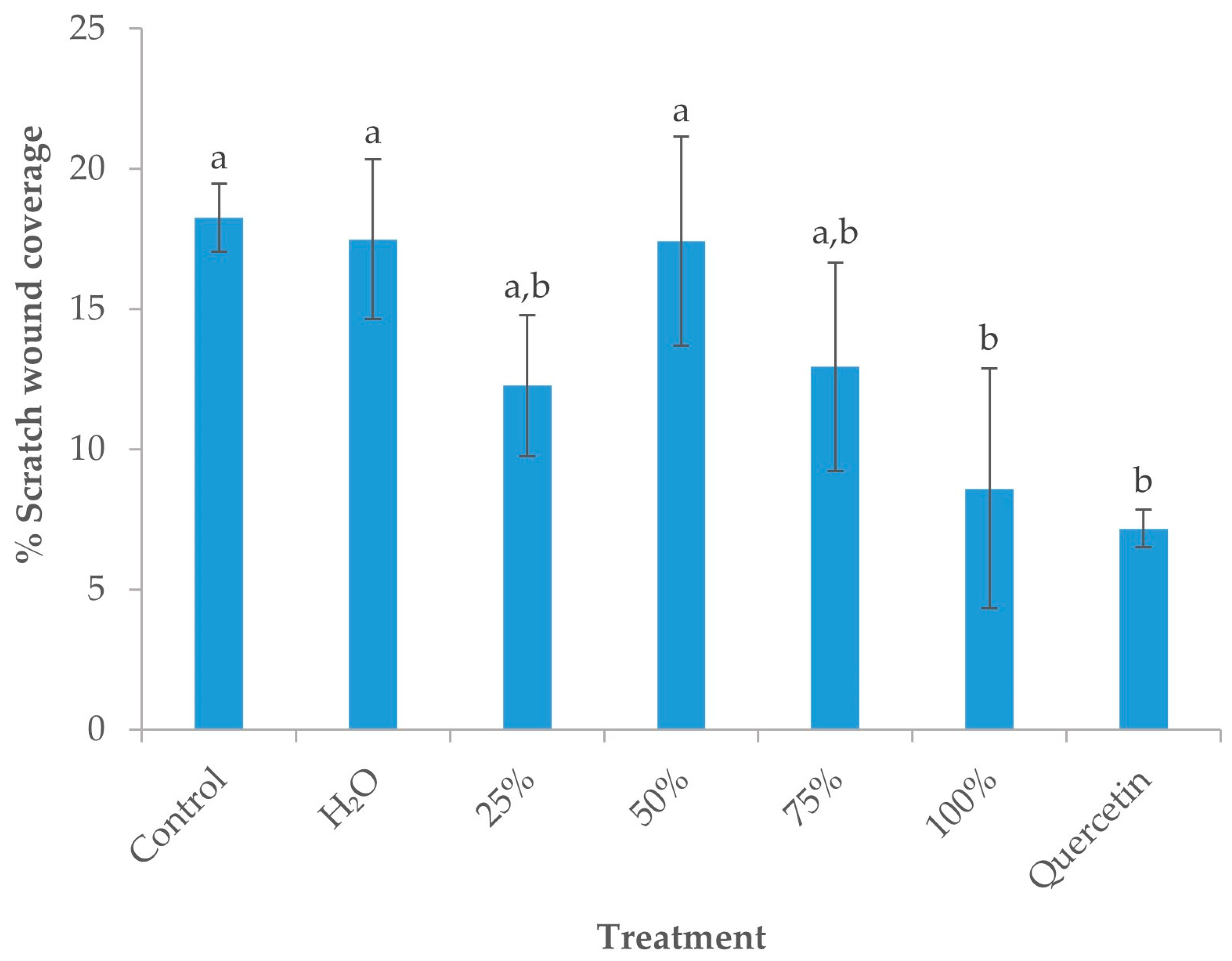
The scratch assay was used for the determination of the migratory potential of MCF-7 cells. Cells were incubated with 100 μg/mL of OS extract or quercetin for 24 h and then images were captured through the inverted microscope Wilovert Standard PH40 (Helmut Hund GmbH, Germany (magnification, ×100) at 0 and 24 h and analyzed. Significant inhibition of wound healing compared to the untreated cells (19.65 ± 1.5% wound healing) was shown when the cells were treated with 100 μg/mL of the 25% extract (1.48 ± 0.92%) (Figure 3). Other treatments such as the H2O extract (22.1 ± 8%), the 50% extract (17.89 ± 8.71%), the 75% extract (14.18 ± 3.24%), the 100% extract (8.60 ± 4.27%) and quercetin (8.93 ± 4.21%) did not cause a statistically significant change of the migratory potential of the cells and the healing of the wound when compared to the untreated cells. 100% Ethanol extract has shown the same effect as quercetin in the inhibition of cell migration. Quercetin reduces the migration of MCF-7 via the reduction in MMP-9 expression [51] or via GSK3β/β-catenin/ZEB1 signaling pathway [52].

3.5.3. Effect of OS Crude Extracts on Antioxidant Gene Expression

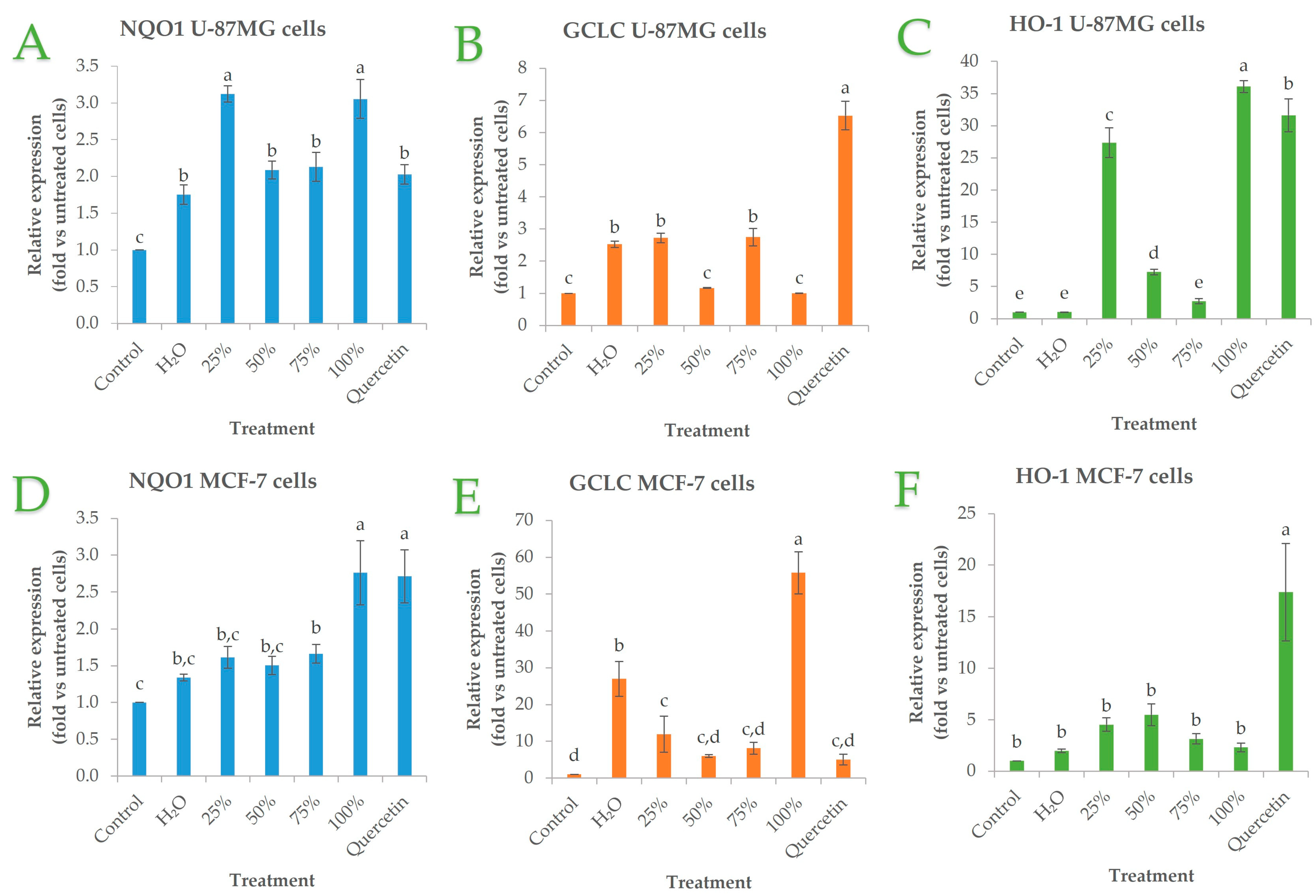
In order to assess the effect of OS crude extracts on antioxidant genes expression (NQO1, GCLC, and HO-1), cells were treated with 100 μg/mL OS crude extracts. The expression levels after each treatment were compared with the untreated cells as well as with cells treated with quercetin. The expression of HO-1 mRNA was significantly higher in U-87 MG after the treatment with the 25% extract (27-fold, p < 0.03), 50% extract (7-fold, p < 0.002), 75% extract (2.7-fold, p < 0.02) and the 100% ethanol extract (36-fold, p < 0.0002) compared to untreated cells. In addition, there was no statistically significant difference in the upregulation caused by the 100% extract in comparison to the upregulation caused by the quercetin treatment (31.5-fold, p < 0.0002 vs. untreated cells) (Figure 4). MCF-7 showed a significant upregulation of HO-1 when incubated with the 25% extract (4.5-fold, p < 0.01), 50% extract (5.5-fold, p < 0.02), 75% extract (3.1-fold, p < 0.02) and the 100% ethanol extract (2.3-fold, p < 0.04) compared to untreated cells. The alteration of HO-1 caused by the rest of the extracts was statistically non-significant when compared to the untreated cells (Figure 4). NQO1 gene expression is upregulated in cells treated with quercetin in both cell lines. OS crude extracts also increased the expression of the NQO1 gene at a higher degree than quercetin in U-87 MG cells (25% ethanol extract, p < 0.0005; 100% ethanol extract, p < 0.01) and comparable levels in MCF-7 cells. OS crude extracts increased GCLC gene expression, especially the water (27-fold, p < 0.01) and 100% ethanol extract (55-fold, p < 0.004) in U-87 MG cells. In MCF-7 cells, OS crude extracts also increased compared to untreated cells (water, 2.5-fold; 25% ethanol, 2.7-fold; 75% ethanol, 2.7-fold) but to a much less extent than quercetin (6.5-fold, p < 0.002 vs. untreated cells) (Figure 4). Quercetin is known to increase NQO1 [53], GCLC [54], and HO-1gene expression [55].

3.5.4. Cytotoxic Activity of OS Crude Extracts and Cell Viability in MCF-7 Spheroids

To assess whether OS crude extracts could affect cancer stem cells, spheroids were grown in hanging drops treated with extracts at 100 μg/mL. After 3 and 5 days of growth without treatment, the average spheroid area was about 412 µm2 and 610 µm2, respectively. Extracts showed different effects on the spheroid area compared to the control (Figure 5). Quercetin (25 μΜ) served as a positive control. Extracts 25, 75, and 100% showed the strongest ability to reduce the spheroid area to 294 µm2, 322 µm2, and 233 µm2 for 3-day treatment and quercetin showed a reduction to 221 µm2. On day 5, extracts 25, and 100% showed the ability to reduce the spheroid area to 474 µm2 and 346 µm2, respectively, when at the same time quercetin reduced the spheroid area to 357 µm2. In conclusion, 100% ethanol exerts the strongest effect on spheroid formation comparable to the effect of quercetin. The cell death induced by the cell extracts was analyzed by fluorescein diacetate and measured in a microplate fluorescence reader. Cell viability in spheroids is shown in Figure 5. There is a reduction in fluorescent intensity in all treatments but compared to untreated spheroids is not statistically significant. Quercetin reduces the migration of triple-negative breast cancer cells in spheroids via dysregulation of PI3K/AKT activities [56].

4. Conclusions

This study evaluated the TPC and TFC, as well as the bioactive and therapeutic properties of OS crude extracts obtained using different solvents. The results revealed significant variations in TPC and TFC values among the solvent extracts, with ethanol exhibiting the highest extraction efficiency. The extracts exhibited potent antioxidant activity, as determined by DPPH, FRAP, and H2O2 scavenging assays, with the highest values observed in the 75% ethanol extract. The difference in the observed antioxidant activity and the TPC content could be the result of different antioxidant activities exhibited by the extracted polyphenols, due to their different structures. Regarding the anti-inflammatory properties, all OS crude extracts displayed varying degrees of inhibition, with the 75% ethanol extract exhibiting the highest activity. The higher the ethanol content, the higher the anti-inflammatory activity, probably due to the extraction of a broader range of bioactive compounds with anti-inflammatory properties. Furthermore, the OS crude extracts exhibited antimicrobial activity against various pathogens. The lack of antibacterial activity from the ethanol extract could be due to the inefficient extraction of antibacterial compounds with this solvent. In terms of anticancer properties, the OS crude extracts and quercetin exhibited dose-dependent inhibitory effects on the viability and proliferation of U-87 MG and MCF-7 cells. Moreover, the OS crude extracts inhibited to various extents the migratory potential of MCF-7 cells, as observed in the scratch assay. The expression of the antioxidant gene HO-1 was significantly upregulated in U-87 MG cells treated with the OS crude extracts, particularly the 100% ethanol extract. The results highlight the potential of OS as a valuable source of bioactive compounds with antioxidant, anti-inflammatory, antimicrobial, and anticancer properties. In conclusion, the results of this study provide strong support for our research hypothesis. The crude extracts derived from OS using different solvents exhibited significant variations in their antioxidant, antimicrobial, and anticancer properties, validating our initial hypothesis. The findings revealed that certain solvents, such as ethanol, resulted in extracts with higher levels of phenolic compounds, greater antioxidant activity, stronger antimicrobial effects against specific microorganisms, and increased cytotoxicity against cancer cells. These results confirm the influence of the extraction solvent on the bioactivity of OS extracts and emphasize the importance of solvent selection in maximizing extraction efficiency and obtaining extracts with the desired bioactive properties. The results of this study contribute to expanding our understanding of the bioactive potential of OS waste and provide valuable insights for its utilization in the development of functional food ingredients, nutraceuticals, and pharmaceuticals.

Supplementary Materials

The following supporting information can be downloaded at: https://www.mdpi.com/article/10.3390/su151511599/s1.

Author Contributions

Conceptualization, E.B., I.S.P., T.C. and D.P.M.; methodology, E.B., I.-S.P. and V.A.; software, T.C., I.S.P. and V.A.; validation, E.B., I.S.P., T.C. and S.I.L.; formal analysis, E.B., I.-S.P., I.S.P. and D.P.; investigation, E.B., I.-S.P. and I.S.P.; resources, E.B., I.-S.P. and S.I.L.; data curation, T.C., D.P. and V.A.; writing—original draft preparation, E.B., T.C. and I.-S.P.; writing—review and editing, E.B., I.-S.P., I.S.P., T.C., D.P., V.A., S.I.L., A.C. and D.P.M.; visualization, T.C. and V.A.; supervision, D.P.M., S.I.L. and A.C.; project administration, I.-S.P., S.I.L., A.C. and D.P.M. All authors have read and agreed to the published version of the manuscript.

Funding

This research received no external funding.

Institutional Review Board Statement

Not applicable.

Informed Consent Statement

Not applicable.

Data Availability Statement

Data are available upon request.

Acknowledgments

The authors would like to thank Spyridon Konteles, Department of Food Science and Technology, University of West Attica, and Nikolaos Solomakos, Faculty of Veterinary Sciences, University of Thessaly for providing the tested microorganisms.

Conflicts of Interest

The authors declare no conflict of interest.

References

  1. Osojnik Črnivec, I.G.; Skrt, M.; Šeremet, D.; Sterniša, M.; Farčnik, D.; Štrumbelj, E.; Poljanšek, A.; Cebin, N.; Pogačnik, L.; Smole Možina, S.; et al. Waste streams in onion production: Bioactive compounds, quercetin and use of antimicrobial and antioxidative properties. Waste Manag. 2021, 126, 476–486. [Google Scholar] [CrossRef] [PubMed]
  2. Bhat, S.G.; Sukumaran, R.K. Enzyme Technologies for Bioconversion of Food Processing by-Products; CRC Press: Boca Raton, FL, USA, 2012; ISBN 9781439848876. [Google Scholar]
  3. Sagar, N.A.; Kumar, Y.; Singh, R.; Nickhil, C.; Kumar, D.; Sharma, P.; Om Pandey, H.; Bhoj, S.; Tarafdar, A. Onion waste based-biorefinery for sustainable generation of value-added products. Bioresour. Technol. 2022, 362, 127870. [Google Scholar] [CrossRef] [PubMed]
  4. Ganesh, K.S.; Sridhar, A.; Vishali, S. Utilization of fruit and vegetable waste to produce value-added products: Conventional utilization and emerging opportunities-A review. Chemosphere 2022, 287, 132221. [Google Scholar] [CrossRef]
  5. Versino, F.; Ortega, F.; Monroy, Y.; Rivero, S.; López, O.V.; García, M.A. Sustainable and Bio-Based Food Packaging: A Review on Past and Current Design Innovations. Foods 2023, 12, 1057. [Google Scholar] [CrossRef]
  6. Paesa, M.; Nogueira, D.P.; Velderrain-Rodríguez, G.; Esparza, I.; Jiménez-Moreno, N.; Mendoza, G.; Osada, J.; Martin-Belloso, O.; Rodríguez-Yoldi, M.J.; Ancín-Azpilicueta, C. Valorization of Onion Waste by Obtaining Extracts Rich in Phenolic Compounds and Feasibility of Its Therapeutic Use on Colon Cancer. Antioxidants 2022, 11, 733. [Google Scholar] [CrossRef]
  7. Choi, I.S.; Cho, E.J.; Moon, J.H.; Bae, H.J. Onion skin waste as a valorization resource for the by-products quercetin and biosugar. Food Chem. 2015, 188, 537–542. [Google Scholar] [CrossRef] [PubMed]
  8. Vojvodić Cebin, A.; Šeremet, D.; Mandura, A.; Martinić, A.; Komes, D. Onion Solid Waste as a Potential Source of Functional Food Ingredients. Eng. Power Bull. Croat. Acad. Eng. 2020, 15, 7–13. [Google Scholar]
  9. Bello, M.O.; Olabanji, I.O.; Abdul-Hammed, M.; Okunade, T.D. Characterization of domestic onion wastes and bulb (Allium cepa L.): Fatty acids and metal contents. Int. Food Res. J. 2013, 20, 2153–2158. [Google Scholar]
  10. Babbar, N.; Baldassarre, S.; Maesen, M.; Prandi, B.; Dejonghe, W.; Sforza, S.; Elst, K. Enzymatic production of pectic oligosaccharides from onion skins. Carbohydr. Polym. 2016, 146, 245–252. [Google Scholar] [CrossRef]
  11. Santiago, B.; Arias Calvo, A.; Gullón, B.; Feijoo, G.; Moreira, M.T.; González-García, S. Production of flavonol quercetin and fructooligosaccharides from onion (Allium cepa L.) waste: An environmental life cycle approach. Chem. Eng. J. 2020, 392, 123772. [Google Scholar] [CrossRef]
  12. Sagar, N.A.; Pareek, S.; Gonzalez-Aguilar, G.A. Quantification of flavonoids, total phenols and antioxidant properties of onion skin: A comparative study of fifteen Indian cultivars. J. Food Sci. Technol. 2020, 57, 2423–2432. [Google Scholar] [CrossRef]
  13. Albishi, T.; John, J.A.; Al-Khalifa, A.S.; Shahidi, F. Antioxidative phenolic constituents of skins of onion varieties and their activities. J. Funct. Foods 2013, 5, 1191–1203. [Google Scholar] [CrossRef]
  14. Chadorshabi, S.; Hallaj-Nezhadi, S.; Ghasempour, Z. Red onion skin active ingredients, extraction and biological properties for functional food applications. Food Chem. 2022, 386, 132737. [Google Scholar] [CrossRef] [PubMed]
  15. Abbaszadeh, S.; Rashidipour, M.; Khosravi, P.; Shahryarhesami, S.; Ashrafi, B.; Kaviani, M.; Sarabi, M.M. Biocompatibility, cytotoxicity, antimicrobial and epigenetic effects of novel chitosan-based quercetin nanohydrogel in human cancer cells. Int. J. Nanomed. 2020, 15, 5963–5975. [Google Scholar] [CrossRef]
  16. Mihaylova, D.; Lante, A. Water an Eco-Friendly Crossroad in Green Extraction: An Overview. Open Biotechnol. J. 2019, 13, 155–162. [Google Scholar] [CrossRef] [Green Version]
  17. Gil-Martín, E.; Forbes-Hernández, T.; Romero, A.; Cianciosi, D.; Giampieri, F.; Battino, M. Influence of the extraction method on the recovery of bioactive phenolic compounds from food industry by-products. Food Chem. 2022, 378, 131918. [Google Scholar] [CrossRef] [PubMed]
  18. Sun, C.; Wu, Z.; Wang, Z.; Zhang, H. Effect of Ethanol/Water Solvents on Phenolic Profiles and Antioxidant Properties of Beijing Propolis Extracts. Evid. Based Complement. Altern. Med. 2015, 2015, 595393. [Google Scholar] [CrossRef] [Green Version]
  19. Kaltsa, O.; Grigorakis, S.; Lakka, A.; Bozinou, E.; Lalas, S.; Makris, D.P. Green Valorization of Olive Leaves to Produce Polyphenol-Enriched Extracts Using an Environmentally Benign Deep Eutectic Solvent. Agriengineering 2020, 2, 226–239. [Google Scholar] [CrossRef] [Green Version]
  20. Chatzimitakos, T.; Athanasiadis, V.; Kotsou, K.; Palaiogiannis, D.; Bozinou, E.; Lalas, S.I. Optimized Isolation Procedure for the Extraction of Bioactive Compounds from Spent Coffee Grounds. Appl. Sci. 2023, 13, 2819. [Google Scholar] [CrossRef]
  21. Kasouni, A.I.; Chatzimitakos, T.G.; Troganis, A.N.; Stalikas, C.D. Citric acid-based carbon dots: From revealing new insights into their biological properties to demonstrating their enhanced wound healing potential by in vitro and in vivo experiments. Mater. Today Commun. 2021, 26, 102019. [Google Scholar] [CrossRef]
  22. Bozinou, E.; Lakka, A.; Poulianiti, K.; Lalas, S.; Makris, D.P. Cyclodextrins as high-performance green co-solvents in the aqueous extraction of polyphenols and anthocyanin pigments from solid onion waste. Eur. Food Res. Technol. 2021, 247, 2831–2845. [Google Scholar] [CrossRef]
  23. Lakka, A.; Lalas, S.; Makris, D.P. Development of a low-temperature and high-performance green extraction process for the recovery of polyphenolic phytochemicals fromwaste potato peels using hydroxypropyl β-cyclodextrin. Appl. Sci. 2020, 10, 3611. [Google Scholar] [CrossRef]
  24. Kasouni, A.I.; Chatzimitakos, T.G.; Stalikas, C.D.; Trangas, T.; Papoudou-Bai, A.; Troganis, A.N. The Unexplored Wound Healing Activity of Urtica dioica L. Extract: An In Vitro and In Vivo Study. Molecules 2021, 26, 6248. [Google Scholar] [CrossRef]
  25. Guleria, I.; Kumari, A.; Lacaille-Dubois, M.A.; Saini, A.K.; Kumar, V.; Saini, R.V.; Lal, U.R.; Gaur, N.A.; Kumari, S.; Seth, A.; et al. In-vitro antimicrobial, antioxidant, anti-inflammatory, and cytotoxic activities of Populus ciliata bark and leaves: A comparative study. S. Afr. J. Bot. 2022, 148, 238–250. [Google Scholar] [CrossRef]
  26. Maritan, S.M.; Lian, E.Y.; Mulligan, L.M. An Efficient and Flexible Cell Aggregation Method for 3D Spheroid Production. J. Vis. Exp. 2017, 2017, e55544. [Google Scholar] [CrossRef]
  27. Lagies, S.; Schlimpert, M.; Neumann, S.; Wäldin, A.; Kammerer, B.; Borner, C.; Peintner, L. Cells grown in three-dimensional spheroids mirror in vivo metabolic response of epithelial cells. Commun. Biol. 2020, 3, 246. [Google Scholar] [CrossRef] [PubMed]
  28. Pappas, I.S.; Siomou, S.; Bozinou, E.; Lalas, S.I. Moringa oleifera leaves crude aqueous extract down-regulates of BRCA1, mta-1 and oncogenes c-myc and p53 in AsPC-1, MCF-7 and HTC-116 cells. Food Biosci. 2021, 43, 101221. [Google Scholar] [CrossRef]
  29. Georgiou-Siafis, S.K.; Tsiftsoglou, A.S. Activation of KEAP1/NRF2 stress signaling involved in the molecular basis of hemin-induced cytotoxicity in human pro-erythroid K562 cells. Biochem. Pharmacol. 2020, 175, 113900. [Google Scholar] [CrossRef]
  30. Yeligar, S.M.; Machida, K.; Kalra, V.K. Ethanol-induced HO-1 and NQO1 are differentially regulated by HIF-1α and Nrf2 to attenuate inflammatory cytokine expression. J. Biol. Chem. 2010, 285, 35359–35373. [Google Scholar] [CrossRef] [Green Version]
  31. Sakasai-Sakai, A.; Takeuchi, M.; Takata, T. Intracellular toxic advanced glycation end-products promote the production of reactive oxygen species in HEPG2 cells. Int. J. Mol. Sci. 2020, 21, 4861. [Google Scholar] [CrossRef] [PubMed]
  32. Subbiah, V.; Ebrahimi, F.; Agar, O.T.; Dunshea, F.R.; Barrow, C.J.; Suleria, H.A.R. Comparative Study on the Effect of Phenolics and Their Antioxidant Potential of Freeze-Dried Australian Beach-Cast Seaweed Species upon Different Extraction Methodologies. Pharmaceuticals 2023, 16, 773. [Google Scholar] [CrossRef] [PubMed]
  33. Shi, L.; Zhao, W.; Yang, Z.; Subbiah, V.; Suleria, H.A.R. Extraction and characterization of phenolic compounds and their potential antioxidant activities. Environ. Sci. Pollut. Res. 2022, 29, 81112–81129. [Google Scholar] [CrossRef] [PubMed]
  34. Hikmawanti, N.P.E.; Fatmawati, S.; Asri, A.W. The effect of ethanol concentrations as the extraction solvent on antioxidant activity of Katuk (Sauropus androgynus (L.) Merr.) leaves extracts. IOP Conf. Ser. Earth Environ. Sci. 2021, 755, 012060. [Google Scholar] [CrossRef]
  35. Lopez-Corona, A.V.; Valencia-Espinosa, I.; González-Sánchez, F.A.; Sánchez-López, A.L.; Garcia-Amezquita, L.E.; Garcia-Varela, R. Antioxidant, Anti-Inflammatory and Cytotoxic Activity of Phenolic Compound Family Extracted from Raspberries (Rubus idaeus): A General Review. Antioxidants 2022, 11, 1192. [Google Scholar] [CrossRef]
  36. Bakrim, S.; El Omari, N.; El Hachlafi, N.; Bakri, Y.; Lee, L.H.; Bouyahya, A. Dietary Phenolic Compounds as Anticancer Natural Drugs: Recent Update on Molecular Mechanisms and Clinical Trials. Foods 2022, 11, 3323. [Google Scholar] [CrossRef]
  37. Singh, B.N.; Singh, B.R.; Singh, R.L.; Prakash, D.; Singh, D.P.; Sarma, B.K.; Upadhyay, G.; Singh, H.B. Polyphenolics from various extracts/fractions of red onion (Allium cepa) peel with potent antioxidant and antimutagenic activities. Food Chem. Toxicol. 2009, 47, 1161–1167. [Google Scholar] [CrossRef]
  38. Kumar, M.; Barbhai, M.D.; Hasan, M.; Punia, S.; Dhumal, S.; Radha; Rais, N.; Chandran, D.; Pandiselvam, R.; Kothakota, A.; et al. Onion (Allium cepa L.) peels: A review on bioactive compounds and biomedical activities. Biomed. Pharmacother. 2022, 146, 112498. [Google Scholar] [CrossRef]
  39. Kumar, S.; Sandhir, R.; Ojha, S. Evaluation of antioxidant activity and total phenol in different varieties of Lantana camara leaves. BMC Res. Notes 2014, 7, 560. [Google Scholar] [CrossRef] [Green Version]
  40. Balasundram, N.; Sundram, K.; Samman, S. Phenolic compounds in plants and agri-industrial by-products: Antioxidant activity, occurrence, and potential uses. Food Chem. 2006, 99, 191–203. [Google Scholar] [CrossRef]
  41. Tawaha, K.; Alali, F.Q.; Gharaibeh, M.; Mohammad, M.; El-Elimat, T. Antioxidant activity and total phenolic content of selected Jordanian plant species. Food Chem. 2007, 104, 1372–1378. [Google Scholar] [CrossRef]
  42. Kim, W.J.; Lee, K.A.; Kim, K.T.; Chung, M.S.; Cho, S.W.; Paik, H.D. Antimicrobial effects of onion (Allium cepa L.) peel extracts produced via subcritical water extraction against Bacillus cereus strains as compared with ethanolic and hot water extraction. Food Sci. Biotechnol. 2011, 20, 1101–1106. [Google Scholar] [CrossRef]
  43. Sagar, N.A.; Pareek, S. Antimicrobial assessment of polyphenolic extracts from onion (Allium cepa L.) skin of fifteen cultivars by sonication-assisted extraction method. Heliyon 2020, 6, e05478. [Google Scholar] [CrossRef]
  44. Lee, K.A.; Kim, K.T.; Nah, S.Y.; Chung, M.S.; Cho, S.W.; Paik, H.D. Antimicrobial and antioxidative effects of onion peel extracted by the subcritical water. Food Sci. Biotechnol. 2011, 20, 543–548. [Google Scholar] [CrossRef]
  45. Santhosh, A.; Theertha, V.; Prakash, P.; Smitha Chandran, S. From waste to a value added product: Green synthesis of silver nanoparticles from onion peels together with its diverse applications. Proc. Mater. Today Proc. 2019, 46, 4460–4463. [Google Scholar] [CrossRef]
  46. Oyawoye, O.M.; Olotu, T.M.; Nzekwe, S.C.; Idowu, J.A.; Abdullahi, T.A.; Babatunde, S.O.; Ridwan, I.A.; Batiha, G.E.; Idowu, N.; Alorabi, M.; et al. Antioxidant potential and antibacterial activities of Allium cepa (onion) and Allium sativum (garlic) against the multidrug resistance bacteria. Bull. Natl. Res. Cent. 2022, 46, 214. [Google Scholar] [CrossRef]
  47. Yüksel, T.N.; Bozgeyik, E.; Yayla, M. The Effect of Quercetin and Quercetin-3-d-xyloside on Breast Cancer Proliferation and Migration. J. Basic Clin. Health Sci. 2022, 6, 569–578. [Google Scholar] [CrossRef]
  48. Asgharian, P.; Tazekand, A.P.; Hosseini, K.; Forouhandeh, H.; Ghasemnejad, T.; Ranjbar, M.; Hasan, M.; Kumar, M.; Beirami, S.M.; Tarhriz, V.; et al. Potential mechanisms of quercetin in cancer prevention: Focus on cellular and molecular targets. Cancer Cell Int. 2022, 22, 257. [Google Scholar] [CrossRef] [PubMed]
  49. Pan, H.-C.; Jiang, Q.; Yu, Y.; Mei, J.-P.; Cui, Y.-K.; Zhao, W.-J. Quercetin promotes cell apoptosis and inhibits the expression of MMP-9 and fibronectin via the AKT and ERK signalling pathways in human glioma cells. Neurochem. Int. 2015, 80, 60–71. [Google Scholar] [CrossRef]
  50. Tang, J.; Oroudjev, E.; Wilson, L.; Ayoub, G. Delphinidin and cyanidin exhibit antiproliferative and apoptotic effects in MCF7 human breast cancer cells. Integr. Cancer Sci. Ther. 2015, 2, 82–86. [Google Scholar] [CrossRef] [Green Version]
  51. Lin, C.-W.; Hou, W.-C.; Shen, S.-C.; Juan, S.-H.; Ko, C.-H.; Wang, L.-M.; Chen, Y.-C. Quercetin inhibition of tumor invasion via suppressing PKC /ERK/AP-1-dependent matrix metalloproteinase-9 activation in breast carcinoma cells. Carcinogenesis 2008, 29, 1807–1815. [Google Scholar] [CrossRef] [Green Version]
  52. Chen, B.; Li, X.; Wu, L.; Zhou, D.; Song, Y.; Zhang, L.; Wu, Q.; He, Q.; Wang, G.; Liu, X.; et al. Quercetin Suppresses Human Glioblastoma Migration and Invasion via GSK3β/β-catenin/ZEB1 Signaling Pathway. Front. Pharmacol. 2022, 13, 963614. [Google Scholar] [CrossRef] [PubMed]
  53. Valerio, L.G.; Kepa, J.K.; Pickwell, G.V.; Quattrochi, L.C. Induction of human NAD(P)H:quinone oxidoreductase (NQO1) gene expression by the flavonol quercetin. Toxicol. Lett. 2001, 119, 49–57. [Google Scholar] [CrossRef] [PubMed]
  54. Kang, J.-H.; Chang, S.-Y.; Jang, H.-J.; Cho, J.M.; Kim, D.-B.; Lee, S.S.; Ko, S.H.; Park, Y.-M.; Needs, P.W.; Jo, Y.-H.; et al. Quercetin-induced upregulation of human GCLC gene is mediated by cis -regulatory element for early growth response protein-1 (EGR1) in INS-1 beta-cells. J. Cell. Biochem. 2009, 108, 1346–1355. [Google Scholar] [CrossRef] [PubMed]
  55. Lee, Y.-J.; Song, J.-H.; Oh, M.-H.; Lee, Y.-J.; Kim, Y.-B.; Im, J.-H.; Lee, S.-H. ERK1/2 activation in quercetin-treated BEAS-2B cell plays a role in Nrf2-driven HO-1 expression. Mol. Cell. Toxicol. 2011, 7, 347–355. [Google Scholar] [CrossRef]
  56. Shahi Thakuri, P.; Gupta, M.; Singh, S.; Joshi, R.; Glasgow, E.; Lekan, A.; Agarwal, S.; Luker, G.D.; Tavana, H. Phytochemicals inhibit migration of triple negative breast cancer cells by targeting kinase signaling. BMC Cancer 2020, 20, 4. [Google Scholar] [CrossRef] [PubMed] [Green Version]
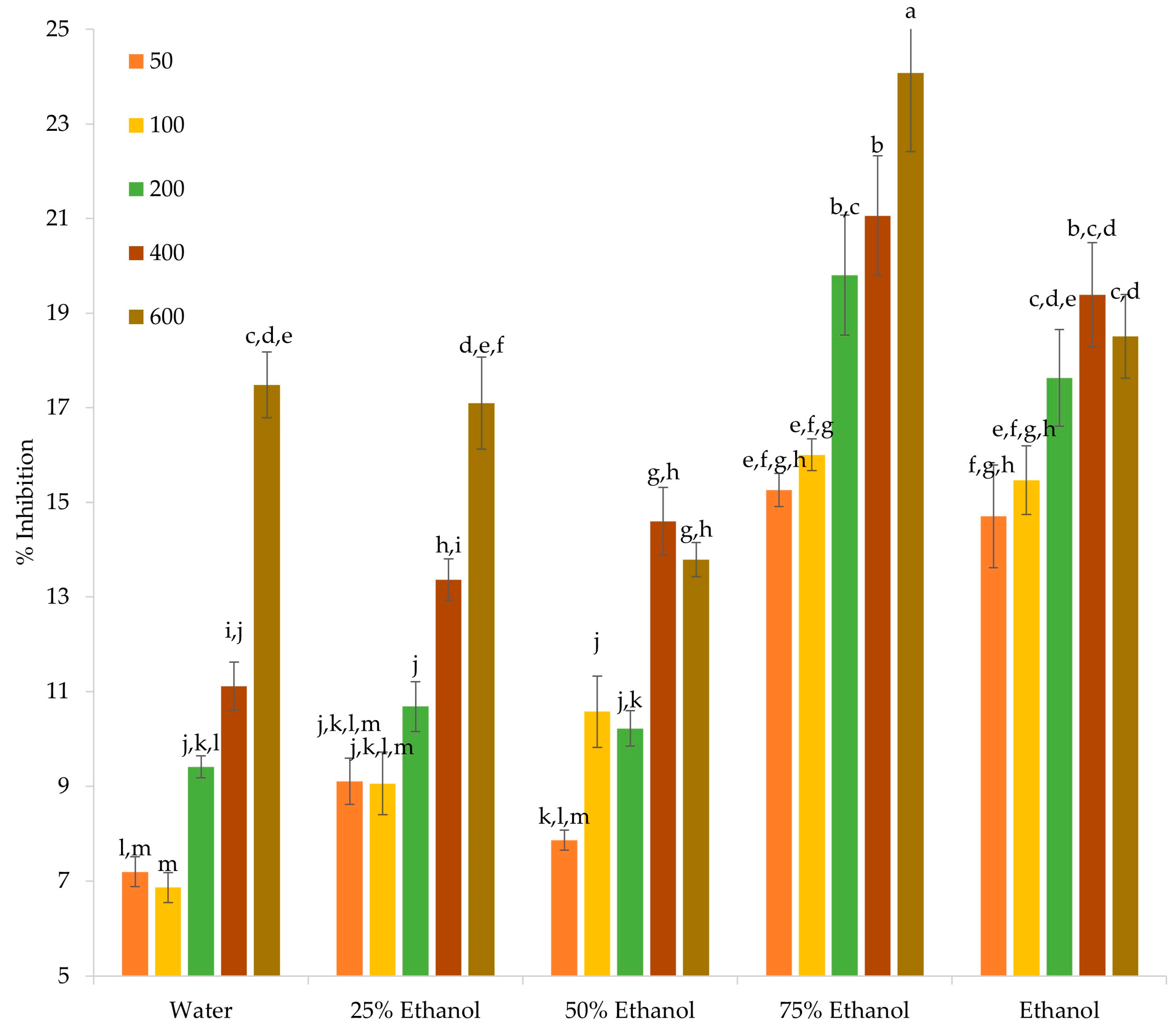
Figure 1. Percent inhibition of albumin denaturation achieved by various concentrations of OS crude extracts. Statistically significant differences (p < 0.05) are stated with different superscript letters (e.g., a–m).
Sustainability 15 11599 g001
Figure 2. Cytotoxic effect of OS crude extracts and quercetin on U-87 MG and MCF-7 cells incubated with various concentrations. Capital letters (i.e., A–D) denote statistically significant differences (p < 0.05) among the same concentration of the various extracts. Small letters (i.e., a–d) denote statistically significant differences (p < 0.05) for the various concentrations of the same extract. Data are presented as mean ± standard deviation.
Sustainability 15 11599 g002
Figure 3. Percent scratch wound coverage of MCF-7 cells upon incubation with the various OS crude extracts and quercetin. Small letters (a, b) denote statistically significant (p < 0.05) differences between samples. Data are presented as mean ± standard deviation.
Sustainability 15 11599 g003
Figure 4. Fold change of NQO1 (A,D), GCLC (B,E), and HO-1 (C,F) mRNA expression compared to untreated U-87 MG (AC) and MCF-7 (DF) cells. Small letters (a–e) denote statistically significant (p < 0.05) differences between samples.
Sustainability 15 11599 g004
Figure 5. Effect of OS crude extracts on the formation of MCF-7 spheroids on day 3 (A) and 5 (B), as well as the cell viability (C). Small letters (a–d) in graphs (A,B) denote statistically significant (p < 0.05) differences between samples. In graph (C), no statistically significant differences (p > 0.05) were observed.
Sustainability 15 11599 g005
Table 1. TPC, TFC, and antioxidant activity (DPPH, FRAP, and H2O2 scavenging) of the OS crude extracts.
SolventsTPC (mg GAE/g dw)TFC (mg RtE/g dw)DPPH (μmol DPPH/g dw)FRAP (μmol AAE/g dw)AAHP (μmol AAE/g dw)
Water348.71 ± 7.85 d11.16 ± 0.51 d177.15 ± 2.8 d127.77 ± 5.86 e25.66 ± 0.11 c,d
25% Ethanol377.37 ± 10.98 c20.73 ± 0.04 c299.84 ± 0.81 c230.95 ± 3.57 d27.09 ± 0.8 c
50% Ethanol505.57 ± 28.63 b32.14 ± 0.22 b378.45 ± 0.76 b356.87 ± 5.06 a32.71 ± 0.8 b
75% Ethanol522.1 ± 25.58 b36.2 ± 0.26 a396.01 ± 7.78 a334.91 ± 1.23 b55.04 ± 2.07 a
Ethanol795.11 ± 19.01 a31.11 ± 1.3 b299.19 ± 2.49 c304.21 ± 2.31 c23.09 ± 1 d
Within each column, statistically significant differences (p < 0.05) are denoted with different superscript letters (e.g., a–e).
Table 2. Concentration (mg/L) of the detected polyphenols in the OS crude extracts.
CompoundsWater25% Ethanol50% Ethanol75% EthanolEthanol
Protocatechuic acid42.04 ± 1.72 a24.53 ± 0.93 b25.28 ± 0.91 b27.27 ± 0.95 b25.7 ± 0.98 b
Spiraeoside141.12 ± 5.64 c137.27 ± 5.22 c267.94 ± 11.25 b356.46 ± 11.41 a348.2 ± 12.54 a
Quercetin29.63 ± 1.24 c143.08 ± 5.87 b259.27 ± 9.59 a241.1 ± 7.47 a257.76 ± 10.83 a
Cyanidin 3-O-glucoside11.35 ± 0.37 d13.53 ± 0.55 c22.27 ± 0.82 a23.51 ± 0.92 a16.81 ± 0.71 b
Delphinidin 3,5-di-O-galactoside7.27 ± 0.24 c9.83 ± 0.3 b12.31 ± 0.52 a11.97 ± 0.47 a9.19 ± 0.31 b
Delphinidin 3,5-di-O-glucoside76.08 ± 2.81 d114.54 ± 4.12 c138.98 ± 5.28 b158 ± 6 a111.52 ± 3.46 c
Cyanidin 3-O-(6″-malonylglucoside)26.35 ± 0.95 e35.66 ± 1.32 c41.98 ± 1.55 b49.21 ± 1.77 a30.5 ± 1.13 d
Within each row, statistically significant differences (p < 0.05) are denoted with different superscript letters (e.g., a–e).
Table 3. Zones of inhibition (mm) from the agar disk diffusion assay for various concentrations of OS crude extracts against various bacterial species.
Bacterial SpeciesSolvents25 mg/L50 mg/L100 mg/L
L. monocytogenesWater10 ± 0.1 b20 ± 0.3 a,b32.5 ± 12.6 a
25% Ethanol20 ± 14.1 a25 ± 10 a30 ± 14.1 a
50% Ethanol10 ± 0.1 a,b15 ± 10 a
75% Ethanol27.5 ± 15 a30 ± 0.3 a40 ± 0.4 a
Ethanol
E. coli O157:H7Water8 ± 1.4 c23 ± 2.4 b45 ± 3.6 a
25% Ethanol32.5 ± 5 a35 ± 17.3 a50 ± 0.8 a
50% Ethanol10 ± 0.3 b17.5 ± 5 a,b22.5 ± 5 a
75% Ethanol25 ± 5.8 b37.5 ± 12.6 a,b42.5 ± 5 a
Ethanol
B. cereusWater17.5 ± 5
25% Ethanol
50% Ethanol
75% Ethanol
Ethanol
E. faecalisWater12.5 ± 5 b30 ± 14.1 a,b32.5 ± 5 a
25% Ethanol30 ± 14.1 a37.5 ± 15 a45 ± 10 a
50% Ethanol32.5 ± 5 a35 ± 23.8 a40 ± 0.9 a
75% Ethanol40 ± 18.3 a42.5 ± 18.9 a52.5 ± 18.9 a
Ethanol10 ± 0
P. aeruginosaWater25 ± 10 a30 ± 14.1 a
25% Ethanol20 ± 0.7 a22.5 ± 5 a25 ± 3.6 a
50% Ethanol10 ± 0.6 b15 ± 2.2 b25 ± 5.8 a
75% Ethanol20 ± 0.4 b30 ± 0.9 a
Ethanol30 ± 7.1 a35 ± 17.3 a37.5 ± 15 a
S. aureusWater
25% Ethanol30 ± 0.3
50% Ethanol20 ± 0.6 a20 ± 0.7 a
75% Ethanol25 ± 5.8 a30 ± 4.1 a35 ± 10 a
Ethanol
S. typhimuriumWater12.5 ± 5
25% Ethanol15 ± 5.8
50% Ethanol20 ± 0.8 a25 ± 5.8 a30 ± 7.1 a
75% Ethanol20 ± 0.2 a20 ± 0.1 a
Ethanol20 ± 0.6 a20 ± 0.5 a20 ± 0.7 a
Y. enterocoliticaWater12.5 ± 5 a17.5 ± 5 a25 ± 10 a
25% Ethanol15 ± 10 a27.5 ± 15 a32.5 ± 6.5 a
50% Ethanol20 ± 4.1 a25 ± 5.8 a30 ± 20 a
75% Ethanol20 ± 3.4 a22 ± 4 a25 ± 10 a
Ethanol12.5 ± 5 b20 ± 0.3 a,b27.5 ± 9.6 a
Within each row, statistically significant differences (p < 0.05) are denoted with different superscript letters (e.g., a–c).
Disclaimer/Publisher’s Note: The statements, opinions and data contained in all publications are solely those of the individual author(s) and contributor(s) and not of MDPI and/or the editor(s). MDPI and/or the editor(s) disclaim responsibility for any injury to people or property resulting from any ideas, methods, instructions or products referred to in the content.