
Role of Patient-Specific 3D-Printed Models for Complex Pediatric Craniocervical Junction Surgery: Case Description and Systematic Literature Review
 , ,
, ,
Abstract
1. Introduction
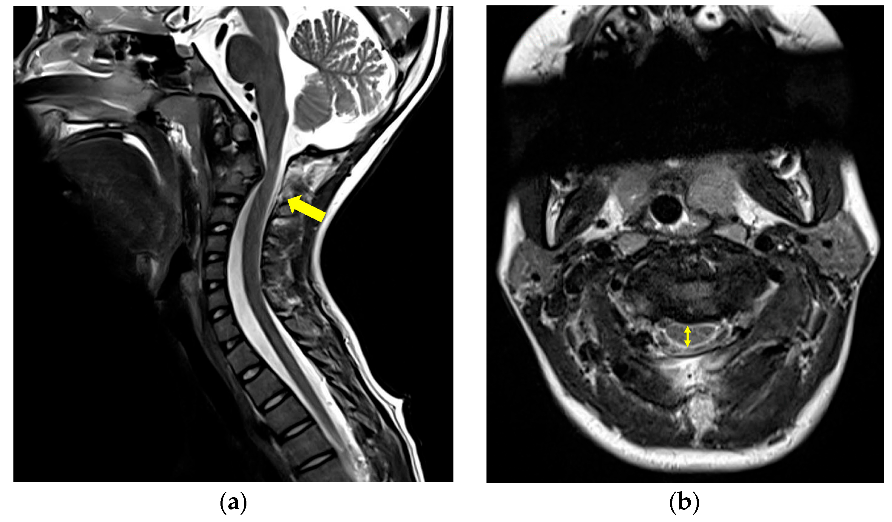
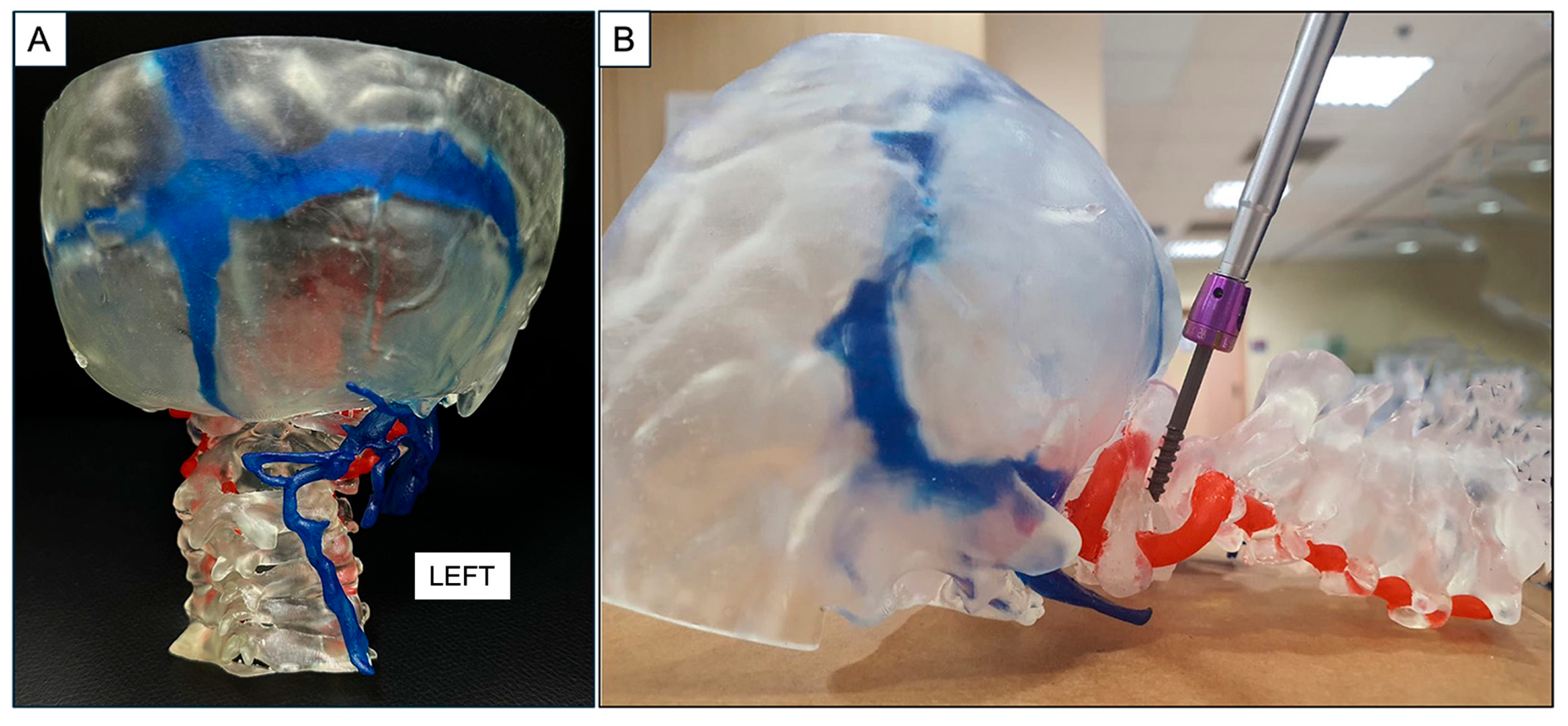
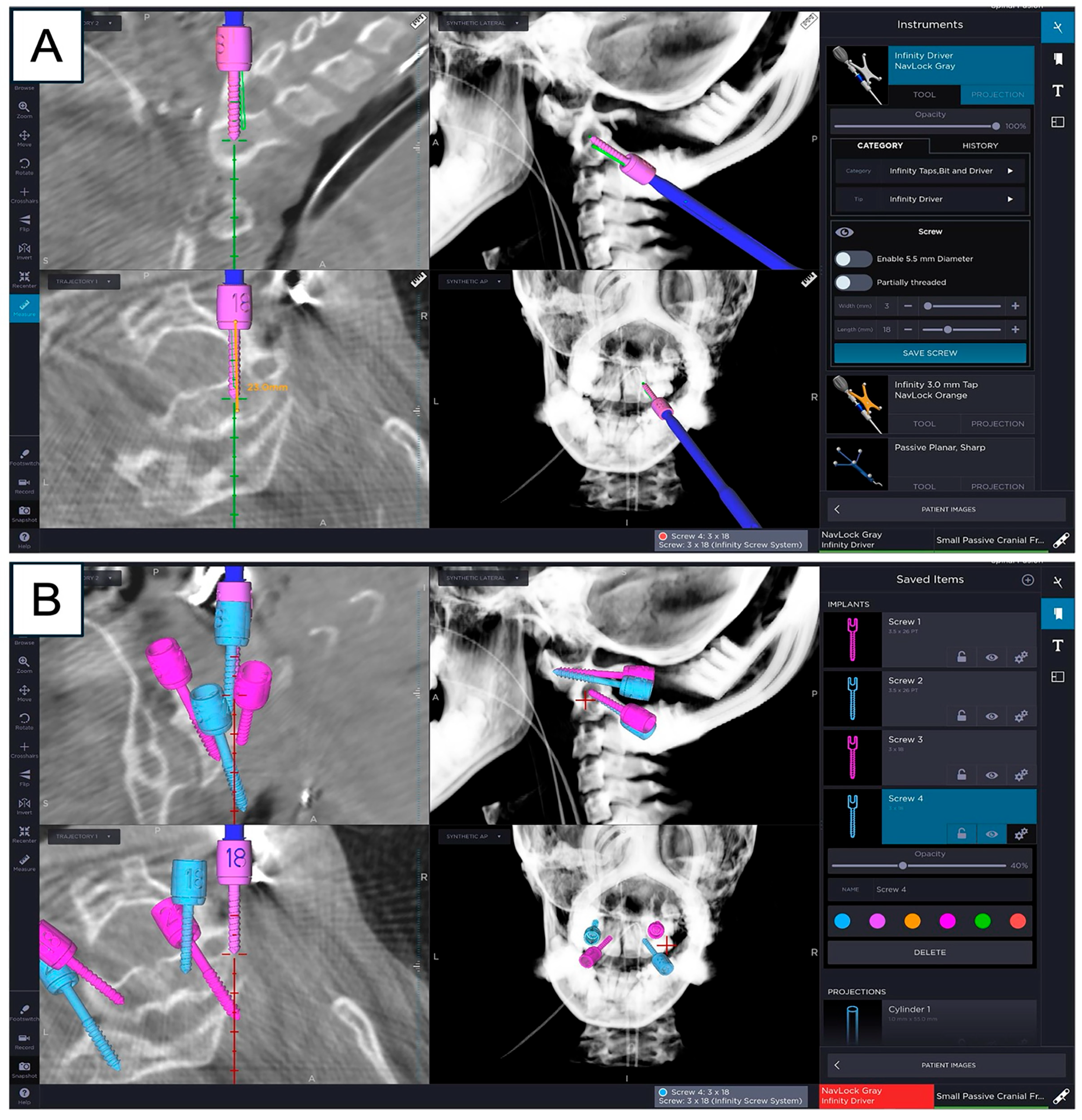
2. Case Description
3. Systematic Literature Review
3.1. Database Search
3.2. Eligibility Criteria and Study Selection
3.3. Data Extraction
3.4. Results
4. Discussion
4.1. Overview of Craniocervical Junction Surgery in the Pediatric Population
4.2. The Role of 3D Printing in Neurosurgery
4.3. Critique of Systematic Literature Review
4.4. Study Reflections and Future Directions
5. Conclusions
Author Contributions
Funding
Institutional Review Board Statement
Informed Consent Statement
Data Availability Statement
Acknowledgments
Conflicts of Interest
References
- Ghanem, I.; El Hage, S.; Rachkidi, R.; Kharrat, K.; Dagher, F.; Kreichati, G. Pediatric cervical spine instability. J. Child. Orthop. 2008, 2, 71–84. [Google Scholar] [CrossRef] [PubMed]
- Garg, B.; Mehta, N. Current status of 3D printing in spine surgery. J. Clin. Orthop. Trauma. 2018, 9, 218–225. [Google Scholar] [CrossRef] [PubMed]
- Jimenez, J.E.; Shaffer, A.D.; Hammersley, E.; Ghodadra, A.; Stapleton, A.L. Use of patient-specific 3D printed models in pre-operative counseling for pediatric skull base surgery. Int. J. Pediatr. Otorhinolaryngol. 2023, 171, 111655. [Google Scholar] [CrossRef] [PubMed]
- Joseph, F.J.; Vanluchene, H.E.R.; Goldberg, J.; Bervini, D. 3D-Printed Head Model in Patient’s Education for Micro-Neurosurgical Aneurysm Clipping Procedures. World Neurosurg. 2023, 175, e1069–e1074. [Google Scholar] [CrossRef]
- Smith-Bindman, R.; Lipson, J.; Marcus, R.; Kim, K.P.; Mahesh, M.; Gould, R.; Berrington de González, A.; Miglioretti, D.L. Radiation dose associated with common computed tomography examinations and the associated lifetime attributable risk of cancer. Arch. Intern. Med. 2009, 169, 2078–2086. [Google Scholar] [CrossRef]
- Alvord, E.C., Jr.; Lofton, S. Gliomas of the optic nerve or chiasm. Outcome by patients’ age, tumor site, and treatment. J. Neurosurg. 1988, 68, 85–98. [Google Scholar] [CrossRef]
- Liberati, A.; Altman, D.G.; Tetzlaff, J.; Mulrow, C.; Gøtzsche, P.C.; Ioannidis, J.P.A.; Clarke, M.; Devereaux, P.J.; Kleijnen, J.; Moher, D. The PRISMA statement for reporting systematic reviews and meta-analyses of studies that evaluate health care interventions: Explanation and elaboration. J. Clin. Epidemiol. 2009, 62, e1–e34. [Google Scholar] [CrossRef]
- Page, M.J.; McKenzie, J.E.; Bossuyt, P.M.; Boutron, I.; Hoffmann, T.C.; Mulrow, C.D.; Shamseer, L.; Tetzlaff, J.M.; Akl, E.A.; Brennan, S.E.; et al. The PRISMA 2020 statement: An updated guideline for reporting systematic reviews. bmj 2021, 372, n71. [Google Scholar] [CrossRef]
- Agarwal, P.; Chopra, S.; Sinha, V.D.; Kataria, R. Three-Dimensional-Printed Model-Assisted Management of Craniovertebral Junction Abnormalities: An Institutional Experience with Literature Review. Asian Spine J. 2020, 14, 204–211. [Google Scholar] [CrossRef]
- Chhabra, S.; Chopra, S.; Kataria, R.; Sinha, V.D. Use of 3D printer model to study vertebral artery anatomy and variations in developmental craniovertebral junction anomalies and as a preoperative tool-an institutional experience. J. Spine Surg. 2017, 3, 572–579. [Google Scholar] [CrossRef]
- Coote, J.D.; Nguyen, T.; Tholen, K.; Stewart, C.; Verter, E.; McGee, J.; Celestre, P.; Sarkar, K. Three-Dimensional Printed Patient Models for Complex Pediatric Spinal Surgery. Ochsner J. 2019, 19, 49–53. [Google Scholar] [CrossRef] [PubMed]
- Goel, A.; Jankharia, B.; Shah, A.; Sathe, P. Three-dimensional models: An emerging investigational revolution for craniovertebral junction surgery. J. Neurosurg. Spine 2016, 25, 740–744. [Google Scholar] [CrossRef] [PubMed]
- He, S.; Ye, C.; Zhong, N.; Yang, M.; Yang, X.; Xiao, J. Customized anterior craniocervical reconstruction via a modified high-cervical retropharyngeal approach following resection of a spinal tumor involving C1-2/C1-3. J. Neurosurg. Spine 2020, 32, 432–440. [Google Scholar] [CrossRef] [PubMed]
- Kagami, Y.; Nakashima, H.; Segi, N.; Ito, S.; Ouchida, J.; Shinjo, R.; Imagama, S. A novel technique for C1-C2 posterior screw insertion using patient-specific guides created by CT-based 3D printing. Nagoya J. Med. Sci. 2024, 86, 487–496. [Google Scholar] [CrossRef]
- Malikov, A.; Secen, A.E.; Divanlioglu, D.; Gunerhan, G.; Ocal, O.; Gunduz, U.K. The Feasibility of Creating Image-Based Patient-Specific Drill Guides for the Atlantoaxial Instabilities Using Open-Source CAD Software and Desktop 3D Printers. World Neurosurg. 2022, 163, e377–e383. [Google Scholar] [CrossRef]
- Pijpker, P.A.J.; Kuijlen, J.M.A.; Kaptein, B.L.; Pondaag, W. Three-Dimensional-Printed Drill Guides for Occipitothoracic Fusion in a Pediatric Patient with Occipitocervical Instability. Oper. Neurosurg. 2021, 21, 27–33. [Google Scholar] [CrossRef]
- Rashim, K.; Verma Pawan, K.; Sinha, V.D. Increasing the safety of surgical treatment for complex Cranio-vertebral anomalies using customized 3D printed models. J. Clin. Neurosci. 2018, 48, 203–208. [Google Scholar] [CrossRef]
- Sakai, T.; Tezuka, F.; Abe, M.; Yamashita, K.; Takata, Y.; Higashino, K.; Nagamachi, A.; Sairyo, K. Pediatric Patient with Incidental Os Odontoideum Safely Treated with Posterior Fixation Using Rod-Hook System and Preoperative Planning Using 3D Printer: A Case Report. J. Neurol. Surg. A Cent. Eur. Neurosurg. 2017, 78, 306–309. [Google Scholar] [CrossRef]
- Texeira da Silva, L.E.C.; Cuéllar, D.J.; Campos de Barros, A.G.; Khan, A.A. Three-dimensional biomodel use in the surgical management of basilar invagination with congenital cervical scoliosis; correction by unilateral C1-C2 facet distraction. J. Craniovertebr. Junction Spine 2020, 11, 347–350. [Google Scholar] [CrossRef]
- Vakharia, V.N.; Smith, L.; Tahir, Z.; Sparks, R.; Ourselin, S.; Tucker, S.; Thompson, D. Occipitocervical instrumented fixation utilising patient-specific C2 3D-printed spinal screw trajectory guides in complex paediatric skeletal dysplasia. Child’s Nerv. Syst. 2021, 37, 2643–2650. [Google Scholar] [CrossRef]
- Wang, J.; Zhu, C.; Xia, H. Management of Unique Basilar Invagination Combined with C1 Prolapsing into the Foramen Magnum in Children: Report of 2 Cases. World Neurosurg. 2019, 127, 92–96. [Google Scholar] [CrossRef]
- Wang, Y.T.; Yang, X.J.; Yan, B.; Zeng, T.H.; Qiu, Y.Y.; Chen, S.J. Clinical application of three-dimensional printing in the personalized treatment of complex spinal disorders. Chin. J. Traumatol. 2016, 19, 31–34. [Google Scholar] [CrossRef] [PubMed]
- Xu, N.; Wei, F.; Liu, X.; Jiang, L.; Cai, H.; Li, Z.; Yu, M.; Wu, F.; Liu, Z. Reconstruction of the Upper Cervical Spine Using a Personalized 3D-Printed Vertebral Body in an Adolescent with Ewing Sarcoma. Spine 2016, 41, E50–E54. [Google Scholar] [CrossRef] [PubMed]
- Jug, M.; Tomaževič, M.; Cimerman, M. 3D model-assisted instrumentation of the pediatric spine: A technical note. J. Orthop. Surg. Res. 2021, 16, 586. [Google Scholar] [CrossRef] [PubMed]
- Malikov, A.; Secen, A.E.; Ocal, O.; Divanlioglu, D.; Belen, A.D.; Dalgic, A. Accuracy of Axis Drill Guides in the Cases of Atlantoaxial Instabilities Associated with High-Riding Vertebral Arteries, Narrow Pedicles, and Complex Deformities: Comparison of 3 Fixation Methods. World Neurosurg. 2022, 168, e336–e343. [Google Scholar] [CrossRef]
- Dogan, S.; Safavi-Abbasi, S.; Theodore, N.; Horn, E.; Rekate, H.L.; Sonntag, V.K. Pediatric subaxial cervical spine injuries: Origins, management, and outcome in 51 patients. Neurosurg. Focus 2006, 20, E1. [Google Scholar] [CrossRef]
- Richards, O.; Saeed, F.; Derham, C.; Tyagi, A. Craniocervical fusion in the paediatric population—Case series of 21 patients. Br. J. Neurosurg. 2024, 38, 116–121. [Google Scholar] [CrossRef]
- Pang, D.; Li, V. Atlantoaxial rotatory fixation: Part 1—Biomechanics of normal rotation at the atlantoaxial joint in children. Neurosurgery 2004, 55, 614–625, discussion 625–616. [Google Scholar] [CrossRef]
- Pang, D.; Li, V. Atlantoaxial rotatory fixation: Part 2—New diagnostic paradigm and a new classification based on motion analysis using computed tomographic imaging. Neurosurgery 2005, 57, 941–953. [Google Scholar] [CrossRef]
- Pang, D. Atlantoaxial rotatory fixation. Neurosurgery 2010, 66, 161–183. [Google Scholar] [CrossRef]
- Salunke, P.; Behari, S.; Kirankumar, M.V.; Sharma, M.S.; Jaiswal, A.K.; Jain, V.K. Pediatric congenital atlantoaxial dislocation: Differences between the irreducible and reducible varieties. J. Neurosurg. 2006, 104, 115–122. [Google Scholar] [CrossRef][Green Version]
- Wang, C.; Yan, M.; Zhou, H.; Wang, S.; Dang, G. Atlantoaxial transarticular screw fixation with morselized autograft and without additional internal fixation: Technical description and report of 57 cases. Spine 2007, 32, 643–646. [Google Scholar] [CrossRef] [PubMed]
- Menezes, A.H. Evaluation and treatment of congenital and developmental anomalies of the cervical spine. Invited submission from the Joint Section Meeting on Disorders of the Spine and Peripheral Nerves, March 2004. J. Neurosurg. Spine 2004, 1, 188–197. [Google Scholar] [CrossRef] [PubMed]
- Ahmed, R.; Traynelis, V.C.; Menezes, A.H. Fusions at the craniovertebral junction. Child’s Nerv. Syst. 2008, 24, 1209–1224. [Google Scholar] [CrossRef] [PubMed]
- Cacciola, F.; Phalke, U.; Goel, A. Vertebral artery in relationship to C1-C2 vertebrae: An anatomical study. Neurol. India 2004, 52, 178–184. [Google Scholar]
- Tian, Y.; Xu, N.; Yan, M.; Chen, J.; Hung, K.-L.; Hou, X.; Wang, S.; Li, W. Vertebral Artery Variations at the Craniovertebral Junction in “Sandwich” Atlantoaxial Dislocation Patients. Neurospine 2021, 18, 770–777. [Google Scholar] [CrossRef]
- Lee, H.J.; Lee, J.J.; Hong, J.T.; Kim, J.T. Quantification of pediatric cervical growth: Anatomical changes in the sub-axial spine. J. Korean Neurosurg. Soc. 2015, 57, 185–191. [Google Scholar] [CrossRef]
- Niu, G.; Chen, H.; Liu, L.; Zhou, G.; Zhou, Q.; Li, C.; Dai, J.; Nie, H.; Bai, J.; Zhang, J. Surgical treatment for upper cervical deformity with atlantoaxial joint dislocation using individualized 3D printing occipitocervical fusion instrument: A case report and literature review. Medicine 2021, 100, e25202. [Google Scholar] [CrossRef]
- Shree Kumar, D.; Ampar, N.; Wee Lim, L. Accuracy and reliability of spinal navigation: An analysis of over 1000 pedicle screws. J. Orthop. 2020, 18, 197–203. [Google Scholar] [CrossRef]
- Flynn, J.M.; Sakai, D.S. Improving safety in spinal deformity surgery: Advances in navigation and neurologic monitoring. Eur. Spine J. 2013, 22, 131–137. [Google Scholar] [CrossRef]
- Munusamy, T.; Thien, A.; Anthony, M.G.; Bakthavachalam, R.; Dinesh, S.K. Computed tomographic morphometric analysis of cervical pedicles in a multi-ethnic Asian population and relevance to subaxial cervical pedicle screw fixation. Eur. Spine J. 2015, 24, 120–126. [Google Scholar] [CrossRef] [PubMed]
- Weinstein, J.N.; Spratt, K.F.; Spengler, D.; Brick, C.; Reid, S. Spinal pedicle fixation: Reliability and validity of roentgenogram-based assessment and surgical factors on successful screw placement. Spine 1988, 13, 1012–1018. [Google Scholar] [CrossRef] [PubMed]
- Liang, S.; Teo, J.C.; Coyuco, B.C.; Cheong, T.M.; Lee, N.K.; Low, S.Y.Y. Mixed Reality for Pediatric Brain Tumors: A Pilot Study from a Singapore Children’s Hospital. Surgeries 2023, 4, 354–366. [Google Scholar] [CrossRef]
- Tagaytayan, R.; Kelemen, A.; Sik-Lanyi, C. Augmented reality in neurosurgery. Arch. Med. Sci. 2018, 14, 572–578. [Google Scholar] [CrossRef]
- Mansour, N.K.; Callera, A.; Potere, F.; Micalizzi, S.; Costantino, M.L.; De Gaetano, F.; Oliva, P. Circular economy and 3D printing in the healthcare sector. Front. Bioeng. Biotechnol. 2025, 13, 1548550. [Google Scholar] [CrossRef]
- Vukicevic, M.; Puperi, D.S.; Jane Grande-Allen, K.; Little, S.H. 3D Printed Modeling of the Mitral Valve for Catheter-Based Structural Interventions. Ann. Biomed. Eng. 2017, 45, 508–519. [Google Scholar] [CrossRef]
- Pacione, D.; Tanweer, O.; Berman, P.; Harter, D.H. The utility of a multimaterial 3D printed model for surgical planning of complex deformity of the skull base and craniovertebral junction. J. Neurosurg. 2016, 125, 1194–1197. [Google Scholar] [CrossRef]
- Liew, Y.; Beveridge, E.; Demetriades, A.K.; Hughes, M.A. 3D printing of patient-specific anatomy: A tool to improve patient consent and enhance imaging interpretation by trainees. Br. J. Neurosurg. 2015, 29, 712–714. [Google Scholar] [CrossRef]
- Waran, V.; Narayanan, V.; Karuppiah, R.; Owen, S.L.; Aziz, T. Utility of multimaterial 3D printers in creating models with pathological entities to enhance the training experience of neurosurgeons. J. Neurosurg. 2014, 120, 489–492. [Google Scholar] [CrossRef]
- Aimar, A.; Palermo, A.; Innocenti, B. The Role of 3D Printing in Medical Applications: A State of the Art. J. Healthc. Eng. 2019, 2019, 5340616. [Google Scholar] [CrossRef]
- Zhang, F.; Zhang, H.; Zhao, H.; He, Z.; Shi, L.; He, Y.; Ju, N.; Rong, Y.; Qiu, J. Design and fabrication of a personalized anthropomorphic phantom using 3D printing and tissue equivalent materials. Quant. Imaging Med. Surg. 2019, 9, 94–100. [Google Scholar] [CrossRef]
- Vezirska, D.; Milev, M.; Laleva, L.; Nakov, V.; Spiriev, T. Three-Dimensional Printing in Neurosurgery: A Review of Current Indications and Applications and a Basic Methodology for Creating a Three-Dimensional Printed Model for the Neurosurgical Practice. Cureus 2022, 14, e33153. [Google Scholar] [CrossRef]
- Govsa, F.; Ozer, M.A.; Biceroglu, H.; Karakas, A.B.; Cagli, S.; Eraslan, C.; Alagoz, A.K. Creation of 3-Dimensional Life Size: Patient-Specific C1 Fracture Models for Screw Fixation. World Neurosurg. 2018, 114, e173–e181. [Google Scholar] [CrossRef]
- Thayaparan, G.K.; Owbridge, M.G.; Thompson, R.G.; D’Urso, P.S. Patient-specific processes for occipitocervical fixation using biomodelling and additive manufacturing. J. Clin. Neurosci. 2020, 71, 251–256. [Google Scholar] [CrossRef]
- Iqbal, J.; Zafar, Z.; Skandalakis, G.; Kuruba, V.; Madan, S.; Kazim, S.F.; Bowers, C.A. Recent advances of 3D-printing in spine surgery. Surg. Neurol. Int. 2024, 15, 297. [Google Scholar] [CrossRef]
- Li, H.; Alkahtani, M.E.; Basit, A.W.; Elbadawi, M.; Gaisford, S. Optimizing environmental sustainability in pharmaceutical 3D printing through machine learning. Int. J. Pharm. 2023, 648, 123561. [Google Scholar] [CrossRef]
- Malik, A.; Ul Haq, M.I.; Raina, A.; Gupta, K. 3D printing towards implementing Industry 4.0: Sustainability aspects, barriers and challenges. Ind. Robot 2022, 49, 491–511. [Google Scholar] [CrossRef]
- De Jesus Encarnacion Ramirez, M.; Chmutin, G.; Nurmukhametov, R.; Soto, G.R.; Kannan, S.; Piavchenko, G.; Nikolenko, V.; Efe, I.E.; Romero, A.R.; Mukengeshay, J.N.; et al. Integrating Augmented Reality in Spine Surgery: Redefining Precision with New Technologies. Brain Sci. 2024, 14, 645. [Google Scholar] [CrossRef] [PubMed]



| S/N | Author/Year | Number of Pediatric Patients in Study (% Total Patients) | Clinical Pathology | Use Case | Key Findings |
|---|---|---|---|---|---|
| 1 | Goel et al., 2016 [12] | 3 (27.3%) | CCJ anomalies |
|
|
| 2 | Xu et al., 2016 [23] | 1 (100%) | C2 spinal tumor |
|
|
| 3 | Wang et al., 2016 [22] | 2 (50%) | 1 AAI and 1 high-cervical eosinophilic granuloma |
|
|
| 4 | Chhabra et al., 2017 [10] | 1 (10%) | CCJ anomalies |
|
|
| 5 | Sakai et al., 2017 [18] | 1 (100%) | AAI |
|
|
| 6 | Rashim et al., 2018 [17] | 2 (15.4%) | AAI |
|
|
| 7 | Coote et al., 2019 [11] | 1 (33.3%) | AAI and cervical stenosis (DS) |
|
|
| 8 | He et al., 2020 [13] | 1 (14.3%) | C1–C3 spine tumor |
|
|
| 9 | Wang et al., 2019 [21] | 2 (100%) | CCJ anomalies |
|
|
| 10 | Agarwal et al., 2020 [9] | 6 (33.3%) | CCJ anomalies |
|
|
| 11 | da Silva et al., 2020 [19] | 1 (100%) | CCJ anomalies |
|
|
| 12 | Jug et al., 2021 [24] | 1 (50%) | Grisel’s syndrome |
|
|
| 13 | Pijpker et al., 2021 [16] | 1 (100%) | CCJ anomalies |
|
|
| 14 | Vakharia et al., 2021 [20] | 2 (100%) | CCJ anomalies |
|
|
| 15 | Malikov et al., 2022 (a) 1 [15] | 4 (25%) | CCJ anomalies and traumatic spine injury |
|
|
| 16 | Malikov et al., 2022 (b) [25] | 8 (26.7%) | CCJ anomalies, high-cervical spine tumor and traumatic spine injury |
|
|
| 17 | Kagami et al., 2024 [14] | 1 (16.7%) | AAI |
|
|
| Application | 3D-Printed Model | Image-Fused Neuro-Navigation |
|---|---|---|
| Pre-operative counseling | Able to use in outpatient clinic as part of consultation with patient and caregivers | Computer stealth-station bulky to transport to outpatient clinic |
| Pre-operative planning by surgical team | Allows physical 3D visuospatial orientation of complex anatomy and trial placement of spinal implants | Allows 2D visualization of anatomy for planning only |
| Intraoperative use | Physical model does not allow real-time changes during implant insertion [55] | Allows intraoperative decisions changes during implant insertion |
| Neurosurgical education | Able to create multiple models for hands-on training to increase deeper understanding of complex spine anatomy | Not applicable |
| Added benefits of technology | Potential for bio-engineering of patient-specific spinal implants and drug-delivery systems [2,55] | Not applicable |
Disclaimer/Publisher’s Note: The statements, opinions and data contained in all publications are solely those of the individual author(s) and contributor(s) and not of MDPI and/or the editor(s). MDPI and/or the editor(s) disclaim responsibility for any injury to people or property resulting from any ideas, methods, instructions or products referred to in the content. |
© 2025 by the authors. Licensee MDPI, Basel, Switzerland. This article is an open access article distributed under the terms and conditions of the Creative Commons Attribution (CC BY) license.
Share and Cite
Mak, D.S.K.; Lo, Y.T.; Tan, M.B.W.; Kumar, D.S.; Low, S.Y.Y. Role of Patient-Specific 3D-Printed Models for Complex Pediatric Craniocervical Junction Surgery: Case Description and Systematic Literature Review. Surg. Tech. Dev. 2026, 15, 1. https://doi.org/10.3390/std15010001
Mak DSK, Lo YT, Tan MBW, Kumar DS, Low SYY. Role of Patient-Specific 3D-Printed Models for Complex Pediatric Craniocervical Junction Surgery: Case Description and Systematic Literature Review. Surgical Techniques Development. 2026; 15(1):1. https://doi.org/10.3390/std15010001
Chicago/Turabian StyleMak, David S. K., Yu Tung Lo, Mark B. W. Tan, Dinesh S. Kumar, and Sharon Y. Y. Low. 2026. "Role of Patient-Specific 3D-Printed Models for Complex Pediatric Craniocervical Junction Surgery: Case Description and Systematic Literature Review" Surgical Techniques Development 15, no. 1: 1. https://doi.org/10.3390/std15010001
APA StyleMak, D. S. K., Lo, Y. T., Tan, M. B. W., Kumar, D. S., & Low, S. Y. Y. (2026). Role of Patient-Specific 3D-Printed Models for Complex Pediatric Craniocervical Junction Surgery: Case Description and Systematic Literature Review. Surgical Techniques Development, 15(1), 1. https://doi.org/10.3390/std15010001