Article

Biological Control of Seedling Damping-Off of Pepper Caused by Rhizoctonia solani Using Bacillus sp. JB005 Isolated from Tidal Flats


by
Chae Yeon Hwang
1,
Murugesan Chandrasekaran
2 and
Se Chul Chun
1,*
1
Department of Environmental and Health Science, Konkuk University, Seoul 05029, Republic of Korea
2
Department of Food Science and Biotechnology, Sejong University, 209-Neundon-ro, Gwangjin-gu, Seoul 05006, Republic of Korea
*
Author to whom correspondence should be addressed.
Microbiol. Res. 2026, 17(8), 148; https://doi.org/10.3390/microbiolres17080148
Submission received: 14 May 2026 / Revised: 22 July 2026 / Accepted: 28 July 2026 / Published: 30 July 2026
(This article belongs to the Topic New Challenges on Plant–Microbe Interactions)

Abstract

Pepper (Capsicum annuum L.) is a major vegetable cultivated for seasoning in Korea. Peppers are typically grown as seedlings and transplanted into fields. Infection with Rhizoctonia solani immediately before transplantation can cause significant damage and loss during cultivation. The effectiveness of chemical fungicides is becoming increasingly limited owing to pesticide resistance, emphasizing the need for sustainable agricultural practices. This study aimed to identify a novel biocontrol agent by investigating tidal flat microorganisms capable of controlling damping-off disease in pepper caused by Rhizoctonia solani. Furthermore, we studied the inhibitory effects of different Bacillus species on the mycelial growth of R. solani AG-4 (KACC 40141) for the biological control of damping-off disease. Of the 116 bacterial strains from the tidal flats tested, five (including Bacillus sp. JB005) showed strong inhibitory activity in dual-culture assays against R. solani AG-4. These strains also demonstrated siderophore production, suggesting their potential to suppress damping-off in pepper. We screened useful agricultural traits (e.g., biofilm formation, IAA production, and phosphate solubilizing activity); the most promising strains were used for greenhouse experiments. The results indicated that Bacillus sp. JB005 strongly inhibited damping-off in pepper, suggesting that Bacillus sp. JB005 could serve as a biocontrol agent against R. solani infections.

1. Introduction

Pepper (Capsicum annuum) is an important vegetable grown worldwide for consumption and processing purposes. Furthermore, it is considered an important horticultural crop because of its nutritional value, flavor, and adding of color and texture to fresh and processed products for consumption [1,2,3]. However, it is highly susceptible to various pathogens during its early and later stages of growth [2,4]. During the cultivation of pepper, both under greenhouse and field conditions, it can be exposed to several plant diseases such as phytophthora blight (caused by Phytophthora capsici) [5], bacterial spot (caused by Xanthomonas euvesicatoria) [6], anthracnose (bitter rot) (caused by Colletotrichum acutatum) [2], mosaics and mottling (caused by potato virus Y) [7] and so on. The infections caused by these pathogens result in various diseases such as blight, stunting, systemic vein-clearing, leaf mosaic or mottling, and bacterial wilt [7,8,9].
Among different microbial diseases that affect pepper plants, the damping-off disease caused by Rhizoctonia solani or Pythium ultimum [10] is the most devastating disease in terms of seedling mortality at very early growth stages and results in economic and yield losses for farmers. During the seedling stage, peppers are particularly vulnerable to damping-off diseases. Moreover, R. solani can affect chili peppers at any stage of growth, but it is especially damaging during the seedling stage [11]. R. solani is a soil-borne pathogen that thrives in cold and waterlogged soils causing seed decay before germination or attacking the base or roots of the stem, leading to rot.
Pathogens are microscopic disease-causing agents such as viruses, bacteria and fungi that infect the plant and cause diseases, whereas pests are macroscopic and multicellular organisms such as insects and so on that cause damage or disturb the plants physically. Therefore, the demand for sustainable agriculture has led to the emergence of biocontrol methods. Biocontrol refers to the use of organisms or biologically derived compounds to manage plant diseases. The organisms or substances used in these control methods are commonly referred to as biocontrol agents, or BCAs [12]. These agents can be employed against a wide range of plant pathogens, including viruses, bacteria, fungi, insects, and nematodes. The mechanisms of BCA-based control include competition with plant pathogens for space and nutrients, parasitism, antibiosis, and the stimulation of systemic-induced resistance in host plants. Compared to chemical fungicides, BCAs are considered more environmentally friendly and are thought to limit the emergence of resistant strains [13,14,15]. Commonly used biocontrol agents include microbial species such as Bacillus [1,16], Trichoderma [2], Pseudomonas [5], and so on. These microorganisms play a vital role in protecting plants from pathogens through various mechanisms. Successful biological control of seedling diseases of vegetable crops like pepper have also been well documented [17].
The isolation of BCAs can be conducted in various environments. Collecting BCAs from suppressive soils is a traditional method [18,19,20]. However, BCAs can also be found in environments such as lakes, marine ecosystems, deserts, salt fields, and tidal flats [17,21,22,23,24,25]. The tidal flat is an intertidal zone that is exposed during low tide and submerged during high tide [26]. Tidal flats act as a boundary between marine and terrestrial environments, receiving a variety of materials from land. These materials undergo processes such as sedimentation, dissolution, deposition, and resuspension, contributing to the decomposition of various organic substances [27]. Moreover, this unique environment fosters a microbiome that thrives under fluctuating conditions, including high salinity, pressure changes, temperature variations, and erratic oxygen levels, resulting in a highly diverse ecosystem that enables unique physiological and metabolic processes [28,29,30]. The tidal flat supports microorganisms with diverse biological characteristics [31].
Bacillus species is frequently used as a biological control agent due to its ability to form endospores, allowing it to survive unfavorable conditions such as high temperatures, osmotic stress, and extreme pH levels [32]. A variety of antimicrobial compounds, including iturin, fengycin, and bacillomycin [33,34], trigger induced systemic resistance in plants under pathogen attack [35]. Bacillus subtilis produces antimicrobial substances such as iturin and fengycin, which inhibit the growth of R. solani and protect plants from pathogen attacks [33,34]. Previous studies also showed that different microbial species had biocontrol activity against for R. solani [36,37,38,39]. Each of these microorganisms has shown effectiveness in suppressing R. solani under greenhouse and field conditions, suggesting their potential for development as bio fungicides.
Despite these advances, research exploring the use of tidal or mud flat microbiomes as BCAs is limited. Tidal flats, characterized by their unique environmental conditions, harbor diverse microbial communities with potentially novel biocontrol traits. Therefore, further research is necessary to explore and develop effective biocontrol agents in this context. However, no registered bio fungicide or chemical fungicide specifically targeting R. solani-induced damping-off in chili peppers exists in Korea. This gap emphasizes the need for further exploration of underutilized microbial sources, such as those from tidal flats, to develop effective and sustainable BCAs.
The objective of the present study was to isolate and screen the bacterial species from mud flats. Further, we studied the mud flats’ isolated strains for their plant-growth-promoting abilities and also biocontrol efficiency against R. solani in pepper plants.

2. Materials and Methods

2.1. Collection and Isolation of the Bacterial Strains from the Tidal Flat

At 2 km off the coast of Jang-bong Island, Ganghwa-gun, Incheon Metropolitan City, Republic of Korea, the mud of habitation of the Venus clam (Cyclina sinensis) was used to isolate bacteria. The tidal flat mud samples were collected using polyvinyl chloride (PVC). Ten grams of mud was dissolved in 100 mL of sterilized distilled water and stirred in a flask shaker for 24 h. Subsequently, the samples were cultured on a Zobell medium (Thane, Maharashtra, India) (5 g of bacto peptone, 1 g of bacto yeast extract, 0.1 g of ferric citrate, and 15 g of difco nutrient agar (NA, Difco, Ditroit, MN, USA) in 1 L of seawater (pH 7.63) for 72 h at 30 °C). The isolated bacterial strains were stocked at a −70 °C in a deep freezer and sub-cultured using tryptic soy agar.

2.2. Characterization

2.2.1. Antagonistic Effects

In this study, we used the fungal strain R. solani AG-4 from the Korea Association of Culture Collection (KACC-40141). For the dual-culture method, we placed a mycelial plug of 4 mm (in diameter) on one side of a potato dextrose agar (PDA) plate. Simultaneously, each bacterial strain was streaked on another side of the plate. The inoculated plate was incubated at 25 °C until control plate growth (only mycelial plug). Two days after the inoculation of the bacterial strains, the length of the inhibition zone (antagonistic effects of the bacterial strains) was measured. All experiments were conducted in triplicate.

2.2.2. Siderophore Production

We used the method of Milgres et al. [40] and Ji et al. [41], with modifications to detect siderophore. In addition, we used blue agar medium with chrome azurol S (CAS) [40]; the prepared media was poured into a Petri dish (9 cm diameter). The inoculum strain was pre-cultured in Tryptic Soy Broth (TSB) at 30 °C with 180 rpm for 2 days. A paper disk (1 cm in diameter) was then placed in the center of the agar plate and inoculated with the 40 µL (approximately 108 CFU/mL) of bacterial suspension. The plates were wrapped up and incubated in dark conditions at 30 °C for 2 days. The positive and negative controls were P. aeruginosa and JS082, respectively. The diameter of the halo zone was determined 2 days after incubation. The presence of an orange halo around the colony indicated the production of siderophores.

2.2.3. Protease Production

The detection of protease secretion was tested using skim milk agar assay [42]. Skim milk agar (10 g of skim milk powder, 15 g of agar, and 1 L of distilled water) was sterilized at 121 °C for 20 min, cooled down until the temperature reached 50 °C, and poured into a Petri dish (9 cm diameter). The paper disk (1 cm diameter) was then placed on the three spots of the surface of skim milk agar and inoculated with the 40 µL (approximately 108 CFU/mL) of bacterial suspension over the disks. Subsequently, the plates were sealed with a sealing film and incubated at 30 °C for 2 days, after which the diameter of the halo zone was determined.

2.2.4. Cellulase Production

Cellulase production was determined using carboxymethylcellulose (CMC) agar assay [43]. The prepared CMC agar (10 g of CMC, 1 g of KH2PO4, 0.5 g of NaCl, 0.4 g of MgSO4∙7H2O, 1 g of K2HPO4, 0.0125 g of FeSO4∙7H2O, 18 g of agar, and 1 L of distilled water) was sterilized at 121 °C for 20 min. CMC agar was then poured into a Petri dish and paper disks were placed at three spots and inoculated with the 40 µL (approximately 108 CFU/mL) of bacterial suspension. The plates were incubated at 30 °C for 2 days, and then, the CMC agar plates were stained with 0.1% Congo red solution for 10–15 min and washed out using 1 M of NaCl. Thereafter, the diameters of the halo zones were determined.

2.3. Effect of Plant-Growth Promotion

2.3.1. Indole Acetic Acid (IAA) Production

Each strain was incubated for 2 days in King’s B broth (per 1 L: 20 g of protease peptone, 1.15 g of K2HPO4, 1.5 g of MgSO4, and 15 g of glycerol) supplemented with 0.1% (w/v) L-tryptophan as precursor of IAA. After 2 days, the optical density of the culture suspension was determined at 600 nm and the culture suspension was centrifugated at 4 °C for 15 min at 8000 rpm. The supernatant was mixed with Salkowski reagent (35% HClO4, 50 mL; 0.5 M FeCl3 1 mL) with a ratio of 1:2 (supernatant: Salkowski reagent), and the reaction mixtures were left in the dark for at least 30 min until a color reaction developed. When the color developed, the absorbance of the mixtures was determined at 530 nm. The IAA concentration was calculated based on the IAA standard curve (Figure S1). First, 6.4 mg of IAA (Kisan bio-MB-I5897, Seoul, Republic of Korea) powder was dissolved in 10 mL of distilled water, and a dilution series was prepared for standardization with King’s B broth.

2.3.2. Phosphate Solubilization

A method of Mehta and Nautiyal [44] was used to test the ability to solubilize phosphate. Each experimental strain was pre-cultured with nutrient broth (NB) for 2 days before inoculation. Thereafter, 50 µL of culture suspension (1–2 × 10⁹ CFU/mL) was inoculated into 5 mL of NBRIP-BPB medium (per 1 L; 10 g of glucose, 5 g of Ca3(PO4)2, 5 g of MgCl2∙6H2O, 0.25 g of MgSO4∙7H2O, 0.2 g of KCl, 0.1 g of (NH4)2SO4, and 0.025 g of Bromophenol Blue; pH was adjusted to 7.0). It was then incubated at 30 °C at 250 rpm for 3 days, and then, the cultures were centrifuged at 10,000 rpm for 10 min to pellet the cell. Then the supernatant was taken, and the absorbance was determined at 600 nm. The concentration of solubilized phosphate was calculated based on the standard curve (Figure S2). The phosphate solution was prepared using KH2PO4. First, KH2PO4 (21.965 g) was dissolved into 500 mL of distilled water to obtain a 10,000 ppm solution. NBRIP-BPB was then added to obtain each concentration. Similarly to the culture suspension, the sample was centrifuged at 10,000 rpm for 10 min, and the absorbance was estimated at 600 nm.

2.3.3. Biofilm Formation

Bacterial strains, including Agrobacterium tumefaciens C58, which was used as a positive control, were cultured overnight at 28 °C in NB. The following day, the culture suspension was diluted to 1:100 in fresh TSB for a biofilm assay. A total of 100 µL of the diluted suspension was added to each well of a 96-well microtiter plate, and the plate was incubated at 28 °C for 24 h. After incubation, the liquid in the wells was discarded by inverting the plate and shaking out the contents. The plate was then gently submerged in a tub of water for rinsing, and the water was shaken off. This rinsing process was repeated twice. To each well, 125 µL of 0.1% crystal violet (diluted with water) was added, and the plate was incubated at room temperature for 10–15 min for staining. Following staining, the plate was rinsed three or four times with water by submerging it in a tub, shaking off the excess liquid after each rinse. After the final rinse, the plate was inverted and allowed to dry overnight. The next day, 125 µL of 30% acetic acid was added to each well to solubilize the crystal violet. The plate was incubated at room temperature for 10–15 min. Subsequently, 125 µL of the solubilized solution was transferred to a new microtiter plate. The absorbance was measured at 550 nm using a spectrophotometer (Molecular Devices, San Jose, CA, USA), with 30% acetic acid serving as a blank control. All experiments, including those using the positive control A. tumefaciens C58, were performed in quintuplicate. The experiment was conducted as described by O’Toole [45].

2.4. Greenhouse Experiment

2.4.1. Preparation of the Inoculum

R. solani AG-4 (KACC number 40141) was cultured on PDA for 7 d. Unhulled barley seeds (150 g) were washed with distilled water and briefly soaked in sufficient distilled water to ensure absorption. The barley seeds were then sterilized in an autoclave at 121 °C for 20 min, twice. The sterilized barley was cooled sufficiently. Using a cork borer, mycelial plugs of a fresh culture of R. solani AG-4 were uniformly inoculated over the barley seeds. The mixture, placed in a sterile glass bottle, was incubated at 27 °C for 10 d. Subsequently, the mycelia were transferred onto aluminum foil to promote further growth. The barley seeds were stored at 4 °C until the pepper plants were inoculated.

2.4.2. Preparation of the Pepper Plants

First, the soil (Gumok, Gyeongju, Republic of Korea) was sterilized in an autoclave at 121 °C for 20 min, twice. The sterilized soil was then placed in a 50-hole plug tray and sufficiently irrigated. Once the soil cooled, a Shin-Hong seed (Asiaseed, Seoul, Republic of Korea) was planted in each hole of the plug tray. After planting, the soil was irrigated when dry, and the seeds were allowed to germinate and grow adequately.

2.4.3. Inoculation of R. solani AG-4 on Pepper

Bacillus strains were cultured on NA for 24 h. After incubation, the bacterial cultures were harvested using distilled water, and the OD of the suspension was adjusted to 0.5 at 600 nm, to approximately 107–108 CFU/mL. A total of 25 mL of the bacterial suspension was poured into each of the 10 pepper plants with about two to four true leaves. One day later, the inoculated unhulled barley seeds were planted at depths of 1 cm and 1 cm from the crown of the pepper plants. The pepper plants were maintained under greenhouse conditions and observed for 10 d. After the 10 d, the height of the whole plants, including fresh and dry weights, was measured for all the plants. The experiments were performed in quadruplicate.

2.4.4. Measurement of Plant Height, Fresh Weight, and Dry Weight of Pepper

The pepper plants from the plug pot were removed and gently washed 10 d after treatment. The distance from the top of the leaf to the ends of the roots was measured. After the fresh weights were measured, the pepper plants were placed in a dry oven for 3 d. When the plants were completely dry, the dry weight of the pepper plant was determined.

2.5. Partial Identification of Tidal Flat Bacteria by 16S rRNA Gene Analysis

The primer sequences used were fD1 (5′-AGAGTTTGATCCTGGCTCAG-3′) and rP2 (5′-ACGGCTACCTTGTTACGACTT-3′) [46]. A PCR mixture was prepared by adding 2 µL of 10× reaction buffer, 0.5 µL of 10 mM dNTPs, 0.2 µL of 5 U/µL Taq polymerase, 1 µL of 10 pmol of each primer (fD1 and rP2), and 1 µL of genomic DNA to the PCR tube. The final volume of the PCR mixture was adjusted to 20 µL by adding third-distilled water. The PCR conditions were as follows; initial denaturation at 94 °C for 1 min, followed by 30 cycles of denaturation at 94 °C for 1 min, annealing at 56 °C for 1 min, and extension at 72 °C for 1 min. A final extension was performed at 72 °C for 10 min, and the PCR product was maintained at 4 °C before removal from the thermal cycler (Bioer Technology, Hangzhou, China). Electrophoresis was performed using 1% agarose gel (Lonza, Rockland, ME, USA), and the DNA of the PCR product was stained with RedSafe (iNtRON, Seongnam, Republic of Korea). The electrophoresis was performed at 100 V for 40 min, and the PCR products were sequenced through Macrogen Inc. (Seoul, Republic of Korea). The sequence of the 16S rRNA gene from the bacterial strain was analyzed through a search of Genbank BLAST 2.16.0 of NCBI (http://blast.ncbi.nlm.nih.gov, accessed on 10 December, 2025).

2.6. Statistical Analysis

All statistical analyses were performed using one-way ANOVA tests in R (version 4.4.1; R Core Team, [47]). All data were checked for normality and homogeneity of variance using the Shapiro–Wilk test and Levene’s test. The post hoc test used the Tukey honesty–significance–difference test and Fisher’s least-significant-difference test. Data were visualized using R version 4.4.1 (R Core Team 2024) and Microsoft Excel. All values are presented as mean and standard deviation.

3. Results

3.1. Isolation and Inhibition Activity of Tidal Flat Bacterial Strains

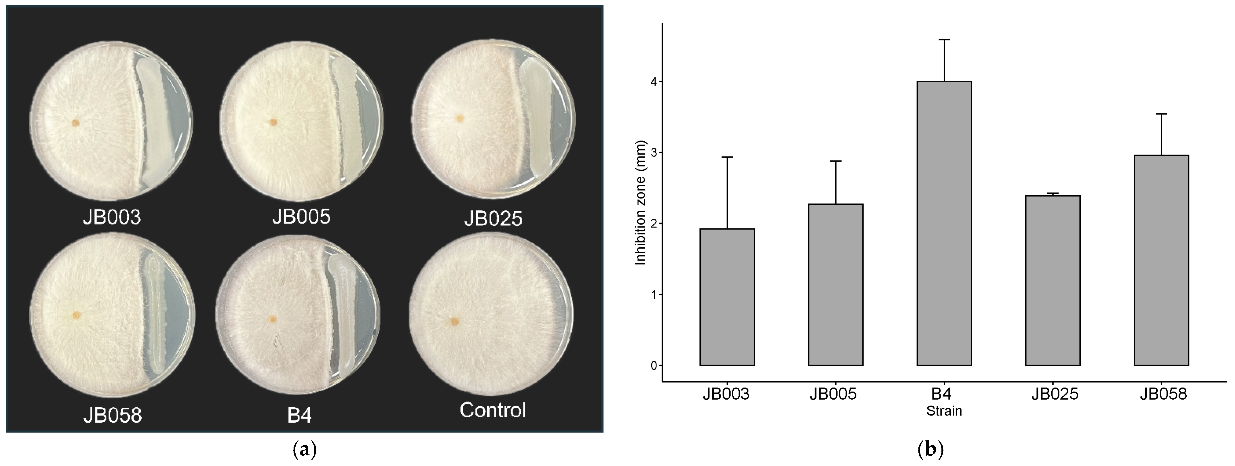
A total of 116 bacterial strains were isolated from the tidal flat of Jang-bong Island, Incheon City, Republic of Korea [48]. The microbiome isolated from the tidal flat was tested against R. solani AG-4 using a dual-culture method. Among the selected strains, five bacterial strains have the strongest antifungal activity against R. solani AG-4, when compared to the control [Figure 1a]. The largest distance of the inhibition zone between bacteria and fungi is found in strain B4 (4.0 mm), followed by strain JB058 (3.0 mm). Strains JB003, JB005, and JB025 show inhibition zones of 1.9 mm, 2.3 mm, and 2.4 mm, respectively (Figure 1b).

3.2. Production of IAA

An IAA-production activity test was conducted using King’s B broth supplemented with l-tryptophan, which is the precursor of IAA. Only two strains show IAA production. IAA is produced by strains B114 (7.54 µg/mL) and B115 (5.47 µg/mL) (Table S1). The positive control is P. aeruginosa, and the IAA concentration was determined using the formula of a standard curve (Figure S1).

3.3. Siderophore Production in Plate Assay

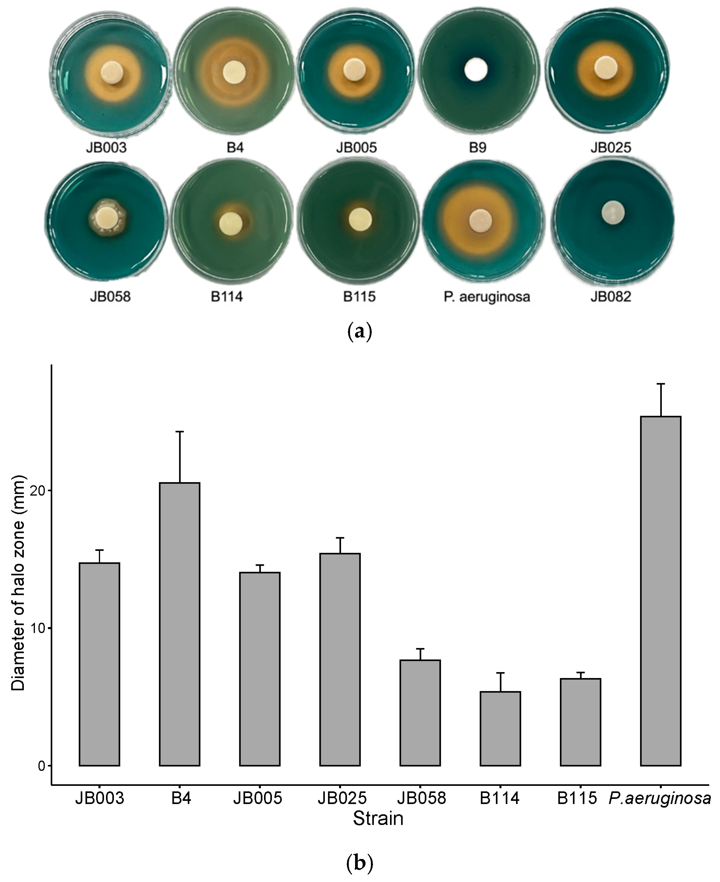
Eight strains show color changes on CAS blue agar (Figure 2a). Strain B4 has the largest halo zone diameter (20.54 mm) followed by JB025 (15.41 mm) (Figure 2b). Moreover, strains JB003 (14.7 mm), JB005 (14.0 mm), JB058 (7.7 mm), B115 (6.3 mm), and B114 (5.4 mm) also demonstrate siderophore production. The JB082 strain and Pseudomonas aeruginosa were negative and positive controls, respectively. The diameter of the halo observed for the positive control, P. aeruginosa is 25.36 mm, and that for the negative control, strain JB082, is 0 mm.

3.4. Protease and Cellulase Production

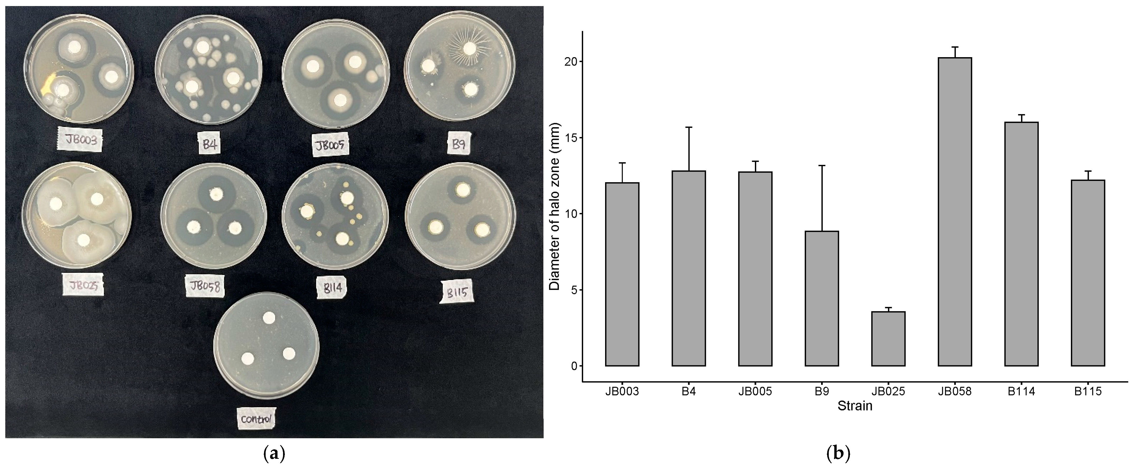
Protease production analysis showed that eight strains have a halo zone on skim milk agar (Figure 3a). Strain JB058 has the largest halo zone of 20.2 mm, followed by strain B114 (16 mm), B4 (12.8 mm), JB005 (12.7 mm), B115 (12.2 mm), JB003 (12.0 mm), and B9 (8.9 mm). JB025 is associated with the smallest halo zone (3.5 mm) (Figure 3b).
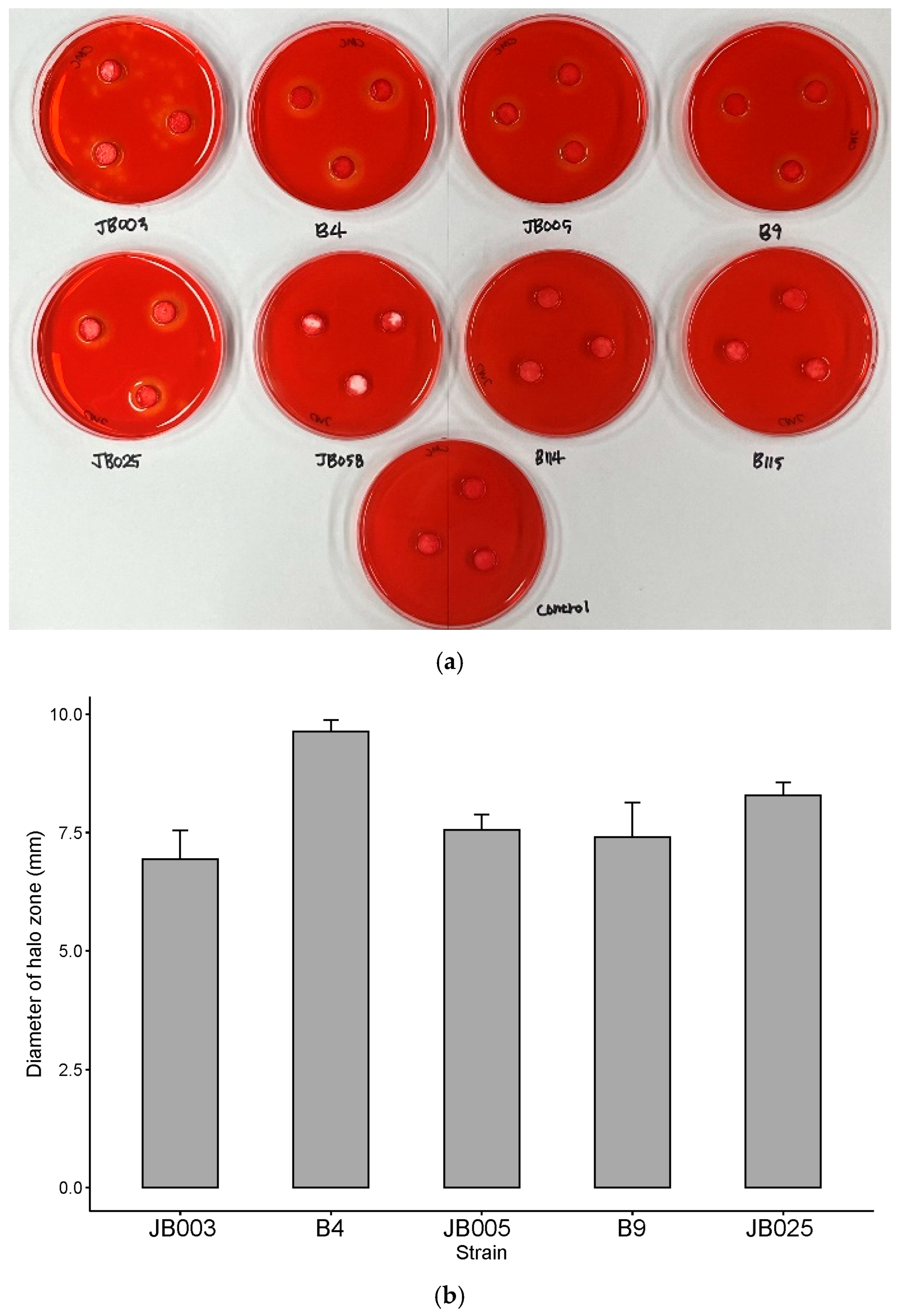
Cellulase production was screened using the CMC agar. Five strains show a halo zone on CMC agar (Figure 4a), which results from the degradation of cellulose by hydrolases secreted by the bacteria. Strain B4 has the largest halo zone (9.6 mm), followed by strains JB025 (8.3 mm), JB005 (7.6 mm), B9 (7.4 mm), and JB003 (6.9 mm) (Figure 4b).

3.5. Phosphate Solubilization and Biofilm formation

Phosphate solubilizing activity tests were conducted in NBRIP-BPB broth. Only one strain shows phosphate solubilizing ability (Table S2). The concentration of phosphate solubilized by strain B9 is 2.05 mg/mL. P. aeruginosa is used as the positive control. Solubilized phosphate is calculated using a standard curve (Figure S2). Among the different strains, JB058 has a significant ability to form biofilms (Table S3 and Figure S3). The absorbance of the solubilized crystal violet from JB058 is 2.366 (Table S3), and the positive control, P. aeruginosa, shows an absorbance value of 0.299.

3.6. Biocontrol of R. solani

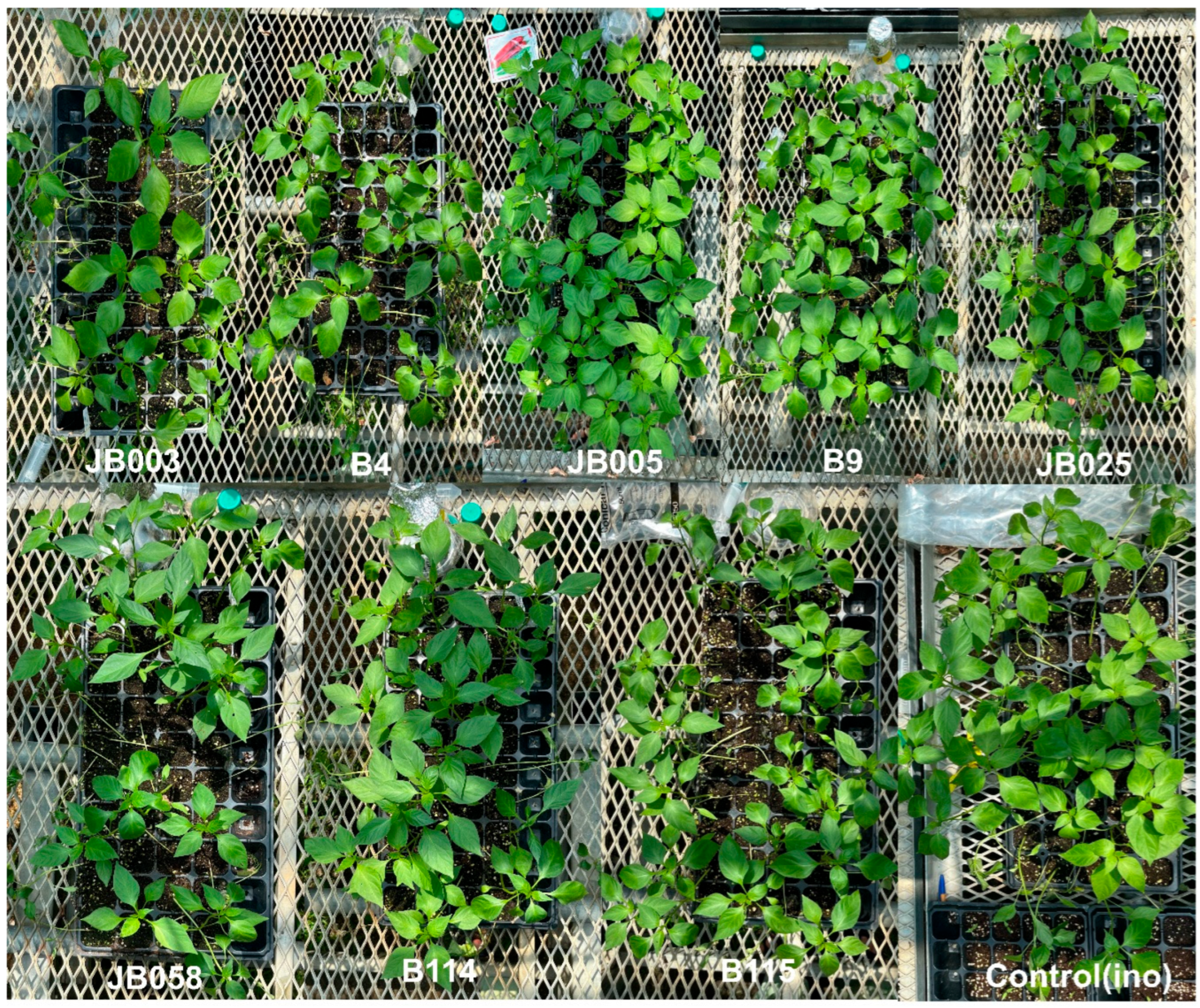
The damping-off rates 3 d after treatment are not significantly different between the treated strains, except for strain JB005 and the uninoculated treatment (Table 1). Strain JB005 and uninoculated treatment show 0% damping-off. Regarding the damping-off rate on the final day of the experiment, JB005 shows the highest suppression of R. solani AG-4 (Table 1). The bacteria JB005 shows 0%, 0%, 0%, 2.5%, 5%, 12.5%, 12.5%, 17.5%, 17.5%, and 17.5% of seedling damping-off rates on each day, respectively. The inoculated treatment shows 0%, 22.5%, 32.5%, 45%, 45%, 45%, 45%, 45%, 45%, 45%, 45%, and 45% of damping-off rates for each day, respectively. The highest damping-off rate is observed for strain JB003. It shows 0%, 17.5%, 32.5%, 67.5%, 70%, 70%, 72.5%, 72.5%, 72.5%, and 72.5% of damping-off rates for each day, respectively. Strain JB005 also shows a strong controlling effect on the seedling damping-off caused by R. solani AG-4 (Figure 5 and Figure 6). In addition, the combination of strains JB005 and B9 shows a significant control effect compared to that of the inoculated treatment (Table 2), although there is no synergistic effect on disease control.

3.7. Promotion of Plant Growth

The heights of the pepper plants in each treatment were measured on the final day of the experiment. The uninoculated treatment shows the greatest height among all treatments (Table 3). The mean size of the uninoculated is 46.218 cm. The lowest observed height in the JB058 strain reaches a mean value of 39.888 cm. The other treatments do not show any significant differences. The highest plant weight is shown by strain B9 (4.99 g), followed by the uninoculated treatment (46.28 g). Nonsignificant results of the other treatments, obtained when comparing them with the inoculated ones, are presented in Table 3. The pepper treated with strain B9 has the highest fresh and dry weights (Table 3). In this experiment, strain B9 does not show high fresh or dry weights, revealing consistent variation between the inoculated and uninoculated treatments (Table 3). The height is significantly different between the treatments; hence, the dry weight was adjusted by dividing by the height. As a result, there are no significant differences among the treatments. Thus, these treatments demonstrate the ability of strain B9 to establish non-reproducibility. Except for strain B9, all treatments show similar results to the previous experimental results.

3.8. Identification of Bacillus Isolates

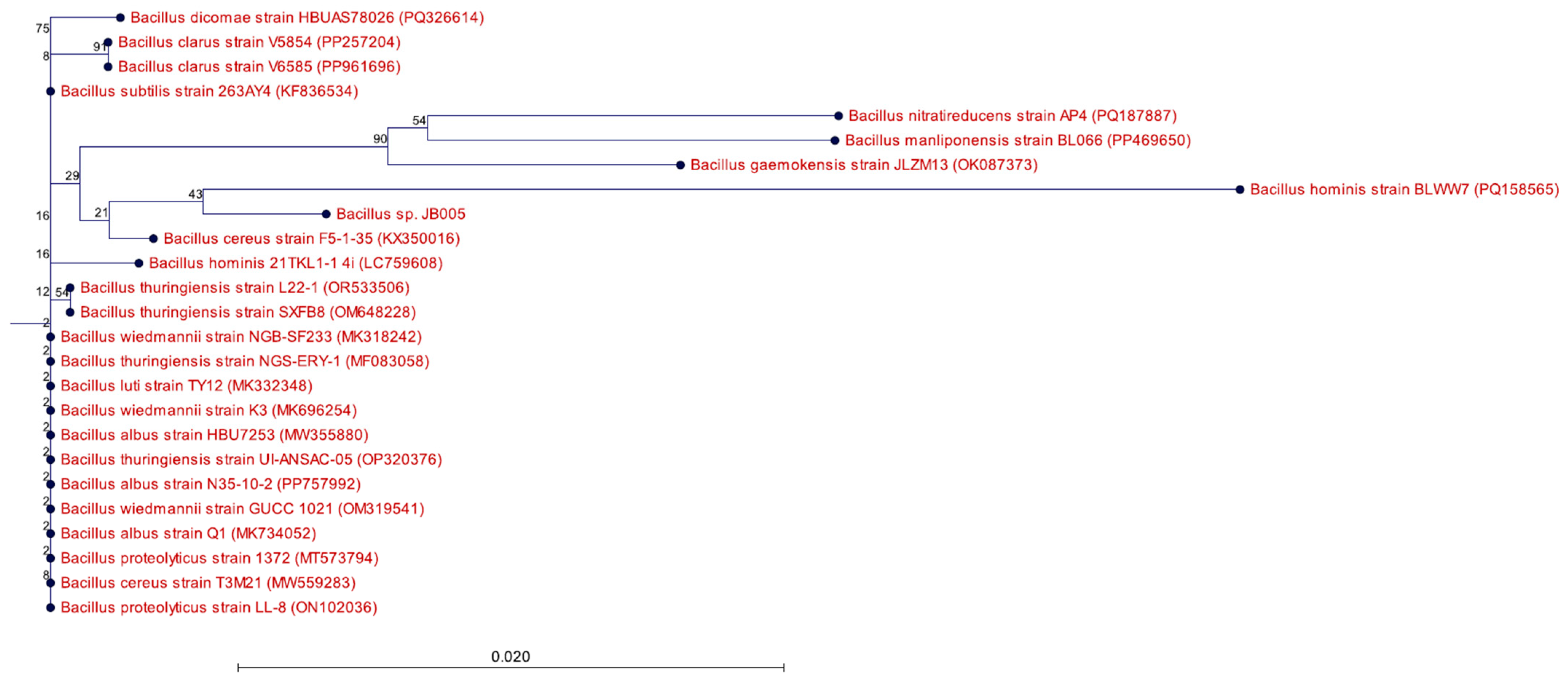
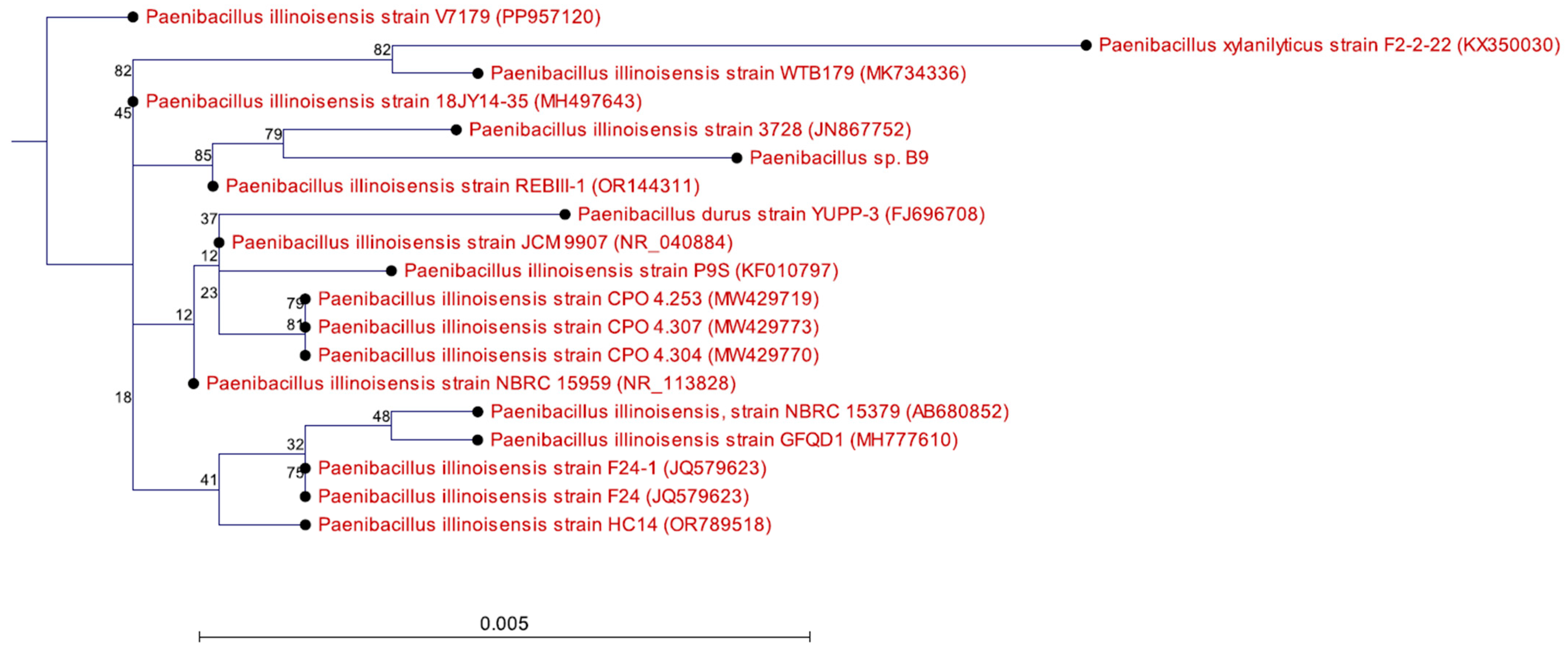
We used 16S rRNA gene locus for cladograms. A phylogenetic tree was created for Bacillus sp. JB005 and B9 for comparative identification with similarity to other strains. The strain JB005 is grouped with Bacillus hominis. However, the bootstrap value of this group is low (Figure 6), which confirms that the construction of polygenetic tree is only accurate 43% out of 100 times. Based on this tree analysis, this bacteria cannot be identified at the species level. Strain B9 is identified to be Paenibacillus illonoisensis (Figure 7).

4. Discussion

Useful agricultural traits and antifungal activities of bacterial strains isolated from tidal flats were ascertained in vitro (using dual-culture antagonistic methods, siderophores, proteases, cellulases, IAA production, phosphate solubilization, biofilm formation, and the detection of the lipopeptide biosynthetic gene). Furthermore, greenhouse experiments were conducted to determine the biological control effect of the tidal isolated bacteria on seedling damping-off disease in pepper, as well as its effect on plant-growth promotion.
In the dual-culture experiments, the strains JB003, JB005, JB025, JB058, and B4 showed effective inhibition zones against R. solani AG-4. The results suggest the five strains could be potential biocontrol agents for sustainable benefits. Silva et al. [49] reported that Trichoderma species with antifungal activities inhibited the formation of sclerotia of Sclerotinia sclerotiorum by approximately 50% and promoted the plant growth of cotton [49]. Dual-culture methods are conventionally used in screening the potential capabilities of BCAs [50]. The methodology evaluates direct antimicrobial activity caused by diffusible extracellular compounds including non-volatile antimicrobial compounds (lipopeptides of surfactin, iturin, or fengycin) or various volatile compounds (1,3,5,7-cyclooctatetraene or annulenen) [51]. However, the limitations concern ambiguities in host-antagonist-pathogen interacting factors and other prominent growth mechanisms (root colonization, induction of systemic resistance, and competition of nutrients and niches in soil) [52,53,54,55].
In the siderophore production test, the strains JB003, B4, JB005, JB025, JB058, B114, and B115 showed halo zones on the CAS blue agar plates. Of these strains, only the strain of Bacillus sp. JB005 reduced the disease incidence caused by R. solani. Siderophore production may not be the only mechanism of biological control but also related to several mechanisms including site competition of infection court and adhesion capability, etc. Gilbert et al. [56] suggested that disease suppression might be related to altered development of rhizosphere communities on UW85n1 (Bacillus cereus)-treated roots.
The inherent physiology upon growing root invasion in the soil habitat disturbs the microbial community causing a sudden abundant nutrient supply. Bacillus sp. JB005 reduced disease incidence. This could be attributed to both siderophore production and other capabilities like site competition or adhesion. Most organisms including fungi need iron as an essential element to maintain the function of various metabolic systems and informational cellular pathways [57]. Siderophores are combined with Fe3+ transported to cells and re-secreted to low molecular weight Fe3+. The production of siderophore by biocontrol agents could limit Fe3+ bioavailability for plant pathogenic fungi or bacteria. Therefore, the production of siderophore contributes to the important mechanisms for controlling phytopathogens. Siderophore production also significantly affects plant growth and inhibits plant pathogen growth through the competition of iron deficiency with PGPR (plant-growth-promoting rhizobacteria) in soils [58,59]. Hence, siderophore production serves as one of the key mechanisms for comprehensively controlling plant pathogens [60].
Protease and cellulase production are implicated in major roles in the biocontrol of plant pathogens [61]. These extracellular hydrolytic enzymes degrade plant pathogenic microbes orchestrating the direct cell death of plant pathogens [62]. Protease production assay showed that the strains JB003, B4, JB005, B9, JB025, JB058, B114, and B115 showed halo zones on the skim milk agar plates.
Cellulase enzyme hydrolyzes cellulose and aids in the indirect degradation of plant pathogenic fungi cell walls. Nonetheless, the cellulase activity acts in conjunction with other hydrolyzing enzymes (chitinase, glucanase, and protease). Thus, cellulase has been attributed to the important hydrolytic enzyme contributing to the degradation of plant pathogenic cell walls [63]. Cellulase production tests revealed the strains JB003, B4, JB005, B9, and JB025 indicating halo zones on CMC agar. The above experiments signified the antifungal efficacy of strain B4 as the most promising strain. Strain B4 was ranked highest among the experimental strains in the dual-culture test, siderophore production test, and cellulase production test. Nevertheless, strain B4 was not top-ranked in the protease production test, with scarce protease production.
The production of enzymes involved in IAA production and phosphate solubilization corresponds to effective plant-growth-promoting traits [64]. IAA also helps in root elongation aided by the increased number of root hairs and root laterals involved in nutrient uptake mechanisms [65,66]. A biocontrol agent synthesizing IAA renders higher utility of plant growth for inciting plant processes like tissue development, cell growth, and division [67].
P (phosphorus) along with N (nitrogen), K (potassium), S (sulfur), Mg (magnesium), and Ca (calcium) are essential macronutrients for plants. P concentrations are regarded as an indispensable element in nucleic acid compositions underlying the adenosine triphosphate (ATP) transfer in energy and carbohydrate transfer between organelles in the leaf [68]. P exists in soil in various forms and is very stable in the natural environment hampering plant uptake and emphasizing degradation into appropriate waste [69]. Biocontrol agents enable phosphate solubilization to improve crop growth and yield [70]. Although the phosphate solubilization activities are not related to biocontrol activities against plant fungal pathogens, this trait could provide BCA not only for biocontrol activities but also a plant-growth-promoting activity. Further, continued crop cultivation results in the deprivation of P fixation and precipitation, aggravating P deficiency [71]. P deficiency accounts for critical damages in crop cultivation, and suboptimal P levels result in yield losses of 5–15% amongst maximum crop yields [72]. Hence, the soil microbiomes contribute to IAA production and phosphate solubilization promoting plant-growth processes. The IAA-production test with strains of B114 and B115 showed positive results and the phosphate solubilizing test indicated that only strain B9 obtained a positive result.
Biofilm formation corresponds to persistent root colonization stressing the necessary process for controlling plant pathogens [73]. In the biofilm formation assay, the strain JB058 showed the highest ability of biofilm formation. The first greenhouse experiment exhibited strain JB005 with the strongest antifungal activity against R. solani AG-4. Treatment of strain B9 displayed strong antifungal activity followed by strain JB005, subsequently causing the lowest seedling to damp-off and conversely higher fresh weight and dry weight. However, other strains described negligible effects on R. solani AG-4 and plant-growth promotion in pepper.
It was assumed that strong adherence to the plants through the formation of biofilm could result in an increase in biocontrol activities against R. solani. However the production of high amounts of biofilm was not related to biocontrol activities, unexpectedly. The mechanism of biocontrol activities is influenced by several other factors such as antibiosis and site competition, among others. This study suggested that the biofilm production could not ensure the biocontrol activities against plant pathogens.
In vitro experiments evaluated the beneficial agricultural traits of each strain and were not associated with authentic and reproducible results as compared to in vivo experiments. The intricate variability is affirmed by environmental factors comprising humidity, temperature, and light differences, emphasizing [74] bacterial vulnerability rather than fungi in combating unfavorable conditions. Moreover, the interactions between biocontrol agents and plants in the soil environment create complexity contributing to the inconsistent lab-to-field effects [75].
The strain JB005 showed inhibition ability in dual-culture experiments and siderophore production. Extracellular compounds including siderophore, protease, and cellulase production by strain JB005 established inhibitory effects on R. solani AG-4. Thus, the hypothesis of high concentrations of the bacteria in secreting higher extracellular compounds and consequent robust disease suppression was hypothesized. Further, another greenhouse experiment using a low-concentration treatment of strain JB005 was conducted to assess the hypothesis’s effectiveness. Thus, the efficacy of the control effect through the combination of both strain B9 and strain JB005 against R. solani AG-4 was assumed for an increase in mixed treatment of the strains B9 and JB005. Hence, the result of three different concentrations of strain JB005 was confronted with the above hypothetical experiment. The combination treatment of the two strains of B9 and JB005 indicated a different result than that of the initial greenhouse experiment. Strain B9 showed a significant difference in fresh weight and dry weight even with a single treatment as compared to the healthy control. The growth-promotion activities of the B9 strain in the greenhouse were varied depending on the time of experiment periods. This might be due to environmental conditions the of greenhouse such as the direction of sunlight coming from the window and temperatures affected by sunlight. The environmental control of the greenhouse was very difficult. Although the strain JB005 did not show growth-promotion activities, this strain was the best potential biocontrol agent, showing very promising disease control activities.
Combination treatment also corroborated similar results as the single-treatment experiments. It was speculated that the result might indicate the involvement of environmental factors and appropriate conditions coercing host plants or microbial interactions in the soil [74,75]. Thus, the effect of strain B9 was not reproducible in the subsequent experiment. Also, two bacteria JB005 and B9 could be incompatible and antagonistic to each other in the soil environment. The incompatibilities of biocontrol agents have been observed when more than two bacteria could be combined to use for biocontrol [76,77]. However, the combination treatment showed the lowest damping-off rate followed by a single treatment of strain JB005. Hence, the antifungal activity of the strain JB005 was active over even lower values, implying seedling damping-off or systemic resistance induction.
In the present study, the mechanism of the antibiosis in the agar plates was studied. The supernatants of the culture extracts of the B. hominis JB005 were TLC-assayed to find out what compounds could be related to antibiotic activities on the agar plate. However we could not find any specific known compound in references like iturin and fengycin of cyclic lipopeptides, etc. In addition, lipopeptide biosynthetic genes were investigated using PCR. In the lane loaded with the iturin biosynthetic gene, a band was detected close to the target size through electrophoresis. Although we tried to search thoroughly in NCBI BLAST, it depicted that the gene of strain JB005 did not match with iturin biosynthetic gene. Hence, the band illustrated in the gel was considered as a non-specific band. Considering all these, the antifungal activity of strain JB005 could be attributed to the activities due to siderophore, protease, and cellulase production. Siderophore, protease, and cellulase have long been known for their antibiosis to the fungus, as discussed previously.

5. Conclusions

Our study suggests that Bacillus sp. JB005, isolated from the tidal flat of Jang Bong Island, demonstrates strong antifungal activity against R. solani AG-4, resulting in reduced seedling damping-off in two separate greenhouse experiments. The disease suppression patterns are primarily attributed to siderophore production correlated to the limitation of iron availability in R. solani growth inhibition. The strain JB005 consistently exhibits the lowest damping-off rate, reinforcing biocontrol potentials against R. solani AG-4. Further experiments with the strain JB005 affirmed production of extracellular enzymes such as protease and cellulase for antifungal activity. The combination of the above traits is postulated to explain the strain JB005’s efficiency as a multifaceted mechanism in combating plant pathogens. Moreover, strain JB005’s consistent performance in both in vitro and in vivo experiments highlights its robust potential for agricultural applications. Overall, strain JB005 poses a promising candidate for biological control of R. solani AG-4.

Supplementary Materials

The following supporting information can be downloaded at: https://www.mdpi.com/article/10.3390/microbiolres17080148/s1, Table S1: Determination of IAA production of 2 bacterial isolates; Table S2: Determination of phosphate solubilization of strain B9; Table S3: Determination of absorbance of solubilized crystal violet at 550nm; Figure S1: Standard curve for the quantification of indole acetic acid; Figure S2: Phosphate solubilizing standard curve; Figure S3: Top-down view of biofilm formation assay.

Author Contributions

C.Y.H. did experiments and wrote the manuscript. M.C. assisted and commented on the experiments. S.C.C. conceptualized, designed and guided the entirety of the experiments. All authors have read and agreed to the published version of the manuscript.

Funding

This research received no external funding.

Institutional Review Board Statement

Not applicable.

Informed Consent Statement

Not applicable.

Data Availability Statement

The original contributions presented in this study are included in the article/Supplementary Materials. Further inquiries can be directed to the corresponding author.

Conflicts of Interest

The authors declare no conflicts of interest.

References

  1. Shin, J.H.; Park, B.S.; Kim, H.Y.; Lee, K.H.; Kim, K.S. Antagonistic and plant growth-promoting effects of Bacillus velezensis BS1 isolated from rhizosphere soil in a pepper field. Plant Pathol. J. 2021, 37, 307. [Google Scholar] [CrossRef] [Scilit] [PubMed]
  2. Heo, Y.; Han, G.; Mun, H.Y.; Lee, C.S.; Cheon, W. Biocontrol Potential and Growth-Promoting Effects of Freshwater Trichoderma Strains against Plant Pathogenic Fungi in Red Pepper. Plant Pathol. J. 2025, 41, 392. [Google Scholar] [CrossRef] [Scilit] [PubMed]
  3. Krasnow, C.S.; Cohen, F.; Sadhasivam, S.; Raphael, G.; Sionov, E.; Ziv, C. Sweet Pepper cv. Lai Lai ripeness stage influences susceptibility to mycotoxinogenic Alternaria alternata causing black mold. Plant Pathol. J. 2025, 41, 266–279. [Google Scholar] [CrossRef] [Scilit] [PubMed]
  4. Wang, L.; Hu, J.; Li, D.; Reymick, O.O.; Tan, X.; Tao, N. Isolation and control of Botrytis cinerea in postharvest green pepper fruit. Sci. Hortic. 2022, 302, 111159. [Google Scholar] [CrossRef] [Scilit]
  5. Volynchikova, E.; Kim, K.D. Anti-oomycete activity and pepper root colonization of Pseudomonas plecoglossicida YJR13 and Pseudomonas putida YJR92 against Phytophthora capsici. Plant Pathol. J. 2023, 39, 123. [Google Scholar] [CrossRef] [Scilit] [PubMed]
  6. Kyeon, M.S.; Son, S.H.; Noh, Y.H.; Kim, Y.E.; Lee, H.I.; Cha, J.S. Xanthomonas euvesicatoria causes bacterial spot disease on pepper plant in Korea. Plant Pathol. J. 2016, 32, 431. [Google Scholar] [CrossRef] [Scilit] [PubMed]
  7. Karavina, C.; Jacques, D.I.; Augustine, G. Potato virus Y isolates infecting bell pepper from parts of Southern Africa display distinct recombination patterns. Physiol. Mol. Plant Pathol. 2021, 114, 101638. [Google Scholar] [CrossRef] [Scilit]
  8. Quesada-Ocampo, L.M.; Parada-Rojas, C.H.; Hansen, Z.; Vogel, G.; Smart, C.; Hausbeck, M.K.; Carmo, R.M.; Huitema, E.; Naegele, R.P.; Kousik, C.S.; et al. Phytophthora capsici: Recent progress on fundamental biology and disease management 100 years after its description. Ann. Rev. Phytopathol. 2023, 61, 185–208. [Google Scholar] [CrossRef] [Scilit] [PubMed]
  9. Mamphogoro, T.P.; Babalola, O.O.; Aiyegoro, O.A. Sustainable management. strategies for bacterial wilt of sweet peppers (Capsicum annuum) and other Solanaceous crops. J. Appl. Microbiol. 2020, 129, 496–508. [Google Scholar] [CrossRef] [Scilit] [PubMed]
  10. Jo, E.J.; Kang, B.G.; Jang, K.S.; Choi, Y.H.; Kim, J.C.; Choi, G.J. Control efficacy of serenade formulation against Rhizoctonia and Pythium damping-off diseases. Res. Plant Dis. 2014, 20, 201–205. [Google Scholar] [CrossRef] [Scilit]
  11. Mannai, S.; Jabnoun-Khiareddine, H.; Nasraoui, B.; Daami-Remadi, M. Rhizoctonia root rot of pepper (Capsicum annuum): Comparative pathogenicity of causal agent and biocontrol attempt using fungal and bacterial agents. J. Plant Pathol. Microbiol. 2018, 9, 431–439. [Google Scholar] [CrossRef]
  12. Raymaekers, K.; Ponet, L.; Holtappels, D.; Berckmans, B.; Cammue, B.P. Screening for novel biocontrol agents applicable in plant disease management–a review. Biol. Control 2020, 144, 104240. [Google Scholar] [CrossRef] [Scilit]
  13. Geiger, F.; Bengtsson, J.; Berendse, F.; Weisser, W.W.; Emmerson, M.; Morales, M.B.; Ceryngier, P.; Liira, J.; Tscharntke, T.; Winqvist, C.; et al. Persistent negative effects of pesticides on biodiversity and biological control potential on European farmland. Basic Appl. Ecol. 2010, 11, 97–105. [Google Scholar] [CrossRef] [Scilit]
  14. Bardin, M.; Ajouz, S.; Comby, M.; Lopez-Ferber, M.; Graillot, B.; Siegwart, M.; Nicot, P.C. Is the efficacy of biological control against plant diseases likely to be more durable than that of chemical pesticides? Front. Plant Sci. 2015, 6, 566. [Google Scholar] [CrossRef] [Scilit] [PubMed]
  15. Borel, B. When the pesticides run out. Nature 2017, 543, 302–304. [Google Scholar] [CrossRef] [Scilit] [PubMed]
  16. Han, J.H.; Shim, H.; Shin, J.H.; Kim, K.S. Antagonistic activities of Bacillus spp. strains isolated from tidal flat sediment towards anthracnose pathogens Colletotrichum acutatum and C. gloeosporioides in South Korea. Plant Pathol. J. 2015, 31, 165. [Google Scholar] [CrossRef] [Scilit] [PubMed]
  17. Heo, Y.M.; Lee, H.; Kim, K.; Kwon, S.L.; Park, M.Y.; Kang, J.E.; Kim, G.H.; Kim, B.S.; Kim, J.J. Fungal diversity in intertidal mudflats and abandoned solar salterns as a source for biological resources. Mar. Drugs 2019, 17, 601. [Google Scholar] [CrossRef] [Scilit] [PubMed]
  18. Wu, Z.; Huang, Y.; Li, Y.; Dong, J.; Liu, X.; Li, C. Biocontrol of Rhizoctonia solani via induction of the defense mechanism and antimicrobial compounds produced by Bacillus subtilis SL-44 on pepper (Capsicum annuum L.). Front. Microbiol. 2019, 10, 2676. [Google Scholar] [CrossRef] [Scilit] [PubMed]
  19. Zheng, Y.; Han, X.; Zhao, D.; Wei, K.; Yuan, Y.; Li, Y.; Liu, M.; Zhang, C.S. Exploring biocontrol agents from microbial keystone taxa associated to suppressive soil: A new attempt for a biocontrol strategy. Front. Plant Sci. 2021, 12, 655673. [Google Scholar] [CrossRef] [Scilit] [PubMed]
  20. Chandrashekara, C.; Bhatt, J.C.; Kumar, R.; Chandrashekara, K.N. Suppressive soils in plant disease management. Eco-Friendly Innov. Approaches Plant Dis. Manag. 2012, 1, 241–256. [Google Scholar]
  21. Knudsen, I.M.B.; Hockenhull, J.; Jensen, D.F.; Gerhardson, B.; Hökeberg, M.; Tahvonen, R.; Teperi, E.; Sundheim, L.; Henriksen, B. Selection of biological control agents for controlling soil and seed-borne diseases in the field. Eur. J. Plant Pathol. 1997, 103, 775–784. [Google Scholar] [CrossRef] [Scilit]
  22. Wekesa, T.B.; Wekesa, V.W.; Onguso, J.M.; Wafula, E.N.; Kavesu, N. Isolation and Characterization of Bacillus velezensis from Lake Bogoria as a Potential Biocontrol of Fusarium solani in Phaseolus vulgaris L. Bacteria 2022, 1, 279–293. [Google Scholar] [CrossRef] [Scilit]
  23. Shih, S.Y.; Huang, Y.S.; Chou, K.R.; Wu, H.Y.; Tsai, H. Isolation and genome characterization of Paenibacillus polymyxa 188, a potential biocontrol agent against fungi. J. Appl. Microbiol. 2024, 135, lxae075. [Google Scholar] [CrossRef] [Scilit] [PubMed]
  24. Zhang, L.; Wu, G.L.; Wang, Y.; Dai, J.; Fang, C.X. Bacillus deserti sp. nov., a novel bacterium isolated from the desert of Xinjiang, China. Antonie Van Leeuwenhoek 2011, 99, 221–229. [Google Scholar] [CrossRef] [Scilit] [PubMed]
  25. Shi, R.; Yin, M.; Tang, S.K.; Lee, J.C.; Park, D.J.; Zhang, Y.J.; Kim, C.J.; Li, W.J. Bacillus luteolus sp. nov., a halotolerant bacterium isolated from a salt field. Int. J. Syst. Evol. Microbiol. 2011, 61, 1344–1349. [Google Scholar] [CrossRef] [Scilit] [PubMed]
  26. Dyer, K.R. The typology of intertidal mudflats. Geol. Soc. Lond. Spec. Publ. 1998, 139, 11–24. [Google Scholar] [CrossRef] [Scilit]
  27. Capone, D.G.; Kiene, R.P. Comparison of microbial dynamics in marine and freshwater sediments: Contrasts in anaerobic carbon catabolism 1. Limnol. Oceanogr. 1988, 33, 725–749. [Google Scholar] [CrossRef] [Scilit]
  28. Moon, K.; Chung, B.; Shin, Y.; Rheingold, A.L.; Moore, C.E.; Park, S.J.; Park, S.; Lee, S.K.; Oh, K.B.; Shin, J.; et al. Pentacyclic antibiotics from a tidal mud flat-derived actinomycete. J. Nat. Prod. 2015, 78, 524–529. [Google Scholar] [PubMed]
  29. Jeong, H.; Jo, S.J.; Bae, M.; Kim, Y.R.; Moon, K. Actinoflavosides B–D, flavonoid type glycosides from tidal mudflat-derived actinomyces. Mar. Drugs 2022, 20, 565. [Google Scholar] [CrossRef] [Scilit] [PubMed]
  30. Hyun, J.H.; Mok, J.S.; Cho, H.Y.; Cho, B.C.; Choi, J.K. Anaerobic mineralization of organic matter and sulfate reduction in summer at Ganghwa intertidal flat, Korea. J. Wetl. Res. 2004, 6, 117–132. [Google Scholar]
  31. Ben Slama, H.; Triki, M.A.; Chenari Bouket, A.; Ben Mefteh, F.; Alenezi, F.N.; Luptakova, L.; Cherif-Silini, H.; Vallat, A.; Oszako, T.; Gharsallah, N.; et al. Screening of the high-rhizosphere competent Limoniastrum monopetalum’culturable endophyte microbiota allows the recovery of multifaceted and versatile biocontrol agents. Microorganisms 2019, 7, 249. [Google Scholar] [CrossRef] [Scilit] [PubMed]
  32. Ashwini, N.; Srividya, S. Potentiality of Bacillus subtilis as biocontrol agent for management of anthracnose disease of chilli caused by Colletotrichum gloeosporioides OGC1. 3 Biotech 2014, 4, 127–136. [Google Scholar] [CrossRef] [Scilit]
  33. Yaraguppi, D.A.; Bagewadi, Z.K.; Patil, N.R.; Mantri, N. Iturin: A promising cyclic lipopeptide with diverse applications. Biomolecules 2023, 13, 1515. [Google Scholar] [CrossRef] [Scilit] [PubMed]
  34. Deleu, M.; Paquot, M.; Nylander, T. Effect of fengycin, a lipopeptide produced by Bacillus subtilis, on model biomembranes. Biophys. J. 2008, 94, 2667–2679. [Google Scholar] [CrossRef] [Scilit] [PubMed]
  35. Kloepper, J.W.; Ryu, C.-M.; Zhang, S. Induced systemic resistance and promotion of plant growth by Bacillus spp. Phytopathology 2004, 94, 1259–1266. [Google Scholar] [CrossRef] [Scilit] [PubMed]
  36. Huang, Y.; Wu, Z.; He, Y.; Ye, B.C.; Li, C. Rhizospheric Bacillus subtilis exhibits biocontrol effect against Rhizoctonia solani in pepper (Capsicum annuum). BioMed Res. Inter. 2017, 2017, 9397619. [Google Scholar] [CrossRef] [Scilit] [PubMed]
  37. Das, M.M.; Haridas, M.; Sabu, A. Biological control of black pepper and. ginger pathogens, Fusarium oxysporum, Rhizoctonia solani and Phytophthora capsici, using Trichoderma spp. Biocatal. Agric. Biotechnol. 2019, 17, 177–183. [Google Scholar] [CrossRef] [Scilit]
  38. Rajkumar, M.; Lee, K.J.; Freitas, H. Effects of chitin and salicylic acid on biological control activity of Pseudomonas spp. against damping off of pepper. S. Afr. J. Bot. 2008, 74, 268–273. [Google Scholar] [CrossRef] [Scilit]
  39. Suwannarach, N.; Kumla, J.; Bussaban, B.; Lumyong, S. Biocontrol of Rhizoctonia solani AG-2, the causal agent of damping-off by Muscodor cinnamomi CMU-Cib 461. World J. Microbiol. Biotechnol. 2012, 28, 3171–3177. [Google Scholar] [CrossRef] [Scilit] [PubMed]
  40. Milagres, A.M.; Machuca, A.; Napoleao, D. Detection of siderophore production from several fungi and bacteria by a modification of chrome azurol S (CAS) agar plate assay. J. Microbiol. Methods 1999, 37, 1–6. [Google Scholar] [CrossRef] [Scilit] [PubMed]
  41. Ji, S.H.; Gururani, M.A.; Chun, S.C. Isolation and characterization of plant growth promoting endophytic diazotrophic bacteria from Korean rice cultivars. Microbiol. Res. 2014, 169, 83–98. [Google Scholar] [CrossRef] [Scilit] [PubMed]
  42. Palin, T.; Kang, D.H.; Schmidt, K.; Fung, D.Y. Detection of extracellular bound proteinase in EPS-producing lactic acid bacteria cultures on skim milk agar. Lett. Appl. Microbiol. 2001, 33, 45–49. [Google Scholar] [CrossRef] [Scilit] [PubMed]
  43. Meddeb-Mouelhi, F.; Moisana, J.K.; Beauregard, M. A comparison of plate assay methods for detectingextracellular cellulase and xylanase activity. Enzym. Microb. Technol. 2014, 66, 16–19. [Google Scholar] [CrossRef] [Scilit] [PubMed]
  44. Mehta, S.; Nautiyal, C.S. An efficient method for qualitative screening of phosphate-solubilizing bacteria. Curr. Microbiol. 2001, 43, 51–56. [Google Scholar] [CrossRef] [Scilit] [PubMed]
  45. O’Toole, G.A. Microtiter dish biofilm formation assay. JoVE 2011, 47, e2437. [Google Scholar] [CrossRef] [Scilit]
  46. Weisburg, W.G.; Barns, S.M.; Pelletier, D.A.; Lane, D.J. 16S ribosomal DNA amplification for phylogenetic study. J. Bacteriol. 1991, 173, 697–703. [Google Scholar] [CrossRef] [Scilit] [PubMed]
  47. R Core Team. RA Language and Environment for Statistical Computing, R Foundation for Statistical; R Foundation for Statitical Computing: Vienna, Austria, 2020. [Google Scholar]
  48. Noh, H. Isolation and Characterization of Sea-Mud Microorganisms for Utilizing Bio-Resources. Ph.D. Thesis, Konkuk University, Seoul, Republic of Korea, 2015. [Google Scholar]
  49. Silva, L.G.; Camargo, R.C.; Mascarin, G.M.; Nunes, P.S.D.O.; Dunlap, C.; Bettiol, W. Dual functionality of Trichoderma: Biocontrol of Sclerotinia sclerotiorum and biostimulant of cotton plants. Front. Plant Sci. 2022, 13, 983127. [Google Scholar] [CrossRef] [Scilit] [PubMed]
  50. Wang, B.; Li, L.; Lin, Y.; Shen, D.; Shao, X.; Zhong, C.; Qian, G. Targeted isolation of biocontrol agents from plants through phytopathogen co-culture and pathogen enrichment. Phytopathol. Res. 2022, 4, 19. [Google Scholar] [CrossRef] [Scilit]
  51. Cao, Y.; Pi, H.; Chandrangsu, P.; Li, Y.; Wang, Y.; Zhou, H.; Xiong, H.; Helmann, J.D.; Cai, Y. Antagonism of two plant-growth promoting Bacillus velezensis isolates against Ralstonia solanacearum and Fusarium oxysporum. Sci. Rep. 2018, 8, 4360. [Google Scholar] [CrossRef] [Scilit] [PubMed]
  52. Stinson, M.; Ezra, D.; Hess, W.M.; Sears, J.; Strobel, G. An endophytic Gliocladium sp. of Eucryphia cordifolia producing selective volatile antimicrobial compounds. Plant Sci. 2003, 165, 913–922. [Google Scholar] [CrossRef] [Scilit]
  53. Lugtenberg, B.J.; Dekkers, L.; Bloemberg, G.V. Molecular determinants of rhizosphere colonization by Pseudomonas. Annu. Rev. Phytopathol. 2001, 39, 461–490. [Google Scholar] [CrossRef] [Scilit] [PubMed]
  54. Bakker, P.A.H.M.; Ran, L.X.; Pieterse, C.M.J.; Van Loon, L.C. Understanding the involvement of rhizobacteria-mediated induction of systemic resistance in biocontrol of plant diseases. Can. J. Plant Pathol. 2003, 25, 5–9. [Google Scholar] [CrossRef] [Scilit]
  55. Kamilova, F.; Leveau, J.H.; Lugtenberg, B. Collimonas fungivorans, an unpredicted in vitro but efficient in vivo biocontrol agent for the suppression of tomato foot and root rot. Environ. Microbiol. 2007, 9, 1597–1603. [Google Scholar] [CrossRef] [Scilit] [PubMed]
  56. Gilbert, G.S.; Clayton, M.K.; Handelsman, J.; Parke, J.L. Use of cluster and discriminant analyses to compare rhizosphere bacterial communities following biological perturbation. Microb. Ecol. 1996, 32, 123–147. [Google Scholar] [CrossRef] [Scilit] [PubMed]
  57. Miethke, M.; Marahiel, M.A. Siderophore-based iron acquisition and pathogen control. Microbiol. Mol. Biol. Rev. 2007, 71, 413–451. [Google Scholar] [CrossRef] [Scilit] [PubMed]
  58. Ghosh, S.K.; Bera, T.; Chakrabarty, A.M. Microbial siderophore–A boon to agricultural sciences. Biol. Control 2020, 144, 104214. [Google Scholar] [CrossRef] [Scilit]
  59. Gupta, C.; Dubey, R.; Maheshwari, D. Plant growth enhancement and suppression of Macrophomina phaseolina causing charcoal rot of peanut by fluorescent Pseudomonas. Biol. Fertil. Soils 2002, 35, 399–405. [Google Scholar] [CrossRef] [Scilit]
  60. Wandersman, C.; Delepelaire, P. Bacterial iron sources: From siderophores to hemophores. Annu. Rev. Microbiol. 2004, 58, 611–647. [Google Scholar] [CrossRef] [Scilit] [PubMed]
  61. Arora, N.K.; Tewari, S.; Singh, R. Multifaceted plant-associated microbes and their mechanisms diminish the concept of direct and indirect PGPRs. In Plant Microbe Symbiosis: Fundamentals and Advances; Springer: New Delhi, India, 2013; pp. 411–449. [Google Scholar]
  62. Shaikh, S.S.; Sayyed, R.Z. Role of plant growth-promoting rhizobacteria and their formulation in biocontrol of plant diseases. In Plant Microbes Symbiosis: Applied Facets; Springer: New Delhi, India, 2014; pp. 337–351. [Google Scholar]
  63. Jadhav, H.P.; Shaikh, S.S.; Sayyed, R.Z. Role of hydrolytic enzymes of rhizoflora in biocontrol of fungal phytopathogens: An overview. Rhizotrophs Plant Growth Promot. Bioremediat. 2017, 2, 183–203. [Google Scholar] [CrossRef] [Scilit]
  64. Raddadi, N.; Cherif, A.; Boudabous, A.; Daffonchio, D. Screening of plant growth promoting traits of Bacillus thuringiensis. Ann. Microbiol. 2008, 58, 47–52. [Google Scholar] [CrossRef] [Scilit]
  65. Woodward, A.W.; Bartel, B. Auxin: Regulation, action, and interaction. Ann. Bot. 2005, 95, 707–735. [Google Scholar] [CrossRef] [Scilit] [PubMed]
  66. Datta, C.; Basu, P.S. Indole acetic acid production by a Rhizobium species from root nodules of a leguminous shrub, Cajanus cajan. Microbiol. Res. 2000, 155, 123–127. [Google Scholar] [CrossRef] [Scilit] [PubMed]
  67. Buensanteai, N.; Yuen, G.Y.; Prathuangwong, S. The biocontrol bacterium Bacillus amyloliquefaciens KPS46 produces auxin, surfactin and extracellular proteins for enhanced growth of soybean plant. Thai J. Agric. Sci. 2008, 41, 101–116. [Google Scholar]
  68. Hawkesford, M.J.; Cakmak, I.; Coskun, D.; De Kok, L.J.; Lambers, H.; Schjoerring, J.K.; White, P.J. Functions of macronutrients. In Marschner’s Mineral Nutrition of Plants; Academic Press: Cambridge, MA, USA, 2023; pp. 201–281. [Google Scholar]
  69. Holford, I.C.R. Soil phosphorus: Its measurement, and its uptake by plants. Soil Res. 1997, 35, 227–240. [Google Scholar] [CrossRef] [Scilit]
  70. Mitra, D.; Anđelković, S.; Panneerselvam, P.; Senapati, A.; Vasić, T.; Ganeshamurthy, A.N.; Chauhan, M.; Uniyal, N.; Mahakur, B.; Radha, T.K. Phosphate-solubilizing microbes and biocontrol agent for plant nutrition and protection: Current perspective. Commun. Soil Sci. Plant Anal. 2020, 51, 645–657. [Google Scholar] [CrossRef] [Scilit]
  71. Zaidi, A.; Khan, M.; Ahemad, M.; Oves, M. Plant growth promotion by phosphate solubilizing bacteria. Acta Microbiol. Immunol. Hung. 2009, 56, 263–284. [Google Scholar] [CrossRef] [Scilit] [PubMed]
  72. Aziz, T.; Sabir, M.; Farooq, M.; Maqsood, M.A.; Ahmad, H.R.; Warraich, E.A. Phosphorus deficiency in plants: Responses, adaptive mechanisms, and signaling. Plant Signal. Underst. Mol. Crosstalk 2014, 7, 133–148. [Google Scholar] [CrossRef] [Scilit]
  73. Zhu, M.L.; Wu, X.Q.; Wang, Y.H.; Dai, Y. Role of biofilm formation by Bacillus pumilus HR10 in biocontrol against pine seedling damping-off disease caused by Rhizoctonia solani. Forests 2020, 11, 652. [Google Scholar] [CrossRef] [Scilit]
  74. Bhuiyan, S.A.; Ryley, M.J.; Galea, V.J.; Tay, D. Evaluation of potential biocontrol agents against Claviceps africana in vitro and in vivo. Plant Pathol. 2003, 52, 60–67. [Google Scholar] [CrossRef] [Scilit]
  75. Besset-Manzoni, Y.; Joly, P.; Brutel, A.; Gerin, F.; Soudiere, O.; Langin, T.; Prigent-Combaret, C. Does in vitro selection of biocontrol agents guarantee success in planta? A study case of wheat protection against Fusarium seedling blight by soil bacteria. PLoS ONE 2019, 14, e0225655. [Google Scholar] [CrossRef] [Scilit] [PubMed]
  76. Klein, T.A.; Ahmad, S.; Whitney, J.C. Contact-dependent interbacterial antagonism mediated by protein secretion machines. Trends Microbiol. 2020, 28, 387–400. [Google Scholar] [CrossRef] [Scilit] [PubMed]
  77. Long, L.; Li, L.; Min, T.; Qianhua, W.; Longteng, Z.; Bozhen, W.; Limin, W.; Xiaolong, S.; Caihong, Z.; Guolian, Q. Assembly of an active microbial consortium by engineering compatible combinations containing foreign and native biocontrol bacteria of kiwifruit. Comput. Struct. Biotech. Jour. 2023, 21, 3672–3679. [Google Scholar] [CrossRef] [Scilit] [PubMed]
Figure 1. (a) Effect of the selected antagonistic strain on R. solani AG-4 using the dual-culture assay. The mycelial agar plug of the R. solani AG-4 was placed and the bacteria were streaked on the other side of the plate. These were incubated at 25 °C for 2 days. After 2 days the inhibition zone between bacterial strain and R. solani AG-4 was observed. B4 strain shows the highest antifungal activity in R. solani AG-4. (b) Inhibition effect of bacterial strains on R. solani AG-4 in dual-culture assay. Mycelial agar plugs of the R. solani AG-4 were placed and the bacteria were streaked on the other side of the plate. The plates were incubated at 25 °C for 2 days. The error bar indicates the standard deviation for each strain.
Microbiolres 17 00148 g001
Figure 2. (a) Siderophore production of different bacterial strains on CAS blue agar. A total of 8 strains show color change on CAS blue agar. Strain B4 shows the highest zone of halo. These CAS blue agar assays were triplicated. P. aeruginosa and JB082 are used as positive control and negative control, respectively. (b) The formation of halo zone produced on CAS blue agar showing siderophore production. The error bar indicates the standard deviation for each strain. Strain B4 shows the largest halo zone next to the positive control of P. aeruginosa. Siderophore production activity was tested in triplicate.
Microbiolres 17 00148 g002
Figure 3. (a) Protease production shows halo zone on skim milk agar. A total of eight strains result in halo zones on skim milk agar. Strain JB058 shows the largest halo zone among the tested strains. These skim milk agar assays were triplicated. Uninoculated treatment is used as a negative control. (b) Formation of halo zones of the different bacteria on skim milk agar indicating protease production. The error bar indicates the standard deviation. Strain JB058 shows the largest halo zone.
Microbiolres 17 00148 g003
Figure 4. (a) Cellulase production of different bacteria on CMC agar. A total of five strains show halo zones on the CMC agar. Strain B4 shows the largest halo zone among experimental strains. These CMC agar assays were triplicated. (b) Formation of halo zones of the different bacteria on CMC agar indicating cellulase production. The error bar indicates the standard deviation. Strain B4 has the largest halo zone among the five strains. These CMC agar assays were triplicated.
Microbiolres 17 00148 g004
Figure 5. Pepper seedling after treatment. Pepper treated with strain JB005 is the healthiest and shows the lowest mortality among the inoculated treatments with R. solani.
Microbiolres 17 00148 g005
Figure 6. Phylogenetic trees of strain JB005 conducted by neighbor-joining method. The scale bar represents the number of nucleotide substitutions per 1000 nucleotides of the sequence. GeneBank accession numbers are next to the name of each strain.
Microbiolres 17 00148 g006
Figure 7. Phylogenetic tree of strain B9 conducted by the neighbor-joining method. The scale bar represents the number of nucleotide substitutions per 1000 nucleotides of the sequence. GeneBank accession numbers are next to the name of each strain.
Microbiolres 17 00148 g007
Table 1. Percentage of seedling damping-off.
StrainPercent Seedling Damping-Off z (3rd Day)Percent Seedling Damping-Off (10th Day)
JB00333.5 ± 1883.7 a ± 12.6
B428.0 ± 1078.8 ab ± 9.53
JB0050.0 ± 0.017.7 d ± 4.89
B917.8 ± 17.644.4 cd ± 8.41
JB02515.0 ± 5.8455.9 bc ± 7.7
JB05839.0 ± 18.967.0 abc ± 1.6
B11430 ± 25.552.6 bc ± 4.74
B11520.4 ± 18.846.7 c ± 3.22
Inoculated33.2 ± 10.147.3 c ± 9.49
Uninoculated0.0 ± 0.00 ± 0.0
z Seedling damping-off rates were transformed into arcsine for statistical analysis. Data are represented as mean and standard deviation. There is no significant difference in percentages of seedling damping-off among the treatments at 3 days after treatment (p < 0.05, F-test). The means with the same letters of superscript are not significantly different at p < 0.05 (Tukey HSD) at 10 days after treatment.
Table 2. Mixed inoculation efficiency on percentage of seedling damping-off.
StrainPercent Seedling Damping-Off zDry Weight z (g)/Height (cm)
JB0057.5 b ± 0.090.005 ± 0.001
B925.0 a ± 0.120.006 ± 0.006
JB005 + B910 b ± 0.080.005 ± 0.001
Inoculated25.0 a ± 0.050.006 ± 0.001
Uninoculated0 ± 0.00.006 ± 0.001
z The mixed treatment of strain JB005 and B9 on the 10th day. Data are represented as mean and standard deviation. The means with superscripts of the same letter are not significantly different in the percent seedling damping-off at p < 0.05 (LSD). There is no significant difference in the dry weights among the treatments. These tests were conducted in quadruplicate.
Table 3. Biological control agents on pepper plant growth.
StrainFresh Weight z (g)Dry Weight z (g)Height (cm) z
JB0034.258 c ± 0.7260.393 bc ± 0.09640.91 bc ± 5.302
B45.759 bc ± 1.6590.454 bc ± 0.20445.467 ab ± 1.97
JB0054.735 c ± 0.9460.477 bc ± 0.10644.152 ab ± 3.832
B97.269 a ± 0.8440.654 a ± 0.09343.99 abc ± 4.364
JB0255.664 bc ± 1.2930.52 b ± 0.10342.811 abc ± 3.599
JB0584.504 c ± 0.9530.385 c ± 0.08639.888 c ± 2.937
B1144.45 c ± 1.6470.432 bc ± 0.11243.055 abc ± 4.497
B1154.626 c ± 0.7620.411 bc ± 0.09442.55 bc ± 3.635
Ino5.643 bc ± 1.3770.491 bc ± 0.16145.17 ab ± 6.291
Non5.839 b ± 1.0030.487 bc ± 0.09946.218 a ± 3.932
z Data are represented as mean and standard deviation. The means with the same superscripts are not significantly different at p < 0.05 (Tukey) and SD denotes standard deviation. These tests were done in quadruplicate.
Disclaimer/Publisher’s Note: The statements, opinions and data contained in all publications are solely those of the individual author(s) and contributor(s) and not of MDPI and/or the editor(s). MDPI and/or the editor(s) disclaim responsibility for any injury to people or property resulting from any ideas, methods, instructions or products referred to in the content.