Article

Emerging Resistance in Oral Candida Isolates from Patients with Periodontal Disease

by
Claudia Berenice Tinoco-Cabral
1,2,
Luis Alfonso Muñoz-Miranda
3,
Manuel R. Kirchmayr
4,
Vianeth Martínez-Rodríguez
2,
Miguel Padilla-Rosas
5,
Maricarmen Iñiguez-Moreno
6,
Suchiquil Rangel-Velázquez
7,
Fabiola Berenice Hernández-Reyes
7,
Claudia Lisette Charles-Niño
8 and
Cesar Arturo Nava-Valdivia
3,*
1
Programa de Doctorado en Microbiología Médica, Departamento de Microbiología y Patología, Centro Universitario de Ciencias de la Salud, Universidad de Guadalajara, Guadalajara 44340, Jalisco, Mexico
2
Especialidad de Periodoncia, Departamento de Clínicas Odontológicas Integrales, Centro Universitario de Ciencias de la Salud, Universidad de Guadalajara, Guadalajara 44340, Jalisco, Mexico
3
Centro de Investigación en Enfermedades Infectocontagiosas, Departamento de Microbiología y Patología, Centro Universitario de Ciencias de la Salud, Universidad de Guadalajara, Guadalajara 44340, Jalisco, Mexico
4
Unidad de Biotecnología Industrial, Centro de Investigación y Asistencia en Tecnología y Diseño del Estado de Jalisco A.C. (CIATEJ), Subsede Zapopan, Zapopan 45019, Jalisco, Mexico
5
Programa de Maestría en Patología y Medicina Oral, Departamento de Clínicas Odontológicas Integrales, Centro Universitario de Ciencias de la Salud, Universidad de Guadalajara, Guadalajara 44340, Jalisco, Mexico
6
Department of Physics and Mathematics, School of Engineering and Technology, Universidad de Monterrey, San Pedro Garza Garcia 66238, Nuevo Leon, Mexico
7
Programa de Maestría en Microbiología Médica, Departamento de Microbiología y Patología, Centro Universitario de Ciencias de la Salud, Universidad de Guadalajara, Guadalajara 44340, Jalisco, Mexico
8
Department of Oral Biology, University of Florida College of Dentistry, 1395 Center Drive, DG-48, P.O. Box 100424, Gainesville, FL 32610, USA
*
Author to whom correspondence should be addressed.
Microbiol. Res. 2026, 17(4), 80; https://doi.org/10.3390/microbiolres17040080
Submission received: 9 March 2026 / Revised: 3 April 2026 / Accepted: 7 April 2026 / Published: 10 April 2026
(This article belongs to the Special Issue Host–Microbe Interactions in Health and Disease)

Abstract

Candida species can shift from commensal organisms to opportunistic pathogens. Both Candida albicans and non-albicans Candida (NAC) species colonize oral biofilms and periodontal pockets, where they may contribute to inflammation and the progression of periodontal disease. This study aimed to determine the prevalence and antifungal susceptibility profiles of Candida species in individuals with different stages of periodontal disease. A cross-sectional study was conducted in 100 participants whose periodontal status was clinically evaluated. Saliva samples were cultured on chromogenic agar for yeast isolation, species identification was confirmed by MALDI-TOF MS, and antifungal susceptibility to fluconazole, clotrimazole, nystatin, and amphotericin B was assessed. Candida spp. was detected in 35% of participants, where C. albicans was the most prevalent species, followed by Nakaseomyces glabratus (formerly Candida glabrata), Candida parapsilosis, Candida dubliniensis, and Candida tropicalis. Species distribution varied according to periodontal status, with N. glabratus predominating in early periodontitis and C. albicans appeared more frequently in higher severe stages of periodontitis. Susceptibility testing showed resistance of C. albicans to clotrimazole (63.6%) and nystatin (22.7%), whereas amphotericin B and fluconazole remained effective. NAC species, particularly N. glabratus, exhibited resistance to nystatin and variable resistance to clotrimazole but remained susceptible to amphotericin B. These findings underscore the importance of early detection and personalized antifungal strategies for managing periodontal disease complicated by Candida colonization.

1. Introduction

The oral cavity is an active ecosystem inhabited by a diverse community of microorganisms, mainly bacteria and yeasts, that form organized biofilms on surfaces such as teeth, prostheses, and implants. Although most of these microorganisms are innocuous, imbalances in microbial communities can lead to pathogenic behavior, causing the development and progression of various oral diseases [1]. Within this microbiota, the genus Candida includes nearly 200 species, of which only a few are opportunistic human pathogens that typically cause infections when the immune system is suppressed. Candida albicans is the most common commensal and the leading opportunistic fungal pathogen in the oral cavity. However, other species like Nakaseomyces glabratus (formerly Candida glabrata), Candida tropicalis, Candida parapsilosis, and Pichia kudriavzevii (formerly Candida krusei), are often found in healthy individuals. Collectively, these five species account for over 90% of Candida-related infections worldwide [2].
The transition of Candida species from commensals to pathogenic organisms involves complex and multifactorial mechanisms that remain incompletely understood. NAC species are widely recognized as significant etiological agents in oral and systemic infections [3]. Candida species express multiple virulence factors that facilitate colonization of the oral mucosa and periodontal pockets, including adhesion to epithelial surfaces and coaggregation with bacterial species. Their persistence in periodontal tissues can induce inflammatory responses leading to tissue attachment loss, bone resorption, and disease progression. Elevated levels of C. albicans have been particularly associated with periodontitis and are frequently detected in subgingival biofilms [4].
Clinically, Candida infections are mainly treated using four classes of systemic antifungal agents, with azoles and polyenes being the most common [5]. In 2022, the World Health Organization (WHO) classified Candida as a Priority Fungal Pathogen, highlighting the genus as a major global health concern. Six Candida species were included in this list: C. krusei (medium priority), N. glabratus, C. parapsilosis, and C. tropicalis (high priority), and C. albicans and C. auris (critical priority) [6]. Similarly, the U.S. Centers for Disease Control and Prevention (CDC) reported in 2019 that drug-resistant Candida infections cause more than 34,000 cases and 1700 deaths annually, emphasizing the urgent need for effective surveillance and management strategies [7].
Antifungal resistance in oral Candida species, particularly among NAC isolates, is an emerging problem that compromises treatment outcomes and may influence both oral and systemic inflammatory conditions. Resistance can be intrinsic, as in P. kudriavzevii (fluconazole-resistant), or acquired, as in N. glabratus (echinocandin-resistant), often resulting from prolonged exposure to antifungals. Furthermore, biofilm formation enhances antifungal tolerance by restricting drug penetration and promoting the persistence of resistant subpopulations [2,8,9]. Despite the increasing clinical relevance of antifungal resistance, many diagnostic laboratories lack the capacity to identify emerging Candida species or determine their susceptibility profiles accurately. This limitation delays the development of targeted antifungal therapies and the monitoring of resistance trends, especially within commensal microbiota associated with common oral conditions such as periodontitis. Therefore, the present study aimed to evaluate the prevalence and species distribution of oral Candida, as well as their antifungal susceptibility patterns across different stages of periodontal disease in a representative population sample.

2. Materials and Methods

2.1. Study Design and Population

A cross-sectional study was conducted involving 100 adult participants by non-probability sampling by consecutive cases, ≥18 years, either sex, who received clinical care at the Periodontics Service of the Comprehensive Dental Clinics, University of Guadalajara. Periodontal status was assessed through a comprehensive clinical examination by calibrated professionals, and the stage of periodontal disease was determined using the 2017 World Workshop classification system for periodontal diseases [10]. All participants provided written informed consent prior to participation.

2.2. Sample Collection and Culture

Unstimulated saliva samples (3 mL) were collected under standardized conditions into sterile 15 mL conical tubes. A 300 µL aliquot of each undiluted sample was inoculated by mass diffusion onto CHROMagar™ Candida (Becton Dickinson, Heidelberg, Germany) and incubated at 37 °C for 48–72 h. Colony morphology and color were recorded for preliminary phenotypic identification: C. albicans (light green), N. glabratus (mauve), C. tropicalis (blue-gray), and P. kudriavzevii (pink with white margins). Colonies with indeterminate pigmentation were reported as Candida spp.

2.3. Species Identification

Species identification was confirmed by matrix-assisted laser desorption/ionization time-of-flight mass spectrometry (MALDI-TOF MS) [11]. Samples were spotted in duplicate onto MALDI MSP 96® polished steel target plates (Bruker Daltonics, Bremen, Germany). Each spot was overlaid with 1 µL of 70% formic acid and subsequently with 1 µL of α-cyano-4-hydroxycinnamic acid (HCCA) matrix solution. After drying, samples were analyzed using the MicroFlex LT® system (Bruker Daltonics, Bremen, Germany) operated with FlexControl v3.4 software. Spectral profiles obtained from six laser shots per spot were compared against the BDAL v10® database in MBT Compass 4.1 software. Identification scores ≥ 1.7 were considered reliable for genus-level identification of yeasts, whereas scores ≥ 2.0 were deemed reliable for species-level identification [12].

2.4. Antifungal Susceptibility Testing

Antifungal susceptibility testing was performed for all isolates following the Clinical and Laboratory Standards Institute (CLSI) M27-A4 reference microdilution method for yeasts [13]. The assay medium consisted of RPMI-1640 buffered with MOPS and supplemented with 2% glucose (Sigma Chemical Co., St. Louis, MO, USA). Serial twofold dilutions were prepared for each antifungal agent: fluconazole (64–0.12 µg/mL), clotrimazole (16–0.03 µg/mL), nystatin (16–0.03 µg/mL), and amphotericin B (16–0.03 µg/mL).
Yeast inoculum was standardized to a 0.5 McFarland turbidity and dispensed into sterile 96-well microplates. Plates were incubated at 37 °C, and minimum inhibitory concentrations (MICs) were determined spectrophotometrically at 405 nm (Multiskan FC, Thermo Scientific Inc., Tijuana, B.C., Mexico) after 24 and 48 h. MIC endpoints were defined as follows: a 50% reduction in absorbance relative to the growth control for fluconazole and clotrimazole, and a 90% reduction for nystatin and amphotericin B [14]. Interpretation was performed according to CLSI guidelines: amphotericin B (S ≤ 1 µg/mL, R > 1 µg/mL) and fluconazole (S ≤ 8 µg/mL, SDD = 16–32 µg/mL, R ≥ 64 µg/mL). For clotrimazole and nystatin, resistance thresholds were defined as R ≥ 0.5 µg/mL and R ≥ 1 µg/mL, respectively [15].

2.5. Statistical Analysis

Descriptive data were expressed as means ± standard deviation (SD) or percentages (%). All measurements were performed in triplicate. Differences in means were assessed using independent-samples Student’s t-tests, and proportions were analyzed using the Chi-square test when comparing participants with periodontitis (stages I–IV) versus non-periodontitis (including periodontal health and gingivitis). Additionally, comparisons across all periodontal stages were conducted using one-way ANOVA. A logistic regression model was constructed to identify factors associated with the presence of Candida spp. as the dependent variable. Covariates included variables with a p-value < 0.2 in the univariate analysis, as well as those considered biologically plausible contributors to Candida presence. Odds ratios (ORs) with 95% confidence intervals (95% CI) were estimated, and variables were entered into the model using a stepwise selection approach to evaluate their contribution as risk factors. A p-value ≤ 0.05 was considered statistically significant. Data analysis was conducted using IBM SPSS Statistics v27.0 (IBM Corp., Chicago, IL, USA) and GraphPad Prism v10.4.1 (GraphPad Software, La Jolla, CA, USA).

3. Results

3.1. Participant Characteristics

A total of 100 participants were enrolled in the study, with a predominance of females (58%). The mean age was 44 ± 15 years, and the average body mass index (BMI) was 27.3 ± 5.0 kg/m2, indicating that most participants were overweight (65%), while 32% had a normal BMI and 3% were classified as obese. Lifestyle factors revealed that 14% of participants were smokers and 30% reported alcohol consumption. Regarding systemic health conditions, hypertension (19%) and diabetes mellitus (12%) were the most frequent comorbidities, followed by rheumatoid arthritis (5%) and human immunodeficiency virus (HIV) infection (3%) (Table 1).

3.2. Periodontal Status

Based on the 2017 World Workshop classification, most individuals exhibited periodontal alterations of varying severity. Gingivitis was the most prevalent condition (38%), followed by mild (stage I) periodontitis in 17%, stage II in 13%, and advanced stages (III and IV) in 14% each. Only 4% of participants presented with periodontal health. This means that this study has a representative sample of the clinical spectrum of periodontal disease (Table 1).

3.3. Candida Isolation and Species Distribution

Candida species were detected in 35% of participants, yielding a total of 45 isolates. C. albicans was the most frequently identified species (48.9%), followed by N. glabratus (24.4%), C. parapsilosis (13.3%), C. dubliniensis (8.9%), and C. tropicalis (4.4%; Table 2). The predominance of C. albicans supports its role as the main oral commensal and opportunistic yeast, while the presence of NAC species in nearly half of the isolates highlights a notable species diversity within the oral cavity. The relatively high frequency of N. glabratus further suggests an emerging trend toward colonization by NAC species, which are often associated with increased antifungal resistance and chronic progression of oral disease.

3.4. Demographic and Clinical Characteristics Across Periodontal Disease Stages

The 100 participants were distributed among the periodontal health group (n = 4), the gingivitis group (n = 38), and the four periodontitis stages (I–IV; n = 58). Although females represented the majority in all groups, the proportion declined progressively from periodontal health (75.0%) to stage IV periodontitis (28.6%), showing a non-significant trend (p = 0.192). Age increased significantly with disease severity (p < 0.001), with mean values ranging from 31 ± 8 years in the healthy group to approximately 50 years in advanced stages of periodontitis. In contrast, BMI did not differ significantly among groups (p = 0.422), although overweight individuals predominated in all disease stages (ranging from 65.8% to 78.6%). The prevalence of smoking and alcohol consumption was relatively low overall and showed no significant association with disease severity (p > 0.05). Regarding systemic comorbidities, only hypertension exhibited an increase with periodontal disease progression (p = 0.003), affecting more than half (57.1%) of participants with stage IV periodontitis. The frequencies of diabetes mellitus, rheumatoid arthritis, and human immunodeficiency virus infection were low and did not differ significantly among groups (Table 3).
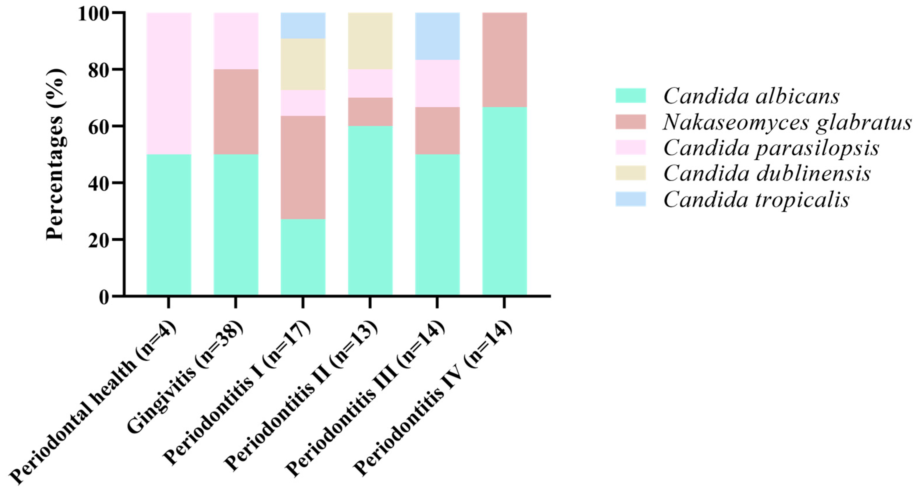
Candida detection showed no statistically significant differences among stages, although colonization tended to be higher in early and moderate periodontitis (47.1–61.5%) compared with gingivitis (21.1%) and healthy individuals (50.0%). The distribution of Candida isolates also varied without significance. Among the isolates, C. albicans was the most prevalent species across all groups, representing 27–67% of isolates. N. glabratus was the second most frequent species, particularly in mild to moderate periodontitis. Other NAC, such as C. parapsilosis, C. dubliniensis, and C. tropicalis, appeared sporadically and without a clear association with disease stage.
C. albicans was the predominant species in all clinical conditions, representing roughly half of the isolates across most stages. Its frequency appeared particularly high in advanced periodontitis (stages III–IV). N. glabratus was the second most prevalent yeast, showing moderate proportions across gingivitis and early periodontitis. The presence of C. parapsilosis, C. dubliniensis, and C. tropicalis was less consistent and limited to a few disease stages, with no clear progression trend (Figure 1). The composition of Candida species did not show a statistically significant difference among periodontal stages, although a trend toward higher diversity was observed in mild to moderate periodontitis (stages I–II). This pattern suggests that Candida colonization may increase in diversity during initial inflammatory stages, while advanced disease is dominated by C. albicans and N. glabratus.
Logistic regression analysis was performed using a stepwise adjusted approach over seven steps. The predictors included periodontitis, age, smoking, alcohol consumption, hypertension, diabetes mellitus, rheumatoid arthritis, and HIV status, with the presence of Candida spp. as the dependent variable. In the adjusted model (Model B), only age (B coefficient = −0.035, OR = 0.965, 95% CI: 0.937–0.994, p = 0.020) remained significantly associated with Candida spp. presence. The remaining variables included in the model were not statistically significant (Supplementary Table S1).
The antifungal susceptibility of 45 Candida isolates revealed marked interspecies differences in response to the four antifungal agents evaluated (Table 4). Nystatin resistance was observed in nearly half of all isolates (48.9%), with minimum inhibitory concentrations (MICs) ranging from 0.5 to 16 µg/mL. N. glabratus and C. parapsilosis demonstrated the highest resistance rates (100% and 83.3%, respectively), whereas C. dubliniensis remained fully susceptible.
For amphotericin B, most isolates were highly susceptible (95.6%), with MIC values between 0.031 and 4 µg/mL. Resistance was only observed in 4.4% of the isolates (one C. albicans and one C. parapsilosis strain). Regarding clotrimazole, resistance was notably high across species (82.2% overall), with MICs ranging from 0.31 to 8 µg/mL. All isolates of N. glabratus, C. parapsilopsis, C. dubliniensis, and C. tropicalis were resistant to this azole, while only 36.4% of C. albicans strains remained susceptible. In contrast, fluconazole exhibited better overall activity, with 80% of isolates classified as susceptible (MICs: 0.25–64 µg/mL). However, resistance (≥64 µg/mL) was detected in 15.6% of strains, mainly C. albicans (18.2%) and C. parapsilopsis (16.7%), while dose-dependent sensitivity was observed in two isolates (4.4%). All C. tropicalis isolates showed high MIC values (64 µg/mL), indicating resistance. N. glabratus and C. parapsilopsis exhibited the broadest resistance patterns, especially to nystatin and clotrimazole, whereas C. dubliniensis showed the most favorable susceptibility profile to all antifungals tested. These findings show the increasing occurrence of azole-resistant Candida species in the oral cavity.
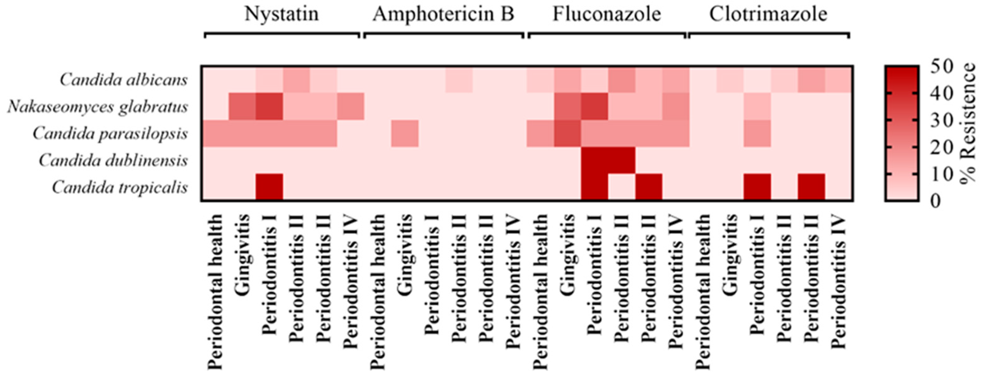
C. albicans remained the most prevalent species in all clinical conditions, but its resistance to nystatin and clotrimazole was more evident in subjects with periodontitis compared to those with periodontal health. In contrast, N. glabratus and C. parapsilosis exhibited high resistance frequencies even at earlier disease stages, particularly to nystatin and fluconazole, indicating their inherent reduced susceptibility. The presence of C. dubliniensis and C. tropicalis was limited to more severe periodontal conditions and was accompanied by strong resistance patterns (Figure 2).

4. Discussion

The role of oral yeasts in the onset and progression of periodontal disease remains insufficiently elucidated. Several studies have reported a higher prevalence of Candida spp., particularly C. albicans, in deeper periodontal pockets of patients with periodontitis compared to periodontally healthy individuals. Factors such as dysbiosis, inflammation, altered pH, local or systemic immunological impairment, and previous antifungal exposure may contribute to the increased recovery of Candida spp. isolates. The microbial competition between Candida and anaerobic periodontopathogens may also shape this distribution, as the latter are better adapted to inflammatory and oxygen-depleted environments, whereas Candida spp. display limited proliferation under extreme conditions [16,17].
It has been reported that virulence factors of key periodontopathogens can modulate up to 35% of fungal adhesion, suggesting that high bacterial virulence activity may inversely correlate with Candida colonization [18]. Nevertheless, recent research has highlighted an increasing involvement of Candida spp. in periodontal pathology. Various species of this genus have been recovered from individuals with gingivitis and periodontitis, indicating a potential contribution to disease progression. In particular, C. albicans is predominant in early periodontal alterations, where it may exacerbate gingival inflammation and disrupt microbial homeostasis [19]. In advanced stages, NAC species such as N. glabratus and C. tropicalis are increasingly detected and may enhance biofilm virulence, while C. parapsilosis and C. dubliniensis tend to predominate in severe diseases, often displaying heightened antifungal resistance and promoting tissue damage.
Epidemiological data suggest that Candida carriage increases the risk of developing periodontitis by approximately 1.76-fold. Patients with periodontal disease consistently exhibit higher Candida prevalence compared to those with healthy gums. However, evidence regarding the direct pathogenic role of N. glabratus, P. kudriavzevii, and C. tropicalis remains inconclusive [20]. In our cohort, a subset of participants presented comorbidities such as hypertension, diabetes mellitus, HIV infection, and rheumatoid arthritis. A recent systematic review reported a significant association between diabetes mellitus and Candida colonization, predominantly involving C. albicans. Similarly, HIV-positive individuals with periodontal disease displayed increased Candida prevalence [19]. Our findings align with these reports: all diabetic patients in our study exhibited some degree of periodontitis, and multiple Candida species were isolated. This supports the hypothesis of a synergistic relationship between systemic metabolic disorders and Candida-associated periodontal deterioration.
Species identification in our study was performed using CHROMagar Candida medium and confirmed via MALDI-TOF/MS, yielding a concordance rate of 77.8%. This discrepancy reflects the limitations of chromogenic media for species-level differentiation. In comparison, Taverna et al. (2023) reported a 94% agreement between both methods, reinforcing the need for confirmatory MALDI-TOF/MS identification [21,22]. Indeed, subsequent studies have demonstrated 100% genus- and species-level accuracy using MALDI-TOF/MS for clinical Candida isolates [23].
Interestingly, NAC species accounted for 51.1% of all isolates, surpassing the prevalence of C. albicans (48.9%). Although C. albicans remains the most frequently recovered oral yeast, an increasing prevalence of NAC species has been recognized in recent years [24,25]. Commonly reported oral NAC species include C. dubliniensis, N. glabratus, C. parapsilosis, M. guilliermondii, and C. kefyr [21,26]. It should be noted that advances in diagnostic technologies, particularly MALDI-TOF/MS, have likely enhanced the detection of previously under-recognized species [27,28].
In this study, the antifungal susceptibility of isolates was evaluated against clotrimazole, fluconazole, nystatin, and amphotericin B—agents commonly prescribed for oral candidiasis [29,30]. Twenty-two isolates exhibited resistance to nystatin, including all N. glabratus strains and several isolates of C. albicans (n = 5), C. parapsilosis (n = 5), and C. tropicalis (n = 1). In the absence of standardized breakpoints, an MIC ≤ 1 µg/mL for topical nystatin was considered indicative of susceptibility, whereas values > 1 µg/mL were interpreted as resistance, following the criteria proposed by Richter et al. [31]. Although nystatin is traditionally considered the first-line therapy for oral candidiasis, emerging resistance has been documented, particularly among immunocompromised hosts. This resistance is frequently linked to alterations in fungal membrane composition, which diminishes drug efficacy [32,33].
Currently, neither EUCAST nor CLSI have established standardized MIC breakpoints for clotrimazole. Based on previous reports, an MIC ≥ 0.5 µg/mL for C. albicans is generally considered indicative of resistance [14,15]. In our study, all NAC isolates exceeded this threshold, with 18% showing MICs between 1–8 µg/mL. Conversely, 82% of isolates remained susceptible to fluconazole. Three isolates (C. parapsilosis and C. tropicalis) exhibited cross-resistance to both clotrimazole (1.0–8.0 µg/mL) and fluconazole (16.0–64.0 µg/mL), while 9.1% of N. glabratus strains were classified as susceptible dose-dependent. Among C. albicans isolates, 64% were resistant to clotrimazole and 18.2% to fluconazole. These findings are consistent with previous studies reporting strong cross-resistance between azoles, particularly clotrimazole, fluconazole, and itraconazole [34,35,36,37]. C. dubliniensis remained susceptible to all antifungal agents except clotrimazole, in line with prior evidence showing general azole and polyene sensitivity, though resistance has occasionally emerged under prolonged drug exposure [26,38,39]. Mechanistically, resistance to clotrimazole may involve reduced target enzyme affinity and altered membrane permeability [40,41,42]. This phenomenon underscores the value of considering increases in MIC as early indicators of antifungal adaptation, even in the absence of formal clinical breakpoints.
Both C. tropicalis isolates displayed azole resistance, corroborating recent reports of increasing resistance rates in Asia. Data from the Taiwan Surveillance of Antimicrobial Resistance of Yeasts program revealed that two-thirds of fluconazole-resistant C. tropicalis isolates were recovered from patients without prior azole exposure, suggesting intrinsic or rapidly acquired mechanisms. Mutations and overexpression of the ERG11 and UPC2 genes have been implicated in these resistance profiles [43,44,45,46].
An emerging concern is the potential for cross-resistance between polyenes (e.g., nystatin) and azoles, given their shared ergosterol target. Mutations in the ergosterol biosynthesis pathway can reduce susceptibility to both classes, complicating therapy and sometimes necessitating alternative agents such as amphotericin B, which, despite its efficacy, is associated with higher costs and toxicity [45]. Positively, most Candida species remain highly susceptible to amphotericin B, with global resistance rates below 2% [47]. In this study, the observed resistance rate was slightly higher (4.4%) yet remained within a comparable range; all isolates were susceptible except one C. parapsilosis and one C. albicans strain, each with an MIC of 2.0 µg/mL.

5. Conclusions

The findings of this study highlight the complex ecological and clinical role of Candida species within the periodontal environment. The coexistence of C. albicans and NAC species such as N. glabratus, C. parapsilosis, C. tropicalis, and C. dubliniensis across different stages of periodontal disease suggests that fungal colonization is dynamic and may reflect local immunological and ecological changes rather than a purely opportunistic event. The high prevalence of NAC isolates, many of which exhibited resistance to azoles and polyenes, highlights a growing concern regarding antifungal tolerance in the oral cavity. These data reinforce the importance of integrating fungal screening into the clinical evaluation of periodontal disease, particularly in patients with metabolic or immunocompromising conditions. Furthermore, the emerging cross-resistance patterns between nystatin and azoles warrant close surveillance and rational antifungal management to prevent treatment failure. Future studies should explore the molecular mechanisms underlying antifungal resistance in oral Candida strains and their interaction with bacterial biofilms, aiming to clarify their contribution to periodontal pathogenesis and to inform targeted therapeutic strategies.

Supplementary Materials

The following supporting information can be downloaded at: https://www.mdpi.com/article/10.3390/microbiolres17040080/s1, Table S1. Factors associated with presence of Candida spp. in the logistic regression.

Author Contributions

All authors contributed to the study’s conception and design. Conceptualization: C.B.T.-C., L.A.M.-M., C.L.C.-N. and C.A.N.-V.; Formal analysis: L.A.M.-M., M.R.K. and C.A.N.-V.; Investigation: C.B.T.-C., M.R.K., S.R.-V. and F.B.H.-R.; Clinical assessment: C.B.T.-C., V.M.-R. and M.P.-R.; Supervision: L.A.M.-M. and C.A.N.-V.; Visualization: C.A.N.-V.; Writing—review & editing: C.B.T.-C., L.A.M.-M. and M.I.-M.; Writing—original draft preparation: C.B.T.-C., L.A.M.-M., M.I.-M. and C.A.N.-V. All authors have read and agreed to the published version of the manuscript.

Funding

This research received no external funding.

Institutional Review Board Statement

The study was conducted in accordance with the Declaration of Helsinki and approved by Ethics Committees of the University of Guadalajara with the approval number CI-09520, accepted on 11 December 2020.

Informed Consent Statement

Informed consent was obtained from all subjects involved in the study.

Data Availability Statement

The original contributions presented in this study are included in the article. Further inquiries can be directed to the corresponding author.

Acknowledgments

We would like to thank the Dental Clinics of the University of Guadalajara, as well as the LQFB. Juan Carlos Díaz Hernández for the technical support provided during the study.

Conflicts of Interest

The authors declare no conflicts of interest.

Abbreviations

NACnon-albicans Candida
WHOWorld Health Organization
CDCCenters for Disease Control and Prevention
HCCAα-cyano-4-hydroxycinnamic acid
CLSIClinical and Laboratory Standards Institute
MICsMinimum Inhibitory Concentrations
BMIBody Mass Index
HIVHuman Immunodeficiency Virus
EUCASTEuropean Committee on Antimicrobial Susceptibility Testing

References

  1. Aggarwal, N.; Kitano, S.; Puah, G.R.Y.; Kittelmann, S.; Hwang, I.Y.; Chang, M.W. Microbiome and Human Health: Current Understanding, Engineering, and Enabling Technologies. Chem. Rev. 2023, 123, 31–72. [Google Scholar] [CrossRef] [Scilit] [PubMed]
  2. Ordaya, E.E.; Clement, J.; Vergidis, P. The role of novel antifungals in the management of candidiasis: A clinical perspective. Mycopathologia 2023, 188, 937–948. [Google Scholar] [CrossRef] [Scilit] [PubMed]
  3. Černáková, L.; Líšková, A.; Lengyelová, L.; Rodrigues, C.F. Prevalence and antifungal susceptibility profile of oral Candida spp. isolates from a hospital in Slovakia. Medicina 2022, 58, 576. [Google Scholar] [CrossRef] [Scilit] [PubMed]
  4. Lomeli-Martínez, S.M.; González-Hernández, L.A.; Villanueva, J.F.A.; Valentín-Gómez, E.; Ratkovich-González, S.; Álvarez-Zavala, M.; Sanchez-Reyes, K.; Cabrera-Silva, R.I.; Varela-Hernández, J.J. In Vitro azole antifungals susceptibility of Candida spp. isolates from HIV-infected patients with periodontitis. J. Med. Mycol. 2022, 32, 101294. [Google Scholar] [CrossRef] [Scilit]
  5. Pappas, P.G.; Kauffman, C.A.; Andes, D.R.; Clancy, C.J.; Marr, K.A.; Ostrosky-Zeichner, L.; Reboli, A.C.; Schuster, M.G.; Vazquez, J.A.; Walsh, T.J.; et al. Clinical practice guideline for the management of candidiasis: 2016 update by the Infectious Diseases Society of America. Clin. Infect. Dis. 2016, 62, e1–e50. [Google Scholar] [CrossRef] [Scilit]
  6. World Health Organization. WHO Fungal Priority Pathogens List to Guide Research, Development and Public Health Action; World Health Organization: Geneva, Switzerland, 2022; Available online: https://iris.who.int/server/api/core/bitstreams/69af4379-4f27-4ac4-8973-74d7b52af7bd/content (accessed on 25 July 2025).
  7. Centers for Disease Control and Prevention. Drug-Resistant Candida Species. U.S. Department of Health & Human Services. Centers for Disease Control and Prevention. 2019. Available online: https://www.cdc.gov/drugresistance/pdf/threats-report/candida-508.pdf (accessed on 20 January 2025).
  8. Černáková, L.; Light, C.; Salehi, B.; Rogel-Castillo, C.; Victoriano, M.; Martorell, M.; Sharifi-Rad, J.; Martins, N.; Rodrigues, C.F. Novel therapies for biofilm-based Candida spp. infections. Adv. Exp. Med. Biol. 2019, 1214, 93–123. [Google Scholar] [CrossRef] [Scilit]
  9. Rodrigues, C.F.; Henriques, M. Liposomal and deoxycholate amphotericin B formulations: Effectiveness against biofilm infections of Candida spp. Pathogens 2017, 6, 62. [Google Scholar] [CrossRef] [Scilit]
  10. Caton, J.G.; Armitage, G.; Berglundh, T.; Chapple, I.L.C.; Jepsen, S.; Kornman, K.S.; Mealey, B.L.; Papapanou, P.N.; Sanz, M.; Tonetti, M.S. A new classification scheme for periodontal and peri-implant diseases and conditions: Introduction and key changes from the 1999 classification. J. Clin. Periodontol. 2018, 45, S1–S8. [Google Scholar] [CrossRef] [Scilit]
  11. Oviaño, M.; Rodríguez-Sánchez, B. MALDI-TOF mass spectrometry in the 21st century clinical microbiology laboratory. Enfermedades Infecc. Microbiol. Clin. 2021, 39, 192–200. [Google Scholar] [CrossRef] [Scilit]
  12. Zhu, H.-H.; Liu, M.-M.; Boekhout, T.; Wang, Q.-M. Improvement of a MALDI-TOF database for the reliable identification of Candidozyma auris (formally Candida auris) and related species. Microbiol. Spectr. 2025, 13, e0144424. [Google Scholar] [CrossRef] [Scilit]
  13. CLSI. Reference Method for Broth Dilution Antifungal Susceptibility Testing of Yeasts, 4th ed.; CLSI Standard M27; Clinical and Laboratory Standards Institute: Wayne, PA, USA, 2017. [Google Scholar]
  14. Borman, A.M.; Muller, J.; Walsh-Quantick, J.; Szekely, A.; Patterson, Z.; Palmer, M.D.; Fraser, M.; Johnson, E.M. MIC distributions for amphotericin B, fluconazole, itraconazole, voriconazole, flucytosine and anidulafungin and 35 uncommon pathogenic yeast species from the UK determined using the CLSI broth microdilution method. J. Antimicrob. Chemother. 2020, 75, 1194–1205. [Google Scholar] [CrossRef] [Scilit]
  15. Pelletier, R.; Peter, J.; Antin, C.; Gonzalez, C.; Wood, L.; Walsh, T.J. Emergence of resistance of Candida albicans to clotrimazole in human immunodeficiency virus-infected children: In vitro and clinical correlations. J. Clin. Microbiol. 2000, 38, 1563–1568. [Google Scholar] [CrossRef] [Scilit]
  16. Du, Q.; Ren, B.; Zhou, X.; Zhang, L.; Xu, X. Cross-kingdom interaction between Candida albicans and oral bacteria. Front. Microbiol. 2022, 13, 911623. [Google Scholar] [CrossRef] [Scilit]
  17. Krüger, W.; Vielreicher, S.; Kapitan, M.; Jacobsen, I.D.; Niemiec, M.J. Fungal-bacterial interactions in health and disease. Pathogens 2019, 8, 70. [Google Scholar] [CrossRef] [Scilit]
  18. Bartnicka, D.; Karkowska-Kuleta, J.; Zawrotniak, M.; Satała, D.; Michalik, K.; Zielinska, G.; Bochenska, O.; Kozik, A.; Ciaston, I.; Koziel, J.; et al. Adhesive protein-mediated cross-talk between Candida albicans and Porphyromonas gingivalis in dual species biofilm protects the anaerobic bacterium in unfavorable oxic environment. Sci. Rep. 2019, 9, 4376. [Google Scholar] [CrossRef] [Scilit] [PubMed]
  19. Suresh Unniachan, A.; Krishnavilasom Jayakumari, N.; Sethuraman, S. Association between Candida species and periodontal disease: A systematic review. Curr. Med. Mycol. 2020, 6, 63–68. [Google Scholar] [CrossRef] [Scilit] [PubMed]
  20. Slazhneva, E.; Tikhomirova, E.; Tsarev, V.; Orekhova, L.; Loboda, E.; Atrushkevich, V. Candida species detection in patients with chronic periodontitis: A systematic review and meta-analysis. Clin. Exp. Dent. Res. 2022, 8, 1354–1375. [Google Scholar] [CrossRef] [Scilit]
  21. Patel, M. Oral cavity and Candida albicans: Colonisation to the development of infection. Pathogens 2022, 11, 335. [Google Scholar] [CrossRef] [Scilit] [PubMed]
  22. Taverna, C.G.; Vivot, M.E.; Arias, B.A.; Irazu, L.; Canteros, C.E. Evaluation of the CHROMagar Candida Plus medium for presumptive identification of yeasts and MALDI-TOF MS identification. Mycoses 2023, 66, 977–983. [Google Scholar] [CrossRef] [Scilit]
  23. Panda, A.; Ghosh, A.K.; Mirdha, B.R.; Xess, I.; Paul, S.; Samantaray, J.C.; Srinivasan, A.; Khalil, S.; Rastogi, N.; Dabas, Y. MALDI-TOF mass spectrometry for rapid identification of clinical fungal isolates based on ribosomal protein biomarkers. J. Microbiol. Methods 2015, 109, 93–105. [Google Scholar] [CrossRef] [Scilit]
  24. Nucci, M.; Queiroz-Telles, F.; Alvarado-Matute, T.; Tiraboschi, I.N.; Cortes, J.; Zurita, J.; Guzman-Blanco, M.; Santolaya, M.E.; Thompson, L.; Sifuentes-Osornio, J.; et al. Epidemiology of candidemia in Latin America: A laboratory-based survey. PLoS ONE 2013, 8, e59373. [Google Scholar] [CrossRef] [Scilit] [PubMed]
  25. Redding, S.W.; Dahiya, M.C.; Kirkpatrick, W.R.; Coco, B.J.; Patterson, T.F.; Fothergill, A.W.; Rinaldi, M.G.; Thomas, C.R. Candida glabrata is an emerging cause of oropharyngeal candidiasis in patients receiving radiation for head and neck cancer. Oral Surg. Oral Med. Oral Pathol. Oral Radiol. Endodontol. 2004, 97, 47–52. [Google Scholar] [CrossRef] [Scilit]
  26. Pesee, S.; Samaranayake, L.; Roytrakul, S.; Paaopanchon, C.; Phantumvanit, P. Prevalence and susceptibility profiles of oral yeast species isolated from a healthy adult Thai cohort. Arch. Oral Biol. 2022, 138, 105415. [Google Scholar] [CrossRef] [Scilit]
  27. Berkow, E.L.; Lockhart, S.R. Fluconazole resistance in Candida species: A current perspective. Infect. Drug Resist. 2017, 10, 237–245. [Google Scholar] [CrossRef] [Scilit] [PubMed]
  28. Mäkinen, A.; Nawaz, A.; Mäkitie, A.; Meurman, J.H. Role of non-albicans Candida and Candida albicans in oral squamous cell cancer patients. J. Oral Maxillofac. Surg. 2018, 76, 2564–2571. [Google Scholar] [CrossRef] [Scilit] [PubMed]
  29. Almangour, T.A.; Kaye, K.S.; Alessa, M.; Eljaaly, K.; Sfouq Aleanizy, F.; Alsharidi, A.; Al Majid, F.M.; Alotaibi, N.H.; A Alzeer, A.; Alnezary, F.S.; et al. Efficacy of clotrimazole for the management of oral candidiasis: A meta-analysis of randomized clinical trials. Saudi Pharm. J. 2021, 29, 315–323. [Google Scholar] [CrossRef] [Scilit] [PubMed]
  30. Kessler, S.Q.S.; Lang, P.M.; Dal-Pizzol, T.S.; Montagner, F. Resistance profiles to antifungal agents in Candida albicans isolated from human oral cavities: Systematic review and meta-analysis. Clin. Oral Investig. 2022, 26, 6479–6489. [Google Scholar] [CrossRef] [Scilit]
  31. Richter, S.S.; Galask, R.P.; Messer, S.A.; Hollis, R.J.; Diekema, D.J.; Pfaller, M.A. Antifungal susceptibilities of Candida species causing vulvovaginitis and epidemiology of recurrent cases. J. Clin. Microbiol. 2005, 43, 2155–2162. [Google Scholar] [CrossRef] [Scilit] [PubMed]
  32. Keyvanfar, A.; Najafiarab, H.; Talebian, N.; Tafti, M.F.; Adeli, G.; Ghasemi, Z.; Tehrani, S. Drug-resistant oral candidiasis in patients with HIV infection: A systematic review and meta-analysis. BMC Infect. Dis. 2024, 24, 546. [Google Scholar] [CrossRef] [Scilit]
  33. Lyu, X.; Zhao, C.; Yan, Z.M.; Hua, H. Efficacy of nystatin for the treatment of oral candidiasis: A systematic review and meta-analysis. Drug Des. Dev. Ther. 2016, 10, 1161–1171. [Google Scholar] [CrossRef] [Scilit]
  34. Chen, T.C.; Chen, Y.H.; Chen, Y.C.; Lu, P.L. Fluconazole exposure rather than clonal spreading is correlated with the emergence of Candida glabrata with cross-resistance to triazole antifungal agents. Kaohsiung J. Med. Sci. 2012, 28, 306–315. [Google Scholar] [CrossRef] [Scilit]
  35. Costa, C.; Ribeiro, J.; Miranda, I.M.; Silva-Dias, A.; Cavalheiro, M.; Costa-de-Oliveira, S.; Rodrigues, A.G.; Teixeira, M.C. Clotrimazole drug resistance in Candida glabrata clinical isolates correlates with increased expression of the drug:H(+) antiporters CgAqr1, CgTpo1_1, CgTpo3, and CgQdr2. Front. Microbiol. 2016, 7, 526. [Google Scholar] [CrossRef] [Scilit]
  36. Frej-Mądrzak, M.; Golec, S.; Włodarczyk, K.; Choroszy-Król, I.; Nawrot, U. Susceptibility to clotrimazole of Candida spp. isolated from the genitourinary system—A single center study. Pathogens 2021, 10, 1142. [Google Scholar] [CrossRef] [Scilit]
  37. Panackal, A.A.; Gribskov, J.L.; Staab, J.F.; Kirby, K.A.; Rinaldi, M.; Marr, K.A. Clinical significance of azole antifungal drug cross-resistance in Candida glabrata. J. Clin. Microbiol. 2006, 44, 1740–1743. [Google Scholar] [CrossRef] [Scilit]
  38. Martinez, M.; López-Ribot, J.L.; Kirkpatrick, W.R.; Coco, B.J.; Bachmann, S.P.; Patterson, T.F. Replacement of Candida albicans with Candida dubliniensis in human immunodeficiency virus-infected patients with oropharyngeal candidiasis treated with fluconazole. J. Clin. Microbiol. 2002, 40, 3135–3139. [Google Scholar] [CrossRef] [Scilit] [PubMed]
  39. Pfaller, M.A.; Messer, S.A.; Gee, S.; Joly, S.; Pujol, C.; Sullivan, D.J.; Coleman, D.C.; Soll, D.R. In vitro susceptibilities of Candida dubliniensis isolates tested against the new triazole and echinocandin antifungal agents. J. Clin. Microbiol. 1999, 37, 870–872. [Google Scholar] [CrossRef] [Scilit] [PubMed]
  40. Ayadi, R.; Sitterlé, E.; d’Enfert, C.; Dannaoui, E.; Bougnoux, M.E. Candida albicans and Candida dubliniensis show different trailing effect patterns when exposed to echinocandins and azoles. Front. Microbiol. 2020, 11, 1286. [Google Scholar] [CrossRef] [Scilit] [PubMed]
  41. Moran, G.P.; Sullivan, D.J.; Henman, M.C.; McCreary, C.E.; Harrington, B.J.; Shanley, D.B.; Coleman, D.C. Antifungal drug susceptibilities of oral Candida dubliniensis isolates from human immunodeficiency virus (HIV)-infected and non-HIV-infected subjects and generation of stable fluconazole-resistant derivatives in vitro. Antimicrob. Agents Chemother. 1997, 41, 617–623. [Google Scholar] [CrossRef] [Scilit]
  42. Perea, S.; López-Ribot, J.L.; Wickes, B.L.; Kirkpatrick, W.R.; Dib, O.P.; Bachmann, S.P.; Keller, S.M.; Martinez, M.; Patterson, T.F. Molecular mechanisms of fluconazole resistance in Candida dubliniensis isolates from human immunodeficiency virus-infected patients with oropharyngeal candidiasis. Antimicrob. Agents Chemother. 2002, 46, 1695–1703. [Google Scholar] [CrossRef] [Scilit] [PubMed]
  43. Tseng, K.Y.; Liao, Y.C.; Chen, F.C.; Chen, F.J.; Lo, H.J. A predominant genotype of azole-resistant Candida tropicalis clinical strains. Lancet Microbe 2022, 3, e646. [Google Scholar] [CrossRef] [Scilit]
  44. Zhou, Z.L.; Tseng, K.Y.; Chen, Y.Z.; Tsai, D.-J.; Wu, C.-J.; Chen, Y.-C.; Peng, H.-L.; Yang, Y.-L.; Hsieh, L.-Y.; Chen, C.-H.; et al. Genetic relatedness among azole-resistant Candida tropicalis clinical strains in Taiwan from 2014 to 2018. Int. J. Antimicrob. Agents 2022, 59, 106592. [Google Scholar] [CrossRef] [Scilit] [PubMed]
  45. Pristov, K.E.; Ghannoum, M.A. Resistance of Candida to azoles and echinocandins worldwide. Clin. Microbiol. Infect. 2019, 25, 792–798. [Google Scholar] [CrossRef] [Scilit]
  46. Wang, D.; An, N.; Yang, Y.; Yang, X.; Fan, Y.; Feng, J. Candida tropicalis distribution and drug resistance is correlated with ERG11 and UPC2 expression. Antimicrob. Resist. Infect. Control 2021, 10, 54. [Google Scholar] [CrossRef] [Scilit] [PubMed]
  47. Yamin, D.; Akanmu, M.H.; Al Mutair, A.; Alhumaid, S.; Rabaan, A.A.; Hajissa, K. Global prevalence of antifungal-resistant Candida parapsilosis: A systematic review and meta-analysis. Trop. Med. Infect. Dis. 2022, 7, 188. [Google Scholar] [CrossRef] [Scilit] [PubMed]
Figure 1. Distribution of Candida species across periodontal disease stages. Data expressed as percentages of total isolates (n = 45).
Microbiolres 17 00080 g001
Figure 2. Heatmap of the distribution and antifungal resistance of Candida species across periodontal health stages. The x-axis represents periodontal status (healthy, gingivitis, early, moderate, and advanced periodontitis), while the y-axis shows the Candida species identified. Color intensity indicates the frequency or degree of antifungal resistance (darker red = higher resistance).
Microbiolres 17 00080 g002
Table 1. Participant characteristics and Candida species isolation.
CharacteristicsTotal Participants
n = 100
Periodontitis
(n = 58)
Non-Periodontitis
(n = 42)
p
Female, n (%)58 (58.0)31 (53.4)27 (64.3)0.190
Age (years)44 ± 1549 ± 1436 ± 13<0.001
Body Mass Index (kg/m2)27.3 ± 5.027.8 ± 5.226.7 ± 4.80.279
Normal weight, n (%)32 (32.0)17 (29.3)15 (35.7)
Overweight, n (%)65 (65.0)38 (65.5)27 (64.3)0.477
Obesity, n (%)3 (3.0)3 (5.2)0 (0)
Smoking, n (%)14 (14.0)10 (17.2)4 (9.5)0.242
Alcohol consumption, n (%)30 (30.0)14 (24.1)16 (38.1)0.320
Comorbidities
Hypertension, n (%)19 (19.0)15 (25.9)4 (9.5)0.034
Diabetes mellitus, n (%)12 (12.0)9 (15.5)3 (7.1)0.169
Rheumatoid arthritis, n (%)5 (5.0)3 (5.2)2 (4.8)0.650
HIV, n (%)3 (3.0)3 (5.2)0 (0)0.191
Periodontitis degree
Periodontal health, n (%)4 (4.0)---
Gingivitis, n (%)38 (38.0)---
Periodontitis I, n (%)17 (17.0)---
Periodontitis II, n (%)13 (13.0)---
Periodontitis III, n (%)14 (14.0)---
Periodontitis IV, n (%)14 (14.0)---
Candida positive, n (%)35 (35.0)25 (43.1)10 (23.8)0.036
Candida isolates *, n (%)45 (100.0)33 (73.3)12 (26.7)0.004
Candida albicans, n (%)22 (48.9)16 (48.5)6 (50.0)
Nakaseomyces glabratus, n (%)11 (24.4)8 (24.2)3 (25.0)
Candida parasilopsis, n (%)6 (13.3)3 (9.1)3 (25.0)0.425
Candida dublinensis, n (%)4 (8.9)4 (12.1)0 (0)
Candida tropicalis, n (%)2 (4.4)2 (6.1)0 (0)
Quantitative variables are expressed as mean ± standard deviation, and qualitative variables as frequency and percentage. Statistical analyses were performed using the Chi-square test (or Fisher’s exact test, when appropriate) to compare proportions. Differences between means were assessed using independent-samples Student’s t-tests. p-values were calculated by comparing participants with periodontitis at any stage (I–IV) to those without periodontitis, including individuals with periodontal health or gingivitis. HIV: Human Immunodeficiency Virus. * Candida species are reported as proportions of the total isolates. Periodontitis included stages I–IV, non-periodontitis included gingivitis and periodontal health.
Table 2. Candida isolates identified by MALDI-TOF MS.
Candida SpeciesIdentity Index Mean, SDFrequency, (%)
n = 45
Candida albicans2.09 ± 0.09 *22 (48.9)
Nakaseomyces glabratus2.19 ± 0.05 *11 (24.4)
Candida parapsilosis2.00 ± 0.11 *6 (13.3)
Candida dublinensis1.98 ± 0.064 (8.9)
Candida tropicalis2.15 ± 0.12 *2 (4.4)
Quantitative variables are expressed as mean ± standard deviation, and qualitative variables as frequency and percentage. Candida species are reported as proportions of the total isolates. * An identity index ≥ 2.0 represents a high confidence and consistency identification of the identified species.
Table 3. Distribution of Candida species according to periodontal health status.
VariablePeriodontal Health
(n = 4)
Gingivitis
(n = 38)
Periodontitis
I
(n = 17)
II
(n = 13)
III
(n = 14)
IV
(n = 14)
p
Female, n (%)3 (75.0)24 (63.2)12 (70.6)8 (61.5)7 (50.0)4 (28.6)0.192
Age (years)31 ± 837 ± 1446 ± 1654 ± 1548 ± 1049 ± 16<0.001
Body Mass Index (kg/m2)25.7 ± 1.526.8 ± 5.027.4 ± 4.929.3 ± 6.528.8 ± 5.725.9 ± 3.10.422
Normal weight, n (%)2 (50.0)13 (34.2)5 (29.4)2 (15.4)3 (21.4)7 (50.0)0.298
Overweight, n (%)2 (50.0)25 (65.8)12 (70.6)9 (69.2)11 (78.6)6 (42.9)
Obesity, n (%)0 (0.0)0 (0.0)0 (0.0)2 (15.4)0 (0.0)1 (7.1)
Smoking, n (%)1 (25.0)3 (7.9)4 (23.5)1 (7.7)0 (0.0)5 (35.7)0.163
Alcohol consumption, n (%)2 (50.0)14 (36.8)4 (23.5)3 (23.1)2 (14.3)5 (35.7)0.177
Comorbidities
Hypertension, n (%)0 (0.0)4 (10.5)1 (5.9)3 (23.1)3 (21.4)8 (57.1)0.003
Diabetes mellitus, n (%)0 (0.0)3 (7.9)1 (5.9)3 (23.1)3 (21.4)2 (14.3)0.478
Rheumatoid arthritis, n (%)0 (0.0)2 (5.3)0 (0.0)1 (7.7)1 (7.1)1 (7.1)0.904
HIV, n (%)0 (0.0)0 (0.0)1 (7.7)0 (0.0)0 (0.0)2 (14.3)0.095
Candida positive, n (%)2 (50.0)8 (21.1)8 (47.1)8 (61.5)5 (35.7)4 (28.6)0.109
Candida isolates, n (%)2 (4.5)10 (22.2)11 (24.5)10 (22.2)6 (13.3)6 (13.3)0.317
Candida albicans, n (%)1 (50.0)5 (50.0)3 (27.2)6 (60.0)3 (50.0)4 (66.7)0.719
Nakaseomyces glabratus, n (%)0 (0)3 (30.0)4 (36.4)1 (10.0)1 (16.7)2 (33.3)
Candida parasilopsis, n (%)1 (50.0)2 (20.0)1 (9.1)1 (10.0)1 (16.7)0 (0)
Candida dublinensis, n (%)0 (0)0 (0)2 (18.2)2 (20.0)0 (0)0 (0)
Candida tropicalis, n (%)0 (0)0 (0)1 (9.1)0 (0)1 (16.7)0 (0)
Quantitative variables are presented as mean ± standard deviation, while qualitative variables are shown as frequency and percentage. HIV: human immunodeficiency virus. Percentages are based on the total number of isolates (n = 45). Comparisons of qualitative variables were made using the Chi-square test, and comparisons of quantitative variables were performed with one-way ANOVA.
Table 4. Distribution of Candida isolates and antifungal categories across periodontal stage.
AntifungalCandida SpeciesPeriodontal Health
(n = 4)
GingivitisPeriodontitis
I
(n = 17)
II
(n = 13)
III
(n = 14)
IV
(n = 14)
NystatinCandida albicans--1 (4.5)3 (13.6)1 (4.5)-
Nakaseomyces glabratus-3 (27.3)4 (36.4)1 (9.1)1 (9.1)2 (18.2)
Candida parasilopsis1 (16.7)1 (16.7)1 (16.7)1 (16.7)1 (16.7)-
Candida dublinensis------
Candida tropicalis--1 (50.0)---
Amphotericin BCandida albicans---1 (4.5)--
Nakaseomyces glabratus------
Candida parasilopsis-1 (16.7)----
Candida dublinensis------
Candida tropicalis------
ClotrimazoleCandida albicans1 (4.5)3 (13.6)1 (4.5)4 (18.2)2 (9.1)3 (13.6)
Nakaseomyces glabratus-3 (27.3)4 (36.4)1 (9.1)1 (9.1)2 (18.2)
Candida parasilopsis1 (16.7)2 (33.3)1 (16.7)1 (16.7)1 (16.7)1 (16.7)
Candida dublinensis--2 (50.0)2 (50.0)--
Candida tropicalis--1 (50.0)-1 (50.0)-
FluconazoleCandida albicans-1 (4.5)-1 (4.5) *1 (4.5)2 (9.1)
Nakaseomyces glabratus--1 (9.1) *---
Candida parasilopsis--1 (16.7)---
Candida dublinensis------
Candida tropicalis--1 (50.0)-1 (50.0)-
Qualitative variables are presented as frequencies and percentages. Cut-off points for antifungals were based on CLSI guidelines for amphotericin B and fluconazole, and on literature values for nystatin and clotrimazole. * Dose-dependent sensitivity.
Disclaimer/Publisher’s Note: The statements, opinions and data contained in all publications are solely those of the individual author(s) and contributor(s) and not of MDPI and/or the editor(s). MDPI and/or the editor(s) disclaim responsibility for any injury to people or property resulting from any ideas, methods, instructions or products referred to in the content.