Article

Bioactive Secondary Metabolites and Anti-Infective Properties of Two Sordariomycetes Taxa Characterized by HR-ESI-MS Technique


by
Fatma A. Abo Nouh
1,2,
Ahmed M. Abdel-Azeem
1,3,4,
Tamer S. Abdelmoneim
5,
Nivien A. Nafady
6,
Saeed Mohammadi
2,
Najeeb Ur Rehman
2,
Hassan Moghtaderi
2,
Moosa Al Hamadani
2,
Saif Al-Housni
2,
Usama Qayum
2 and
Abdullah M. S. Al-Hatmi
2,*
1
Botany and Microbiology Department, Faculty of Science, University of Suez Canal, Ismailia 41522, Egypt
2
Natural and Medical Sciences Research Center, University of Nizwa, Nizwa 616, Oman
3
BIOGRIP Mineral Node, Centre for Mineral Biogeochemistry, Faculty of Natural and Agricultural Sciences, University of the Free State, Bloemfontein 9301, South Africa
4
Research Institute of the University of Bucharest (ICUB), Splaiul Independentei 91-95, 5th District, 050095 Bucharest, Romania
5
Agricultural Botany Department, Faculty of Agriculture, University of Suez Canal, Ismailia 41522, Egypt
6
Botany and Microbiology Department, Faculty of Science, University of Assiut, Assiut 71515, Egypt
*
Author to whom correspondence should be addressed.
Microbiol. Res. 2026, 17(4), 81; https://doi.org/10.3390/microbiolres17040081
Submission received: 3 March 2026 / Revised: 28 March 2026 / Accepted: 2 April 2026 / Published: 15 April 2026

Abstract

The emergence of antimicrobial resistance and the increasing incidence of cancer have highlighted the urgent need to develop new drugs; therefore, the discovery of new bioactive molecules is an important goal for future research. In this study, freshwater fungi isolated from submerged Phragmites australis from Egypt were screened for antimicrobial and cytotoxic activities. Using ITS1 and ITS4 primers, eight frequently occurring Sordariomycetes taxa were identified and were then selected for further evaluation of bioactivity. Ethyl acetate crude extracts (A–H) were evaluated for antimicrobial activity using the agar disk-diffusion method. Extracts A and E, derived from Chaetomium globosum SCUF0000404 (PX596738) and Chaetomium madrasense SCUF0000401 (PX596735), respectively, showed broad-spectrum activity at 100 mg/mL against bacterial pathogens, including Staphylococcus aureus ATCC 29213 (15.33 and 18.00 mm), Streptococcus pyogenes ATCC 19615 (11.00 mm), Escherichia coli ATCC 35218 (10.33 and 10.67 mm), Klebsiella pneumoniae ATCC 700603 (14.00 and 16.67 mm), and Pseudomonas aeruginosa ATCC 27853 (13.33 and 16.33 mm), and show antifungal activity against Candida albicans ATCC 14053 (20.33 mm), Candida krusei ATCC 6258 (15.67 and 15.33 mm), Trichosporon asahii AMS 187 (17.00 and 17.67 mm), Exserohilum rostratum AMS 1077 (34.00 and 33.67 mm), and Trichophyton indotineae AMS 180 (38.33 and 34.00 mm). Selective cytotoxic effects on the breast cancer cell line MDA-MB-231 were observed by extracts A and E at IC50 = 309 and 277 μg/mL, while non-selective cytotoxic effects on the normal HUVEC cell line were found with IC50 = 919 and 796 μg/mL, respectively. Characterization of the most effective extracts A and E by high-resolution electrospray ionization mass spectrometry (HR-ESI-MS) shows that they have a wide range of secondary metabolites, including cytochalasans, azaphilone alkaloids, steroids, terpenoids, flavonoids, and phenols. These findings underscore the chemical diversity and therapeutic potential of freshwater fungi from Egypt.

1. Introduction

The discovery of novel biologically active natural products has become increasingly challenging since the late 1990s, coinciding with the intensified exploration of natural resources [1]. Bacterial resistance to antibiotics has become a major global health concern, leading to increased morbidity and mortality rates worldwide. The overuse and misuse of antibiotics in human medicine and agriculture have accelerated the emergence of multidrug-resistant (MDR) bacterial strains [2]. The rapid evolution of resistance often outpaces the development of new antibiotics, creating a critical gap in effective therapeutic options. This situation highlights the urgent need for novel antimicrobial agents with unique mechanisms of action to combat resistance [3]. Breast cancer is the most commonly diagnosed cancer in women, accounting for more than 1 in 10 new cancer cases annually, and is the second most common cause of cancer death among women worldwide [4]. The risk factors for breast cancer are well established, and prevention strategies play a vital role in reducing the incidence of breast cancer. Breast cancer continues to be a major global health challenge and places a substantial burden on healthcare systems worldwide [5]. Therefore, innovation in drug discovery and responsible antibiotic stewardship are crucial in combating resistant bacteria and cancer. Natural products, particularly those derived from fungi, have demonstrated remarkable potential as sources of unique bioactive compounds capable of overcoming resistance and exhibiting strong cytotoxic and anticancer activities [3,6].
Freshwater fungi are an ecologically significant group that inhibit freshwater environments around the world. Most of the lignicolous freshwater fungi (fungi that grow on decaying wood) found in freshwater environments belong to the orders Dothideomycetes and Sordariomycetes [7]. Phragmites australis is one of the most important plants in Egypt’s lakes, rivers, and wetlands. This plant has spread quickly throughout Egypt and is now found in all wetland areas, as well as in the Nile Region, Oases, Mediterranean Coast, Desert, Red Sea, and Sinai [8]. In Egypt, only a limited number of studies have examined filamentous freshwater fungi on submerged samples collected from the Nile River and irrigation canals [9,10,11,12]. Typically, research on freshwater fungi is conducted by ecologists, taxonomists and systematists, as opposed to natural-product chemists [13].
Freshwater Sordariomycetes are prolific producers of a variety of diverse biologically active secondary metabolites that have potential for drug-like properties [14]. Members of this class exhibit remarkable metabolic versatility, with numerous species having been reported to synthesize compounds with antimicrobial, anticancer, anti-inflammatory, and other pharmacologically relevant activities [15].
To date, the antimicrobial and anticancer activities and chemical profiles of freshwater fungi from Egyptian aquatic habitats have received little attention. As such, this investigation examined the antimicrobial and cytotoxic potential of secondary metabolites produced by two taxa of Sordariomycetes, specifically C. globosum (extract A) and C. madrasense (extract E), and utilized high-resolution electrospray ionization mass spectrometry (HR-ESI-MS) to metabolically profile the most bioactive extracts.

2. Materials and Methods

2.1. Study Area and Sampling

A total of 720 underwater shoot samples of P. australis were collected from freshwater bodies in six Egyptian states (Ismailia, Dakahlia, Sharqiyah, Gharbia, Port Said and Damietta), as described in Table 1. Samples were put in clean, sterile polyethylene bags with a layer of paper towels that would absorb some of the excess water so they would be moist but not flooded when taken back to the laboratory.
In the laboratory, the submerged shoots were cut into approximately 10 cm sections and sterilized using 70% ethanol to remove loose soil and insect eggs. They were then gently washed with sterile distilled water and placed on a moist sterile filter paper with an agricultural gel in a sterile plastic box at room temperature under a 12 h light/12 h darkness cycle. The boxes were observed daily [13].

2.2. Isolation and Preservation

Substrates were examined monthly for 3–6 months for the presence of sexual (asci/ascospores) and/or asexual (conidia) structures. Spores were then collected using a clean needle and placed onto water agar that contained antibiotics; this was done to limit contamination by bacteria, as seen in many freshwater samples (since bacteria are commonly found in aquatic environments). Germination occurred after 24–48 h, depending on the species, and the culture was then transferred aseptically to potato dextrose agar (PDA) [16]. Pure cultures were maintained on PDA and stored at 4 °C. Representative taxa were deposited in the Fungarium of Suez Canal University (https://ccinfo.wdcm.org/collection/by_id/1180, accessed on 21 August 2025), Botany and Microbiology Department, Faculty of Science, Ismailia 41522, Egypt.

2.3. Phenotypic Identification

Taxonomic identification using morphology characteristics of fungal isolates to the species level on standard media was mainly based on the following identification keys: [17,18] for Penicillium; [19] for Aspergillus; [20,21,22] for Fusarium; [23,24] for miscellaneous fungi; [25] for ascomycetes; [26] for Chaetomium; and [27] for Alternaria. The names of authors of fungal taxa are abbreviated according to [28]. The systematic arrangement follows the latest system of classification appearing in the 10th edition of Ainsworth & Bisby’s Dictionary of the Fungi [29].
Name corrections, authorities, and taxonomic assignments of all taxa reported were checked in the Index Fungorum website database (https://www.indexfungorum.org/). Phenotypic identification of fungal taxa was carried out by Prof. Ahmed M. Abdel-Azeem (Systematic Mycology Laboratory, Botany Department, Faculty of Science, Suez Canal University) based on relevant identification keys.

2.4. Genetic Identification

The genomic DNA was isolated using a CTAB procedure to check DNA integrity after electrophoresis through an 0.8% agarose gel that had been stained with ethidium bromide [30]. Amplification of the ITS region of rDNA was accomplished by using the primers ITS1 (5′-TCCGTAGGTGAACCTGCGG-3′) and ITS4 (5′-TCCTCCGCTTATTGATATGC-3′), as previously described in [31]. A match to amplified sequences obtained from NCBI’s database of ITS sequence data from the same species was used to make identifications of fungi; the BLASTn sequence match routine was used for this purpose.

2.5. Fermentation of Fungal Isolates and Extraction of Active Metabolites

Pure cultures of eight isolates representing the most frequently recovered taxa from sordariomycetes were grown on oatmeal agar for 14 days at 25 °C. Solid-state cultivation was performed in 500 mL Erlenmeyer flasks containing rice medium (40%, w/v) [32] with minor modifications. Briefly, 80 g of rice and 120 mL of distilled water were autoclaved (121 °C, 20 min) and cooled. Under sterile conditions, each flask was inoculated with 5 mL of a spore suspension adjusted to 106 spores/mL (haemocytometer). A flask of autoclaved solid rice medium without inoculum served as the control. Three replicate flasks were prepared per isolate.
Cultures were incubated at 25 ± 2 °C for 21 days and monitored routinely. For extraction, the contents of the three replicate flasks were pooled and homogenized with deionized water (300 mL total; 100 mL per flask). The homogenate was extracted with ethyl acetate (2:1, v/v) in three successive extractions. The combined organic phase was filtered (Whatman No. 1) under vacuum and separated using a separatory funnel. Organic layers were pooled and evaporated to dryness under reduced pressure at 40 °C to yield crude extracts.

2.6. Assessment of Antimicrobial Activity of Fungal Crude Extracts

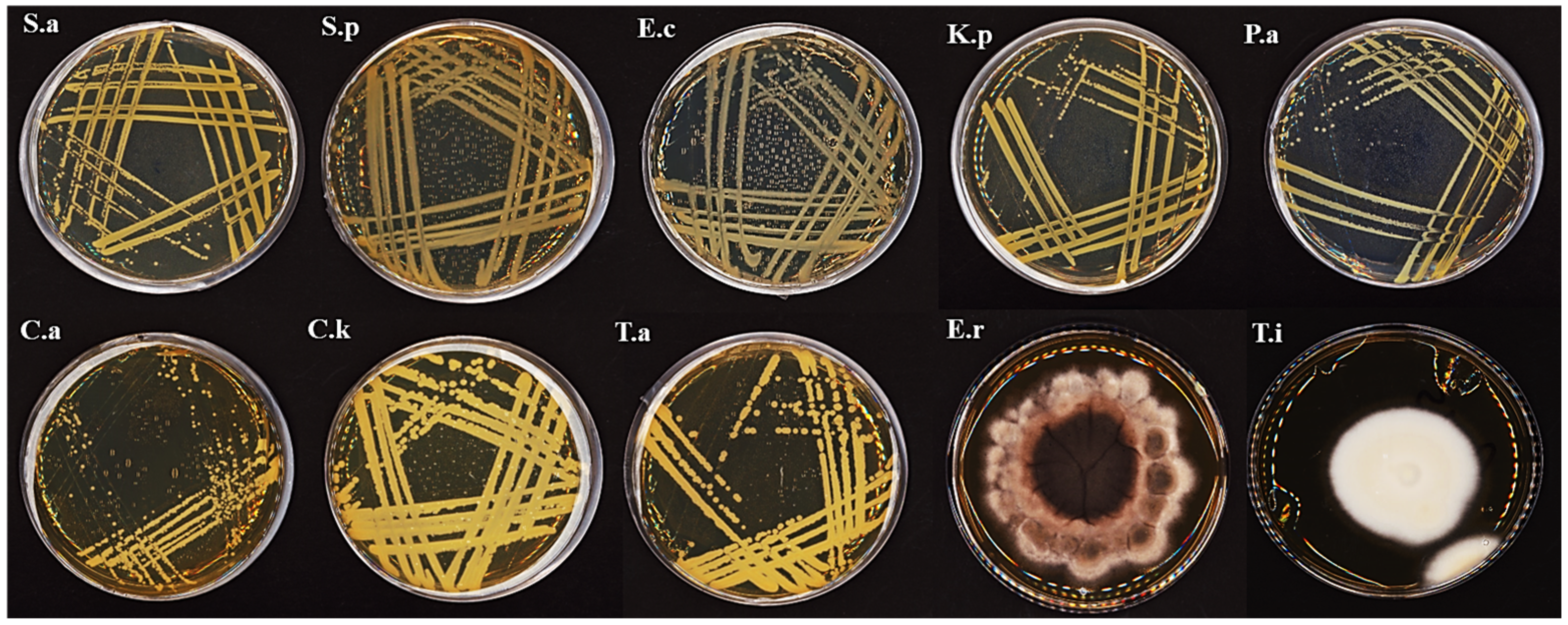
Antimicrobial assays were performed in triplicate following standard recommended by the Clinical and Laboratory Standards Institute [33]. Stock solutions of the eight crude extracts were prepared in 10% dimethyl sulfoxide (DMSO) and serially diluted to 3.12–100 mg/mL. Extracts were screened against Gram-positive bacteria (Staphylococcus aureus ATCC 29213; Streptococcus pyogenes ATCC 19615), Gram-negative bacteria (Escherichia coli ATCC 35218; Klebsiella pneumoniae ATCC 700603; Pseudomonas aeruginosa ATCC 27853), yeasts (Candida albicans ATCC 14053; Candida krusei ATCC 6258), and filamentous fungal pathogens of human origin (Trichosporon asahii AMS 187; Exserohilum rostratum AMS 1077; Trichophyton indotineae AMS 180) from the culture collection of the Microbiology Department, Natural and Medical Sciences Research Center (NMSRC), University of Nizwa, Oman (Figure 1).
Sterile 6 mm Whatman paper disks were impregnated with 20 µL of each extract (100 mg/mL), dried, and placed on inoculated agar plates (disk-diffusion assay). Bacterial suspensions (approximately 108 CFU/mL) were spread on Mueller–Hinton agar, whereas yeasts and filamentous fungi were inoculated on Sabouraud dextrose agar [34]. The negative control was 10% DMSO. The positive control for bacteria was azithromycin (15 mg/mL); for yeasts, it was nystatin (100,000 U/mL); and for filamentous fungi, it was sodium hypochlorite (NaClO, 5.25%). After 24 h at 37 °C (for bacteria), 48 h at 28 °C (for yeasts), and 3 days at 25 °C (for filamentous fungi), the diameter of the zone of inhibition was recorded. Results are expressed as mean ± standard deviation (n = 3). The MIC values were determined by serial dilution of those extracts that inhibited microbial growth.

2.7. Cell Lines and Cytotoxicity Assay

The breast cancer cell line (MDA-MB-231) and human umbilical vein endothelial cell line (HUVEC) were obtained from Sigma-Aldrich (St. Louis, MO, USA) in the United States. The cells were cultured according to the supplier’s suggestions in complete DMEM media with penicillin (100 U/mL), streptomycin (100 µg/mL), and 10% Fetal Bovine Serum (FBS) (Thermo Fisher Scientific Inc., Waltham, MA, USA). All cultures were maintained under standard conditions of temperature (37 °C) and humidity (5% CO2).
The cytotoxicity of the fungal crude extract was evaluated using the MTT assay, following a modified method described by [35]. A stock solution (2 mg/mL) was prepared by dissolving the extract in 1% DMSO and sterilized using a 0.45 µm syringe filter. Serial two-fold dilutions (500–7.81 µg/mL) were prepared in culture medium. Human cells were seeded at 1 × 10⁵ cells/mL in 96-well plates (100 µL/well) and incubated at 37 °C for 24 h.
The medium was then replaced with 100 µL of the extract concentrations (1% DMSO final concentration), while the control wells received only medium. After a further 24 h incubation, the wells were washed with PBS and treated with 100 µL of MTT solution (5 mg/mL in PBS). Plates were shaken (150 rpm, 5 min) and incubated for 4 h at 37 °C/5% CO2 to form formazan crystals. The medium was removed and 50 µL of DMSO was added to dissolve the crystals. Absorbance was measured at 570 nm using a microplate reader. Cell viability was calculated, and IC50 values were determined from dose–response curves. All experiments were conducted in triplicate.
% cell viability = (OD570 treated cells) × 100/(OD570 control).

2.8. Characterization of Secondary Metabolites by Using High-Resolution Electrospray Ionization Mass Spectrometry (HR-ESI-MS)

Crude extracts were analyzed using a high-resolution mass spectrometer (Waters, Milford, MA, USA) equipped with an electrospray ionization source (HR-ESI-MS) described by [36], with minor modifications. Samples were separated using a Waters XBridgeTM C18 column (Waters, Milford, MA, USA) (150 mm × 4.6 mm ID, 5 μm particle size), which had been previously fitted with a matching guard column (Waters XBridgeTM C18, 20 mm × 4.6 mm ID, 5 μm), at 25 °C.
The mobile phase comprised 0.15% formic acid in water as solvent A, and 0.1% formic acid in acetonitrile as solvent B. The columns were pre-equilibrated prior to sample injection for 5 min, and during this time, the chromatographic flow rate of 1.0 mL/min was also set. A 30 min gradient elution profile was developed as follows: The initial composition of the mobile phase was 3% solvent B and was held for 2 min. This was followed by a linear increase in the proportion of solvent B to 20% over 3 min, and to 70% over a further 7 min, where it was maintained for 2 min. The mobile phase was then returned to the starting composition (3% solvent B) within one minute and remained in this state for a further 5 min to allow the column to be fully re-equilibrated.
Each sample (10 μL) was injected onto the column using the Waters autosampler. The MS was operated in positive ESI+ mode, and high-resolution full-scan spectra were recorded across the m/z range of 100–1000 with sufficient resolving power to allow accurate mass measurements to be made to within ±5 ppm of the actual value. Prior to analysis, external calibration of the mass spectrometer was performed to ensure that mass values could be accurately measured. Continuous infusion of standard solution (1 μg/mL) into the mass spectrometer via an automated injector was used to tune and optimize the mass spectrometer performance.

2.9. Statistical Analysis

The data obtained were analyzed by one-way analysis of variance (ANOVA) with SPSS version 25 (IBM). Duncan’s multiple-range test (DMRT) was used to evaluate the significance of differences between treatments, when overall differences were found to be significant using ANOVA, with *, **, and ***, considered significant at p < 0.05, <0.01, <0.001; NS represents nonsignificant at p > 0.05. For all fungi screening tests, standard deviation represents ±SD of three replicates.

3. Results

3.1. Fungal Isolation and Genetic Identification

As the most frequently occurring taxonomic groups, eight fungal isolates were selected for analysis from a group of 86 different species that were isolated from P. australis samples taken from freshwater habitats. Isolates were purified and grown in pure culture to maintain stability and ensure the repeatability of the research.
Sequences generated from the isolates were compared by alignment using BLAST (version 2.16.0+) to GenBank’s database of nucleotide sequences to determine the sequence similarity scores and to identify statistically significant differences within the match. Sequence comparison revealed a high degree of similarity among the isolates, with a level of homology at 100%. Therefore, the isolates have also been formally deposited into GenBank as evidenced by their respective accession numbers listed in Table 2.

3.2. Small-Scale Fermentation

The yield of dry crude extracts was determined gravimetrically by subtracting the weight of the empty beaker from that of the beaker containing the dried extract (Table 3). Considerable variation in metabolite yield was observed among the isolates. The highest yield was obtained from C. madrasense SCUF0000401 (PX596735) (Extract E, 770 mg), followed by C. globosum SCUF0000404 (PX596738) (Extract A), which produced 760 mg of crude extract. Moderate yields were recorded for C. globosum SCUF0000407 (PX596741) (Extract C, 560 mg), C. globosum SCUF0000406 (PX596740) (Extract G, 520 mg), C. globosum SCUF0000405 (PX596739) (Extract D, 500 mg), T. terrophilus SCUF0000403 (PX596737) (Extract B, 460 mg), and C. globosum SCUF0000400 (PX596734) (Extract F, 450 mg). The lowest yield was recorded for S. fimicola SCUF0000402 (PX596736) (Extract H, with 430 mg). Overall, these findings indicate that while all isolates were able to produce secondary metabolites under the applied fermentation conditions, notable differences in productivity were evident both among species and among different isolates.

3.3. Assessment of Antimicrobial Activity of Crude Extracts

3.3.1. Antibacterial Activity Against Bacterial Pathogens

The antimicrobial activity of different crude extracts (A–H) at a concentration of 100 mg/mL was evaluated against five pathogenic bacteria: S. aureus, S. pyogenes, E. coli, K. pneumoniae, and P. aeruginosa. The inhibition zones were presented in Table 4. Extract A exhibited moderate activity with inhibition zones of 11 mm against S. pyogenes, 15.33 mm against S. aureus, 14 mm against K. pneumoniae, 10.33 mm against E. coli, and 13.33 mm against P. aeruginosa. Extract B demonstrated inhibitory activity of 13 mm against S. aureus, 10.33 mm against K. pneumoniae, and 11.67 mm against P. aeruginosa, but no activity against S. pyogenes and E. coli. Extract C was effective with zones of 11 mm against S. aureus, 11.67 mm against K. pneumoniae, and 11.33 mm against P. aeruginosa, but inactive against S. pyogenes and E. coli.
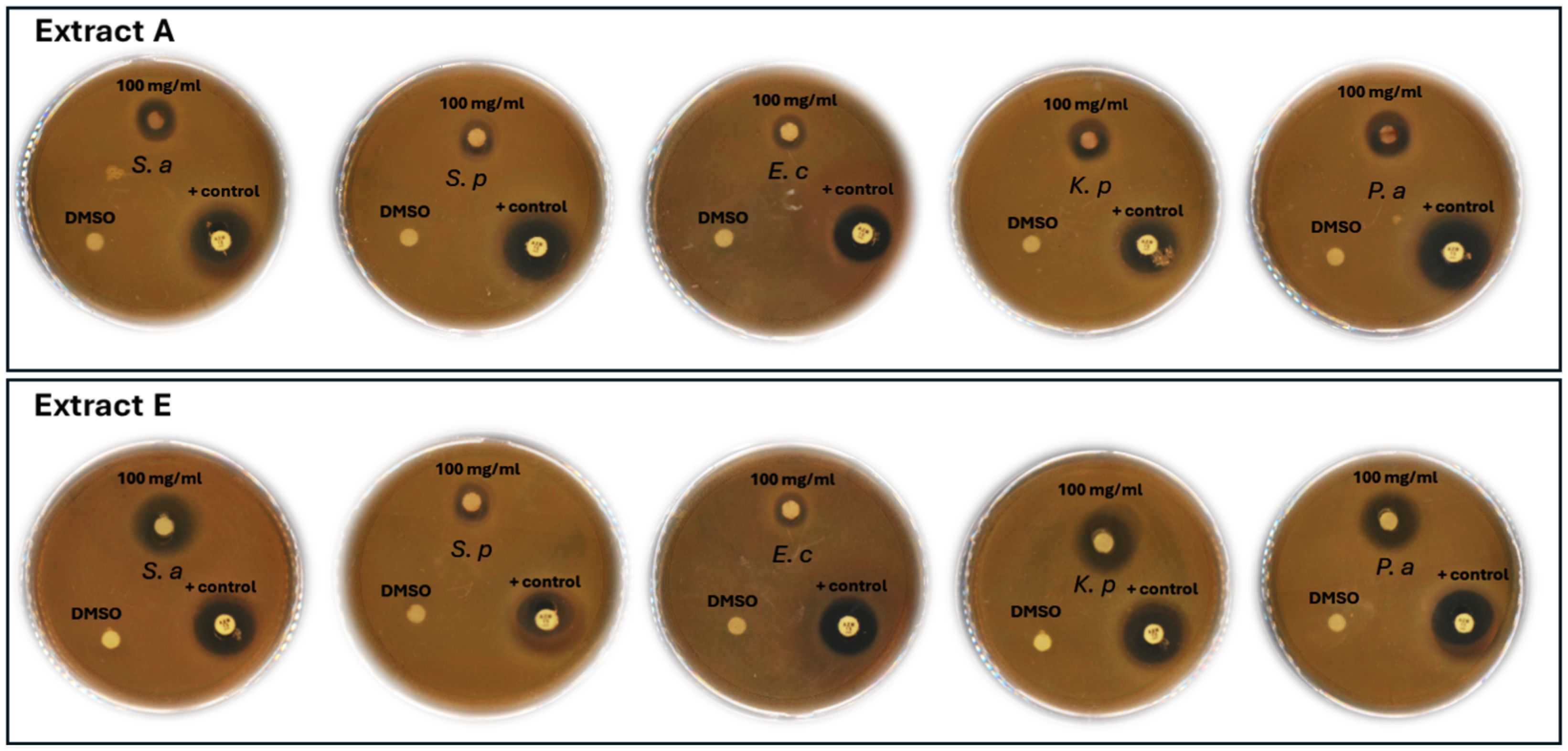
Extract D and H showed weak activity, with inhibition zones of 8.33 mm against S. aureus and K. pneumoniae, only 7.67 mm against K. pneumoniae, and 8.33 mm against P. aeruginosa. Extract E had good activity with inhibition zones of 11 mm against S. pyogenes, 18 mm against S. aureus, 16.67 mm against K. pneumoniae, 10.67 mm against E. coli, and 16.33 mm against P. aeruginosa. Extract F was moderately effective, showing inhibition zones of 15.33 mm against S. aureus, 16.67 mm against K. pneumoniae, and 12.33 mm against P. aeruginosa, but inactive against S. pyogenes and E. coli. Extract G exhibited only weak activity of 7.67 mm against K. pneumoniae and 8.33 mm against P. aeruginosa. Control (Azithromycin 15 mg/mL) showed the highest inhibition zones against all tested organisms, ranging from 18.33 mm (S. pyogenes and E. coli) to 20.33 mm (S. aureus and K. pneumoniae) to 24.33 mm (P. aeruginosa). Overall, extracts A and E exhibited the strongest antibacterial effectiveness, shown in Figure 2.
The crude extracts exhibited variable antibacterial activity across the tested concentrations (50, 25, 12.5, 6.25, and 3.12 mg/mL) against the target bacterial pathogens (Table 5). Extract A demonstrated MIC values of 6.25 mg/mL against S. aureus and K. pneumoniae (9.0 mm inhibition zone), and 6.25 mg/mL against P. aeruginosa (9.33 mm inhibition zone). Extract B showed an MIC of 12.5 mg/mL against S. aureus (8.33 mm) and 25 mg/mL against both K. pneumoniae (8.33 mm) and P. aeruginosa (10.33 mm). Extract C exhibited MIC values of 25 mg/mL for S. aureus (8.6 mm), K. pneumoniae (8.67 mm), and P. aeruginosa (8.33 mm).
Extract E was the most potent among the tested extracts, showing inhibition at the lowest concentration evaluated, with an MIC of 3.12 mg/mL against S. aureus, K. pneumoniae, and P. aeruginosa (8.67 mm inhibition zone for each). Extract F recorded MIC values of 12.5 mg/mL for S. aureus (8.67 mm) and K. pneumoniae (8.33 mm), and 25 mg/mL for P. aeruginosa (9.0 mm). Overall, extract E exhibited the strongest antibacterial effectiveness, maintaining growth inhibition even at the lowest concentration, whereas extracts A, B, C, and F displayed moderate inhibitory potential.

3.3.2. Antifungal Activity Against Fungal Pathogens

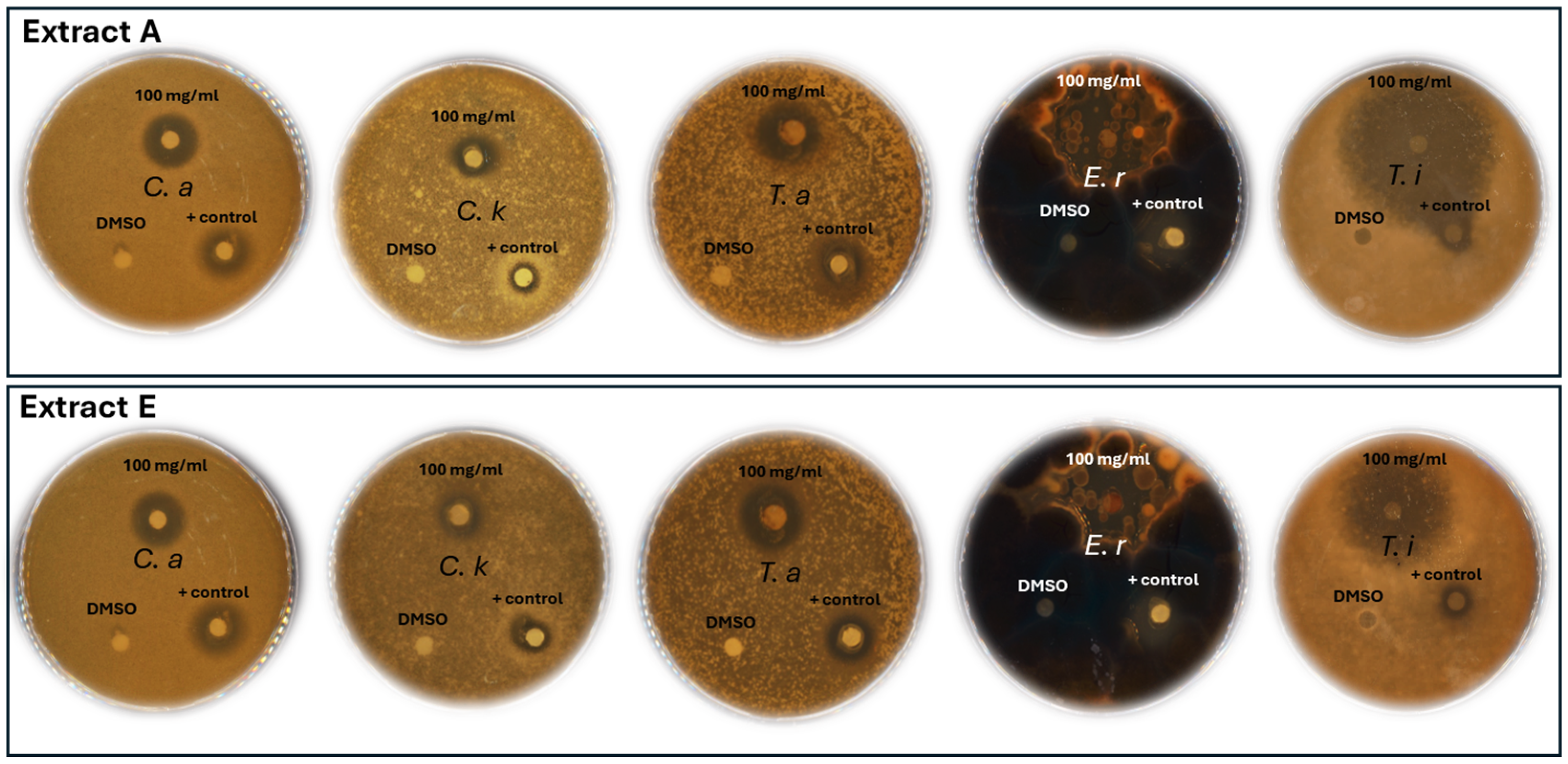
The antifungal activity of different crude extracts (A–H) at a concentration of 100 mg/mL was evaluated against five pathogenic fungi: C. albicans, C. krusei, T. asahii, E. rostratum, and T. indotineae. The inhibition zones are presented in Table 6. Extracts A and E exhibited the most potent antifungal activity, producing inhibition zones of 34.00 mm and 33.67 mm against Exserohilum rostratum, and 38.33 mm and 34.00 mm against Trichophyton indotineae, respectively, exceeding the effects observed in the control. Both extracts also demonstrated strong activity against Candida albicans (20.33 mm), Candida krusei (15.33–15.67 mm), and Trichosporon asahii (17.00–17.67 mm).
Extract F showed moderate antifungal effects, particularly against E. rostratum (20.00 mm) and T. indotineae (21.33 mm). Extract D exhibited selective inhibition, especially against C. krusei (14.33 mm) and T. indotineae (20.33 mm). Extract H showed broad but comparatively lower activity, with inhibition zones ranging from 8.33 mm against C. krusei to 30.33 mm against E. rostratum. In contrast, extracts B and C showed no measurable activity against most tested fungi, except for weak inhibition of E. rostratum by extract B (10.33 mm) and C. krusei by extract C (8.33 mm). Extract G displayed low antifungal activity, producing inhibition zones of 11.33 mm against C. albicans and 9.33 mm against T. asahii and moderate against E. rostratum (15.33 mm). Overall, extracts A and E exhibited the strongest antifungal effectiveness show in Figure 3.
The crude extracts exhibited variable antifungal activity across the tested concentrations (50, 25, 12.5, 6.25, and 3.12 mg/mL) against the selected fungal pathogens presented in Table 7. Extract A demonstrated an MIC value of 12.5 mg/mL against C. albicans with an inhibition zone of 10.50 mm, and against T. asahii with an inhibition zone of 8.67 mm. Extract E also recorded an MIC of 12.5 mg/mL against both C. albicans (10.33 mm) and T. asahii (10.67 mm). Extract A demonstrated the lowest MIC value of 3.12 mg/mL against E. rostratum, with an inhibition zone of 12.00 mm, while also exhibiting an MIC of 12.5 mg/mL against T. indotineae (10.67 mm inhibition zone). Extract D showed MIC values of 25 mg/mL against both E. rostratum (8.67 mm) and T. indotineae (10.33 mm). Extract E exhibited an MIC of 6.25 mg/mL against E. rostratum (10.33 mm) and 12.5 mg/mL against T. indotineae (9.67 mm). Extract F recorded MIC values of 12.5 mg/mL against E. rostratum (13.33 mm) and 25 mg/mL against T. indotineae (9.67 mm). Extracts G and H both presented MICs of 12.5 mg/mL against E. rostratum with inhibition zones of 12.67 mm and 14.67 mm, respectively. Overall, extracts A and E exhibited the strongest antifungal performance, maintaining considerable inhibition at the lowest tested concentrations, whereas extracts D, F, G, and H demonstrated moderate inhibitory potential.

3.4. Anticancer Activity of Crude Extracts

The cytotoxic effects of the crude extracts obtained from C. globosum (A) and C. madrasense (E), which had exhibited the strongest antimicrobial activity and maintained notable inhibition even at the lowest tested concentrations, were evaluated against two cell lines: MDA-MB-231 and HUVEC.
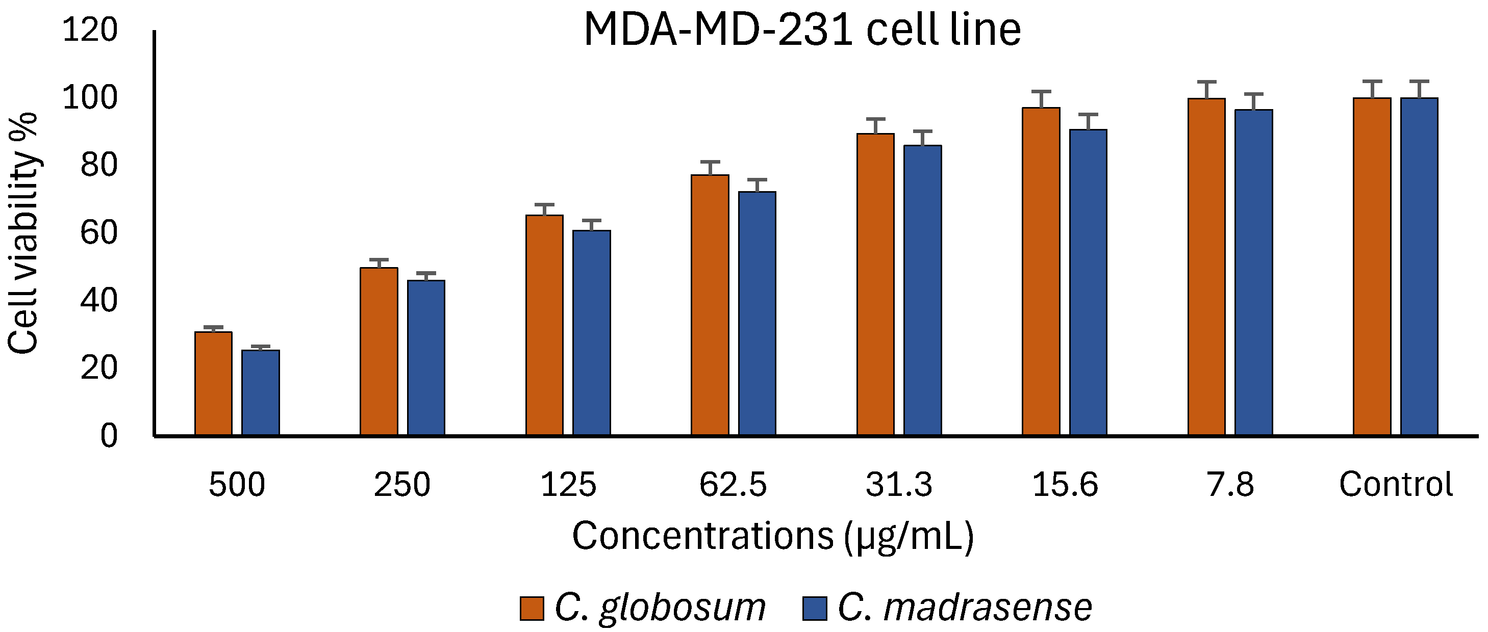
Both the A and E crude extracts exhibited a clear, concentration-dependent cytotoxic effect on the MDA-MB-231 cell line (Figure 4). At the highest tested concentration (500 µg/mL), extract E demonstrated the strongest inhibitory effect, reducing cell viability to approximately 28%, while extract A caused a slightly lower reduction, maintaining about 33% cell viability. At intermediate concentrations (250 and 125 µg/mL), the two extracts continued to show significant cytotoxic effects, with cell viability values ranging between 50% and 70%, respectively. At lower concentrations (62.5–7.8 µg/mL), both extracts exhibited moderate to minimal cytotoxicity, and the cell viability gradually increased to more than 85% at 15.6 µg/mL and reached nearly 100% in the control untreated cells.
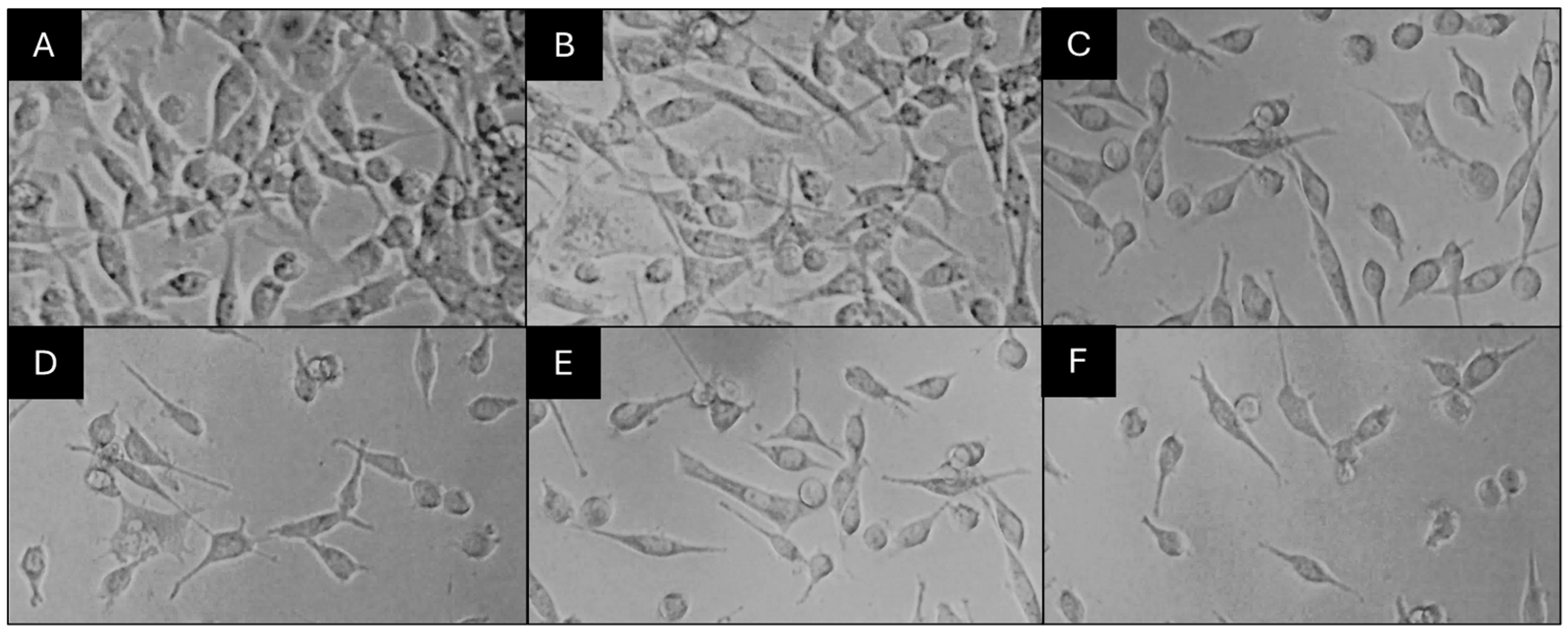
The results revealed that both crude extracts A and E significantly inhibited cancer cell proliferation in a concentration-dependent manner. The IC50 values were approximately 277 µg/mL for extract E and 309 µg/mL for extract A, indicating that extract E exhibited slightly higher cytotoxic potency. Microscopic observations (Figure 5) supported these results, as untreated control cells maintained their normal elongated morphology and dense confluence, whereas treated cells displayed dose-dependent morphological changes, including cell rounding, shrinkage, membrane blebbing, and detachment from the culture surface. Collectively, these findings confirm that both fungal extracts exert notable antiproliferative effects against breast cancer cells, with extract E demonstrating comparatively stronger cytotoxic activity.
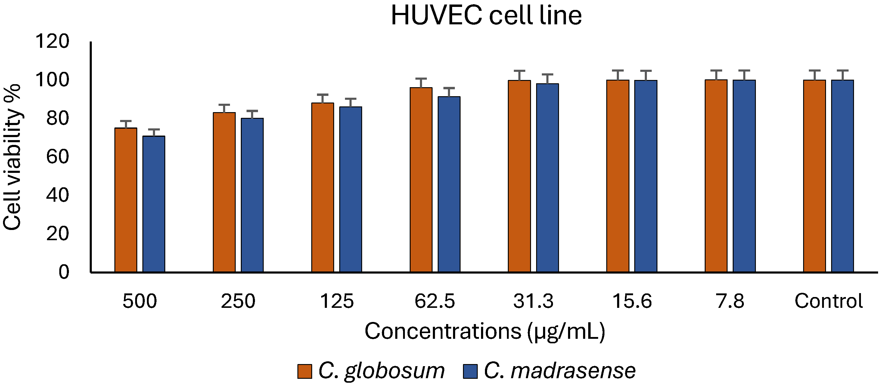
Both crude extracts A and E exhibited a clear concentration-dependent cytotoxic effect on HUVEC cells (Figure 6). At the highest tested concentration (500 µg/mL), extract E showed the strongest inhibitory effect, reducing cell viability to approximately 20%, while extract A caused a slightly lower reduction, maintaining about 29% cell viability. At intermediate concentrations (250 and 125 µg/mL), both extracts continued to demonstrate significant cytotoxicity, with cell viability ranging between 80% and 90%, respectively. At lower concentrations (62.5–7.8 µg/mL), the cytotoxic effects were moderate to minimal, and cell viability gradually increased to over 95% at 15.6 µg/mL, reaching nearly 100% in the untreated control cells.
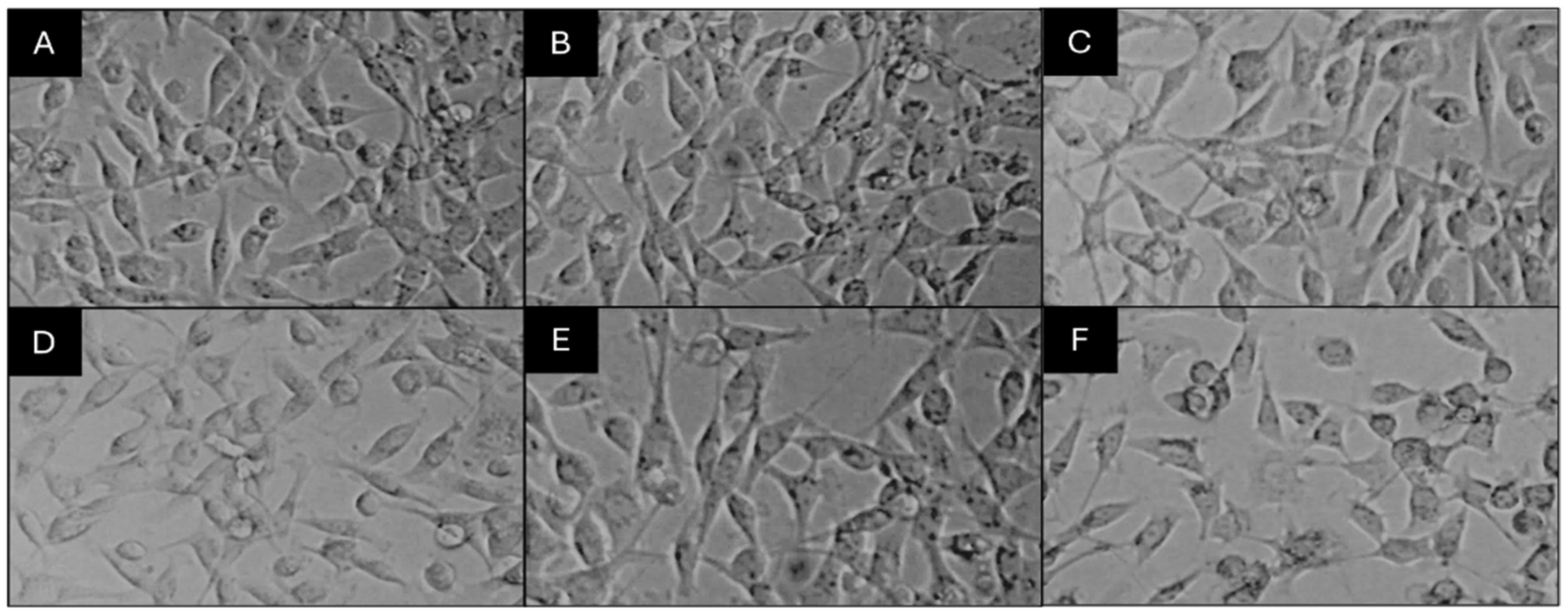
Similarly, both extracts A and E significantly inhibited cancer cell proliferation in a concentration-dependent manner. The calculated IC50 values were approximately 796 µg/mL for extract E and 919 µg/mL for extract A, indicating that extract E exhibited slightly higher cytotoxic potency. Microscopic observations (Figure 7) supported these findings, where control cells exhibited normal elongated morphology and dense confluence, whereas treated cells showed progressive, dose-dependent morphological changes, including cell rounding, shrinkage, membrane blebbing, and detachment from the culture surface. Overall, these results confirm that both fungal extracts possess strong antiproliferative potential against breast cancer cells, with extract E displaying relatively greater cytotoxic efficacy.

3.5. Characterization of Secondary Metabolites from Crude Extracts of C. globosum (A) and C. madrasense (E) by HR-ESI-MS Technique

For the rapid identification of bioactive metabolites correlated with the pronounced antimicrobial and anticancer activities of the isolates C. globosum (A) and C. madrasense (E), HR-ESI-MS analysis was conducted. The metabolic profiling of the fungal crude extracts revealed the presence of numerous bioactive secondary metabolites representing diverse chemical classes, including cytochalasans, azaphilone alkaloids, steroids, terpenoids, flavonoids, and phenolic compounds.

3.5.1. Characterization of Bioactive Compounds of Crude Extract A

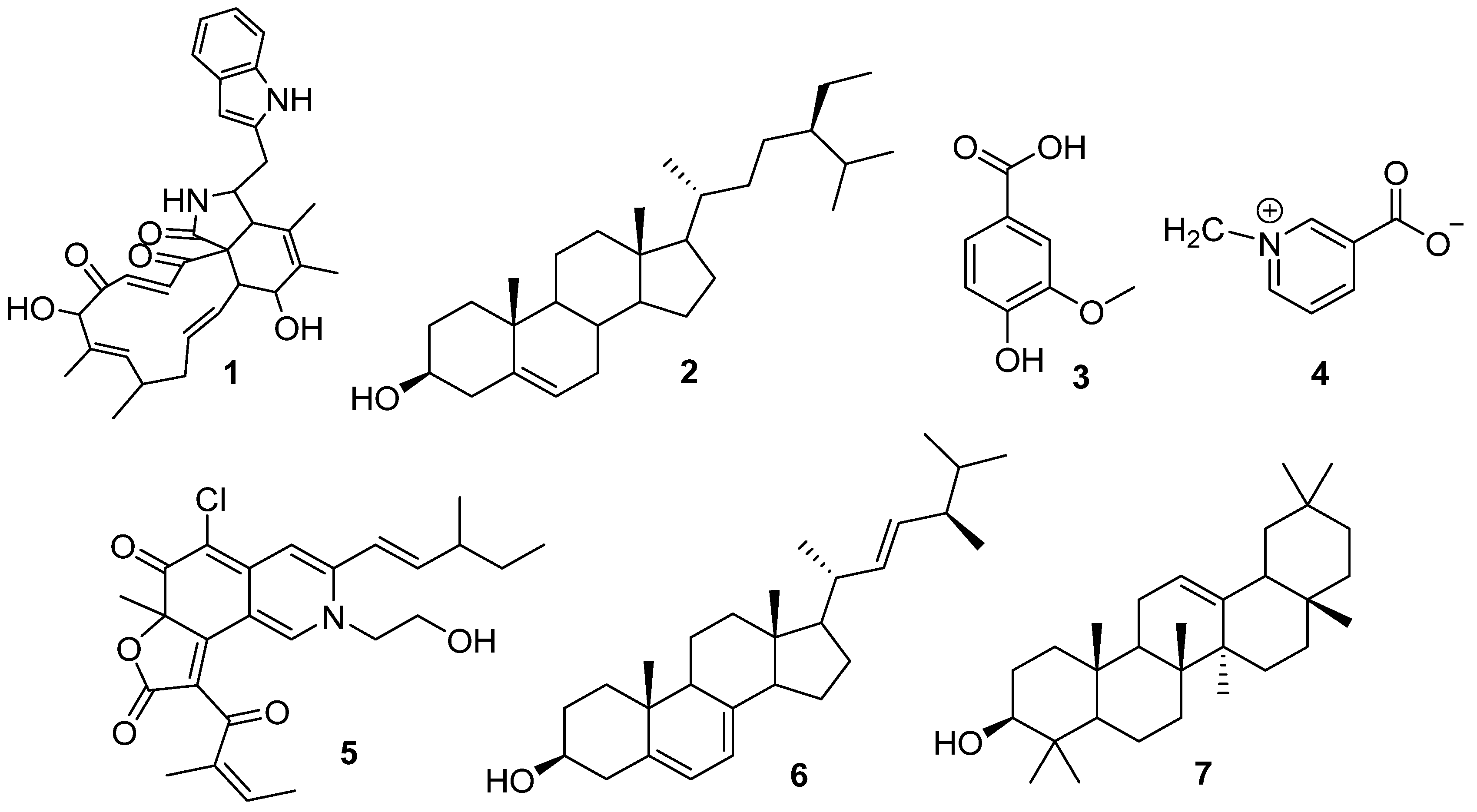
The HR-ESI-MS analysis of crude extract A led to the putative identification of seven bioactive secondary metabolites belonging to diverse chemical classes, including phytosterols, phenolic acids, alkaloids, and triterpenoids, as shown in Table 8 and Figure 8. These compounds are known for their pharmacological and therapeutic significance, particularly in antimicrobial and anticancer contexts.
Chaetoglobosin A is a cytochalasin-type alkaloid mainly synthesized by C. globosum. Chaetoglobosin A is composed of an isoindolone group attached to a cyclic ring of 16 carbon atoms. Chaetoglobosin A has demonstrated the ability to suppress the proliferation of T-24 human bladder cancer cells and has been shown to be capable of suppressing tumor cell migration and inhibiting colony-forming abilities, thus demonstrating its potential as an anticancer drug with strong antimetastatic capabilities [37].
β-Sitosterol is part of the phytosterol (plant sterol) class and is found in various types of plants and fungi. Phytosterols have been shown to have a wide range of pharmacological activities; specifically anticancer, anti-inflammatory, immunomodulatory, and cholesterol-lowering effects. In relation to cancer research, β-sitosterol has been shown to inhibit tumor cell proliferation and induce apoptosis in a variety of human cancer cell lines, including breast, colon, and prostate cancer cells, primarily via caspase-dependent pathways and alterations in cell cycle progression [38].
Vanillic acid is a phenolic acid (benzoic acid derivative) that is found in many traditional Chinese medicinal herbs such as Angelica sinensis and Panax ginseng. Vanillic acid has been shown to have several pharmacological properties, including antioxidant, anti-inflammatory, antimicrobial, and anticancer properties [39].
Trigonelline is a pyridine alkaloid that is most commonly found in coffee beans and fenugreek seeds (Trigonella foenum-graecum). Trigonelline has been shown to demonstrate remarkable anti-invasive and antimetastatic activities against cancer cells. Using the rat ascites hepatoma cell line (AH109A), trigonelline combined with niacin (nicotinic acid and nicotinamide) was able to significantly inhibit tumor cell invasion at concentrations of 2.5–40 μM without altering proliferation [40]. Ergosterol is a steroidal triterpenoid and is the main sterol in fungi and serves the same functional purpose as cholesterol does in animals.
Ergosterol has been shown to have antioxidant, anti-inflammatory, antifungal, and anticancer properties. Ergosterol is capable of inhibiting the proliferation of tumor cells and inducing apoptosis through the disruption of mitochondrial integrity and activation of caspase-mediated pathways, and therefore represents a possible anticancer compound derived from fungi [41].
α-Amyrin is a pentacyclic triterpenoid, which is abundant in higher plants such as Pergularia daemia. α-Amyrin has been shown to possess numerous biological activities, including antioxidant, anti-inflammatory, antinociceptive, antipruritic, gastroprotective, and hepatoprotective effects. α-Amyrin demonstrated a dose-dependent cytotoxic effect against Hep2 human laryngeal carcinoma cells at concentrations ranging from 10 to 320 μmol/mL with an IC50 value of 69.32 μmol/mL [42].
Table 8. Putatively identified compounds from C. globosum (A) crude extract by HR-ESI-MS.
NoCompoundRet. Time
(min)
Mass Chem DrawMass CalculatedMass ObservedPrecursor AdductMolecular FormulaRef.
1Chaetoglobosin A11–12528.26242529.27025529.3663[M + H] +C32H36N2O5[43]
2β-Sitosterol9.5–10.8414.38617397.38343397.5374[M − H2O + H] +C29H50O[44]
3Vanillic acid2.8–3.5168.04226185.04500185.1645[M + OH] +C8H8O4[45]
4Trigonelline0.9–1.5136.03985136.03985136.1343[M] +C7H7NO2[45]
5Chaetomugilides B8.0–8.8457.16560458.17343458.4651[M + H] +C25H28ClNO5[46]
6Ergosterol8.7–9.4396.33922435.30292435.3295[M + K] +C28H44O[47]
7α-Amyrine9.2–10426.38617426.38617426.4694[M] +C30H50O[48]

3.5.2. Characterization of Bioactive Compounds of Crude Extract E

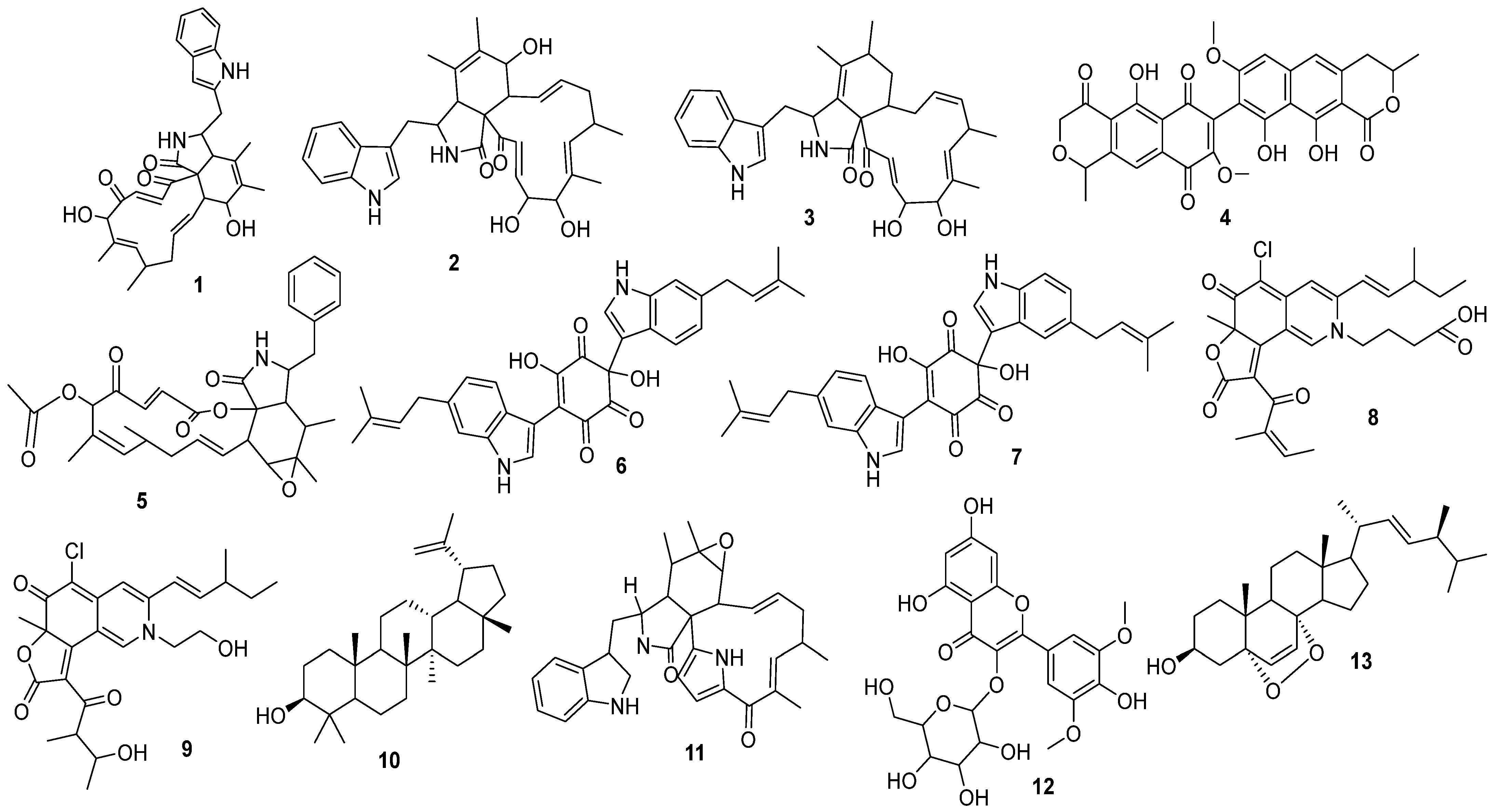
The HR-ESI-MS analysis of the crude extract E led to the putative identification of thirteen bioactive secondary metabolites belonging to diverse chemical classes, including cytochalasans, azaphilone alkaloids, steroids, terpenoids, flavonoids, and phenolics, as presented in Table 9 and Figure 9. These metabolites are known for their pharmacological and therapeutic significance, particularly for their cytotoxic, antimicrobial, and antiproliferative properties.
Chaetoglobosin A was isolated from the culture of an endophytic fungus, C. globosum, which was derived from Nymphaea nouchali. Extraction of the fungus followed by purification led to the isolation of two previously identified cytochalasans (chaetoglobosin A and C). Chaetoglobosin A showed considerable antimicrobial action against S. aureus (MIC 32 µg/mL), Bacillus subtilis (MIC 16 µg/mL), and methicillin-resistant S. aureus (MRSA) MIC 32 µg/mL) [49]. Additionally, seven novel cytochalasan derivatives, specifically Cytoglobosins A-G, were isolated from a culture of C. globosum QEN-14, an endophytic fungus found in association with the marine green alga Ulva pertusa. Cytoglobosins C and D had some level of cytotoxicity against the human lung carcinoma A-549 cell line [50].
Viomellein and Xanthomegnin, both members of the anthraquinone-type polyketide family, were obtained from an active fraction of an Aspergillus sp. derived from a marine source. It is worth noting that viomellein has greater activity towards dormant Mycobacterium bovis BCG cells compared to actively growing cells, and also displayed weak action towards M. smegmatis [51]. Cytochalasans, including cytochalasin L, are fungal macrocyclic alkaloids known to inhibit actin polymerization and thereby disrupt microfilament organization; such disruption in cancer cells leads to inhibited proliferation and induction of apoptosis [52]. Chaetoglobinol A and Chaetoglobinol B, both phenylspirodrimanes, along with five other known secondary metabolites, were isolated from the solid-state fermented rice culture of Chaetomium elatum CIB-412. Both compounds showed strong inhibition of alpha-glucosidase at 2.5 mg/mL, suggesting possible applications for the treatment of diabetes [45]. Three new azaphilone alkaloids referred to as Chaetomugilides A-C, along with three analogous compounds, were isolated from the methanol extract of C. globosum TY1, an endophyte of Ginkgo biloba. These compounds have shown significant cytotoxic activity against the HepG2 human hepatocellular carcinoma cell line, with IC50 values ranging from 1.7 to 53.4 µM [46]. Additionally, lupeol, a pentacyclic triterpenoid isolated from the leaves of Elephantopus scaber L., has been shown to possess significant antiproliferative actions against breast cancer MCF-7 cells, in a dose-dependent fashion, with an IC50 of 80 µM, validating its ability to cause apoptosis in cancerous cells [53].
A new group of cytochalasans, the penochalasins (A, B, and C), were isolated from a Penicillium sp. isolate that was associated with the marine alga Enteromorpha intestinalis. All of these compounds have shown a high degree of cytotoxicity against the cultured P388 murine leukemia cell line [54]. Flavonols, particularly syringetin, and their corresponding glycosides (such as syringetin 3-O-galactoside) are methylated flavonols that are capable of acting as antioxidants and inhibitors of cancer cell proliferation [55]. Finally, Ergosterol peroxide, a steroidal peroxide isolated from the marine basidiomycete Fulvifomes sp., exhibited pronounced cytotoxic activity by inducing apoptosis in the breast cancer MCF-7 cell line. The compound inhibited 50% of cell growth (IC50) at a concentration of 40 μg/mL and induced 90% cell death (IC90) at 80 μg/mL [56].
Table 9. Putatively identified compounds from C. madrasense (E) crude extract by HR-ESI-MS.
NoCompoundsRet. Time
(min)
Mass Chem DrawMass CalculatedMass ObservedPrecursor AdductMolecular FormulaReference
1Chaetoglobosin A11–12528.26242529.27025529.3663[M + H]+C32H36N2O5[43]
2Cytoglobosin A11–11.8529.27025530.27807530.3686[M + H]+C32H36N2O5[50]
3Cytoglobosin D10.6–11.3515.29098515.29098515.3720[M + H]+C32H38N2O4[50]
4Voimellien9–10560.13186561.13969561.3567[M + H]+C30H24O11[51]
5Cytochalasin L8.3–9547.25700547.25700547.3780[M]+C32H37NO7[57]
6Chaetoglobinol A11–12522.21547561.17918561.3567[M + K]+C32H30N2O5[45]
7Chaetoglobinol B 11.5–13522.21547545.20524545.3621[M + Na]+C32H30N2O5[45]
8Chaetomugilides A8.5–9.3499.17617499.17617499.3741[M]+C27H30ClNO6[46]
9Chaetomugilides C10.2–10.9475.17617493.21054493.3396[M + NH4]+C27H34ClNO6[46]
10Lupeol15–18426.38617409.38343409.3396[M- H2O + H]+C30H50O[58]
11Penochalasins A8.7–9.5510.27567511.28349511.3543[M + H]+C32H35N3O3[54]
12Syringetin 3-O-galactoside4.8–5.5508.12169509.12952509.3364[M + H]+C23H24O13[59]
13Ergosterol-5,8-peroside9.2–10.1428.32905446.4265446.3403[M + NH4]+C28H44O3[51]

4. Discussion

The antimicrobial potential of fungal secondary metabolites has prompted significant scientific interest because of the growing worldwide problem of antibiotic resistance and the need to find new bioactive agents [60]. Cancer is one of the top causes of death in the world, and the search for new natural compounds with potent and selective anticancer activity remains a major focus of biomedical research [53]. Fungi of the genus Chaetomium have recently been considered to be promising taxa producing bioactive secondary metabolites with notable cytotoxic and antiproliferative effects.
The crude extracts were prepared from eight (A–H) representative taxa evaluated for their antimicrobial activity using the agar disk-diffusion method. The extracts were tested at concentrations ranging from 3.12 to 100 mg/mL against bacterial and fungal pathogens. The results demonstrated considerable variation in antimicrobial potency among the tested isolates. Notably, the crude extracts of C. globosum (A) and C. madrasense (E) exhibited the most pronounced broad-spectrum activity. At a concentration of 100 mg/mL, they produced inhibition zones of 15.33–18.00 mm against S. aureus, 11.00 mm against S. pyogenes, 10.33–10.67 mm against E. coli, 14.00–16.67 mm against K. pneumoniae, and 13.33–16.33 mm against P. aeruginosa. These findings confirm the strong antimicrobial potential of Chaetomium-derived metabolites. A study by [61] reported that the endophytic fungus Chaetomium sp. NF15, isolated from Justicia adhatoda, exhibited significant antimicrobial activity against both Gram-positive and Gram-negative bacteria. The extract showed inhibition zones ranging from 9.4 to 15.1 mm against S. aureus, E. coli, and P. aeruginosa, demonstrating the inherent antibacterial potential of Chaetomium-derived metabolites. Further supporting these observations, ref. [57] demonstrated that various Chaetomium spp. exhibit strong antibacterial effects against clinically relevant pathogens. In their study, all Chaetomium isolates (1–10) showed inhibitory activity against S. aureus, S. pyogenes, E. coli, and P. aeruginosa, while K. pneumoniae appeared resistant to all extracts. The inhibition zones reported ranged between 10.5 and 26 mm, indicating significant variability in activity among isolates.
Since some pathogenic fungi have shown intrinsic or developing resistance to antifungal drugs, little is known about the activity of fungal extracts against them. Previously, it was reported that C. albicans and C. krusei density is a major cause of candidiasis, with C. krusei having intrinsic resistance to fluconazole [62]. T. asahii is also found to be associated with superficial and systemic infections, often with reduced susceptibility to several antifungal agents [63]. Furthermore, E. rostratum and T. indotinea are emerging pathogens that demonstrate resistance to common antifungal agents, presenting therapeutic challenges [64,65].
In contrast, in our study, Chaetomium extracts exhibited broad-spectrum antifungal activity against most of these resistant species. Crude extracts A and E demonstrated the highest activity against C. albicans (20.33 mm), C. krusei (15.33–15.67 mm), T. asahii (17.00–17.67 mm), E. rostratum (33.67–34.00 mm), and T. indotineae (34.00–38.33 mm). These findings suggest the strong antifungal potential of Chaetomium-derived metabolites. In agreement with our observations, ref. [61] reported that Chaetomium sp. NF15 exhibited a significant antifungal activity against C. albicans, with an inhibition zone of 14.2 mm. Similar findings were observed by [57], indicating that the inhibition zones of extracts from different Chaetomium spp. C. globosum isolates (3, 4, 5, and 8) were 11.3 to 25.6 mm, while that for C. madrasense (10) was 15.5 mm. The variation in inhibition zones among isolates may be attributed to differences in their metabolite profiles.
In diffusion-based screening, inhibition zones provide a rapid qualitative indication of activity; however, they are influenced by extract diffusion, solubility, and the agar matrix, and they do not yield quantitative potency metrics such as MIC/MBC/MFC values. Therefore, disk-diffusion results are best interpreted as an initial prioritization step that should be complemented by standardized broth microdilution and time–kill approaches to confirm spectrum and potency in a concentration-dependent manner [34].
In addition to their antimicrobial efficacy, the crude extracts of C. globosum extract (A) and C. madrasense extract (E) were further evaluated for cytotoxic activity against the MDA-MB-231 cell line and HUVEC cell line using an MTT assay. Both extracts exhibited concentration-dependent cytotoxicity accompanied by distinct morphological alterations in cancer cells, including cell shrinkage, membrane blebbing, and detachment, which are consistent with programmed cell death. The IC50 values indicated that extract E (IC50 = 277 µg/mL) exerted stronger cytotoxic effects than extract A (IC50 = 309 µg/mL), suggesting differences in the type and/or abundance of active metabolites between the two taxa. Importantly, both extracts showed comparatively low cytotoxicity toward HUVEC cells under the tested conditions, suggesting preliminary in vitro selectivity that should be confirmed across a broader panel of non-malignant cell lines. This selectivity is biologically plausible because Chaetomium spp. are well-known producers of cytochalasans, including chaetoglobosins and azaphilones, that can modulate cytoskeletal function and apoptosis-related pathways and have repeatedly been reported as cytotoxic scaffolds in cancer models [66,67,68].
HR-ESI-MS profiling performed on bioactive metabolite extracts revealed a vast diversity of secondary metabolites belonging to major chemical classes of alkaloids, polyketides, triterpenoids, flavonoids, phenolics, and steroids. Seven compounds from C. globosum extract (A) and thirteen compounds from C. madrasense extract (E) were reported, indicative of chemical richness and biological relevance. Chaetoglobosin A, Cytoglobosins A and D, Chaetoglobinol A and B, and Cytochalasin L have been simply reported as belonging to the cytochalasan alkaloids, with strong cytotoxic and antitumor properties. Chaetomugilides A–C azaphilone alkaloids are also reported to show significant cytotoxicity against different human cancer cell lines [46,50,57].
Other metabolites were Lupeol, α-amyrin and β-sitosterol, triterpenoid/steroidal compounds with anti-inflammatory and anticancer properties [44,48,58]. Ergosterol and its oxidized form, Ergosterol-5,8-peroxide, are peroxides of steroids often found in fungi and owe their apoptosis-inducing properties in cancerous cells to this same category [47,51]. Moreover, Syringetin 3-O-galactoside, a flavonol glycoside, demonstrated strong antioxidant and antiproliferative properties, while vanillic acid and trigonelline, classified as phenolic and alkaloid compounds, contribute to the overall antioxidant and antimicrobial profile of the extracts [59]. Altogether, there is strong evidence to show that Chaetomium spp. are prolific sources of varied, bioactive, naturally occurring products [67].
Overall, this work demonstrates that freshwater-associated fungi colonizing submerged P. australis in Egypt are taxonomically diverse and can produce metabolites with potent antimicrobial and selective anticancer activity. The most active Chaetomium isolates represent valuable candidates for further natural-product research. Accordingly, the broader metabolite diversity observed in C. madrasense extract (E) may partially explain its stronger antimicrobial and cytotoxic profiles, although definitive attribution requires purification and bioactivity-guided fractionation [67,68].

5. Conclusions

In conclusion, this study highlights the promising therapeutic potential of native Chaetomium isolates recovered from submerged P. australis freshwater habitats in Egypt. These isolates demonstrated strong antimicrobial and selective anticancer activities, particularly C. globosum (A) and C. madrasense (E) extracts, which showed potent inhibition against clinically relevant pathogens and breast cancer cells. The antimicrobial and cytotoxic activities observed in this study may be attributed to multiple metabolite classes, including cytochalasans, azaphilones, steroids, terpenoids, flavonoids, and phenolic compounds, as revealed by HR-ESI-MS profiling. These metabolites are known for their strong antimicrobial and anticancer effects, supporting the pharmaceutical relevance of the detected chemical constituents. Native Chaetomium spp. from Egyptian aquatic ecosystems show great promise as potential candidates for future drug discovery and innovative applications in medicine, industry, and nanotechnology. Further studies focusing on purification, structural characterization, and advanced formulation approaches are warranted to optimize the therapeutic value of these fungal metabolites.

Author Contributions

Conceptualization, F.A.A.N., A.M.A.-A. and T.S.A.; methodology, F.A.A.N., A.M.A.-A., A.M.S.A.-H. and T.S.A.; software, S.M. and H.M.; validation, N.A.N., N.U.R. and M.A.H.; formal analysis, F.A.A.N., S.A.-H. and U.Q.; investigation, F.A.A.N. and A.M.S.A.-H.; resources, A.M.A.-A. and T.S.A.; data curation, S.M., N.U.R. and H.M.; writing—original draft preparation, F.A.A.N.; writing—review and editing, A.M.A.-A., N.A.N. and A.M.S.A.-H.; visualization, S.A.-H. and U.Q.; supervision, A.M.S.A.-H. and A.M.A.-A.; project administration, A.M.S.A.-H.; funding acquisition, A.M.S.A.-H. All authors have read and agreed to the published version of the manuscript.

Funding

This research was financially supported by grant number BFP/RGP/HSS/25/185 from the Oman Research Council, Oman.

Institutional Review Board Statement

Not applicable.

Informed Consent Statement

Not applicable.

Data Availability Statement

The data presented in this study are available in the article. Genetic sequences have been deposited in the GenBank database under the accession numbers mentioned in the Section 3.

Acknowledgments

The authors thank the Laboratory of Systematic Mycology, Botany and Microbiology Department, Suez Canal University, for providing space and supporting the deposition of the native fungi during this study. Deep thanks are also extended to Natural & Medical Sciences Research Center, University of Nizwa, for unlimited support during this study.

Conflicts of Interest

The authors declare no conflicts of interest.

References

  1. Atanasov, A.G.; Zotchev, S.B.; Dirsch, V.M.; International Natural Product Sciences Taskforce; Supuran, C.T. Natural Products in Drug Discovery: Advances and Opportunities. Nat. Rev. Drug Discov. 2021, 20, 200–216. [Google Scholar] [CrossRef]
  2. Laxminarayan, R.; Duse, A.; Wattal, C.; Zaidi, A.K.M.; Wertheim, H.F.L.; Sumpradit, N.; Vlieghe, E.; Hara, G.L.; Gould, I.M.; Goossens, H.; et al. Antibiotic Resistance-the Need for Global Solutions. Lancet Infect. Dis. 2013, 13, 1057–1098. [Google Scholar] [CrossRef]
  3. Theuretzbacher, U.; Bush, K.; Harbarth, S.; Paul, M.; Rex, J.H.; Tacconelli, E.; Thwaites, G.E. Critical Analysis of Antibacterial Agents in Clinical Development. Nat. Rev. Microbiol. 2020, 18, 286–298. [Google Scholar] [CrossRef]
  4. Watkins, E.J. Overview of Breast Cancer. J. Am. Acad. Physician Assist. 2019, 32, 13. [Google Scholar] [CrossRef]
  5. Sung, H.; Ferlay, J.; Siegel, R.L.; Laversanne, M.; Soerjomataram, I.; Jemal, A.; Bray, F. Global Cancer Statistics 2020: GLOBOCAN Estimates of Incidence and Mortality Worldwide for 36 Cancers in 185 Countries. CA Cancer J. Clin. 2021, 71, 209–249. [Google Scholar] [CrossRef]
  6. Newman, D.J.; Cragg, G.M. Natural Products As Sources of New Drugs over the 30 Years from 1981 to 2010. J. Nat. Prod. 2012, 75, 311–335. [Google Scholar] [CrossRef]
  7. Maharachchikumbura, S.S.N.; Hyde, K.D.; Jones, E.B.G.; McKenzie, E.H.C.; Bhat, J.D.; Dayarathne, M.C.; Huang, S.-K.; Norphanphoun, C.; Senanayake, I.C.; Perera, R.H.; et al. Families of Sordariomycetes. Fungal Divers. 2016, 79, 1–317. [Google Scholar] [CrossRef]
  8. Fouad, A.M.M.; Arnous, M.O.; Zaghloul, M.S.; Moustafa, A.A. Expansion of Phragmites Australis in Response to Climate Change and Human Activities in Wetland Ecosystems, Ismailia, Egypt. Sci. Afr. 2025, 28, e02613. [Google Scholar] [CrossRef]
  9. Abdel-Aziz, F. Diversity of Aquatic Fungi on Phragmites Australis at Lake Manzala, Egypt. Sydowia 2008, 60, 1–14. [Google Scholar]
  10. Abdel-Aziz, F. Freshwater Fungi from the River Nile, Egypt. Mycosphere 2016, 7, 741–756. [Google Scholar] [CrossRef]
  11. Abdel-Raheem, A.M. Colonization Pattern of Aquatic Hyphomycetes on Leaf Packs in Subtropical Stream. Mycopathologia 1997, 138, 163–171. [Google Scholar] [CrossRef]
  12. Khattab, O. Aquatic Pycnidial and Hyphomycetes Fungi from Macrophytes and Riparian Plants in the River Nile. Res. J. Microbiol. 2007, 2, 346–353. [Google Scholar] [CrossRef]
  13. El-Elimat, T.; Raja, H.; Figueroa, M.; Sharie, A.; Bunch, R.; Oberlies, N. Freshwater Fungi as a Source of Chemical Diversity: A Review. J. Nat. Prod. 2021, 84, 898–916. [Google Scholar] [CrossRef]
  14. Noumeur, S.R.; Teponno, R.B.; Helaly, S.E.; Wang, X.-W.; Harzallah, D.; Houbraken, J.; Crous, P.W.; Stadler, M. Diketopiperazines from Batnamyces globulariicola, Gen. & Sp. Nov. (Chaetomiaceae), a Fungus Associated with Roots of the Medicinal Plant Globularia Alypum in Algeria. Mycol. Prog. 2020, 19, 589–603. [Google Scholar] [CrossRef]
  15. Abo Nouh, F.A.; Al-Hatmi, A.M.S.; Abdelmoneim, T.S.; Nafady, N.A.; Abdel-Azeem, A.M. Some Taxa from Sordariomycetes with Potential Secondary Metabolites. Microb. Biosyst. 2025, 10, 1–25. [Google Scholar] [CrossRef]
  16. Senanayake, I.; Rathnayaka, A.; Sandamali, D.; Calabon, M.; Gentekaki, E.; Lee, H.; Pem, D.; Dissanayake, L.; Wijesinghe, S.; Bundhun, D.; et al. Morphological Approaches in Studying Fungi: Collection, Examination, Isolation, Sporulation and Preservation. Mycosphere 2020, 11, 2678–2754. [Google Scholar] [CrossRef]
  17. Raper, K.B.; Thom, C. A Manual of the Penicillia; Williams & Wilkins Co.: Baltimore, MD, USA, 1949. [Google Scholar]
  18. Pitt, J. The Genus Penicillium and Its Teleomorphic States Eupenicillium and Talaromyces; Academic Press: London, UK, 1980; p. 634p. [Google Scholar]
  19. Abdel-Azeem, A.M.; Abu-Elsaoud, A.; Darwish, A.M.G.; Balbool, B.A.; Abo Nouh, F.; Abo Nahas, H.H.; Abd El-Azeem, M.A.; Ali, N.H.; Kirk, P. The Egyptian Ascomycota 1: Genus Aspergillus. Microb. Biosyst. 2020, 5, 61–99. [Google Scholar] [CrossRef]
  20. Ellis, M. Dematiaceous Hyphomycetes; Commonwealth Mycological Institute: Kew, UK, 1971; p. 608p. [Google Scholar]
  21. Ellis, M. More Dematiaceous Hyphomycetes; Commonwealth Mycological Institute: Kew, UK, 1976; p. 507p. [Google Scholar]
  22. Leslie, J.; Summerell, B. The Fusarium Laboratory Manual; Blackwell Publishing: Ames, IA, USA, 2006. [Google Scholar]
  23. von Arx, J. The Genera of Fungi Sporulating in Pure Culture, 3rd ed.; Cramer Vaduz, J., Ed.; Lubrecht & Cramer Ltd.: Port Jervis, NY, USA, 1981; p. 424p. [Google Scholar]
  24. Domsch, K.; Gams, W.; Anderson, T. Compendium of Soil Fungi. Eur. J. Soil. Sci. 2007, 59, 1007. [Google Scholar] [CrossRef]
  25. Guarro, J.; Gene, J.; Stchigel, A.; Figueras, M. Atlas of Soil Ascomycetes; CBS Biodiversity Series, no. 10; CBS-KNAW Fungal Biodiversity Centre: Utrecht, The Netherlands, 2012. [Google Scholar]
  26. Abdel-Azeem, A.M. (Ed.) Taxonomy and Biodiversity of the Genus Chaetomium in Different Habitats. In Recent Developments on Genus Chaetomium; Fungal Biology; Springer International Publishing: Cham, Switzerland, 2020; pp. 3–77. [Google Scholar]
  27. Simmons, E. Alternaria: An Identification Manual; The American Phytopathological Society Press: St. Paul, MN, USA, 2007. [Google Scholar]
  28. Kirk, P.; Ansell, A. Authors of Fungal Names; Index of Fungi Supplement; International Mycological Institute, C.A.B. International: Wallingford, UK, 1992. [Google Scholar]
  29. Kirk, P.; Cannon, P.; Minter, D.; Stalpers, J. Index of Fungi Supplement; International Mycological Institute, C.A.B. International: Wallingford, UK, 2008. [Google Scholar]
  30. Schenk, J.J.; Becklund, L.E.; Carey, S.J.; Fabre, P.P. What Is the “Modified” CTAB Protocol? Characterizing Modifications to the CTAB DNA Extraction Protocol. Appl. Plant Sci. 2023, 11, e11517. [Google Scholar] [CrossRef] [PubMed]
  31. Abdel-Azeem, A.; Balbool, B.; Abu-Elsaoud, A.; Abo Nouh, F.A. Plant-Growth-Promoting Endophytic Fungi of Some Wild Plants from Harsh Habitats in Egypt. Sydowia Horn 2024, 77, 193–209. [Google Scholar]
  32. Ibrahim, M.; Oyebanji, E.; Fowora, M.; Aiyeolemi, A.; Orabuchi, C.; Akinnawo, B.; Adekunle, A.A. Extracts of Endophytic Fungi from Leaves of Selected Nigerian Ethnomedicinal Plants Exhibited Antioxidant Activity. BMC Complement. Med. Ther. 2021, 21, 98. [Google Scholar] [CrossRef]
  33. CLSI M100-ED29; 2021 Performance Standards for Antimicrobial Susceptibility Testing, 30th ed. CLSI: Wayne, PA, USA, 2020.
  34. Balouiri, M.; Sadiki, M.; Ibnsouda, S.K. Methods for in Vitro Evaluating Antimicrobial Activity: A Review. J. Pharm. Anal. 2016, 6, 71–79. [Google Scholar] [CrossRef]
  35. Hassan, M.G.; Elmezain, W.A.; Baraka, D.M.; AboElmaaty, S.A.; Elhassanein, A.; Ibrahim, R.M.; Hamed, A.A. Anti-Cancer and Anti-Oxidant Bioactive Metabolites from Aspergillus Fumigatus WA7S6 Isolated from Marine Sources: In Vitro and In Silico Studies. Microorganisms 2024, 12, 127. [Google Scholar] [CrossRef]
  36. Bateman, A.P.; Nizkorodov, S.A.; Laskin, J.; Laskin, A. High-Resolution Electrospray Ionization Mass Spectrometry Analysis of Water-Soluble Organic Aerosols Collected with a Particle into Liquid Sampler. Anal. Chem. 2010, 82, 8010–8016. [Google Scholar] [CrossRef]
  37. Song, J.; Qiao, J.; Chen, M.; Li, J.; Wang, J.; Yu, D.; Zheng, H.; Shi, L. Chaetoglobosin A Induces Apoptosis in T-24 Human Bladder Cancer Cells through Oxidative Stress and MAPK/PI3K-AKT-mTOR Pathway. PeerJ 2025, 13, e19085. [Google Scholar] [CrossRef]
  38. Awad, A.B.; Roy, R.; Fink, C.S. Beta-Sitosterol, a Plant Sterol, Induces Apoptosis and Activates Key Caspases in MDA-MB-231 Human Breast Cancer Cells. Oncol. Rep. 2003, 10, 497–500. [Google Scholar]
  39. Hao, X.; Gao, Z.; Hu, M. Anti-Tumor Role and Molecular Mechanism of Vanillic Acid. Discov. Oncol. 2025, 16, 20. [Google Scholar] [CrossRef] [PubMed]
  40. Hirakawa, N.; Okauchi, R.; Miura, Y.; Yagasaki, K. Anti-Invasive Activity of Niacin and Trigonelline against Cancer Cells. Biosci. Biotechnol. Biochem. 2005, 69, 653–658. [Google Scholar] [CrossRef] [PubMed]
  41. Song, L.; Wang, S.; Zou, H.; Yi, X.; Jia, S.; Li, R.; Song, J. Regulation of Ergosterol Biosynthesis in Pathogenic Fungi: Opportunities for Therapeutic Development. Microorganisms 2025, 13, 862. [Google Scholar] [CrossRef] [PubMed]
  42. Mirunalini, S.; Isabella, S.; Vaithiyanathan, V. Antiproliferative Effect of Alpha Amyrin on Hep2 Cells by Inducing Cytotoxicity and Oxidant Antioxidant Status Modifications. FoodSci Indian J. Res. Food Sci. Nutr. 2016, 3, 44–47. [Google Scholar] [CrossRef]
  43. Wang, L.-J.; Ma, Z.-Y.; Wang, X.-L.; Wang, K.-L.; Zhang, T.; Han, R.-Y.; Li, J.-J.; Bao, J.; Wang, Y.-Y.; Zhang, H. New Chaetoglobosins with Fungicidal Activity from Chaetomium sp. UJN-EF006 Endophytic in Vaccinium Bracteatum. J. Fungi 2025, 11, 511. [Google Scholar] [CrossRef] [PubMed]
  44. Qin, J.-C.; Gao, J.-M.; Zhang, Y.-M.; Yang, S.-X.; Bai, M.-S.; Ma, Y.-T.; Laatsch, H. Polyhydroxylated Steroids from an Endophytic Fungus, Chaetomium Globosum ZY-22 Isolated from Ginkgo Biloba. Steroids 2009, 74, 786–790. [Google Scholar] [CrossRef]
  45. Yao, C.; Bai, H.-H.; Zhang, Q.; Qian, X.-Q.; Zhang, X.; Wu, L.-W.; Yang, T.; Li, G.-Y. Secondary Metabolites from the Fungus Chaetomium Elatum CIB-412. Chem. Nat. Compd. 2019, 55, 899–901. [Google Scholar] [CrossRef]
  46. Li, X.; Tian, Y.; Yang, S.-X.; Zhang, Y.-M.; Qin, J.-C. Cytotoxic Azaphilone Alkaloids from Chaetomium Globosum TY1. Bioorganic Med. Chem. Lett. 2013, 23, 2945–2947. [Google Scholar] [CrossRef] [PubMed]
  47. Wang, Y.; Xu, L.; Ren, W.; Zhao, D.; Zhu, Y.; Wu, X. Bioactive Metabolites from Chaetomium Globosum L18, an Endophytic Fungus in the Medicinal Plant Curcuma Wenyujin. Phytomedicine 2012, 19, 364–368. [Google Scholar] [CrossRef]
  48. Muffler, K.; Leipold, D.; Scheller, M.-C.; Haas, C.; Steingroewer, J.; Bley, T.; Neuhaus, H.E.; Mirata, M.A.; Schrader, J.; Ulber, R. Biotransformation of Triterpenes. Process Biochem. 2011, 46, 1–15. [Google Scholar] [CrossRef]
  49. Dissanayake, R.K.; Ratnaweera, P.B.; Williams, D.E.; Wijayarathne, C.D.; Wijesundera, R.L.C.; Andersen, R.J.; de Silva, E.D. Antimicrobial Activities of Endophytic Fungi of the Sri Lankan Aquatic Plant Nymphaea Nouchali and Chaetoglobosin A and C, Produced by the Endophytic Fungus Chaetomium Globosum. Mycology 2016, 7, 1–8. [Google Scholar] [CrossRef]
  50. Cui, C.-M.; Li, X.-M.; Li, C.-S.; Proksch, P.; Wang, B.-G. Cytoglobosins A−G, Cytochalasans from a Marine-Derived Endophytic Fungus, Chaetomium Globosum QEN-14. J. Nat. Prod. 2010, 73, 729–733. [Google Scholar] [CrossRef]
  51. Kamiya, K.; Arai, M.; Setiawan, A.; Kobayashi, M. Anti-Dormant Mycobacterial Activity of Viomellein and Xanthomegnin, Naphthoquinone Dimers Produced by Marine- Derived Aspergillus sp. Nat. Prod. Commun. 2017, 12, 579–581. [Google Scholar] [CrossRef]
  52. Formánek, B.; Dupommier, D.; Volfová, T.; Rimpelová, S.; Škarková, A.; Herciková, J.; Rösel, D.; Brábek, J.; Perlíková, P. Synthesis and Migrastatic Activity of Cytochalasin Analogues Lacking a Macrocyclic Moiety. RSC Med. Chem. 2024, 15, 322–343. [Google Scholar] [CrossRef] [PubMed]
  53. Pitchai, D.; Roy, A.; Ignatius, C. In Vitro Evaluation of Anticancer Potentials of Lupeol Isolated from Elephantopus scaber L. on MCF-7 Cell Line. J. Adv. Pharm. Technol. Res. 2014, 5, 179–184. [Google Scholar] [CrossRef]
  54. Numata, A.; Takahashi, C.; Ito, Y.; Minoura, K.; Yamada, T.; Matsuda, C.; Nomoto, K. Penochalasins, a Novel Class of Cytotoxic Cytochalasans from a Penicillium Species Separated from a Marine Alga: Structure Determination and Solution Conformation. J. Chem. Soc. Perkin Trans. 1 1996, 2, 239–245. [Google Scholar] [CrossRef]
  55. Chmiel, M.; Stompor-Gorący, M. The Spectrum of Pharmacological Actions of Syringetin and Its Natural Derivatives—A Summary Review. Nutrients 2022, 14, 5157. [Google Scholar] [CrossRef]
  56. Govindharaj, M.; Arumugam, S.; Nirmala, G.; Bharadwaj, M.; Murugiyan, K. Effect of Marine Basidiomycetes Fulvifomes sp.-Derived Ergosterol Peroxide on Cytotoxicity and Apoptosis Induction in MCF-7 Cell Line. J. Fungi 2019, 5, 16. [Google Scholar] [CrossRef] [PubMed]
  57. Goda, M.S.; El-Kattan, N.; Abdel-Azeem, M.A.; Allam, K.A.M.; Badr, J.M.; Nassar, N.A.; Almalki, A.J.; Alharbi, M.; Elhady, S.S.; Eltamany, E.E. Antimicrobial Potential of Different Isolates of Chaetomium Globosum Combined with Liquid Chromatography Tandem Mass Spectrometry Chemical Profiling. Biomolecules 2023, 13, 1683. [Google Scholar] [CrossRef] [PubMed]
  58. Pathak, A.R.; Patel, S.R.; Joshi, A.G.; Shrivastava, N.; Sindhav, G.; Sharma, S.; Ansari, H. Elicitor Mediated Enhancement of Shoot Biomass and Lupeol Production in Hemidesmus indicus (L.) R. Br. Ex. Schult. and Tylophora indica (Burm. F.) Merrill Using Yeast Extract and Salicylic Acid. Nat. Prod. Res. 2023, 37, 1767–1773. [Google Scholar] [CrossRef]
  59. Mahrous, F.S.M.; Mohammed, H.; Sabour, R.; Ismail, L.D. LC-ESI-QTOF-MS/MS of Holoptelea Integrifolia (Roxb.) Planch. Leaves and In Silico Study of Phenolic Compounds’ Antiviral Activity against the HSV1 Virus. Azhar Int. J. Pharm. Med. Sci. 2021, 1, 91–101. [Google Scholar] [CrossRef]
  60. Ventola, C.L. The Antibiotic Resistance Crisis: Part 1: Causes and Threats. Pharm. Ther. 2015, 40, 277–283. [Google Scholar]
  61. Fatima, N.; Mukhtar, U.; Ihsan-Ul-Haq; Ahmed Qazi, M.; Jadoon, M.; Ahmed, S. Biological Evaluation of Endophytic Fungus Chaetomium sp. NF15 of Justicia adhatoda L.: A Potential Candidate for Drug Discovery. Jundishapur J. Microbiol. 2016, 9, e29978. [Google Scholar] [CrossRef]
  62. Turner, S.; Butler, G. The Candida Pathogenic Species Complex. Cold Spring Harb. Perspect. Med. 2014, 4, a019778. [Google Scholar] [CrossRef]
  63. Singh, S.; Capoor, M.R.; Varshney, S.; Gupta, D.K.; Verma, P.K.; Ramesh, V. Epidemiology and Antifungal Susceptibility of Infections Caused by Trichosporon Species: An Emerging Non-Candida and Non-Cryptococcus Yeast Worldwide. Indian J. Med. Microbiol. 2019, 37, 536–541. [Google Scholar] [CrossRef] [PubMed]
  64. Smith, R.M.; Schaefer, M.K.; Kainer, M.A.; Wise, M.; Finks, J.; Duwve, J.; Fontaine, E.; Chu, A.; Carothers, B.; Reilly, A.; et al. Fungal Infections Associated with Contaminated Methylprednisolone Injections. N. Engl. J. Med. 2013, 369, 1598–1609. [Google Scholar] [CrossRef]
  65. Sonego, B.; Corio, A.; Mazzoletti, V.; Zerbato, V.; Benini, A.; di Meo, N.; Zalaudek, I.; Stinco, G.; Errichetti, E.; Zelin, E. Trichophyton Indotineae, an Emerging Drug-Resistant Dermatophyte: A Review of the Treatment Options. J. Clin. Med. 2024, 13, 3558. [Google Scholar] [CrossRef] [PubMed]
  66. Zhang, J.; Ge, H.M.; Jiao, R.H.; Li, J.; Peng, H.; Wang, Y.R.; Wu, J.H.; Song, Y.C.; Tan, R.X. Cytotoxic Chaetoglobosins from the Endophyte Chaetomium globosum. Planta Medica 2010, 76, 1910–1914. [Google Scholar] [CrossRef]
  67. Dwibedi, V.; Rath, S.K.; Jain, S.; Martínez-Argueta, N.; Prakash, R.; Saxena, S.; Rios-Solis, L. Key Insights into Secondary Metabolites from Various Chaetomium Species. Appl. Microbiol. Biotechnol. 2023, 107, 1077–1093. [Google Scholar] [CrossRef]
  68. Zhang, Q.; Li, H.-Q.; Zong, S.-C.; Gao, J.-M.; Zhang, A.-L. Chemical and Bioactive Diversities of the Genus Chaetomium Secondary Metabolites. Mini Rev. Med. Chem. 2012, 12, 127–148. [Google Scholar] [CrossRef] [PubMed]
Figure 1. Pathogenic microorganisms. S.a = Staphylococcus aureus, S.p = Streptococcus pyogenes, E.c = Escherichia coli, K.p = Klebsiella pneumoniae, P.a = Pseudomonas aeruginosa, C.a = Candida albicans, C.k = Candida krusei, T.a = Trichosporon asahii, E.r = Exserohilum rostratum, T.i = Trichophyton indotineae.
Microbiolres 17 00081 g001
Figure 2. Inhibition zone (mm) by crude extracts (A and E) against bacterial pathogens at 100 mg/mL.
Microbiolres 17 00081 g002
Figure 3. Inhibition zone (mm) by crude extracts (A and E) against fungal pathogens at 100 mg/mL.
Microbiolres 17 00081 g003
Figure 4. The cell viability of crude extracts of C. globosum (A) and C. madrasense (E) was evaluated against the MDA-MB-231 breast cancer cell line.
Microbiolres 17 00081 g004
Figure 5. Microscopic observations of the crude extracts of C. globosum (A) and C. madrasense (E) against the MDA-MB-231 cell line: (A) untreated cell line (control), (B) cell line with 1% DMSO, (C,D) cell line treated with 125 and 500 µg/mL of extract A, (E,F) cell line treated with 125 and 500 µg/mL of extract E, respectively.
Microbiolres 17 00081 g005
Figure 6. The cell viability of crude extracts of C. globosum (A) and C. madrasense (E) were evaluated against the HUVEC cell line.
Microbiolres 17 00081 g006
Figure 7. Microscopic observations of the crude extracts of C. globosum (A) and C. madrasense (E) against the HUVEC cell line: (A) untreated cell line (control), (B) cell line with 1% DMSO, (C,D) cell line treated with 125 and 500 µg/mL of extract A, (E,F) cell line treated with 125 and 500 µg/mL of extract E, respectively.
Microbiolres 17 00081 g007
Figure 8. Chemical structures of metabolites detected by HR-ESI-MS, listed in Table 8.
Microbiolres 17 00081 g008
Figure 9. Chemical structures of metabolites detected by HR-ESI-MS, listed in Table 9.
Microbiolres 17 00081 g009
Table 1. GPS coordinates of collection sites for submerged P. australis from freshwater habitats.
SiteGPS
NorthEast
Ismailia30°36′15.4″32°16′20.1″
Dakahlia31°9′56″31°29′28″
Sharqiyah30°40′22.9″31°9′33.5″
Gharbia30°52′31″31°2′0.6″
Port-Said31°15′45″32°18′22″
Damietta31°24′59″31°48′48″
Note: °, degrees; ′, minutes; ″, seconds.
Table 2. Molecular confirmation of fungal taxa.
CodeFungal IdentificationPlant PartAccession Number
SCUF0000400 Chaetomium globosumStemPX596734.1
SCUF0000401 Chaetomium madrasenseStemPX596735.1
SCUF0000402 Sordaria fimicolaLeavePX596736.1
SCUF0000403 Trichurus terrophilusStemPX596737.1
SCUF0000404 Chaetomium globosumLeavePX596738.1
SCUF0000405 Chaetomium globosumLeavePX596739.1
SCUF0000406 Chaetomium globosumLeavePX596740.1
SCUF0000407 Chaetomium globosumLeavePX596741.1
Table 3. The crude extract of selected fungal taxa.
CodeSpeciesExtractCrude Extract (mg)
SCUF0000404C. globosumA760
SCUF0000403T. terrophilusB460
SCUF0000407C. globosumC560
SCUF0000405C. globosumD500
SCUF0000401C. madrasenseE770
SCUF0000400C. globosumF450
SCUF0000406C. globosumG520
SCUF0000402S. fimicolaH430
Table 4. Inhibition zone (mm) of crude extract against bacterial pathogens at 100 mg/mL.
MicroorganismS. aureusS. pyogenesE. coliK. pneumoniaeP. aeruginosaANOVA
ExtractsMean of Zone Inhibition in mm (Mean ± SD)
A15.33 ± 0.58 c11 ± 0.96 b10.33 ± 0.58 b14 ± 0.96 c13.33 ± 0.58 c<0.001 ***
B13 ± 0.96 dNDND10.33 ± 0.58 d11.67 ± 0.58 d<0.001 ***
C11 ± 0.96 eNDND11.67 ± 0.58 d11.33 ± 0.58 d<0.001 ***
D8.33 ± 0.58 fNDND8.33 ± 0.58 e8.33 ± 0.58 e<0.001 ***
E18 ± 0.96 b11 ± 0.96 b10.67 ± 0.58 b16.67 ± 1.32 b16.33 ± 0.58 b<0.001 ***
F15.33 ± 0.58 cNDND16.67 ± 1.32 b12.33 ± 0.58 d<0.001 ***
GNDNDNDND7.67 ± 0.58 e<0.001 ***
H8.33 ± 0.58 fNDND7.67 ± 0.58 e8.33 ± 0.58 e<0.001 ***
Control20.33 ± 0.58 a18.33 ± 0.58 a18.33 ± 0.58 a20.33 ± 0.58 a24.33 ± 0.58 a<0.001 ***
***, significant at p < 0.001, different letters represent different significant differences according to DMRTs. ND, not detected.
Table 5. Inhibition zone (mm) by crude extract against bacterial pathogens at lower concentrations.
Bacterial
Pathogens
Extracts50
mg/mL
25
mg/mL
12.5
mg/mL
6.25
mg/mL
3.12
mg/mL
ANOVA
S. aureusA12.67 ± 0.58 bc11.67 ± 0.58 b10.67 ± 0.58 b9 ± 0.58 bND<0.001 ***
B11.67 ± 0.58 cd10.33 ± 0.58 c8.33 ± 0.58 cNDND<0.001 ***
C11 ± 0.96 d8.6 ± 0.58 dNDNDND<0.001 ***
E14.33 ± 0.58 a13.33 ± 0.58 a11.67 ± 0.58 a10.33 ± 0.58 a8.67 ± 0.58 a<0.001 ***
F13.33 ± 1.15 ab10.67 ± 0.58 c8.67 ± 0.58 cNDND<0.001 ***
K. pneumoniaeA11.67 ± 0.58 c11.33 ± 0.58 b10.67 ± 0.58 b9 ± 0.58 bND<0.001 ***
B9.67 ± 0.58 d8.33 ± 0.58 cNDNDND<0.001 ***
C9.33 ± 0.58 d8.67 ± 0.58 cNDNDND<0.001 ***
E14.33 ± 0.58 a13.33 ± 0.58 a12.33 ± 0.58 a11.33 ± 0.58 a8.67 ± 0.58 a<0.001 ***
F13 ± 0.96 b11.33 ± 0.58 b8.33 ± 0.58 cNDND<0.001 ***
P. aeruginosaA12.33 ± 0.58 b11.67 ± 0.58 b10.33 ± 0.58 b9.33 ± 0.58 bND<0.001 ***
B12.67 ± 0.58 b10.33 ± 0.58 cNDNDND<0.001 ***
C9.67 ± 0.58 d8.33 ± 0.58 dNDNDND<0.001 ***
E14.33 ± 0.58 a13.33 ± 0.58 a12.67 ± 0.58 a10.67 ± 0.58 a8.67 ± 0.58 a<0.001 ***
F11 ± 0.96 c9 ± 0.58 dNDNDND<0.001 ***
***, significant at p < 0.001, different letters represent different significant differences according to DMRTs. ND, not detected.
Table 6. Inhibition zone (mm) of crude extract against fungal pathogens at 100 mg/mL.
MicroorganismC. albicansC. kruseiT. asahiiE. rostratumT. indotineaeANOVA
ExtractsMean of Zone Inhibition in mm (Mean ± SD)
Control18.33 ± 0.58 b13.33 ± 0.58 c14.33 ± 0.58 c10.33 ± 0.58 e10.33 ± 0.58 d<0.001 ***
A20.33 ± 0.58 a15.67 ± 0.58 a17 ± 0.58 a34 ± 1.20 a38.33 ± 0.58 a<0.001 ***
BNDNDND10.33 ± 0.58 eND<0.001 ***
CND8.33 ± 0.58 dNDNDND<0.001 ***
DND14.33 ± 0.58 bND15.33 ± 0.58 d20.33 ± 0.58 c<0.001 ***
E20.33 ± 0.58 a15.33 ± 0.58 a17.67 ± 0.58 a33.67 ± 1.12 a34 ± 1.50 b<0.001 ***
FND13.33 ± 0.58 c10.33 ± 0.58 d20 ± 0.96 c21.33 ± 0.58 c<0.001 ***
G11.33 ± 1.31 dND9.33 ± 0.58 e15.33 ± 0.58 dND<0.001 ***
H16.33 ± 0.58 c8.33 ± 0.58 d15.33 ± 0.58 b30.33 ± 0.58 b9.67 ± 0.58 d<0.001 ***
***, significant at p < 0.001, different letters represent different significant differences according to DMRTs. ND, not detected.
Table 7. Inhibition zone (mm) by crude extracts against fungal pathogens at lower concentrations.
Fungal
Pathogens
Extracts50
mg/mL
25
mg/mL
12.5
mg/mL
6.25
mg/mL
3.12 mg/mLANOVA
C. albicansA15.33 ± 0.58 a14.33 ± 0.58 a10.5 ± 0.96 aNDND<0.001 ***
E13.33 ± 0.58 b12.33 ± 0.58 b10.33 ± 0.58 abNDND<0.001 ***
H12.67 ± 0.58 b11 ± 0.96 b9.33 ± 0.58 bNDND<0.001 ***
T. asahiiA12.33 ± 0.58 b10.67 ± 0.58 b8.67 ± 0.58 bNDND<0.001 ***
E14.33 ± 0.58 a12.33 ± 0.58 a10.67 ± 0.58 aNDND<0.001 ***
E. rostratumA25.33 ± 0.58 b24.33 ± 0.58 a21.33 ± 0.58 a16 ± 0.96 a12 ± 0.96 a<0.001 ***
D10.33 ± 0.58 e8.67 ± 0.58 eNDNDND<0.001 ***
E30.33 ± 0.58 a25.33 ± 0.58 a19.67 ± 0.58 b10.33 ± 0.58 bND<0.001 ***
F24 ± 0.96 c20 ± 0.96 b13.33 ± 0.58 dNDND<0.001 ***
G21.33 ± 0.58 d15.33 ± 0.58 d12.67 ± 0.58 dNDND<0.001 ***
H25.33 ± 0.58 b17.67 ± 0.58 c14.67 ± 0.58 cNDND<0.001 ***
T. indotineaeA13.67 ± 0.58 a12.33 ± 0.58 a10.67 ± 0.58 aNDND<0.001 ***
D11.67 ± 0.58 c10.33 ± 0.58 cNDNDND<0.001 ***
E12.67 ± 0.58 ab11.33 ± 0.58 ab9.67 ± 0.58 bNDND<0.001 ***
F11.67 ± 0.58 c9.67 ± 0.58 cNDNDND<0.001 ***
***, significant at p < 0.001, different letters represent different significant differences according to DMRTs. ND, not detected.
Disclaimer/Publisher’s Note: The statements, opinions and data contained in all publications are solely those of the individual author(s) and contributor(s) and not of MDPI and/or the editor(s). MDPI and/or the editor(s) disclaim responsibility for any injury to people or property resulting from any ideas, methods, instructions or products referred to in the content.