

Microbiol. Res., Volume 16, Issue 11 (November 2025) – 22 articles
Cover Story (view full-size image):
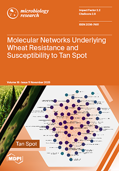
Wheat is a global staple crop, sustaining food security for billions. Understanding how it defends itself against major pathogens is essential for protecting yield and ensuring long-term resilience. In this study, we used RNA-seq and weighted gene co-expression network analysis to uncover coordinated molecular responses to the Tan Spot pathogen Pyrenophora tritici-repentis. By comparing resistant and susceptible cultivars, we identified distinct defense signatures and novel hub genes linked to resistance or susceptibility. We also found common pathogen-triggered responses shared by both wheat lines. These findings reveal previously uncharacterized components of wheat immunity and provide candidates for improving disease-resistant varieties. View this paper
- Issues are regarded as officially published after their release is announced to the table of contents alert mailing list.
- You may sign up for e-mail alerts to receive table of contents of newly released issues.
- PDF is the official format for papers published in both, html and pdf forms. To view the papers in pdf format, click on the "PDF Full-text" link, and use the free Adobe Reader to open them.
Previous Issue
Next Issue