Article

Influence of Mechanical Wounding and Compartmentalization Mechanism on the Suppression of Invasive Plant Species Using the Example of Cherry Laurel (Prunus laurocerasus)

1
Department of Wood Science and Technology, Biotechnical Faculty, University of Ljubljana, 1000 Ljubljana, Slovenia
2
Department of Condensed Matter Physics, Jožef Stefan Institute, 1000 Ljubljana, Slovenia
*
Author to whom correspondence should be addressed.
Forests 2021, 12(12), 1646; https://doi.org/10.3390/f12121646
Submission received: 6 October 2021 / Revised: 24 November 2021 / Accepted: 25 November 2021 / Published: 27 November 2021

Abstract

:
Natural habitats increasingly face the introduction and spread of non-native species. Under the right conditions, non-native species can become invasive over time. This issue is now being addressed by many experts and researchers who are using and developing various approaches and methods to limit and eliminate or suppress problematic plant species. Many invasive plants are already spreading uncontrollably in urban and forestry areas, causing health hazards, environmental and economic damage and negatively impacting natural ecosystems. The use of chemical agents is generally limited, so our only option to control and suppress the problematic species is mechanical removal. In this research suppression by tree stem wounding, i.e., incomplete girdling, was used. This type of injury causes the plant to lose its vitality, become weaker after first year and then die within a few years. Using a research approach, we chronologically monitored the response of cherry laurel (Prunus laurocerasus L.) stem tissue to mechanical wounding of the incomplete girdling. Magnetic resonance imaging (MRI) and light microscopy were used for monitoring moisture content and anatomical changes in different periods after injury. The results of the study showed that cherry laurel, with an intense wound tissue response and other changes, is a species with good compartmentalization potential. The rapid and intense tissue response to injury requires high energy and nutrient consumption and consequently leads to a loss of vigour and mechanical stability, which may result in plant destruction. Results revealed that mechanical wounding by incomplete girdling is a successful method for suppression of non-native and invasive cherry laurel.

1. Introduction

In Slovenia 58% of the territory is forested, which makes Slovenia one of the greenest and most forested countries in Europe. Mixed forests predominate, in which more than 70 native tree species can be found. Due to the consequences of climate change, increasingly frequent natural disasters (windstorms, ice storms, snow breaks…), the appearance of numerous natural enemies (fungi and insects) and of new non-native plant species, our forests and with them native species are seriously threatened. Humans have always unintentionally introduced new species through trade, travel, and nowadays global transportation. A number of these new non-native plant species (trees and shrubs) are now thriving outside their natural range in areas that could not be reached without indirect human assistance [1,2]. After introduction, many non-native plant species die out, but a few survive, become domesticated over time, and begin to spread rapidly and successfully, especially if they do not yet have natural enemies (fungi and insects, etc.) from their native environment. Today, the proportion of non-native tree species in Slovenia is about 1% of the total timber stand, and they are monitored [1,3]. For some of the most problematic non-native species such as tree of heaven (Ailanthus altissima Mill.), black locust (Robinia pseudoacacia L.), staghorn sumac (Rhus typhina L.) as well as cherry laurel (Prunus laurocerasus L.), the spread is so effective that they displace and dominate native species and cause economic damage, which means that non-native species become invasive and even threaten the functioning of the primary natural ecosystem [4,5].
Monitoring and control measures are taken especially for non-native species that have a high potential for invasion. As long as the new species is confined to a small area and identified as invasive in a timely manner, we can attempt to remove it completely. In the USA in particular, chemical control is considered most effective [6], but in Slovenian forests the use of chemical agents is not allowed according to the related regulation [7,8], as it has a negative impact on other species, and also on the functioning of the ecosystem.
If a non-native or even invasive species is already too widespread, complete elimination of the species is impossible. Therefore, with the help of different projects and institutions, we determine its distribution [1,2,4,5], properties [9,10,11] and various possibilities of using them for industrial purposes [12,13,14,15], and thus used to the highest extent for products.
The only option we have left to remove, limit and control fouling is mechanical methods. These include:
  • plucking, where the entire younger plant can be removed along with the root system;
  • digging, where, especially in certain species, we want to remove the entire root system from which a new shoot might emerge;
  • removal by pruning, which is the simplest method but usually less effective as the plant often sprouts even more vegetative buds, so it is crucial to repeat the annual pruning [16] and;
  • various types of bark and cambium removal, known as girdling, applied mainly to species with thicker stems (trunks).
Girdling is a type of mechanical wounding in which the phloem, cambium, and some last cells or xylem barrier around the circumference are removed. Most often, girdling is applied around the entire circumference of the trunk or stem, but some authors suggest that complete girdling is less effective. If a tree or shrub dies too quickly above the girdled part, then many vegetative buds will appear below the wound (as with pruning). To avoid this, it is useful to girdle the plant less, to a maximum of ¾ of the circumference [8,17]. Thus, a mechanically wounded tree or shrub remains alive, but due to reduced transpiration and impaired nutrient transfer its vigour is significantly reduced, and the plant dies over time [18]. There are several examples of successful test use of incomplete girdling on various tree species [17,19,20,21].
The optimal method for removing non-native and invasive plant species therefore depends on the type, size and shape of the mechanical wounding, but it is also important to subsequently monitor the reappearance of the species in the same location [22]. Black locust, for example, is actually highly sought after by forest owners due to its fast growth, high quality wood and strong honey, but on the other hand threatens native vegetation due to its invasiveness [23].
In economic forests, especially when managing native tree species, we want as little mechanical injury as possible, as it greatly reduces the quality and value of the wood for commercial purposes. With weather disasters (due to wind, ice and snowstorms), we have no control over the nature and extent of the wounding. Therefore, the affected forest must be rehabilitated as soon as possible to prevent additional damage that may be caused by the appearance of beetles, fungi and other pests [24]. For non-native, especially invasive woody species, mechanical wounding can generally achieve their limitation, suppression and desired complete removal from the environment.
The aim of this study was to reveal the anatomical features, histochemical changes, and etiology of tissue compartmentalization in invasive woody species. Based on the findings, it should be possible to develop and present the most effective mechanical wounding method for suppressing, particular non-native, invasive woody species. For this study, we chose cherry laurel, which is already grown as an invasive non-native species in the protected Landscape Park in Ljubljana, Slovenia.

2. Materials and Methods

2.1. Plant Species Studied

Cherry laurel (Prunus laurocerasus L.) originates from SW Asia, Bulgaria and Serbia and is a very common non-native ornamental plant in Slovenia, which is already included on the warning list of non-native species [25]. It is found in parks, gardens, hedgerows in urban and suburban centres and already occurs in forests in some places [26]. Cherry laurel is an evergreen shrub resistant to many diseases and various environmental factors, so when it occurs it can quickly form dense stands. The most common method of removal is by sawing off the shrubs, as the strong and branched root system often makes it very difficult to manual excavate the underground parts by hand [27]. When cutting, it is necessary to remove the above-ground parts persistently and regularly over several years until the root system of the plant is completely exhausted and decays.

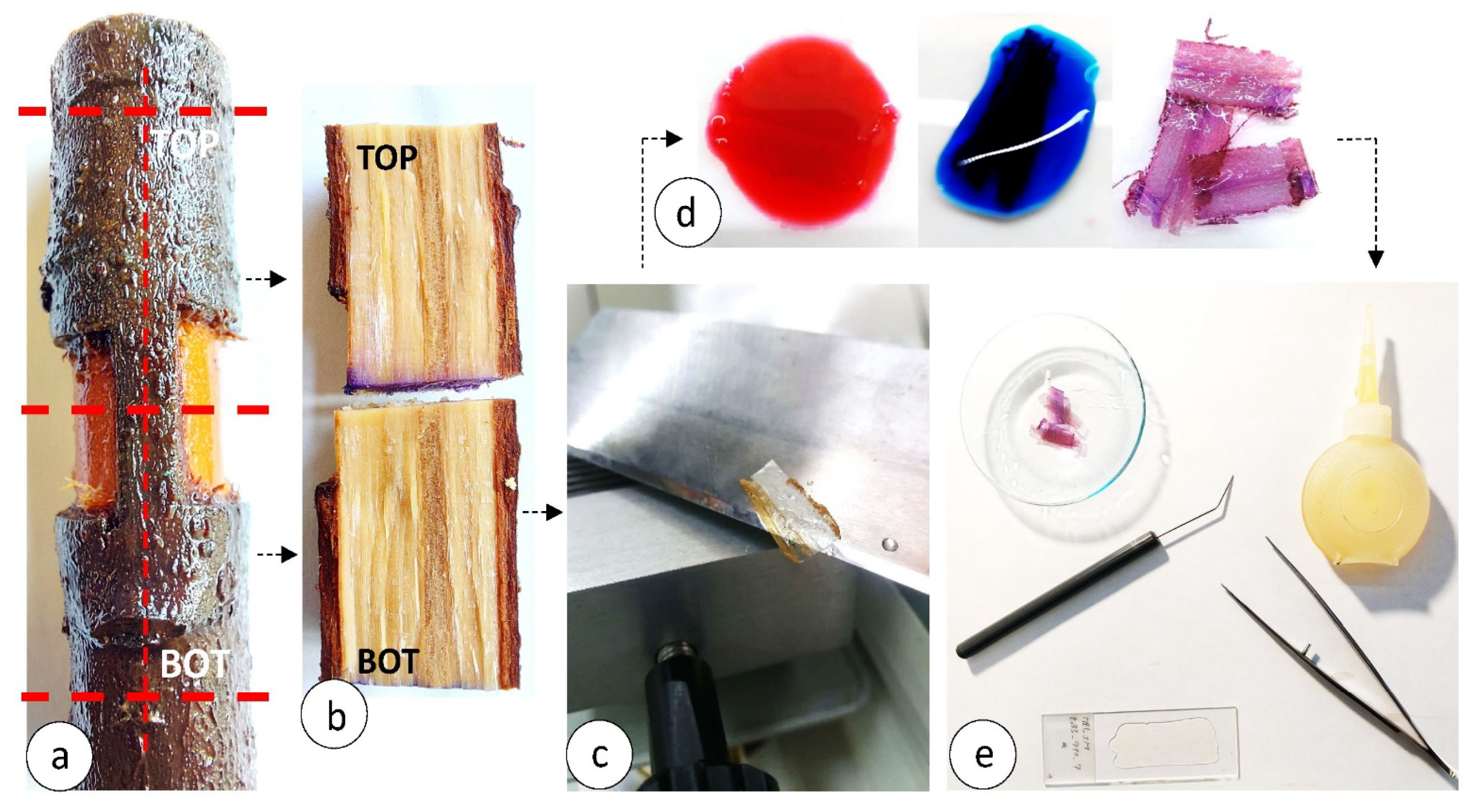
2.2. Seedling Selection, Wounding and Sampling

Cherry laurel is commonly found in private gardens as a cultivated, stand-alone ornamental shrub or used as a hedge on private property. For the purpose of the research, we selected seedlings, aged 4 to 5 years, in the arboriculture. Seedlings were stored in the experimental field of the Biotechnical Faculty in the Department of Wood Science and Technology, where we also conducted injury experiments, response monitoring and sampling. Stems of cherry laurel were mechanically injured with a custom-made tool with two slender blades clamped in parallel and an adjustable depth of cut [28]. In this way, we could arbitrarily adjust (depending on stem diameter and bark thickness) the height and depth of the incision that formed the basis for the mechanical wounding. Type of wounding was partial girdling in which more than 1/10 of the phloem on the stem circumference was left intact so that it could continue to connect the bottom (BOT) and top (TOP) part of the injured stem (Figure 1a and Figure 2a). The wounding of 3 parallel sample plants was carried out in April 2020. Subsequently, samples of wounded tissue were collected at specific time intervals after the injury and detailed examinations of moisture content and anatomical changes were performed. Samples were taken on days 8 (sample PrLa_8), 76 (sample PrLa_76), and 151 (sample PrLa_151) after injury.

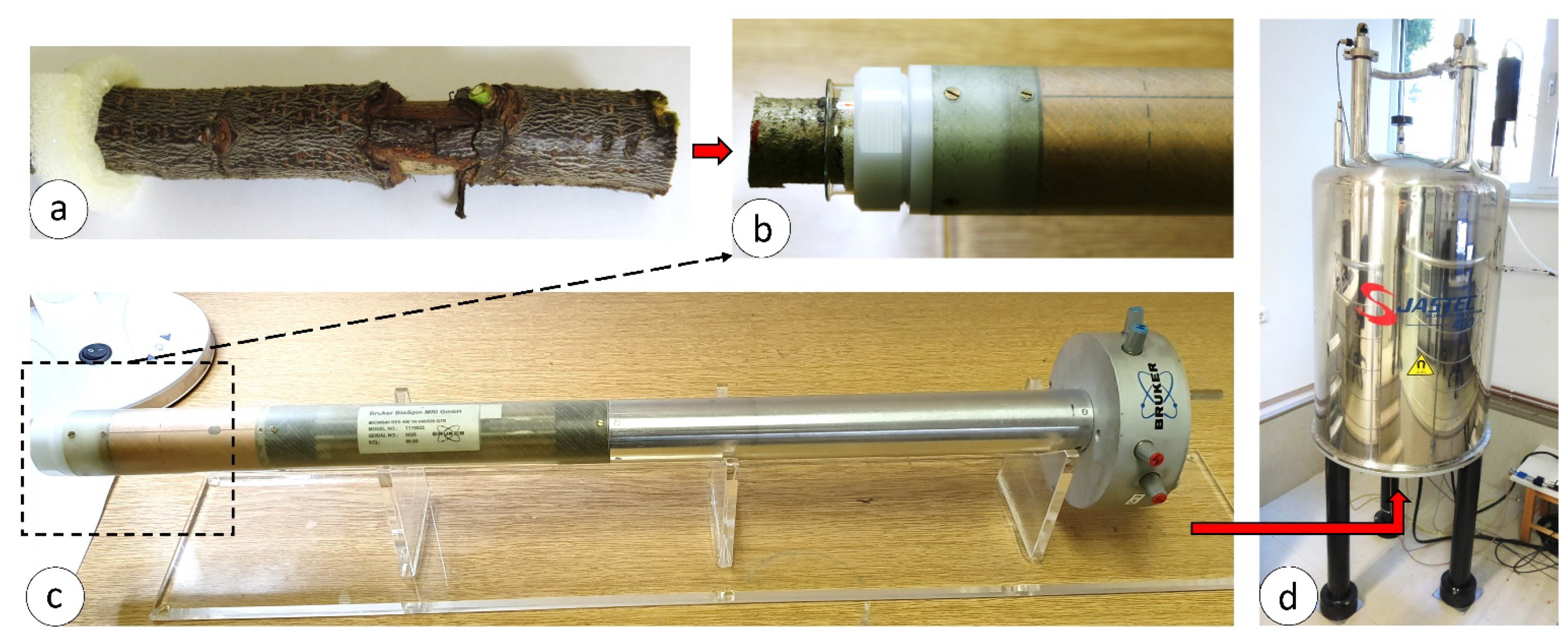
2.3. Magnetic Resonance Imaging and Wood Moisture Determination

To monitor changes in tissue moisture content after injury, we used high-resolution magnetic resonance imaging (MRI), i.e., magnetic resonance microscopy (MRM). For successful MRI, it is important that the observed materials contain hydrogen in a liquid state. Due to its structure and high moisture content, wood is a convenient material for studying of changes in moisture distribution after injury by MRI. With suitably selected MRI parameters the MR signal obtained from the observed wood tissue is directly proportional to the amount and distribution of water [29,30,31,32].
Magnetic resonance imaging was performed on days 8, 76 and 151 after injury. On the day of each MRI, a stem at least 50 cm long was removed so that the wounded tissue was in the middle of the taken stem. Immediately after cutting the injured stem with pruning shears, a coating (Jub akrilin, Ljubljana, Slovenia) was applied to the cut cross surface of the stem tissue and brought into the MR laboratory for immediate MR scanning. During the MRI, the protective coating prevented drying and leakage of water from the cross surfaces of the removed stem. In the MR laboratory, each sample was prepared for imaging by trimming the stem to a length of 20 cm and then placing it in a glass tube 30 mm in diameter and 200 mm in length. To ensure that the sample was aligned and positioned at a specific location in the glass tube, we wrapped the stem with a strip of PU foam to enable better fixation of the sample in the glass tube (Figure 1a). The glass tube with the sample was then inserted into the RF coil of the MR system (Figure 1b,c), and then into the gradient coil of the 400 MHz vertical bore superconducting magnet (Jastec, Kobe, Japan) (Figure 1d).
Magnetic resonance microscopy (MRM) experiments were performed on a Redstone NMR/MRI spectrometer (Tecmag, Houston TX, USA) connected to the magnet (Figure 1d). Stems were imaged in a 30 mm diameter RF coil (Bruker, Ettlingen, Germany). A 2D multi-slice spin-echo MRM sequence was used with the following parameters: field of view (FOV)—60 × 30 mm2, echo time (TE)—10,4 ms; repetition time (TR)—187 ms, imaging matrix—512 × 256, in plane resolution—117 µm isotropic and, slice thickness—1 mm, number of slices—16 (no gap was between the slices), slice orientation was longitudinal with respect to the sample, two signal averages were used and total MR imaging time was 25 min and 33 sec. The parameters of MRI were optimized for fast acquisition and to gain a signal proportional to the water content in wood [30,33].
Since the imaging methods were of a Fourier imaging type, the images were reconstructed by the Fourier transform of the corresponding k-space domain signals [34] to get 2D images of different slices. MRM data were processed and analysed using the spectrometer software TNMR 3.4.27 (Tecmag) and digital image processing program ImageJ 1.44 (National Institute of Health, Bethesda, MD, USA).
To monitor the changes in moisture content of the injured stem tissue as accurately as possible, we needed to match the MR signal intensity with the actual moisture content of the wood tissue in the stem; so, we had to determine the correction factor (CF). In the first phase, the average stem moisture content was calculated on nine parallel, undamaged samples using the gravimetric method [35] (see Table A1 in Appendix A). Secondly the average pixel intensity values (PIV) were calculated from several consecutive slices of 2D MRI of the studied sample on the intact part away from the injury.
The correction factor (CF) between MC and PIV was finally calculated using the following Equation (1). Calculations of CF are attached as Table A2 in Appendix A.
CF = MC/PIV;
where: CF—correction factor; MC—moisture content, [%] and PIV—pixel intensity values.
Since imaging parameters were set in a way that the MR signal was proportional to the amount of water in the wood [33], we were able to calculate the MC for each point of interest inside the examined sample using Equation (2).
MC = CF × PIV; [%]
where: MC—moisture content, [%]; CF—correction factor and PIV—pixel intensity values.

2.4. Wood Anatomical Investigation

The anatomical changes of the wood after injury were observed by light microscopy. All samples imaged by magnetic resonance imaging (MRI) were then prepared for light microscopy. Before sectioning, samples were stored in FAA fixative (mixture of formalin, ethanol and acetic acid) and after 10 days gradually dehydrated in ethanol (30%, 50% and then 70%) according to the protocol of our wood anatomical laboratory [36]. Because the samples were long, we first cut them in half transversely in the middle of the injury and trimmed the bottom (BOT) and the top (TOP) to the estimated length of 15 mm (Figure 2a; red mark). All samples were cut in half with a slender knife over the pith and the rest of the phloem tissue to obtain a radial xylotomic plane (Figure 2b). In the case of the PrLA_151 sample, a transverse xylotome slice was taken at the exact middle of the injury site before radial bisection. Using a sliding microtome (Leica SM2010R, Nussloch, Germany) 18 µm thick histological sections were cut in the radial and transverse xylotomy planes (Figure 2c). Wood slices were stained with a combination of acridine red chrysoidin and astra blue, which is suitable for anatomical studies in bright field and ultraviolet light [37] (Figure 2d). Ultraviolet light (UV) was used for the detection of suberin [36]. Stained wood slices were transferred to an objective glass and permanent anatomical preparations were made using Euparal resin (3C-239 Croma, Chroma 3C-239, Waldeck, Germany) (Figure 2e). Slides were observed and imaged using an E800 light microscope (Nikon, Tokyo, Japan, brightfield, polarization and UV fluorescence), and the image was captured with a Nikon DS Fi1 digital video camera and analysed with NIS Elements BR 3.0.

3. Results and Discussion

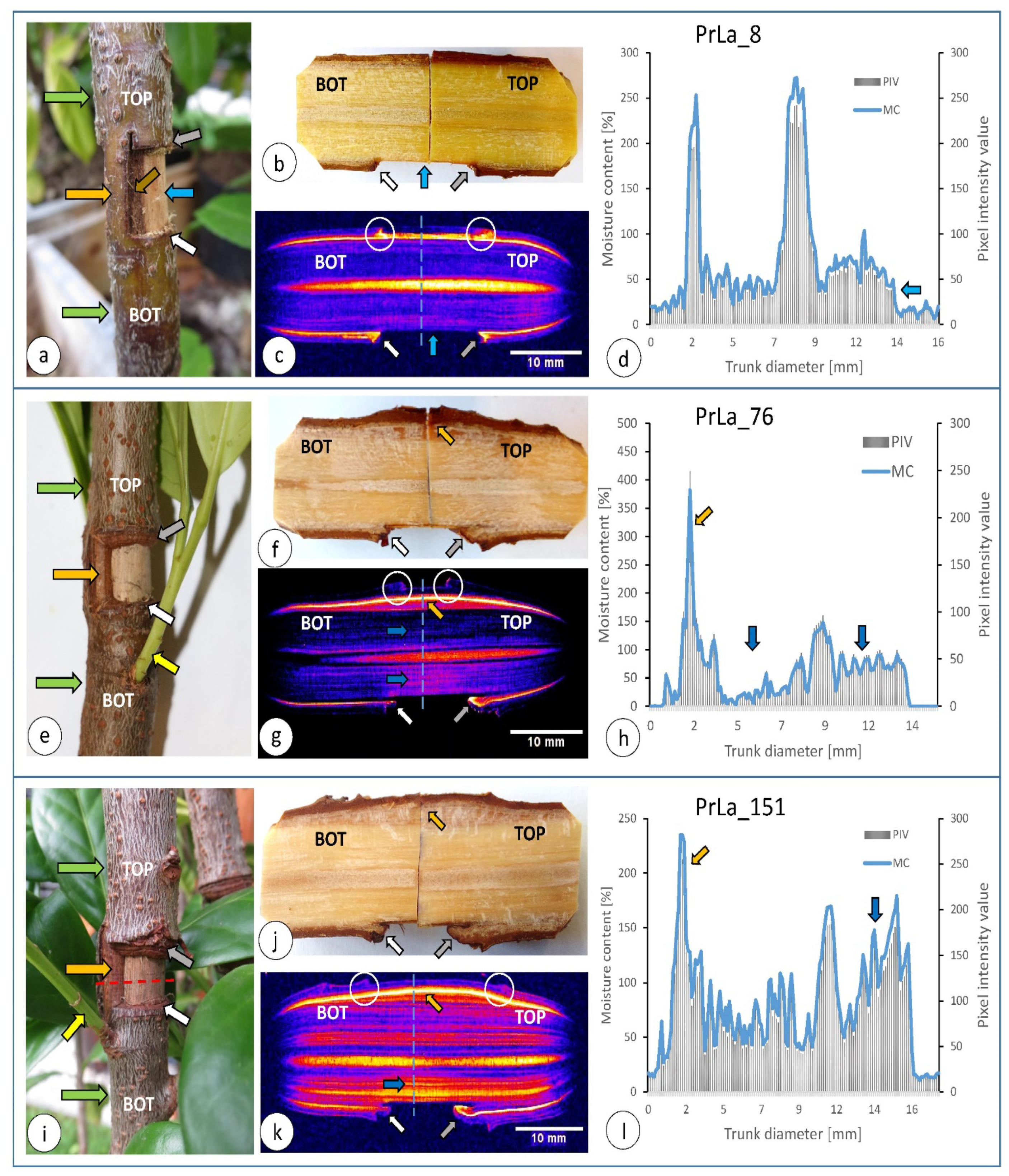
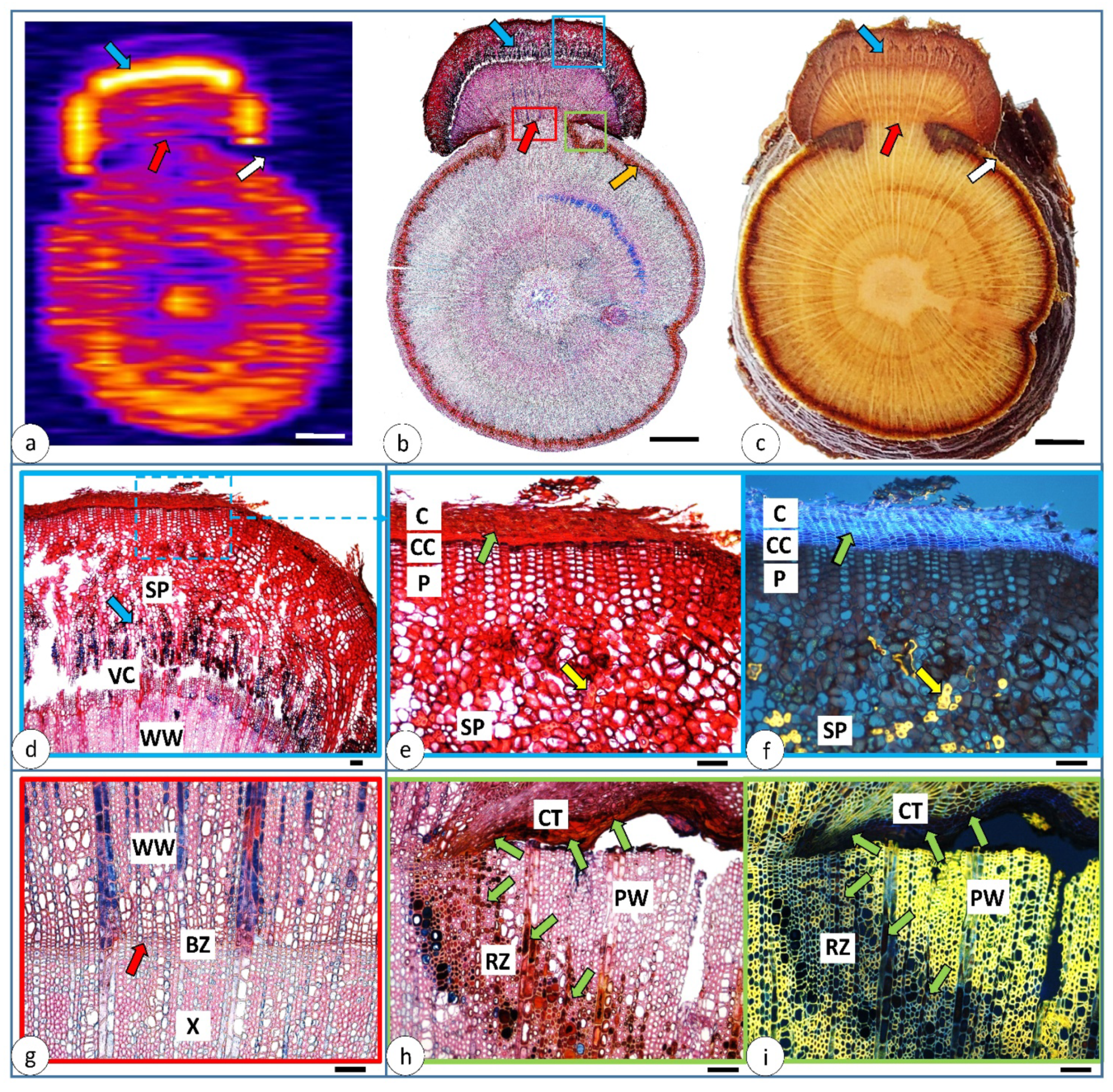
3.1. Morphological and Water Distribution Changes after Injury

The sampling method used enabled us to analyse parallel injured stems at different time periods after mechanical wounding. The main changes in morphology and water content after wounding are shown in Figure 3. Stems were analysed at 8, 76 and 151 days after wounding. Our first observations were made in the field (on the live stem), where each stem was observed and photographed before removing a portion of the stem for sampling (Figure 3a,e,i). MRI of water distribution inside the stems showed some similarities between all three samples, regardless of the time point after mechanical injury. Higher moisture content (represented by brightly pixels on the MR image) is present in the region of the pith and cambium zone (Figure 3c,g,k). Moisture content in the cambium zone ranged from 250% to 350% and in the pith from 150% to 270% (Figure 3d,h,l).
Wounding triggers immediate desiccation and aeration of the exposed tissues. On the macro-image of a stem 8 days after wounding, we can see that the wood has bright brown colour where the bark was removed and wood was exposed to atmospheric air. At the point immediately adjacent to the remaining bark, the wood was coloured dark brown (Figure 3a; brown arrow). Later this discoloured part was identified as a protection wood (see Section 3.3), as defined by Shigo and Marx [38]. MRI of the exposed wood showed that a tiny layer under the exposed wood surface had dried out (Figure 3b–d; blue arrow). Dehydration of the xylem tissue is related to passive protection function, since wood with lower MC is not at risk for fungal infection [31,33,39].
At the place where a bark remnant is present (Figure 3a; orange arrow), we can see that 8 days after injury there are no obvious changes in growth increment compared to normal growth (stem thickening) in the bottom (BOT) and top (TOP) part of the stem (Figure 3a; green arrows).
Although 8 days after injury no intensive growth was detected, the MRI revealed that MC increased significantly along all the edge of the bark remnant (Figure 3a–c; white and grey arrows). Increased moisture content indicates increased cellular activity or increased influx of nutrients to the site where intensive cell division will take place. This fact was confirmed 76 days after injury, where intensive growth of wound wood appeared in the place with high MC (Figure 3e–g; orange, grey and white arrows). By this date after injury wound wood had already completely outgrown the darker coloured protection wood (presented in Figure 3a; brown arrow). Callus and wound wood formation were more intensive in the part above the bark removal (Figure 3e–g; grey arrow) and less intensive on the bottom side of the injury (Figure 3e–g; white arrow).
Some changes also appeared in the remaining bark at the transition between the bottom (BOT) and top (TOP) part of the stem (Figure 3c; white circle), with increased moisture content from the cambium towards the outer bark surface. As a result of the mechanical injury also epicormic bud was activated and a new branch started to grow just under the injury (Figure 3e; yellow arrow). Activation of epicormic buds is usually associated by stand thinning, which leads to an increased exposure to light. But, as in this case, epicormic bud was a result of tree response to mechanical injury—to ensure photosynthetic capacity (nutrition production) for the stem under the injury.
The last field observation and sampling was made on the day 151 after wounding (Figure 3i). On this parallel sample also new branch from epicormic bud had developed under the wounding site (Figure 3l; yellow arrow). Detailed analysis showed that callus and wound wood extensively overgrown the exposed dehydrated wood at all places (Figure 3i; orange, grey and white arrows). Significant growth was evident at the site below the bark remnant (Figure 3i–k; orange arrow) and at the upper part of the stem above the injury (Figure 3i–k; grey arrow). Less new wound wood was formed on the lower part of the stem (under the injury) (Figure 3i–k; white arrow). We assume that the growth increment is more intense on the upper side because the cell supply is not interrupted, and more nutrients are available. These are formed as a product of photosynthesis in the green parts of the plant, which are located in the upper part of the plant (in the canopy). On the lower side (below the injury), the nutrient supply is interrupted due to the removal of the bark, and this results in lower growth increment. MR imaging 151 days after wounding showed high MC of the new wound wood (Figure 3k; orange arrow) and of the wood below the exposed surface (Figure 3k,l; blue arrow). Increased moisture under the dried exposed wood is associated with the formation of the reaction zone [31,33,39,40,41] which is detailed described in Section 3.3.

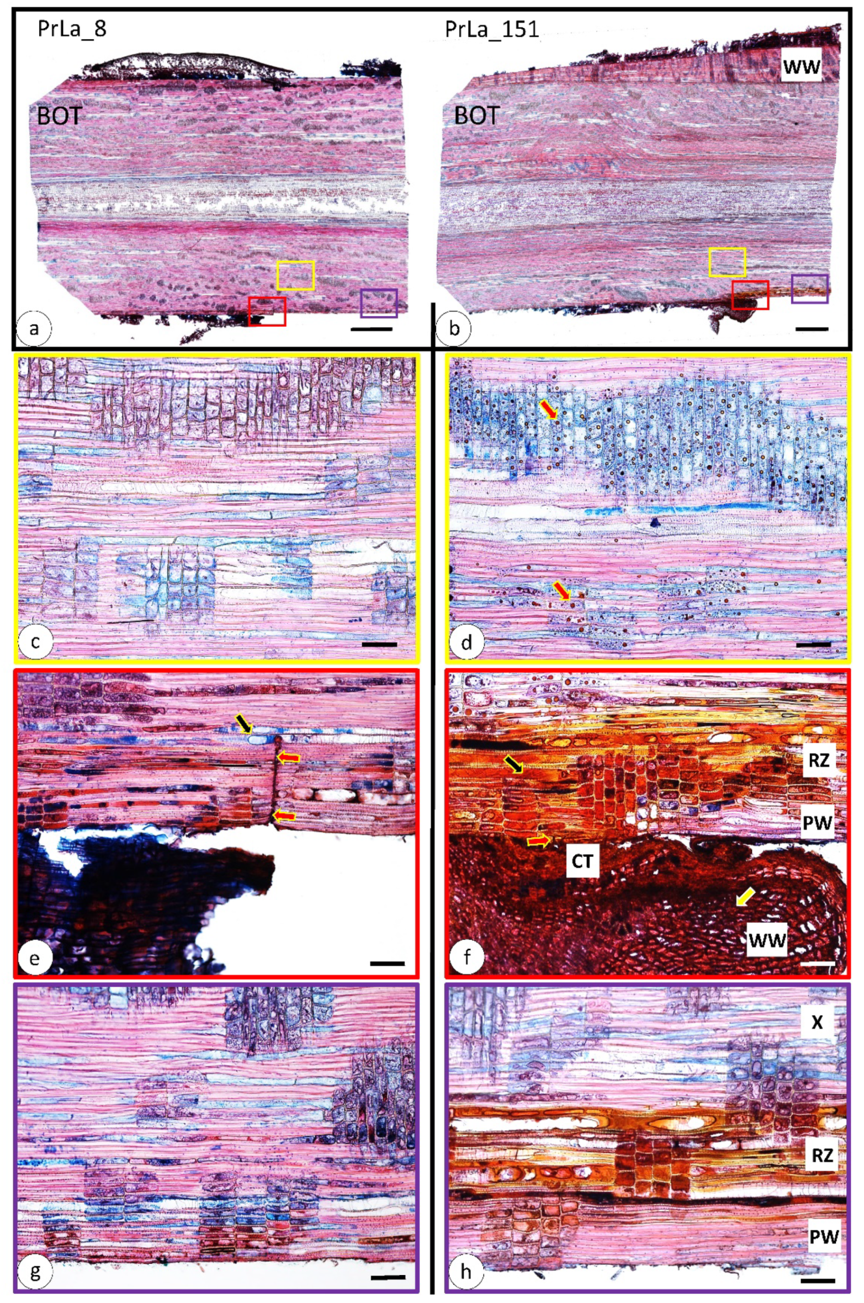
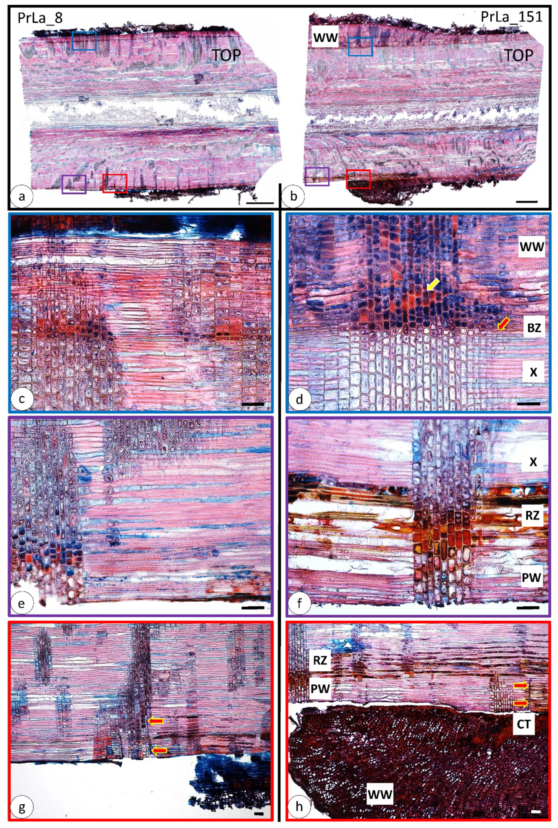
3.2. Wood Anatomical Changes Visible on Radial Section

After stems were scanned by MRI, we performed light microscopic examination on the same stems. For a more transparent anatomical comparison, we combined the lower (BOT) (Figure 4) and the upper parts (TOP) (Figure 5) of the stem 8 and 151 days after wounding.
In lower parts of the stem (TOP), 8 days after wounding (Figure 4c) we can observe intact sound xylem with no significant changes approximately 3 mm deep from the exposed wood surface. In the same location, 151 days after wounding vessels were partly filled with forming tyloses and both, ray and axial, parenchyma cells were filled with polyphenolic compounds in the form of tiny droplets (Figure 4d, red arrows). When we checked the moisture content of this region (Figure 3k), we saw that MC was increased in this part of the stem. These anatomical changes, as well as higher MC corresponds to the initiative formation of reaction zones [38,39,40,41,42].
At the site where we made the cut and removed the bark (Figure 4e, red arrows), the post-injury changes are the most intensive. In 8 days after injury large amount of red and blue-coloured deposits appears in the axial and ray parenchyma, as well as in the vessels, the latter containing also occlusions (such as tyloses) (Figure 4e, black arrow). These changes indicate obstructions of the conducting elements, which reduce the permeability of the tissue and thus prevent the intrusion of atmospheric air and the desiccation of the inner intact sound tissue.
Investigation of the same location 151 days after injury revealed that, approximately 5 mm deep from the cut towards the inside of the stem, all tissues were extensively filled with red to orange polyphenolic deposits (Figure 4f, black arrow). A callus and wound wood (Figure 4f, red and white arrow, respectively) can clearly be seen overgrowing the exposed surface of the wood. A thin layer of initially exposed xylem (Figure 4f), that is overgrown by wound wood, does not contain deposits and MC is low (MRI in Figure 3k). We assume that this part of the wood tissue dried out quickly after bark removal and exposal to atmospheric air, resulting in quick death of the parenchyma. In this case parenchyma cells could not actively respond quick enough to produce the protective components (inclusions such as tyloses and polyphenolic compounds). This part of wood represents the protection wood as it is defined by Shigo and Marx [38].
Exposed wood surface and thin layer of protection wood below the surface is shown on Figure 4g—8 days and Figure 4h–151 days after injury. On the outer part of the stem there is a thin layer of protection wood that does not contain deposits, either tyloses (Figure 4h) and also has low MC (MRI image in Figure 3k). On the inner part between this layer and sound wood highly moist and extensively occluded tissue was formed (polyphenolic compounds and tyloses)—this is the reaction zone (Figure 4h).
Anatomical changes are observed also in the upper part of the stem (TOP), at 8 days (Figure 5a) and 151 days (Figure 5b) after mechanical injury. After 8 days from the injury, observation of the tissue at top part of the stem, below the bark, revealed presence of red and purple polyphenolic deposits present in the radial ray cells (Figure 5c). Rays represent conductive tissue in the radial direction and occlusion in these elements prevents drying in the radial direction. The main conductive elements (vessels) in this area are not filled with occlusions and can normally perform a conductive function, which can also be confirmed in the MRI (Figure 3c).
After 151 days from the injury, it is clearly visible a boundary, namely barrier zone, between the wood before and after injury (Figure 5d, red arrow). The parenchyma cells of the vessel tissue below the barrier zone to the pith direction are unchanged, while the ones in the wound wood tissue (formed after the injury) are wider and completely filled with various polyphenolic deposits (Figure 5d, yellow arrow). On this image we can see strong contrast between wood formed before and after wounding and the barrier zone in between. This reveals how strong and effective the barrier zone (wall 4 according to the CODIT model [38]) really is.
In the area of the exposed surface of the wood (Figure 5e,f), changes in the tissue beginning slightly below the surface of the wood, both after 8 and 151 days from the injury. These changes indicate the layer where reaction zone was developed. After the removal of the bark, the exposed wood surface dried out, also confirmed on the MR image (Figure 3k) and lost its vital function and thus the ability to form new protective substances.
As in the stem below the injury (BOT) (Figure 4e,f), also in this case we observed the most severe anatomical changes above the injury, adjacent to the incision where the bark was removed, both after 8 and 151 days from the injury (Figure 5g,h the incision is indicated by red arrows). After 8 days from wounding (Figure 5g), vessels are partially or completely filled with polyphenolic deposits and tyloses. The ray cells near to the incision are filled with red, purple, and orange polyphenolic deposits. The depth of the incision determines the linear course of the resulting changes in the axial parenchyma, which protects the internal tissue from desiccation. The intensity of these secondary changes decreases with distance from the injury, indeed, far from the injury deposits are present only in the form of small droplets; tyloses are not present.
After 151 days from the injury (Figure 5h) it is clearly seen that intense changes occur in a certain depth where reaction zone was formed. Reaction zone is recognized by intensively occluded ray and axial parenchyma as well as inclusions in vessels (polyphenolic compound as well as tyloses).
Callus and extensive wound wood are present at the site of cut where the bark is removed, overgrowing the exposed, discoloured protection wood (Figure 5h). In the MR image (Figure 3k), the protection wood is not visible and obviously without MR signal, which means that it is dehydrated. At this place of wounded stem, the reaction zone is the main barrier that is protecting inner sound wood form consequences of wounding.

3.3. Changes 151 Days after Injury—The Cross-Section

The cross-section of the MRI of the PrLa_151 sample is presented in Figure 6a. After mechanical injury (removal of the bark), only part of the bark remained (about 1/10 of rim), which then took over the entire physiological growth process along the vascular cambium. Transport of nutrients was only possible through this constricted portion of conductive part of the secondary phloem. As seen on MRI image (Figure 6a, blue arrow) these are tissues with the highest moisture content, with up to 240% of the calculated MC. The tissues in the secondary xylem are moist only in the inner part (sound wood) to the reaction zone (Figure 6b; orange arrow).
On the outside of the reaction zone, there is the protection wood (Figure 6a,c,h,i; white arrow) which is dehydrated (as displayed on MRI—Figure 6a), dysfunctional and dead. With a formation of reaction zone dysfunctional xylem tissue was sharply walled-off of the sound wood.
Figure 6d–f show the situation from the wound wood through the entire periderm, which at the time of wounding remained intact in a proportion of about 1/10 of the circumference. We assume that the flux of organic matter and nutrients (photosynthetic products) in this part was more intense, as evidenced by high MC revealed by MRI (Figure 6a). A polychromatic combination of the dyes acridine red chrysoidine and astra blue in UV light resulted in yellow secondary fluorescence of lignin and light blue to purple fluorescence of suberin. Suberin, a complex polymer of lipids and phenolic components, forms a ligno-suberin layer in the cell wall that is impermeable to aqueous solutions while providing effective, protection against many pathogenic organisms [42,43]. We proved that the outer part of the periderm tissue (cork) is suberized, as indicated by the light bluish colour (Figure 6e,f; green arrow).
Xylem tissues formed before injury and wound wood formed after injury are separated by the specialized tissue called the barrier zone (Figure 6a–c,g, red arrows) that is formed by the cambium after injury. In the CODIT model concept [38], the barrier zone is defined as a compartment wall 4 representing tissues with a high proportion of parenchyma, with a few conducting elements and low lignin content [38,43]. The cell walls in the barrier zone may also be suberized in some species, which has not been confirmed in cherry laurel. According to Shigo and Marx, the barrier zone is the strongest and most effective compartmentalization barrier concept [38].
Figure 6h (light microscopy) and 6i (UV microscopy) show location where the bark remained on the stem at the time of the injury. Light blue to purple suberin layer (marked by green arrows is extending from the periderm cork of wound wood (Figure 6f) along the surface of the callus tissue to the barrier zone in the interior, and to the reaction zone (Figure 6h,i, green arrows) which extends around the entire circumference of the stem.
Living trees respond to wounding in a relatively predictable manner. After injury, the cells on the exposed wood surface dry out, the cells lose their function and die. The damaged tissues are subsequently actively compartmentalised by the formation of physical and chemical barrier, i.e., reaction zone or passively by existing tissues and biochemical compounds and by the high moisture content of the living sapwood. A reaction zone forms at the boundary between inner living, sound and dead xylem, dehydrated cells on the other side. As shown by the MRI, the reaction zone is constantly high in moisture, which prevents infection with pathogens from the outside. In addition to the high moisture in the reaction zone, thick-walled tyloses form in the vessels lumina, the axial and ray parenchyma is filled with orange-, purple-, blue-, and red-coloured deposits, and the cell walls are suberized. The new compounds formed after injury make the tissue less permeable and also have a fungicidal effect. The combination of all histochemical and anatomical changes successfully protects the internal sound tissue from the consequences of wounding which are dehydration and infection by pathogens.

4. Conclusions

Non-native or invasive alien tree species are already spread in natural habitats, and without action their numbers will increase over time, likely affecting the environment and ecosystems as well as biodiversity. Our research showed the potential of application of mechanical wounding by incomplete girdling on the cherry laurel. Results revealed that mechanical wounding by incomplete girdling is a successful method for suppression of non-native and invasive cherry laurel. This specie showed good compartmentalization potential producing intense wound tissue in response to the injury.
The rapid and intense tissue response to wounding required high energy and nutrient consumption. The vitality of the injured stem decreases significantly and almost no new epicormic buds form below the site of incomplete girdling. The mechanically wounded stems usually survive, but their vitality is significantly reduced. The mechanically wounded portion may weaken over time, causing the stem to crack and break off at the site of the mechanical injury. Consequently, the tree loose vigour and mechanical stability, eventually leading to plant mortality.
The results presented in this work are the starting point for understanding compartmentalization processes and their broader application in the control of invasive non-native woody species, where we can be more successful in limiting and controlling problematic species with this type of incomplete girdling.

Author Contributions

Conceptualization, D.P. and M.M.; methodology, D.P., M.M and U.M.; validation, M.M.; formal analysis, D.P. and M.M.; investigation, D.P., M.M and U.M.; resources, D.P., M.M and U.M.; data curation, D.P.; writing—original draft preparation, D.P. and M.M.; writing—review and editing, M.M. and U.M.; visualization, D.P. and M.M.; supervision, M.M. and U.M..; project administration, M.M.; funding acquisition, M.M. All authors have read and agreed to the published version of the manuscript.

Funding

The research was supported by the Program P4-0015, co-financed by the Slovenian Research Agency; LesGoBio V4-2016 financed by Slovenian Research Agency and Ministry of Agriculture, Forestry and Food and APPLAUSE (UIA02-228) project, co-financed by the European Regional Development Fund through the Urban Innovative Actions Initiative (www.ljubljana.si/en/applause/, accessed on 18 June 2021).

Institutional Review Board Statement

Not applicable.

Informed Consent Statement

Not applicable.

Data Availability Statement

Not applicable.

Acknowledgments

The authors wish to thank Igor Serša for his immense help with MR experiment and Angela Balzano and Luka Krže for sample preparation and microscopy.

Conflicts of Interest

The authors declare no conflict of interest.

Appendix A

Appendix A.1. Average Moisture Content

Table A1. Gravimetrically determined moisture content (MC) of nine intact samples of cherry laurel (Prunus laurocerasus—PrLa).
PrLamgreen [g]m0 [g]u [%]
13.1911.86970.7
22.7241.57473.0
32.5361.42977.5
43.4091.86383.0
52.6581.43385.5
62.4801.36082.4
76.0833.55970.9
85.7783.36371.8
96.7923.90873.8
Aver. MC:    76.5

Appendix A.2. Correction Factor

Table A2. Average pixel intensity value (PIV) signal of MR image and corresponding correction factors (CF) calculated from Equation (1). Average moisture content was 76.5%.
MR Image
days after injury876151
samplePrLa8PrLa76PrLa151
PIV685083
CF1.131.530.92

References

  1. Jogan, N.; Kos, I. Poti vnosa, prenosa in širjenja tujerodnih vrst. CRP NeoBiota—Končno Poročilo 2012, 31–42. Available online: http://www.bioportal.si/neobiota.php (accessed on 10 June 2021).
  2. Richardson, D.M.; Hui, C.; Nuñez, M.A.; Pauchard, A. Tree invasions: Patterns, processes, challenges and opportunities. Biol. Invasions 2014, 16, 473–481. [Google Scholar] [CrossRef]
  3. Brus, R.; Pötzelsberger, E.; Lapin, K.; Brundu, G.; Orazio, C.; Straigyte, L.; Hasenauer, H. Extent, distribution and origin of non-native forest tree species in Europe. Scand. J. For. Res. 2019, 34, 533–544. [Google Scholar] [CrossRef]
  4. Castro-Díez, P.; Vaz, A.S.; Silva, J.S.; van Loo, M.; Alonso, Á.; Aponte, C.; Bayón, Á.; Bellingham, P.J.; Chiuffo, M.C.; DiManno, N.; et al. Global effects of non-native tree species on multiple ecosystem services. Biol. Rev. 2019, 94, 1477–1501. [Google Scholar] [CrossRef] [PubMed]
  5. Sádlo, J.; Vítková, M.; Pergl, J.; Pyšek, P. Towards site-specific management of invasive alien trees based on the assessment of their impacts: The case of Robinia pseudoacacia. Neobita 2017, 35, 1–34. [Google Scholar] [CrossRef] [Green Version]
  6. Swearingen, J.; Slattery, B.; Reshetiloff, K.; Zwicker, S. Plant Invaders of Natural Areas, 5th ed.; National Park Service, US Fish and Wildlife Service: Washington, DC, USA, 2014.
  7. Regulation (EU) No 1143/2014 of the European Parliament and of the Council of 22 October 2014 on the Prevention and Management of the Introduction and Spread of Invasive Alien Species. 2014, pp. 35–55. Available online: https://eur-lex.europa.eu/legal-content/EN/TXT/?uri=celex%3A32014R1143 (accessed on 18 June 2021).
  8. Roženbergar, D.; Nagel, T.A.; Urbas, B.; Marion, L.; Brus, R. Nekateri ukrepi za omejevanje širjenja visokega pajesena (Ailanthus altissima (Mill.) Swingle) in smernice za gozdnogojitveno ukrepanje ob vdoru potencialno invazivnih tujerodnih drevesnih vrst v ohranjene gozdove v Sloveniji. Gozdarski Vestn. 2017, 1, 3–20. [Google Scholar]
  9. Gorišek, Ž.; Plavčak, D.; Straže, A.; Merela, M. Technological properties and usability of chinese sumac wood in comparison with ash wood. Les/Wood 2018, 67, 29–44. [Google Scholar] [CrossRef] [Green Version]
  10. Plavčak, D.; Gorišek, Ž.; Straže, A.; Merela, M. Drying characteristics of wood of invasive tree species growing in an urban environment. Les/Wood 2019, 68, 31–43. [Google Scholar] [CrossRef] [Green Version]
  11. Merela, M.; Thaler, N.; Balzano, A.; Plavčak, D. Optimal Surface Preparation for Wood Anatomy Research of Invasive Species by Scanning Electron Microscopy. Drv. Ind. 2020, 71, 117–127. [Google Scholar] [CrossRef]
  12. Merhar, M.; Gornik Bučar, D.; Merela, M. Machinability Research of the Most Common Invasive Tree Species in Slovenia. Forests 2020, 11, 752. [Google Scholar] [CrossRef]
  13. Gornik Bučar, D.; Prislan, P.; Smolnikar, P.; Stare, D.; Krajnc, N. Uporabnost lesnih ostankov tujerodnih invazivnih drevesnih vrst za proizvodnjo peletov. Les/Wood 2021, 70, 45–58. [Google Scholar] [CrossRef]
  14. Horvat, M.; Iskra, J.; Pavlič, M.; Žigon, J.; Merela, M. Wood dyes from invasive alien plants. Les/Wood 2020, 69, 37–48. [Google Scholar] [CrossRef]
  15. Gornik Bučar, D.; Gospodarič, B.; Smolnikar, P.; Stare, D.; Krajnc, N.; Prislan, P. Invasive species as raw material for pellets production. In Proceedings of the 70th Anniversary of Drvna Industrija Journal, Zagreb, Croatia, 12–13 December 2019; pp. 61–68. [Google Scholar]
  16. Burch, P.L.; Zedaker, S.M. Removing the invasive tree Ailanthus Altissima and restoring natural cover. J. Arboric. 2003, 29, 18–24. [Google Scholar]
  17. Moore, G.M. Ring-Barking and Girdling: How Much Vascular Connection Do You Need between Roots and Crown? In Proceedings of the 14th National Street Tree Symposium; University of Adelaide Australia: Adelaide, Australia, 2013; pp. 87–96. [Google Scholar]
  18. Kowarik, I.; Säumel, I. Biological flora of Central Europe: Ailanthus altissima (Mill.) Swingle. Perspect. Plant. Ecol. Evol. Syst. 2007, 8, 207–237. [Google Scholar] [CrossRef]
  19. Noel, A.R.A. The Girdled Tree. Bot. Rev. 1970, 36, 162–195. [Google Scholar] [CrossRef]
  20. Drescher, A.; Ließ, N. Control of alien woody species in the Danube National Park (Austria) the Example of Ailanthus altissima. BfN-Skripten 2006, 184. Available online: https://www.researchgate.net/profile/Anton-Drescher/publication/320357724_Control_of_alien_woody_species_in_the_Danube_National_Park_Austria_The_example_of_Ailanthus_altissima/links/59dfe095a6fdcca98420e68a/Control-of-alien-woody-species-in-the-Danube-National-Park-Austria-The-example-of-Ailanthus-altissima.pdf (accessed on 10 June 2021).
  21. De Schepper, V.; Steppe, K. Tree girdling: A tool to improve our understanding of coupled sugar and water transport. Acta Hortic. 2013, 990, 313–320. [Google Scholar] [CrossRef] [Green Version]
  22. Nyssen, B.; Schmidt, U.E.; Muys, B.; van der Lei, P.B.; Pyttel, P. The history of introduced tree species in Europe in a nutshell. In Introduced Tree Species in European Forests: Opportunities and Challenges; Krumm, F., Vítková, L., Eds.; European Forest Institute: Freiburg, Germany, 2016; p. 423. [Google Scholar]
  23. Rudolf, S.; Brus, R. Razširjenost in invazivnost robinije (Robinia pseudoacacia L.) v severovzhodni Sloveniji. Gozdarski Vestn. 2006, 64, 134–140. [Google Scholar]
  24. De Groot, M.; Ogris, N.; Kobler, A. The effects of a large-scale ice storm event on the drivers of bark beetle outbreaks and associated management practices. For. Ecol. Manag. 2018, 408, 195–201. [Google Scholar] [CrossRef]
  25. Life Artemis Alien Species in Slovenija. Available online: https://www.tujerodne-vrste.info/en/alien-species-slovenia/alien-plants/ (accessed on 18 June 2021).
  26. Rusterholz, H.P.; Schneuwly, J.; Baur, B. Invasion of the alien shrub Prunus laurocerasus in suburban deciduous forests: Effects on native vegetation and soil properties. Acta Oecol. 2018, 92, 44–51. [Google Scholar] [CrossRef]
  27. Applause APPLAUSE—Alien Plant Species from Harmful to Useful with Citizens’ Led Activities. Available online: https://www.uia-initiative.eu/en/uia-cities/ljubljana. (accessed on 18 June 2021).
  28. Plavčak, D.; Balzano, A.; Krže, L.; Merela, M. Wood Response Investigation of Four Selected Non-Native Woody Plants to Various Mechanical Injuries. In Proceedings of the 9th Hardwood Proceedings Part I; University of Sopron: Sopron, Hungary, 2020; pp. 212–217. [Google Scholar]
  29. Merela, M.; Oven, P.; Sepe, A.; Serša, I. Three-dimensional in vivo magnetic resonance microscopy of beech (Fagus sylvatica L.) wood. Magn. Reson. Mater. Phys. Biol. Med. 2005, 18, 171–174. [Google Scholar] [CrossRef] [PubMed]
  30. Mikac, U.; Merela, M.; Oven, P.; Sepe, A.; Serša, I. MR Study of Water Distribution in a Beech (Fagus sylvatica) Branch Using Relaxometry Methods. Molecules 2021, 26, 4305. [Google Scholar] [CrossRef] [PubMed]
  31. Oven, P.; Merela, M.; Mikac, U.; Serša, I. 3D magnetic resonance microscopy of a wounded beech branch. Holzforschung 2008, 62, 322–328. [Google Scholar] [CrossRef]
  32. Oven, P.; Merela, M.; Mikac, U.; Serša, I. Application of 3D magnetic resonance microscopy to the anatomy of woody tissues. IAWA J. 2011, 32, 401–414. [Google Scholar] [CrossRef] [Green Version]
  33. Merela, M.; Oven, P.; Serša, I.; Mikac, U. A single point NMR method for an instantaneous determination of the moisture content of wood. Holzforschung 2009, 63, 348–351. [Google Scholar] [CrossRef]
  34. Hall, L.D.; Rajanayagam, V. Evaluation of the distribution of water in wood by use of three dimensional proton NMR volume imaging. Wood Sci. Technol. 1986, 20, 329–333. [Google Scholar] [CrossRef]
  35. SIST EN 13183-1: Moisture Content of a Piece Of Sawn Timber—Part 1: Determination by oven Dry Method. 2002. Available online: https://standards.iteh.ai/catalog/standards/cen/ebaf1b83-ed78-4bfe-838f-b02fef750459/en-13183-1-2002 (accessed on 18 June 2021).
  36. Prislan, P.; Merela, M.; Zupančič, M.; Krže, L.; Čufar, K. Uporaba izbranih svetlobno mikroskopskih tehnik za raziskave lesa in skorje. Les/Wood 2009, 5, 222–229. [Google Scholar]
  37. Čufar, K.; Merela, M.; Erič, M. A Roman barge in the Ljubljanica river (Slovenia): Wood identification, dendrochronological dating and wood preservation research. J. Archaeol. Sci. 2014, 44, 128–135. [Google Scholar] [CrossRef]
  38. Shigo, A.L.; Marx, H.G. Compartmentalization of Decay in Trees; US Department of Agriculture, Forest Service: Washington, DC, USA, 1977; 73p.
  39. Rayner, A.D.; Boddy, L. Fungal Decomposition of Wood—Its Biology and Ecology, 1st ed.; Wiley: Wiltshire, UK, 1988. [Google Scholar]
  40. Merela, M.; Oven, P.; Serša, I.; Mikac, U. Response of beech trunk tissues to mechanical wouding. In Rational Use of Wood in the Context of Sustainable Forest Management; Humar, M., Krajger, H., Eds.; Studia forestalia Slovenica: Ljubljana, Slovenia, 2009; pp. 89–97. [Google Scholar]
  41. Oven, P.; Zupančič, M.; Merela, M.; Torelli, N. Zgradba reakcijskih con pri bukvi (Fagus sylvatica L.) Anatomy of reaction zones in beech (Fagus sylvatica L.). Zb Gozdarstva Lesar. 2004, 73, 51–62. [Google Scholar]
  42. Oven, P. Odziv drevesnih tkiv na poškodbe in infekcijo. Zb Gozdarstva Lesar. 1998, 55, 113–133. [Google Scholar]
  43. Torelli, N.; Oven, P.; Zupančič, M. Nastanek in značilnosti barierne cone ter lesa nastalega po ranitvi. Zb Gozdarstva Lesar. 1990, 36, 3–16. [Google Scholar]
Figure 1. (a) Sample PrLa_76 (Prunus laurocerasus); (b,c) 30 mm diameter RF coil (Brucker, Ettlingen, Germany) and (d) MRI spectrometer with a superconducting 9.4 T magnet (Jastec 400, Kobe, Japan).
Forests 12 01646 g001
Figure 2. Mechanical injured stem of cherry laurel (Prunus laurocerasus); (a) sample PrLa_8 with preparation red mark; (b) split sample at the bottom (BOT) and top (TOP) at mechanical injury; (c) cutting histological slices for anatomical investigation with a sliding microtome; (d) staining of wood slices, and (e) tools used.
Forests 12 01646 g002
Figure 3. Injured stems of cherry laurel (Prunus laurocerasus): (a,e,i) situation on the day of sampling; (b,f,j) bisected samples on the bottom (BOT) and top (TOP) part; (d,h,l) radial profile of moisture content calculated from MRI pixel intensity values, (c,g,k) Magnetic Resonance Image (MRI) of injured stems.
Forests 12 01646 g003
Figure 4. Cherry laurel (Prunus laurocerasus), radial section: (a) sample PrLa_8, bottom part (BOT), 8 days after injury; (b) sample PrLa_151, bottom part (BOT), 151 days after injury; (c,d) comparison of inner part in 8 and 151 days; (e,f) comparison of wound wood (WW), callus tissue (CT), protection wood (PW) and reaction zone (RZ) formation in 8 and 151 days; (g,h) exposed wood in 8 and 151 days after wounding with protection wood (PW), reaction zone (RZ) and sound secondary xylem (X). ROIs are marked by colour frames of the figures. Scale bars: (a,b) = 2 mm; (ch) = 100 µm.
Forests 12 01646 g004
Figure 5. Cherry laurel (Prunus laurocerasus), radial section: (a) sample PrLa_8, top part (TOP), 8 days after injury; (b) sample PrLa_151, top part (TOP), 151 days after injury; (c,d) comparison of wound wood (WW), barrier zone (BZ) formation and sound secondary xylem (X) in 8 and 151 days; (e,f) comparison of exposed wood, reaction zone (RZ) and protection wood (PW) formation in 8 and 151 days; (g,h) comparison of formation of wound wood (WW), callus tissue (CT), protection wood (PW) and reaction zone (RZ) in 8 and 151 days after wounding. ROIs are marked by colour frames of the figures. Scale bars: (a,b) = 2 mm; (ch) = 100 µm.
Forests 12 01646 g005
Figure 6. Cherry laurel (Prunus laurocerasus); sample PrLa 151—stem cross section (located at the red dashed line in Figure 3i). (a) the MRI cross-section; (b) cross-section of the in bright field; (c) macro image of cross section; (d) wound wood (WW), vascular cambium (VC), secondary phloem (SP) and periderm; (e,f) bright-field periderm and UV fluorescence, detail of secondary phloem (SP) with lignified sclereids, phelloderm (P), cork cambium (CC) and cork (C) that is suberized; (g) secondary xylem before injury (X), barrier zone (BZ) and xylem after injury—wounded wood (WW); (h,i) callus tissue (CT), reaction zone (RZ) and exposed dehydrated tissue i.e., protection wood (PW) in bright field and UV fluorescence. ROIs are marked by colour frames of the figures. Scale bars: (ac) = 2 mm; (di) = 100 µm.
Forests 12 01646 g006
Publisher’s Note: MDPI stays neutral with regard to jurisdictional claims in published maps and institutional affiliations.

