Article

Study of New Glass–Ceramic and Dense Ceramic Containing Biogenic Hydroxyapatite

1
Department of Silicate Technology, University of Chemical Technology and Metallurgy, 8 Kliment Ohridski blvd, 1797 Sofia, Bulgaria
2
Department of Industrial Safety, University of Chemical Technology and Metallurgy, 8 Kliment Ohridski blvd, 1797 Sofia, Bulgaria
*
Author to whom correspondence should be addressed.
Materials 2025, 18(13), 3059; https://doi.org/10.3390/ma18133059
Submission received: 19 May 2025 / Revised: 19 June 2025 / Accepted: 25 June 2025 / Published: 27 June 2025

Abstract

A novel bioactive glass–ceramic was developed using biogenic hydroxyapatite (BHA) synthesized from Rapana venosa (Black Sea) shells and monocalcium phosphate monohydrate [Ca(H2PO4)2·H2O] via solid-state synthesis. The prepared batches were obtained by combining BHA with SiO2, B2O3, and Na2O, melted at 1200 °C and melt-quenched in water to form glass–ceramic materials. Dense biogenic hydroxyapatite-based ceramics were successfully sintered at 1200 °C (2 h hold) using a 25 mass % sintering additive composed of 35 mass % B2O3, 45 mass % SiO2, 10 mass % Al2O3, and 10 mass % Na2O. Structural characterization was carried out using X-ray diffraction (XRD), Fourier-transform infrared spectroscopy (FTIR), and scanning electron microscopy (SEM). The resulting materials consisted of a well-defined crystalline hydroxyapatite phase [Ca10(PO4)6(OH)2] alongside an amorphous phase. In samples with increased SiO2 and reduced B2O3 content (composition 3), a finely dispersed Na3Ca6(PO4)5 crystalline phase appeared, with a reduced presence of hydroxyapatite. Bioactivity was assessed in simulated body fluid (SBF) after 10 and 20 days of immersion, confirming the material’s ability to support apatite layer formation. The main structural units SiO4, PO4, and BO3 are interconnected through Si–O–Si, B–O–B, P–O–P, and mixed Si–O–Al linkages, contributing to both structural stability and bioactivity.

1. Introduction

The development of advanced materials for biomedical applications is among the most important problems faced by modern material science. More and more scientific developments are emerging in the field of biomaterials based on glasses, glass ceramics, and ceramics. Hydroxyapatite (HA) is a calcium phosphate bioceramic and is the main material for applications in bone replacement and exhibits biocompatibility, osteoconductivity, and bioactive behavior, being able to bond to the bone directly. Hydroxyapatite has been widely used in biomedical applications due to its similar composition to that of bone and teeth, and its excellent biocompatibility [1,2,3,4]. Different types of composites on the base of biogenic (BHA) and synthetic hydroxyapatite (SHA) and glass phase were studied and cited in the scientific literature. BHA is obtained from various biological sources like animal bones (bovine, porcine), fish scales, eggshells, and seashells. BHA has a crystallite size that is similar to that found in natural bone. BHA is highly bioactive, meaning it can stimulate biological responses, making it suitable for bone regeneration and other applications. BHA is environmentally friendly and cost-effective [5,6,7]. HA has been reinforced with glasses, glass–ceramics, and ceramics to improve its mechanical properties. One way to improve the properties of the HA ceramics is the addition of glass as a second phase. Glasses used to obtain composites with hydroxyapatite are on the base of the systems Na2O–CaO–SiO2, B2O3–Na2O–CaO–SiO2, Na2O–CaO–P2O5–SiO2, and others. It is known from the scientific literature that glass-reinforced HA shows greater bioactivity compared to commercial HA [8,9,10,11,12]. This occurs because silicate-based glasses release critical concentrations of ions (such as Si, P or Ca) in vivo, which are able to stimulate bone formation and play an important role both in angiogenesis and in neo-vascularization. The disadvantage of HA is its poor mechanical strength. HA is difficult to sinter and thus is mechanically weak. HA is the most thermodynamically stable calcium phosphate ceramic compound at the pH, temperature, and composition of the physiological fluid. Due to the chemical similarity between HA and mineralized bone of human tissue, synthetic HA exhibits a strong affinity to host hard tissues. The formation of a chemical bond with the host tissue offers HA a greater advantage in clinical applications over most other bone substitutes, such as allografts or metallic implants. To improve the mechanical properties of HA, appropriate sintering additives are also applied for dense samples [13,14,15]. The mechanical strength and fracture toughness of hydroxyapatite ceramics can be improved by applying different sintering techniques that include the addition of a low melting secondary phase to achieve liquid phase sintering for better densification, and the incorporation of sintering additives to enhance densification through grain boundary strengthening [16].
This paper aims to obtain new glass–ceramic and ceramic materials containing biogenic hydroxyapatite for potential application in dental and regenerative medicine.

2. Materials and Methods

Biogenic hydroxyapatite powder used in this study was prepared from Black Sea Rapana venosa shells and monocalcium phosphate monohydrate Ca(H2PO4)2·H2O by solid-state synthesis at 1180 °C. The synthesis method and characteristics of hydroxyapatite powder are described in our previous publications [17,18]. Samples marked from 1 to 3 in Table 1 were prepared by homogenization and the melting of chemical reagents as powdered quartz, H3BO3, Na2CO3, and NaNO3, and 30 mass % of BHA powder in a corundum crucible at 1200 °C and melt-quenched in water. For the production of dense ceramic samples (compositions 4 and 5 in Table 1), BHA powder in quantities of 50 mass % and 75 mass % and 50 mass % and 25 mass % were pre-melted and milled with glass powder containing 35 mass % B2O3, 45 mass % SiO2, 10 mass % Al2O3, and 10 mass % Na2O. Samples with dimensions of 3 × 0.5 cm were pressed at a pressure of 50 MPa on a hydraulic press and fired at 1200 °C with 2 h hold. For the analysis of the structure of the obtained samples, X-ray diffraction (DRON 3M diffractometer, Cu Ka radiation, wavelength 1.5418 Å, 28 mA current and 40 kV voltage) was used. The FTIR spectra were recorded in the 4000–400 cm−1 range by using FTIR spectrometer Varian 600-IR (Melbourne, Australia). The samples for these measurements were prepared in the form of KBr–disks. The precision of the absorption maxima was ±3 cm−1. The scanning electron microscope Carl Zeiss GmbH (Jena and Oberkochen, Germany), model EVO 10, brand ZEISS was used for the SEM and EDS (Oxford, UK) analysis. The densities of the glasses were measured by applying the Archimedes principle, using an analytical scale Mettler Toledo New Classic ME 104 (Greifensee, Switzerland) equipped with a density determination kit for solids using distilled water as the immersion liquid. For each glass composition, the density of at least three different samples was measured at least ten times. The bioactivity behavior of the studied BHAp-containing compositions was traced in a simulated body fluid (SBF) medium of pH 7.4 in a water bath at 37 °C for 10 and 20 days. Preparation of SBF is performed by following the Kokubo protocol [19]. SBF solution has ion concentrations nearly equal to human blood plasma and is buffered at pH 7.40 with 50 mM trishydroxymethylaminomethane and 45 mM hydrochloric acid at 36.5 °C.

3. Results and Discussion

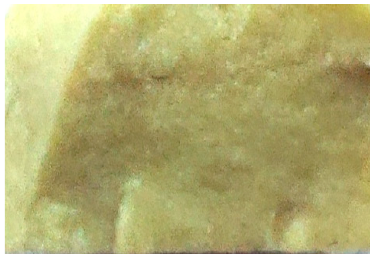
The results obtained show that samples with compositions from 1 to 4 are glass ceramics, and the sample with composition 5 is ceramic. In Figure 1 and Figure 2, the obtained samples with compositions 3 and 5 are shown. The results of the measured densities are presented in Table 1. The density of the glass melted samples increases with decreasing B2O3 content and increasing SiO2 content. The density of pressed ceramic samples increases with increasing hydroxyapatite content.

3.1. X-Ray Powder Diffraction

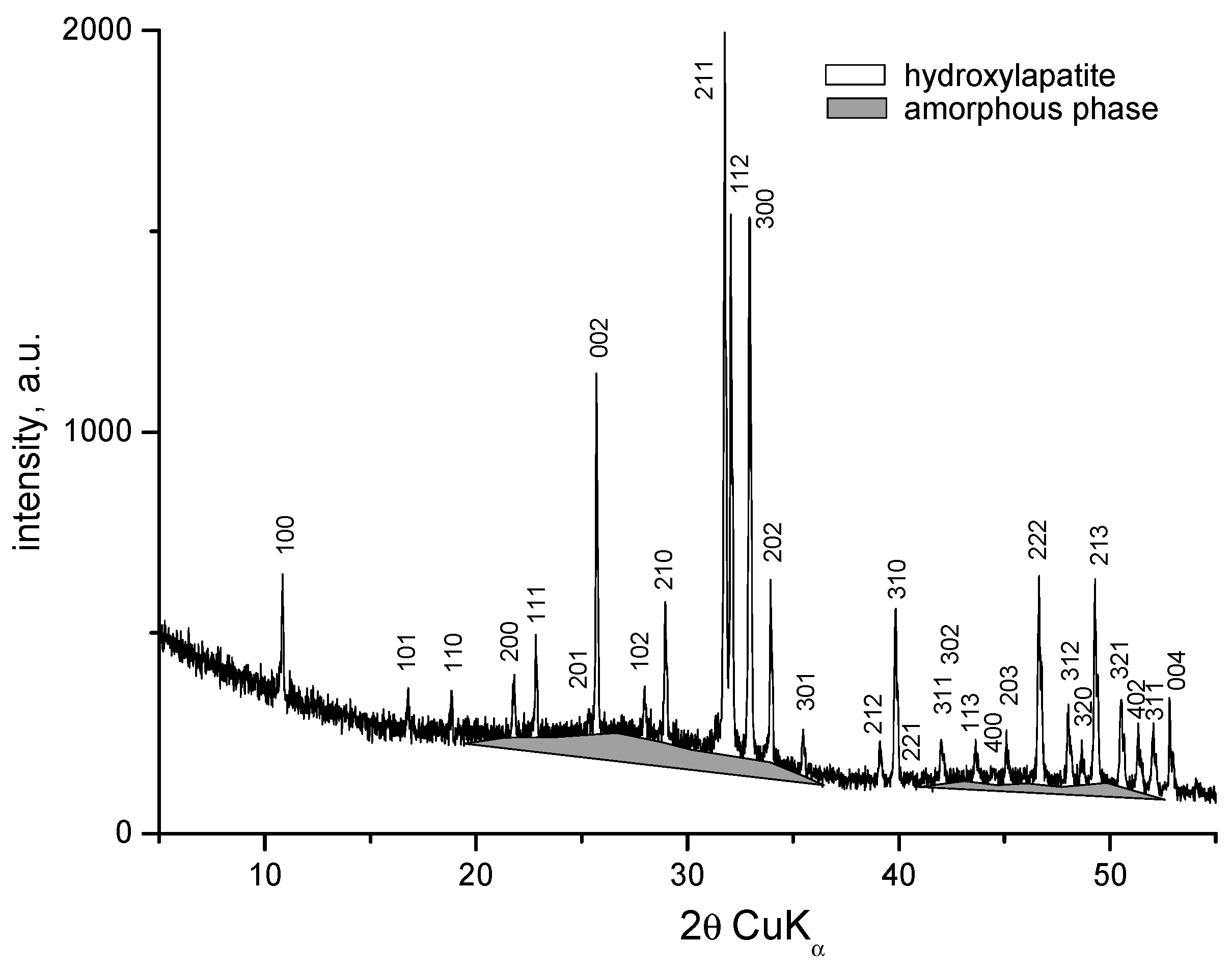
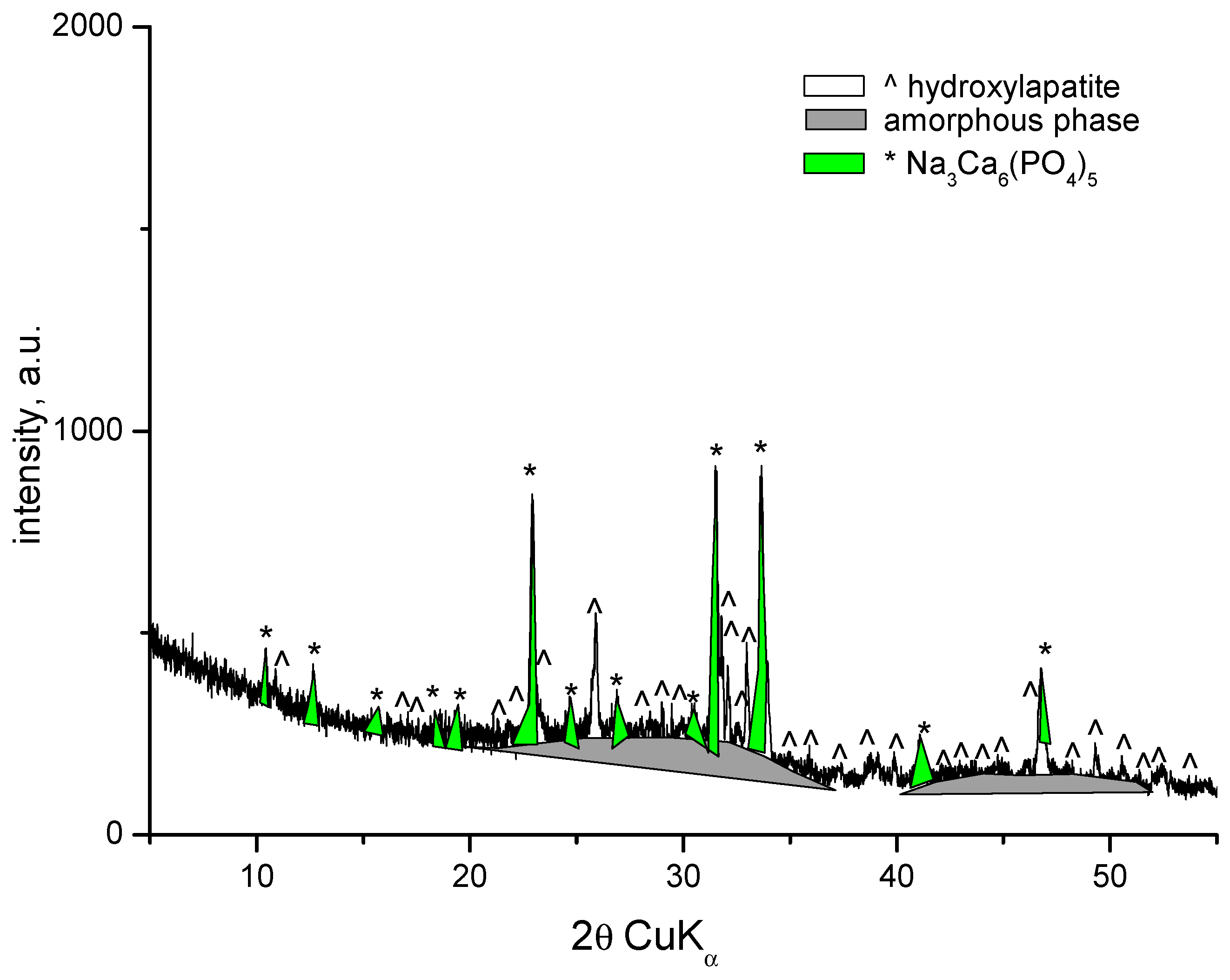
XRD patterns of the samples with compositions 1 and 2 are identical and show amorphous halo and diffraction peaks. The XRD pattern of the sample with composition 2 is presented in Figure 3. Along with the amorphous phase, the main crystalline phase in both compositions is hydroxyapatite (PDF # 01-073-1731). The amorphous phase is more abundant in Sample 2. The X-ray diffractogram of the sample with composition 3 shows the presence of an amorphous phase, a small amount of hydroxyapatite (PDF # 01-073-1731), and the formation of a new fine crystalline phase of sodium calcium phosphate Na3Ca6(PO4)5 (PDF # 11-0236) (Figure 4). In this case, dissolution of hydroxyapatite in the amorphous phase and crystallization of sodium calcium phosphate probably occurs. H. Demirkiran et al. also identified the formation of the Na3Ca6(PO4)5 phase in hydroxyapatite–bioglass composites [20]. In compositions containing higher bioglass content (10 and 25 wt%), additional crystalline phases-namely calcium phosphate silicate (Ca5(PO4)2SiO4) and sodium calcium phosphate (Na3Ca6(PO4)5), were observed, embedded within the amorphous silicate matrices.
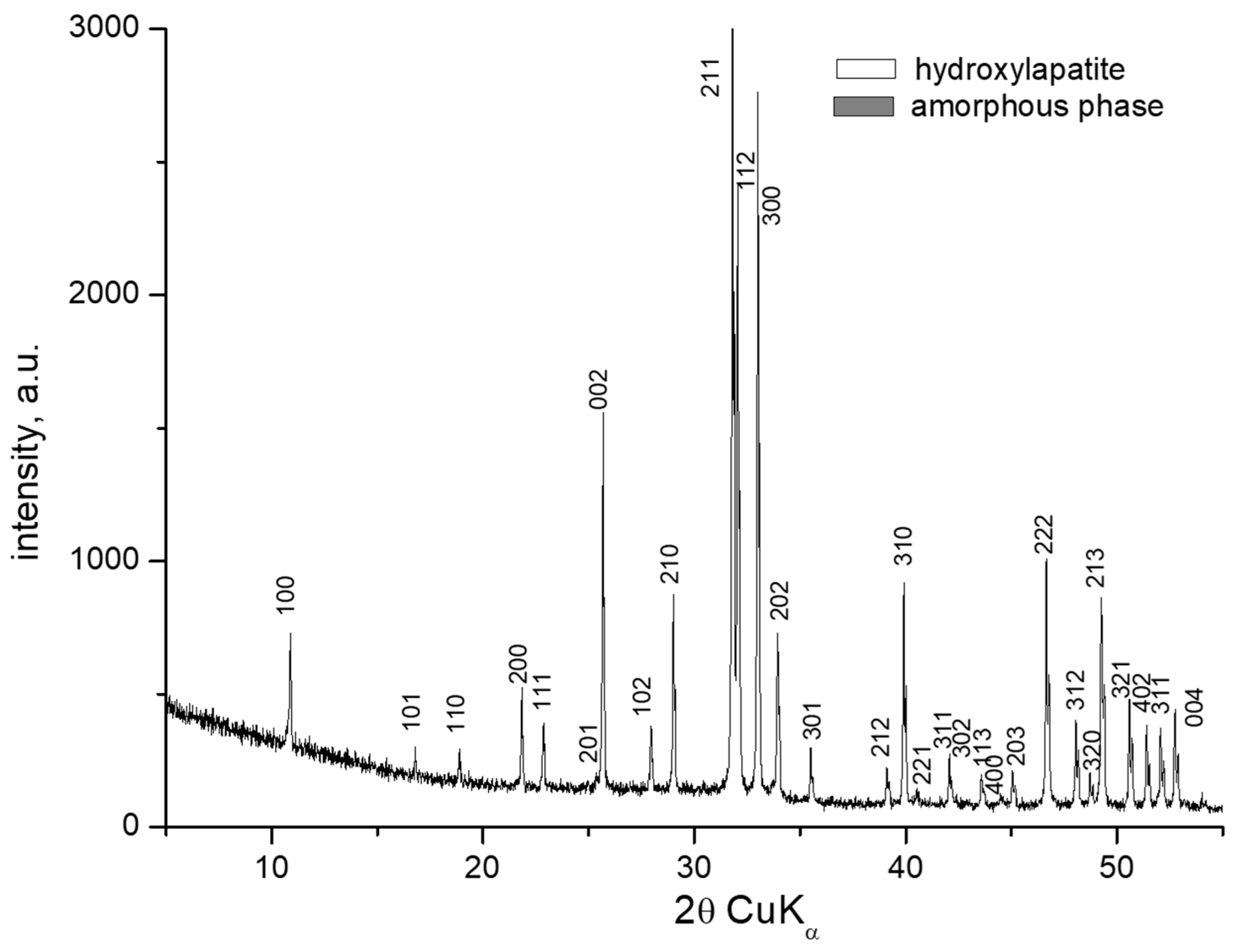
The diffractogram of the sample with composition 4 is similar to those of compositions 1 and 2, containing both hydroxyapatite and amorphous phases. In contrast, the diffractogram of the sample with composition 5 reveals only a hydroxyapatite phase (PDF # 01-073-1731) (Figure 5). Bioactive glasses have been successfully used as sintering additives to produce dense hydroxyapatite (HA) ceramics. These glasses typically contain components such as SiO2, B2O3, Na2O, K2O, Al2O3, P2O5, CaO, CaF2, MgO, and MgF2, among others [21]. Our results are consistent with data reported in the literature; in our case, borosilicate glass effectively facilitated the densification of HA ceramics through viscous flow, promoting liquid-phase sintering. Additionally, it stabilized the hydroxyapatite phase against thermal decomposition by encapsulating it within the glassy matrix.

3.2. FTIR Spectroscopy

The results of the FTIR spectra of the samples are presented in Figure 6. Several spectral ranges are observed high-frequency bands in the 1470–1354 cm−1 range, the 1021–1031 cm−1 range, the 700–750 cm−1 range, as well as bands at around 600 cm−1 and a band around 454–480 cm−1. The band at the high-frequency region becomes narrower and two well-defined bands appear at 1442 cm−1 and 1395 cm−1 in the spectra of Sample 4 and Sample 5. The band at 1021 cm−1 in the spectrum of Sample 1 and Sample 2 shifts to 1031 cm−1 in Samples 3, 4, and 5. A small shoulder at 960 cm−1 is observed in the spectra of all samples except Sample 3. The band at 709 cm−1 in the spectra of Samples 1 and 2 shifts to 699 cm−1 in the spectra of Sample 3, and a double degenerate vibration is observed in the spectra of Samples 4 and 5. The band at 564 cm−1 in the spectra of Sample 3 is double degenerated with bands at 600 cm−1 and 564 cm−1 in the spectra of Samples 1 and 2, and triple degenerated in the spectra of Samples 4 and 5 at 605 cm−1, 569 cm−1, and 544 cm−1. A band at 454 cm−1 with the addition of SiO2 in the spectra of Sample 2 appears; this band shifts up to 480 cm−1 in the spectra of Sample 5.
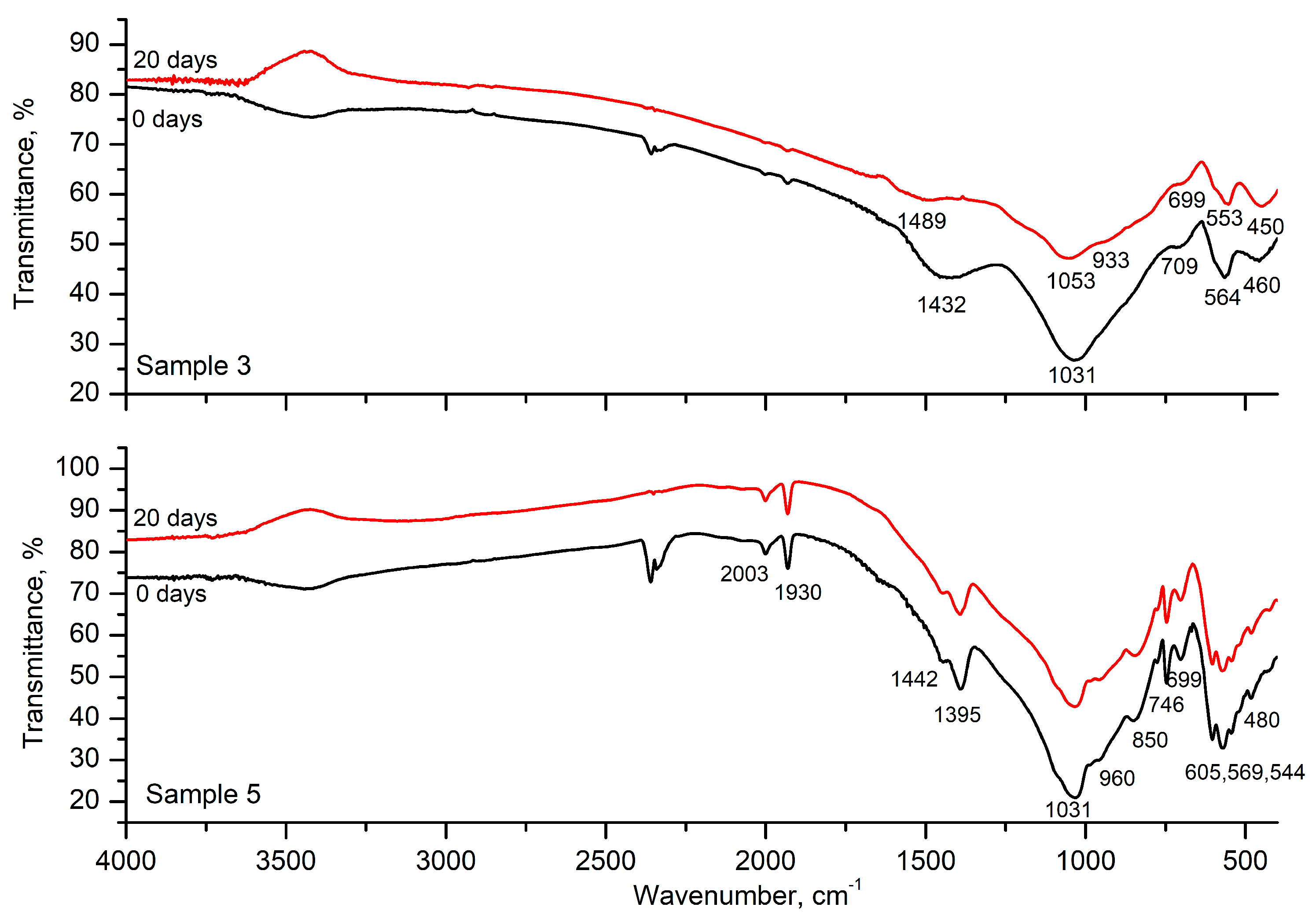
The results of the FTIR spectra of Samples 3 and 5 before and after 20 days in SBF are shown in Figure 7. The spectrum of Sample 3 after 20 days shows small changes. The bands at 1432 cm−1 and 1031 cm−1 are shifted to higher frequencies; a shoulder at 933 cm−1 appeared, and the bands at 709 cm−1, 564 cm−1, and 460 cm−1 are shifted to lower frequencies. No changes in the spectra are observed in the spectra of Sample 5 after 20 days in the SBF.
The structure of the glasses was studied by the FTIR spectroscopy (Figure 6). The vibrations of the isolated PO4 tetrahedra can be used as a starting point for the interpretation of phosphate spectra [16]. The isolated PO4 tetrahedra has four normal vibrations, namely a triple degenerative stretching antisymmetric vibration νdas(F) at 1017 cm−1, a symmetric stretching vibration νs(A1) at 938 cm−1, a triple degenerative bending δd(F) at 567 cm−1, and a double degenerate deformation vibration δd(E) at 420 cm−1. At the highest tetrahedral symmetry (Td), only the first and the third vibrations are active in the IR spectrum. As a result of a decrease in the symmetry of PO4 in the crystal structure, a degeneration of the degenerative vibrations and activation of the inactive vibrations occurs [22,23].
Koutsopoulos synthesized hydroxyapatite crystals from aqueous solutions using three approaches and summarized the analytical data for several calcium phosphates [23]. According to this study, the bands at 1087 cm−1, 1072–1032 cm−1, 962 cm−1, 601 cm−1, 571 cm−1, and 474 cm−1 are attributed to vibrations of the phosphate group, PO4. The peak at 1087 cm−1 in their spectra corresponds to the triple degenerate asymmetric stretching vibration, ν3. The other two components of this vibration appear at 1046 and 1032 cm−1. The peak at 962 cm−1 is assigned to the symmetric stretching mode, ν1, of the P–O bond in the PO4 group. The peaks at 601, 575, and 561 cm−1 are assigned to the O–P–O bond’s triple degenerated bending mode, ν4. The weak peaks at 472 cm−1 and the shoulder at 462 cm−1 are components of the double degenerated bending mode, δ2, of the phosphate group. The FTIR spectra of hydroxyapatite synthesized from Black Sea Rapana venosa shells by Yoleva et al. used in the present study also exhibit the previously mentioned bonds [17].
The addition of SiO2 causes a shift in the band at 1021 cm−1 to 1031 cm−1 and a new band at 454 cm−1 appears. Isolated SiO4 tetrahedra exhibit four normal vibrations [24]. However, only two of them, namely a triple degenerate asymmetric stretching vibration, νdas(F), at 956 cm−1, and a triple degenerate bending vibration, δ(F), at 527 cm−1, remain active when the SiO4 tetrahedra have higher symmetry. It is found that while increasing the degree of linkage of tetrahedra in the order ortho-, pyro-, and meta- to disilicates, the position of the asymmetric stretching vibration νdas(F) is shifted toward higher wavenumbers, and decreasing the symmetry of the tetrahedra induces an activation of the nonactive vibrations [24].
In the structure of borate materials, the boron atom can form coordination polyhedra as either BO3, BO4, or superstructural units built up of both BO3 and BO4 polyhedra [25,26]. The characteristic IR regions of vibrations of B–O bonds are related to the asymmetric B–O stretching vibration of borate tetrahedral (BØ4) around ~800–1200 cm−1 range, the corresponding vibrations of boron oxygen triangular units (BØ3 and BØ2O) at 1200–1650 cm−1, and the deformation vibrations of the network units give rise to weak absorption bands between 550 and 800 cm−1 [24].
Based on the data above, the following assignments of the bands that occurred in our spectra can be made (Table 2). The band at 1021 cm−1 can be assigned to the triple degenerate stretching antisymmetric vibration νdas(F) of PO4, the band at 960 cm−1 to the symmetric stretching vibration νs(A1) of PO4, and the doublet at 600 cm−1 and 564 cm−1 to the triple degenerate bending vibration δd(F) of PO4 unit in the spectra of samples with composition 1 and 2. The degeneracy of the triple degenerative bending vibrations δd(F) of the PO4 unit is observed in the spectra of the samples with compositions 4 and 5, where the triplet at 605 cm−1, 569 cm−1, and 544 cm−1 is observed. These spectral bands are characteristic of the hydroxyapatite phase. In the spectra of the sample with composition 3, the band related to the triple degenerate bending vibrations δd(F) of the PO4 unit is represented by a singlet. This indicates the presence of a different symmetrical form of the PO4 unit. Such an observation follows the results obtained by XRD where a new crystalline phase appears (Na3Ca6(PO4)5).
The bands related to the triple degenerate stretching antisymmetric vibration νdas(F) of SiO4 group overlap with those of PO4 in the high-frequency region. The bending vibrations of Si–O–Si of connected SiO4 units are found at 454 cm−1. However, a further increase in the SiO2 and hydroxyapatite content causes a shift in this band towards higher frequencies, where the double degenerated bending vibrations δd(E) of PO4 and SiO4 tetrahedra are observed [22,23]. The peak at 2000 cm−1 is assigned to a 2.ν3 harmonic overtone or a combination mode, ν1 + ν3 [23]. The broad band at the 1468–1354 cm−1 region becomes narrower with decreasing B2O3 content and two well-defined peaks are formed in the spectra of Samples 4 and 5. These bands could be assigned to the overlapping vibrations of the stretching mode (ν1) of the CO32− group in A-type CAP or the bending mode (ν4 or ν3) of the CO32− group in A- and B-type CAP and the double degenerative asymmetric stretching vibration νdas(E) of borate triangular units BO3 [24,26]. The band at 850 cm−1 could be assigned to the stretching vibrations of the CO32− group in CAP [23]. The degenerate bending vibration δd(E) of BO3 units is at 560 cm−1. The band at 706–701 cm−1 could be assigned to the overlapping of bending vibrations of B–O–B in the [BO3] group and symmetric stretching vibrations of Si–O–Al [24,25].

3.3. SEM and EDS Analysis

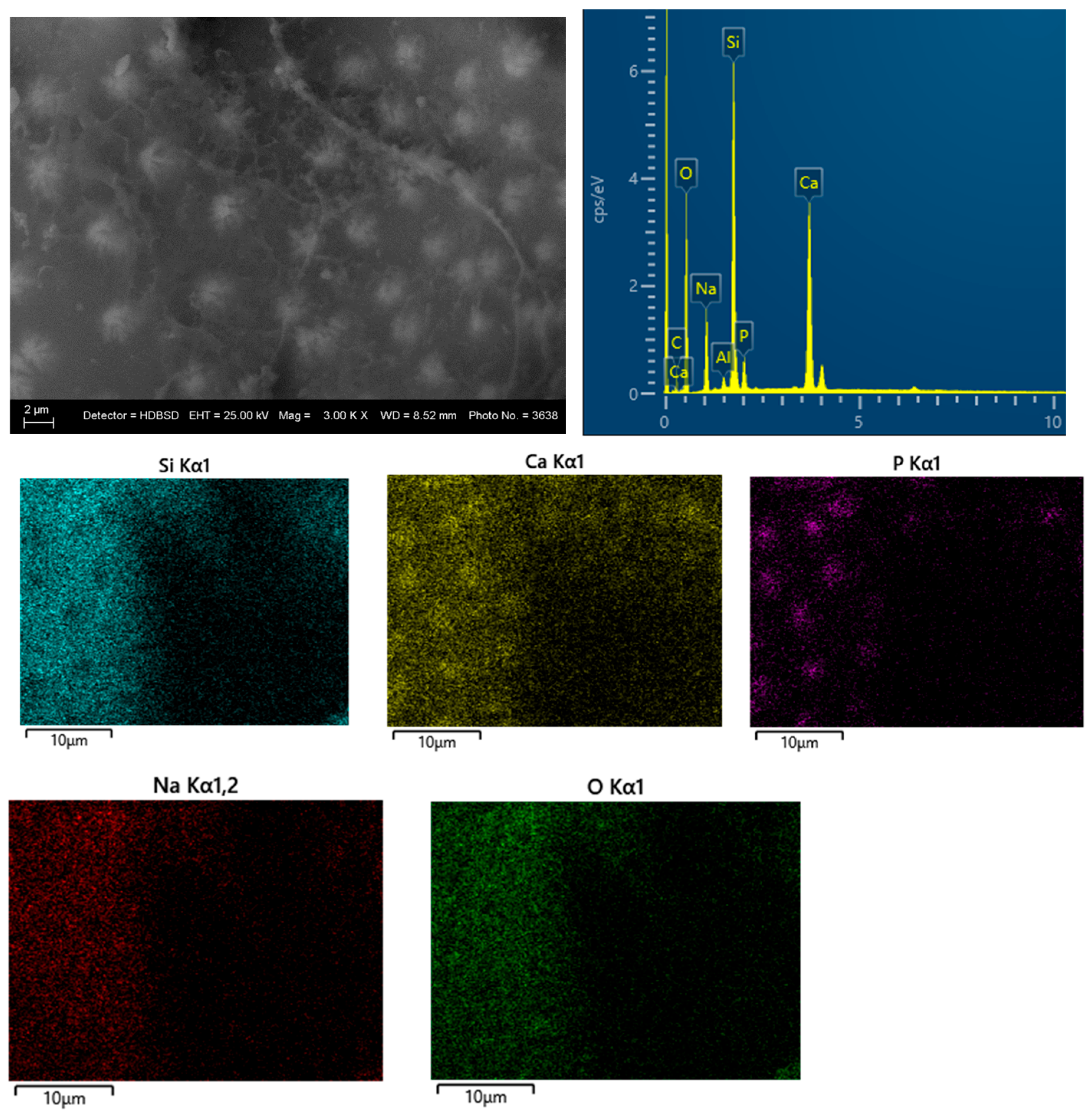
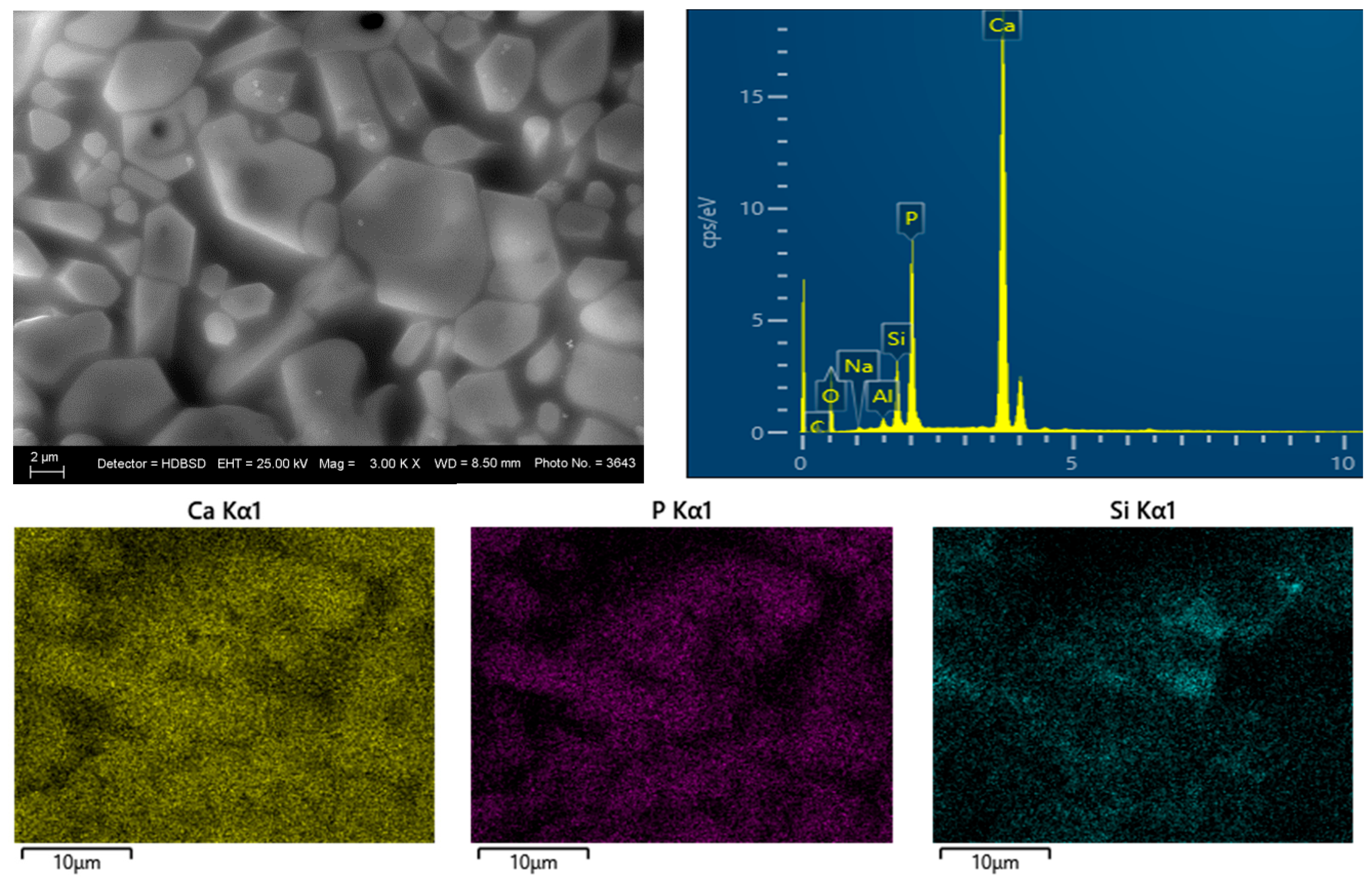
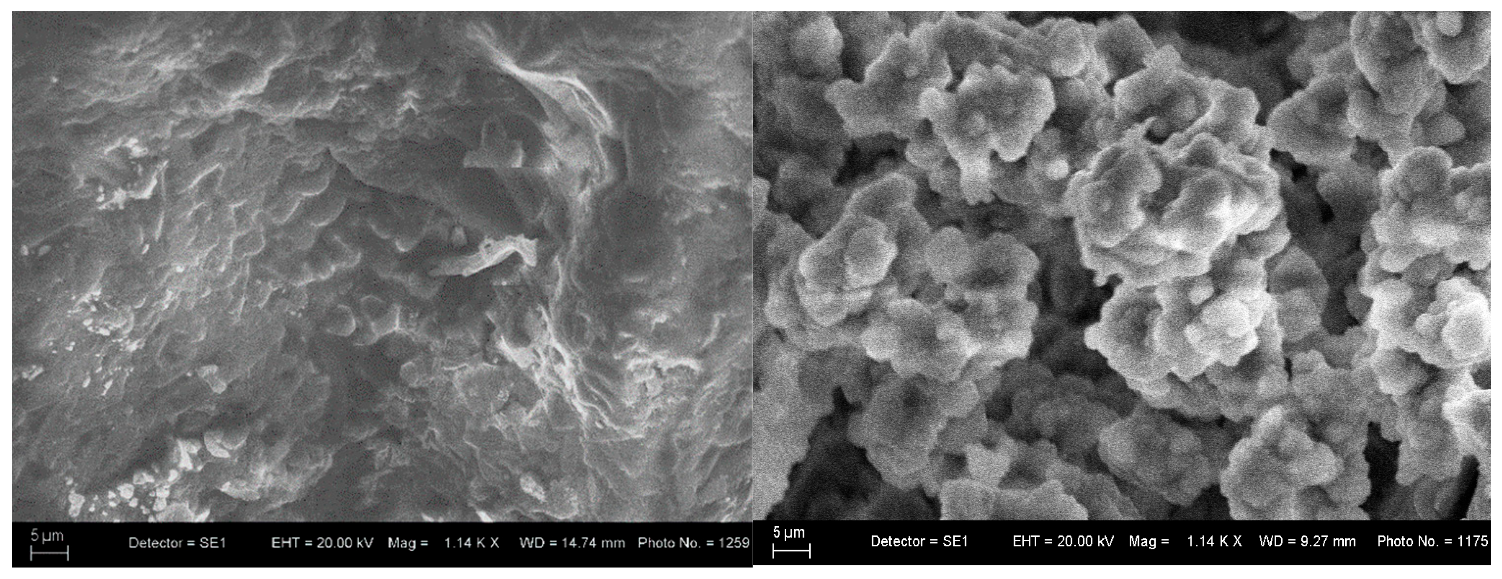
SEM and EDS analyses of the glass–ceramic materials with biogenic hydroxyapatite confirm the results of XRD and FTIR analyses. The results from SEM and EDS analyses of glass–ceramic samples with composition 3 are presented in Figure 8. Uniformly distributed hydroxyapatite and Na3Ca6(PO4)5 are observed in the amorphous matrix. SEM and EDS of ceramic samples with composition 5 are presented in Figure 9. The SEM image of the sample with composition 5 shows hexagonal crystals of hydroxyapatite with a size of 10 µm, sintered with the aid of the glass additive.
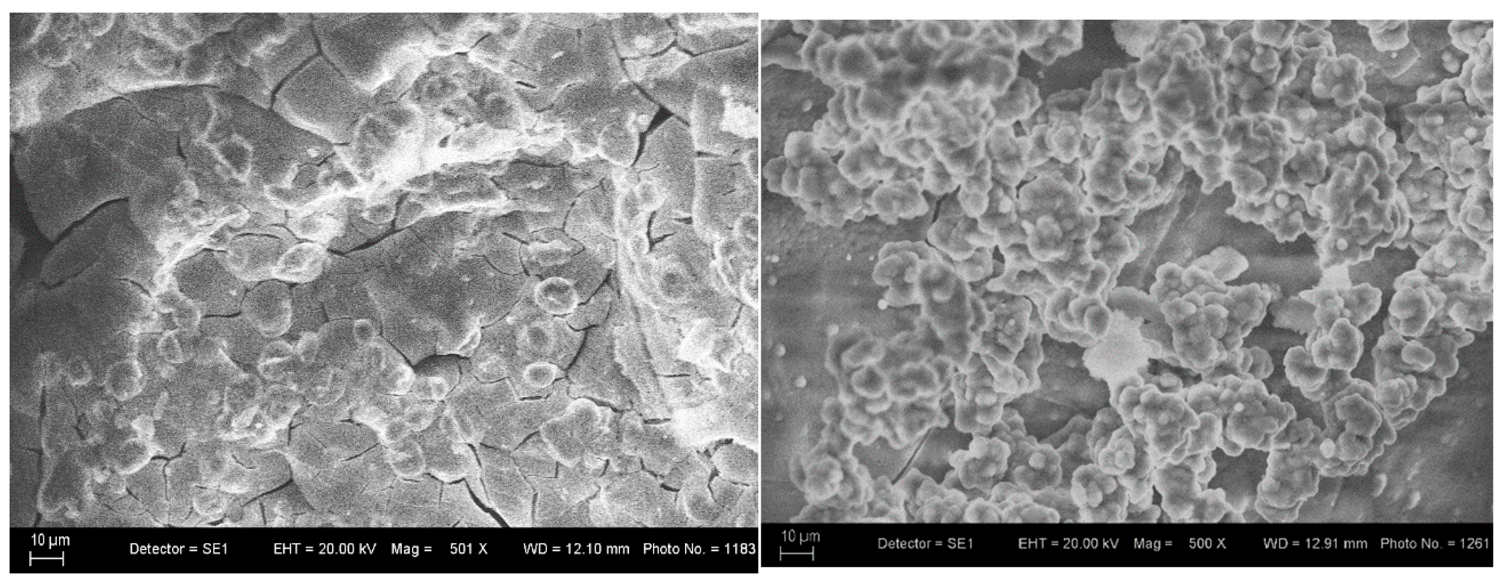
Figure 10 shows the SEM of the glass–ceramic sample with composition 3 after 10 and 20 days in the SBF solution. Figure 10 shows that the glass–ceramic with composition 3 has good bioactivity, since a new powdery layer of apatite is deposited on the glass–ceramic surface from the SBF solution after a stay of 20 days. The behavior of the hydroxyapatite ceramics (Composition 5, Figure 11) is similar. The SEM images of the glass–ceramic and hydroxyapatite ceramic (Figure 10 and Figure 11) illustrate the changes in surface morphology of the samples and hydroxyapatite crystals, which are covered by a layer from apatite after the soaking of the samples for 20 days in SBF solution.
The main glass-forming oxide in composition 1 is B2O3, and the main crystalline phase, which is observed, together with the amorphous one, is hydroxyapatite [Ca10(PO4)6(OH)2]. The addition of 20% by weight at the expense of B2O3 and Na2O does not result in a change in the phase (composition 2). In contrast, in composition 3 (45 wt% SiO2) the main phase is Na3Ca6(PO4)5. This shows that with the same amount of BHA, but with a change in the glass-forming oxide in glass–ceramics (compositions 1–3), the formation of two different crystalline phases is observed. Composition 3 has good bioactivity, since a new powdery layer of apatite is deposited on the glass–ceramic surface by the SBF solution after a stay of 20 days.

4. Conclusions

A new bioactive glass–ceramic was successfully developed by the melting at 1200 °C and the rapid quenching in water of batches combining 30 mass % biogenic hydroxyapatite (BHA), synthesized from Rapana venosa shells and monocalcium phosphate monohydrate [Ca(H2PO4)2·H2O] via solid-state synthesis at 1180 °C, and SiO2, B2O3, and Na2O. The resulting glass–ceramics contain both an amorphous phase and crystalline phases of hydroxyapatite [Ca10(PO4)6(OH)2] and a newly formed, finely dispersed Na3Ca6(PO4)5 phase. With an increase in the content of SiO2 at the expense of B2O3 (composition 3) the quantity of the finely dispersed Na3Ca6(PO4)5 crystalline phase increases and that of hydroxyapatite decreases as a result of the partial dissolution of hydroxyapatite into the glass matrix, followed by crystallization during cooling. After immersing for 20 days in simulated body fluid (SBF), SEM analysis confirmed morphological transformations and that an apatite layer forms on the surface of the glass–ceramic. The main structural units in the glass–ceramic—SiO4, PO4, and BO3—are interconnected via Si–O–Si, B–O–B, P–O–P, and mixed Si–O–Al bonds.
A dense hydroxyapatite ceramic was also produced using a glass-based sintering additive (35 mass % B2O3, 45 mass % SiO2, 10 mass % Al2O3, 10 mass % Na2O) to the biogenic hydroxyapatite. SEM analysis of composition 5 revealed well-formed hexagonal hydroxyapatite crystals (~10 µm). The borosilicate glass facilitated densification via viscous flow and stabilized the hydroxyapatite phase against thermal decomposition by encapsulating it in the glass matrix. After immersion in simulated body fluid (SBF) for 10 and 20 days, SEM analysis confirmed morphological transformations and the formation of an apatite layer on the ceramic surface, indicating bioactivity through ion exchange with the SBF.
Further structural, mechanical, and biological analyses will be performed. The resulting glass–ceramic containing BHA will be tested in future studies for potential application as a biomaterial in dental medicine. As for the sintered dense ceramic, our research will continue in the field of mechanical and biological studies of the resulting ceramic for its potential application in regenerative medicine.

Author Contributions

Conceptualization, A.Y. and T.T.; methodology, A.Y. and T.T.; investigation, A.Y., T.T., J.M. and H.G.; resources, A.Y., T.T., J.M. and H.G.; writing—original draft preparation, A.Y., T.T., J.M. and H.G.; writing—review and editing, A.Y. and T.T.; supervision, A.Y. All authors have read and agreed to the published version of the manuscript.

Funding

This research was funded by contract No.: BG-RRP 2.004-0002-C01, project name: BiOrgaMCT, Procedure BG-RRP-2.004 “Establishing of a network of research higher education institutions in Bulgaria”, funded by BULGARIAN NATIONAL RECOVERY AND RESILIENCE PLAN.

Institutional Review Board Statement

Not applicable.

Informed Consent Statement

Not applicable.

Data Availability Statement

The original contributions presented in this study are included in the article. Further inquiries can be directed to the corresponding author.

Conflicts of Interest

The authors declare no conflicts of interest.

References

  1. Habraken, W.; Habibovic, P.; Epple, M.; Bohner, M. Calcium phosphates in biomedical applications: Materials for the future? Mater. Today 2016, 19, 69–87. [Google Scholar] [CrossRef]
  2. Prakasam, M.; Locs, J.; Salma-Ancane, K.; Loca, D.; Largeteau, A.; Berzina-Cimdina, L. Fabrication, properties, and applications of dense hydroxyapatite: A review. J. Funct. Biomater. 2015, 6, 1099–1140. [Google Scholar] [CrossRef] [PubMed]
  3. Workie, A.B.; Shih, S.J. A study of bioactive glass–ceramic’s mechanical properties, apatite formation, and medical applications. RSC Adv. 2022, 12, 23143–23152. [Google Scholar] [CrossRef] [PubMed]
  4. Pinchuk, N.; Parkhomey, O.; Sych, O. In vitro investigation of bioactive glass-ceramic composites based on biogenic hydroxyapatite or synthetic calcium phosphates. Nanoscale Res. Lett. 2017, 12, 111. [Google Scholar] [CrossRef] [PubMed]
  5. Forero-Sossa, P.A.; Olvera-Alvarez, I.U.; Salazar-Martinez, J.D.; Espinosa-Arbelaez, D.G.; Segura-Giraldo, B.; Giraldo-Betancur, A.L. Biogenic hydroxyapatite powders: Effects of source and processing methodologies on physicochemical properties and bioactive response. Mater. Charact. 2021, 173, 110950–110964. [Google Scholar] [CrossRef]
  6. Fiume, E.; Magnaterra, G.; Rahdar, A.; Vernéand, E.; Baino, F. Hydroxyapatite for Biomedical Applications: A Short Overview. Ceramics 2021, 4, 542–563. [Google Scholar] [CrossRef]
  7. Antoniac, I.; Lesci, I.G.; Blajan, A.I.; Vitioanu, G.; Antoniac, A. Bioceramics and biocomposites from marine sources. Key Eng. Mater. 2015, 672, 276–292. [Google Scholar] [CrossRef]
  8. Cursaru, L.M.; Iota, M.; Piticescu, R.M.; Tarnita, D.; Savu, S.V.; Savu, I.D.; Dumitrescu, G.; Popescu, D.; Hertzog, R.-G.; Calin, M. Hydroxyapatite from Natural Sources for Medical Applications. Materials 2022, 15, 5091. [Google Scholar] [CrossRef]
  9. Bellucci, D.; Sola, A.; Anesi, A.; Salvatori, R.; Chiarini, L.; Cannillo, V. Bioactive glass/hydroxyapatite composites: Mechanical properties and biological evaluation. Mater. Sci. Eng. C 2015, 51, 196–205. [Google Scholar] [CrossRef]
  10. Elbadawi, M.; Wally, Z.J.; Reaney, I.M. Porous hydroxyapatite-bioactive glass hybrid scaffolds fabricated via ceramic honeycomb extrusion. J. Am. Ceram. Soc. 2018, 101, 3541–3556. [Google Scholar] [CrossRef]
  11. Romez-Vega, J.M.; Saiz, E.; Tomsia, A.P. Glass–hydroxyapatite coatings on titanium-based implants. In Bioceramics: Materials and Applications III, 1st ed.; George, L., Rusin, R.P., Fischman, G.S., Janas, V., Eds.; Wiley-American Ceramic Society: Hoboken, NJ, USA, 2000; Volume III, pp. 15–21. [Google Scholar]
  12. Queiroz, A.C.; Santos, J.D.; Monteiro, F.J.; Prado da Silva, M.H. Dissolution studies of hydroxyapatite and glass-reinforced hydroxyapatite ceramics. Mater. Charact. 2003, 50, 197–202. [Google Scholar] [CrossRef]
  13. Rey, C. Calcium phosphate biomaterials and bone mineral. Difference in composition, structure, and properties. Biomaterials 2000, 11, 5–13. [Google Scholar]
  14. Georgiou, G.; Knowles, J.C. Glass-reinforced hydroxyapatite for hard tissue surgery—Part 1. Biomaterials 2001, 22, 2811–2815. [Google Scholar] [CrossRef]
  15. Vargas-Becerril, N.; Sánchez-Téllez, D.A.; Zarazúa-Villalobos, L.; González-García, D.M.; Álvarez-Pérez, M.A.; de León-Escobedo, C.; Téllez-Jurado, L. Structure of biomimetic apatite grown on hydroxyapatite (HA). Ceram. Int. 2020, 46, 28806–28813. [Google Scholar] [CrossRef]
  16. Bhatt, H.A. Synthesis and Characterization of Nanocrystalline Hydroxyapatite Powder; and the Effects of Oxide-Based Sintering Additives on Tricalcium Phosphate. Master’s Thesis, University of Central Florida Orlando, Orlando, FL, USA, 2005. Available online: https://stars.library.ucf.edu/etd/432 (accessed on 24 June 2025).
  17. Yoleva, A.; Mihailova, I.; Djambazov, S. Solid-state synthesis of hydroxyapatite from Black Sea Rapana venosa shells. J. Chem. Technol. Metall. 2023, 58, 385–393. [Google Scholar] [CrossRef]
  18. Mateeva, J.; Yoleva, A.; Djambazov, S. Phase study of Rapana venosa shells from the Black Sea. J. Chem. Technol. Metall. 2023, 58, 38–43. [Google Scholar]
  19. Kokubo, T.; Takadama, H. How useful is SBF in predicting in vivo bone bioactivity? Biomaterials 2006, 27, 2907–2915. [Google Scholar] [CrossRef]
  20. Demirkiran, H.; Mohandas, A.; Dohi, M.; Fuentes, A.; Nguyen, K.; Aswath, P. Bioactivity and mineralization of hydroxyapatite with bioglass as sintering aid and bioceramics with Na3Ca6(PO4)5 and Ca5(PO4)2SiO4 in a silicate matrix. Mater. Sci. Eng. C 2010, 30, 263–272. [Google Scholar] [CrossRef]
  21. Wojciech, S.; Masatomo, Y.; Masato, K.; Masahiro, Y. Hydroxyapatite ceramics with selected sintering additives. Biomoteriols 1997, 18, 923–933. [Google Scholar]
  22. Dimitrov, V.; Dimitriev, Y. Structural Analysis (Spectroscopy), 1st ed.; UCTM: Sofia, Bulgaria, 2010. (In Bulgarian) [Google Scholar]
  23. Koutsopoulos, S. Synthesis and characterization of hydroxyapatite crystals: A review study on the analytical methods. J. Biomed. Mater. Res. 2002, 62, 600–612. [Google Scholar] [CrossRef] [PubMed]
  24. Mačković, M.; Hoppe, A.; Detsch, R.; Mohn, D.; Stark, W.J.; Spiecker, E.; Boccaccini, A.R. Bioactive glass (type 45S5) nanoparticles: In vitro reactivity on nanoscale and biocompatibility. J. Nanopart. Res. 2012, 14, 966. [Google Scholar] [CrossRef]
  25. Yoleva, A.; Tasheva, T.; Djambazov, S.; Batsova, A. Development of multicomponent glasses for application as a glazing layer on dental zirconia. Int. J. Appl. Glass Sci. 2025, 16, e16684. [Google Scholar] [CrossRef]
  26. Tasheva, T.; Valova, G. FT-IR and Raman spectroscopy of ZnO and MgO containing glasses in the B2O3/Na2O/CaO/P2O5 system. J. Chem. Technol. Metall. 2024, 59, 1391–1398. [Google Scholar] [CrossRef]
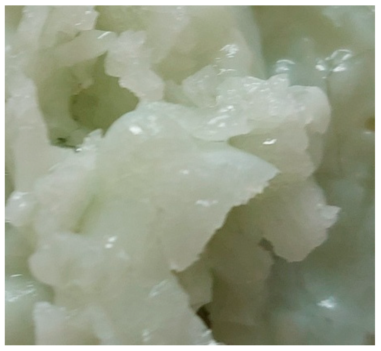
Figure 1. The sample with composition 3, melted at 1200 °C and melt-quenched in water.
Materials 18 03059 g001
Figure 2. Sample with composition 5 pressed on a hydraulic press at 50 MPa and fired for 2 h at 1200 °C.
Materials 18 03059 g002
Figure 3. XRD patterns of the sample with composition 2 melted at 1200 °C and melt-quenched in water.
Materials 18 03059 g003
Figure 4. XRD of the sample with composition 3 melted at 1200 °C and melt-quenched in water.
Materials 18 03059 g004
Figure 5. XRD of the sample with composition 5 pressed on a hydraulic press at 50 MPa and fired for 2 h at 1200 °C.
Materials 18 03059 g005
Figure 6. FTIR spectra of the samples.
Materials 18 03059 g006
Figure 7. FTIR spectra of Sample 3: before and after 20 days in SBF (top) and Sample 5: before and after 20 days in SBF (bottom).
Materials 18 03059 g007
Figure 8. SEM and EDS of glass–ceramic sample with composition 3.
Materials 18 03059 g008
Figure 9. SEM and EDS of the ceramic sample with composition 5.
Materials 18 03059 g009aMaterials 18 03059 g009b
Figure 10. SEM glass–ceramic sample with composition 3 after immersion in SBF for 10 and 20 days.
Materials 18 03059 g010
Figure 11. SEM of the ceramic sample with composition 5 after immersion in SBF for 10 and 20 days.
Materials 18 03059 g011
Table 1. Compositions of the studied biogenic hydroxyapatite samples.
Sample No.Composition, Mass %Density,
g/cm3
SiO2Na2OB2O3BHA
Powder
1-2050302.48 ± 0.04
2201535302.50 ± 0.04
3351025302.54 ± 0.04
450 mass % powdered glass with the following composition in mass %: 35 B2O3, 45 SiO2, 10 Al2O3, 10 Na2O502.60 ± 0.04
525 mass % powdered glass with the following composition in mass %: 35 B2O3, 45 SiO2,10 Al2O3, 10 Na2O752.70 ± 0.04
Table 2. IR spectroscopy data for the samples, bands, and assignments.
Peak, cm−1AssignmentReferences
2003, 19302.ν3 harmonic overtone or to a combination mode, ν1 + ν3[23]
1468–1442Stretching mode (ν1) of the CO32− group in A-type CAP or bending mode (ν4 or ν3) of the CO32− group in A and B-type CAP[25]
1354–1395Asymmetric B–O stretching vibration of borate triangular units BO3[24]
1021–1031Triple degenerate stretching antisymmetric vibration νdas(F) of PO4[17,22,23]
960Symmetric stretching vibration νs(A1) of PO4[22,23]
850Stretching mode of the CO32− group in CAP[23]
709
699, 746
Overlapping of bending vibrations δ of B–O–B in [BO3] and symmetric stretching vibrations νs of Si–O–Al [24,25,26]
564
600, 564
605, 569, 544
Triple degenerative bending δd(F) of PO4[17,22,23]
470–480Overlapping of double degenerated bending vibrations δd(E) of PO4 and SiO4 tetrahedra [23,24]
454Bending vibration δ of Si–O–Si of connected [SiO4] units[24]
Disclaimer/Publisher’s Note: The statements, opinions and data contained in all publications are solely those of the individual author(s) and contributor(s) and not of MDPI and/or the editor(s). MDPI and/or the editor(s) disclaim responsibility for any injury to people or property resulting from any ideas, methods, instructions or products referred to in the content.