The Impact of Eye Dominance on Fixation Stability in School-Aged Children
Abstract
:Introduction
Methods
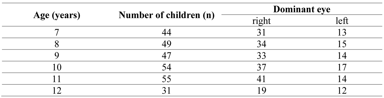
Participants
Procedure
Data analysis
Statistical Analysis
Results
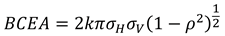
Fixation Stability in the Dominant and Non-dominant Eyes in Each Age Group
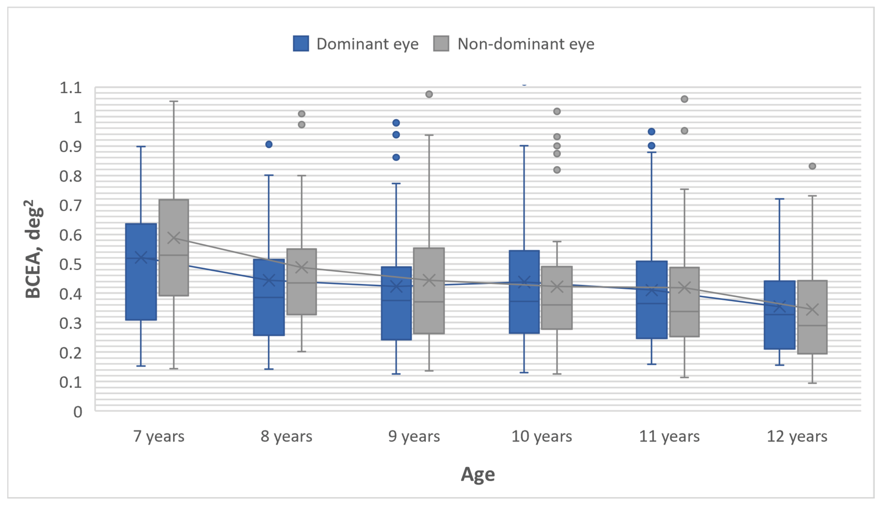
Fixation Stability in the Right and Left Eyes in Each Age Group
Discussion
Conclusion
Ethics and Conflict of Interest
Acknowledgments
References
- Aizenman, A. M., and D. M. Levi. 2021. Fixational stability as a measure for the recovery of visual function in amblyopia. Proceedings. Eye Tracking Research & Applications Symposium 2021: 10. [Google Scholar] [CrossRef]
- Altemir, I., A. Alejandre, A. Fanlo-Zarazaga, M. Ortín, T. Pérez, B. Masiá, and V. Pueyo. 2021. Evaluation of fixational behavior throughout life. Brain sciences 12, 1: 19. [Google Scholar] [CrossRef]
- Aring, E., M. A. Grönlund, A. Hellström, and J. Ygge. 2007. Visual fixation development in children. Graefe's archive for clinical and experimental ophthalmology 245, 11: 1659–1665. [Google Scholar] [CrossRef] [PubMed]
- Bigelow, E. R., and B. E. McKenzie. 1985. Unstable ocular dominance and reading ability. Perception 14, 3: 329–335. [Google Scholar] [CrossRef] [PubMed]
- Carey, D. P. 2001. Vision research: losing sight of eye dominance. Current biology 11, 20: 828–830. [Google Scholar] [CrossRef]
- Crossland, M. D., and G. S. Rubin. 2002. The use of an infrared eyetracker to measure fixation stability. Optometry and vision science 79, 11: 735–739. [Google Scholar]
- Dekker, T. M., M. Farahbakhsh, J. Atkinson, O. J. Braddick, and P. R. Jones. 2020. Development of the spatial contrast sensitivity function (CSF) during childhood: Analysis of previous findings and new psychophysical data. Journal of vision 20, 13: 4. [Google Scholar] [CrossRef]
- González, E. G., A. M. Wong, E. Niechwiej-Szwedo, L. Tarita-Nistor, and M. J. Steinbach. 2012. Eye position stability in amblyopia and in normal binocular vision. Investigative ophthalmology & visual science 53, 9: 5386–5394. [Google Scholar] [CrossRef]
- Hessels, R. S., D. C. Niehorster, C. Kemner, and I. T. C. Hooge. 2017. Noise-robust fixation detection in eye movement data: Identification by two-means clustering (I2MC). Behavior research methods 49, 5: 1802–1823. [Google Scholar] [CrossRef]
- Hu, B., Z. Liu, J. Zhao, L. Zeng, G. Hao, D. Shui, and K. Mao. 2022. The global prevalence of amblyopia in children: a systematic review and meta-analysis. Frontiers in pediatrics 10: 819998. [Google Scholar] [CrossRef]
- Jones, P. R., N. Yasoubi, M. Nardini, and G. Rubin. 2016. Feasibility of macular integrity assessment (MAIA) microperimetry in children: sensitivity, reliability, and fixation stability in healthy observers. Investigative Opthalmology & Visual Science 57, 14: 6349–6359. [Google Scholar] [CrossRef]
- Kim, S. Y., B. Y. Moon, H. G. Cho, and D. S. Yu. 2022. Quantitative evaluation of the association between fixation stability and phoria during short-term binocular viewing. Frontiers in neuroscience 16: 721665. [Google Scholar] [CrossRef] [PubMed]
- Niehorster, D. C., R. Andersson, and M. Nyström. 2020. Titta: A toolbox for creating PsychToolbox and Psychopy experiments with Tobii eye trackers. Behavior research methods 52, 5: 1970–1979. [Google Scholar] [CrossRef]
- Nyong'o, O. L., and M. A. Del Monte. 2008. Childhood visual impairment: normal and abnormal visual function in the context of developmental disability. Pediatric clinics of North America 55, 6: 1403–1415. [Google Scholar] [CrossRef]
- Pueyo, V., J. Yam, T. Perez-Roche, V. Balasanyan, M. Ortin, G. Garcia, E. Prieto, C. Pham, D. Gutiérrez, O. Castillo, B. Masia, A. Alejandre, M. Bakkali, M. Ciprés, E. Esteban-Ibañez, A. Fanlo-Zarazaga, I. Gonzalez, I. Gutiérrez-Luna, X. Pan, and X. Zhang. 2022. Development of oculomotor control throughout childhood: A multicenter and multiethnic study. Journal of vision 22, 13: 4. [Google Scholar] [CrossRef]
- Raveendran, R. N., W. R. Bobier, and B. Thompson. 2019. Binocular vision and fixational eye movements. Journal of vision 19, 4: 9. [Google Scholar] [CrossRef]
- Samet, S., E. G. González, M. S. Mandelcorn, M. H. Brent, and L. Tarita-Nistor. 2018. Changes in fixation stability with time during binocular and monocular viewing in maculopathy. Vision 2, 4: 40. [Google Scholar] [CrossRef] [PubMed]
- Shneor, E., and S. Hochstein. 2005. Effects of eye dominance in visual perception. International congress series 1282: 719–723. [Google Scholar] [CrossRef]
- Subramanian, V., M.R. Jost, and E.E. Birch. 2013. A quantitative study of fixation stability in amblyopia. Investigative ophthalmology & visual science 54, 3: 1998–2003. [Google Scholar] [CrossRef]
- Thaler, L., A.C. Schütz, M.A. Goodale, and K.R. Gegenfurtner. 2013. What is the best fixation target? The effect of target shape on stability of fixational eye movements. Vision research 76: 31–42. [Google Scholar] [CrossRef]
- Ukwade, M. T., and H. E. Bedell. 1993. Stability of oculomotor fixation as a function of target contrast and blur. Optometry and vision science 70, 2: 123–126. [Google Scholar] [CrossRef]
- Vikesdal, G. H., and T. Langaas. 2016. Saccade latency and fixation stability: Repeatability and reliability. Journal of eye movement research 9, 2. [Google Scholar] [CrossRef]
- Wang, Y. Z., S. E. Morale, R. Cousins, and E. E. Birch. 2009. Course of development of global hyperacuity over lifespan. Optometry and vision science 86, 6: 695–700. [Google Scholar] [CrossRef] [PubMed]
- Williams, C. 2009. Amblyopia., 0709. [Google Scholar]

 |
 |
 |
Copyright © 2023. This article is licensed under a Creative Commons Attribution 4.0 International License.
Share and Cite
Serpa, E.; Alecka, M.; Ceple, I.; Krumina, G.; Svede, A.; Kassaliete, E.; Goliskina, V.; Volberga, L.; Berzina, A.; Mikelsone, R.; et al. The Impact of Eye Dominance on Fixation Stability in School-Aged Children. J. Eye Mov. Res. 2023, 16, 1-8. https://doi.org/10.16910/jemr.16.3.6
Serpa E, Alecka M, Ceple I, Krumina G, Svede A, Kassaliete E, Goliskina V, Volberga L, Berzina A, Mikelsone R, et al. The Impact of Eye Dominance on Fixation Stability in School-Aged Children. Journal of Eye Movement Research. 2023; 16(3):1-8. https://doi.org/10.16910/jemr.16.3.6
Chicago/Turabian StyleSerpa, Evita, Madara Alecka, Ilze Ceple, Gunta Krumina, Aiga Svede, Evita Kassaliete, Viktorija Goliskina, Liva Volberga, Asnate Berzina, Rita Mikelsone, and et al. 2023. "The Impact of Eye Dominance on Fixation Stability in School-Aged Children" Journal of Eye Movement Research 16, no. 3: 1-8. https://doi.org/10.16910/jemr.16.3.6
APA StyleSerpa, E., Alecka, M., Ceple, I., Krumina, G., Svede, A., Kassaliete, E., Goliskina, V., Volberga, L., Berzina, A., Mikelsone, R., Ozola, E., Toloka, D., Ruza, T., Klavinska, A., Vasiljeva, S., & Koleda, M. (2023). The Impact of Eye Dominance on Fixation Stability in School-Aged Children. Journal of Eye Movement Research, 16(3), 1-8. https://doi.org/10.16910/jemr.16.3.6