Eye Tracking in Optometry: A Systematic Review

Abstract
:Introduction
Methods
Design
Search Strategy
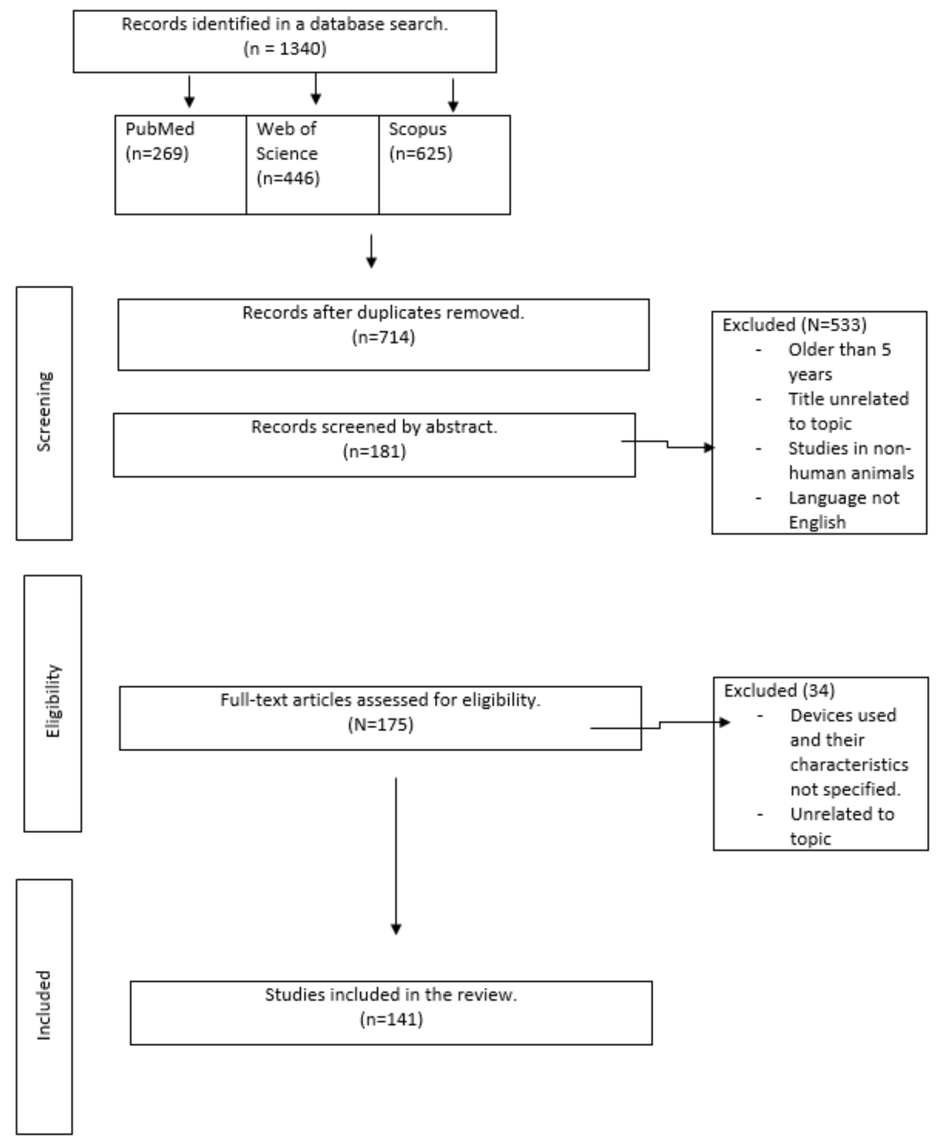
Study Selection
Data Extraction and Quality Assessment
Statistical Analysis
Results
Eye Trackers in the Field of Optometry
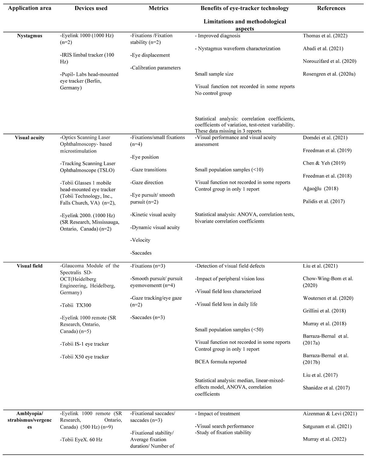
Nystagmus
Visual Acuity
Visual Field
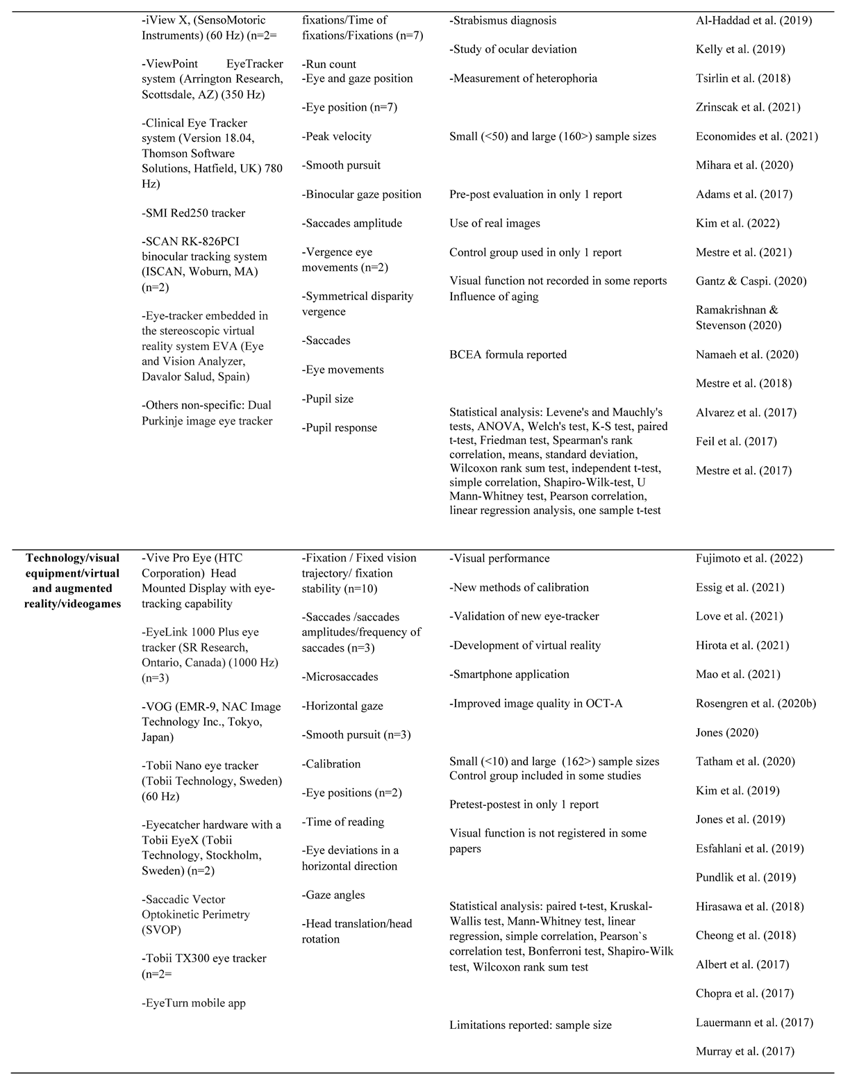
Amblyopia/Strabismus/Vergences
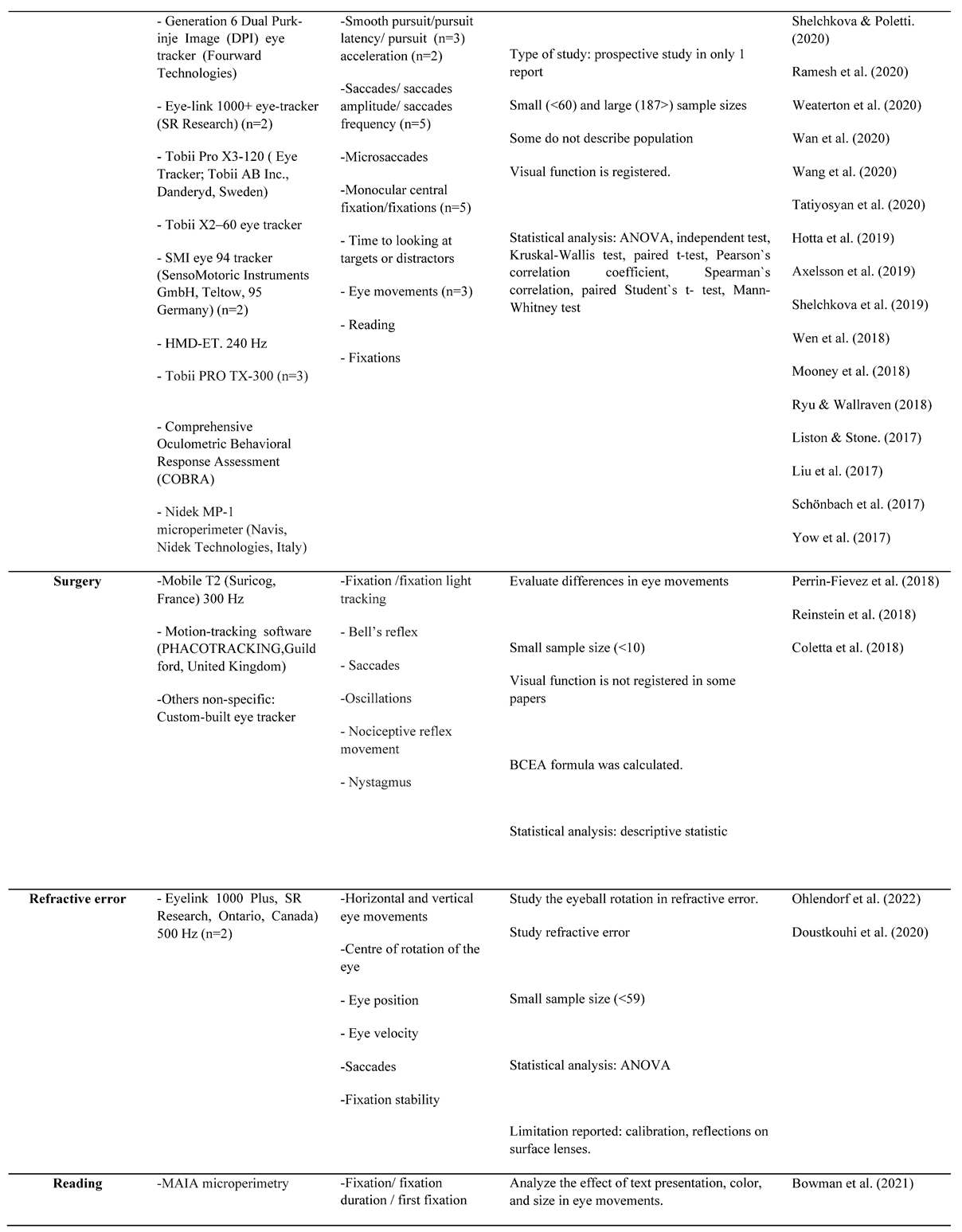
Surgery
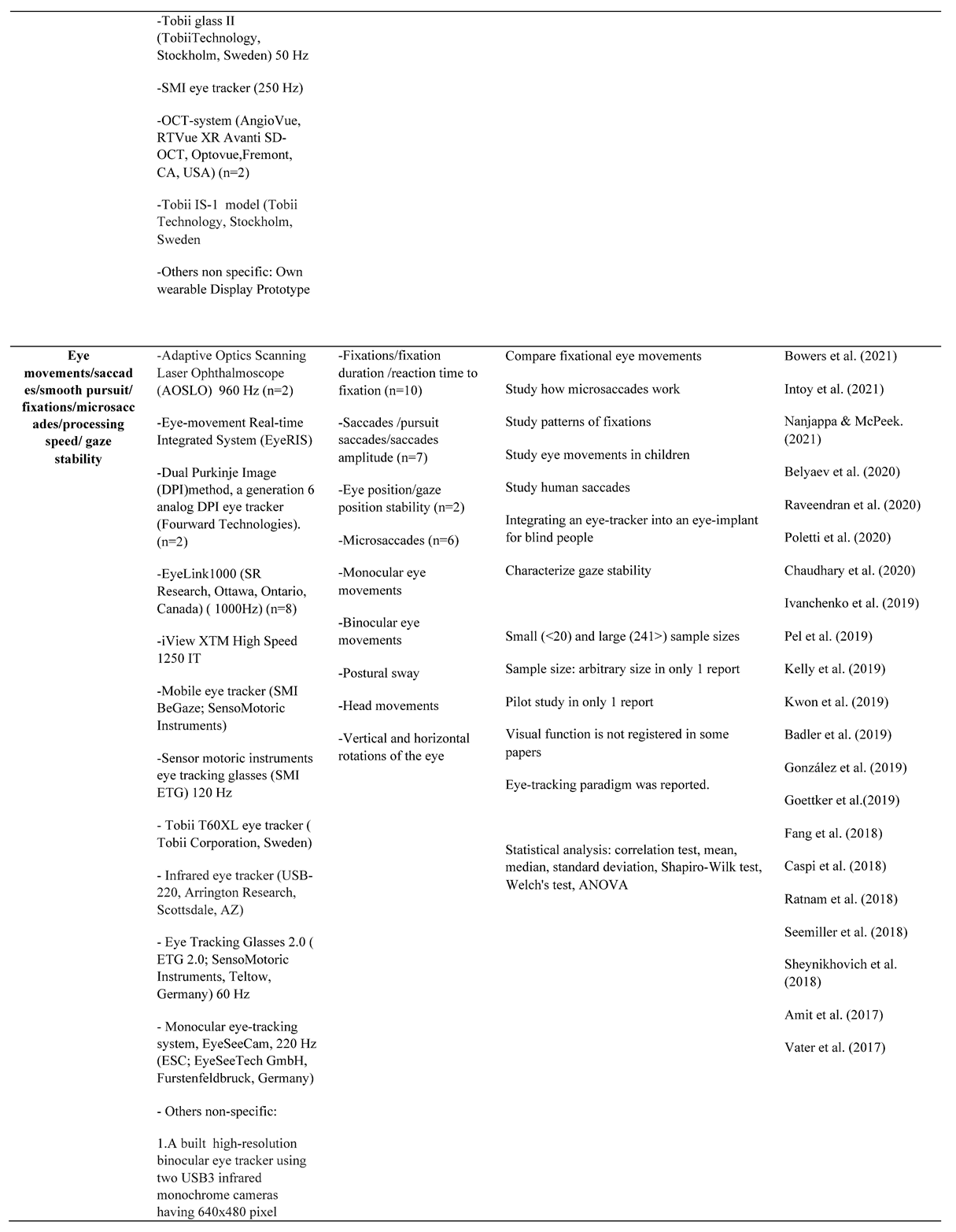
Technology/Visual Equipment/Virtual and Augmented Reality
Ocular Pathology/Low Vision
Assessment/Diagnosis/Rehabilitation/Training
Refractive Error
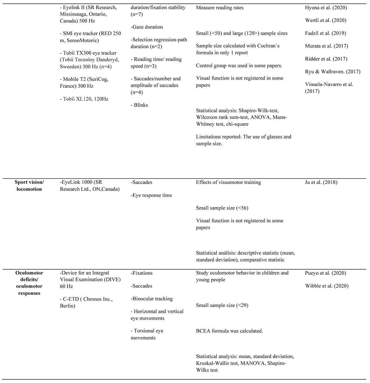
Reading in Optometry Assessment
Sports Vision/Locomotion
Oculomotor Deficits/Oculomotor Responses
General Eye Movements
Methods Used
Main Devices and Their Characteristics.
Stimuli Setups and Recordings
Main Metrics Used and Statistical Analysis
Discussion
Main Areas of Application
Devices Used and Their Characteristics
Main Metrics Used
Methods and Statistical Aspects
Technological Readiness
Conclusions
Ethics and Conflict of Interest
Acknowledgments
Funding sources
Appendix A. Main devices and metrics used in the different areas of optometry

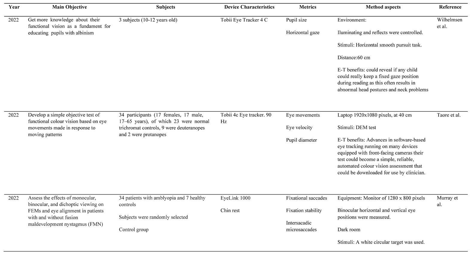
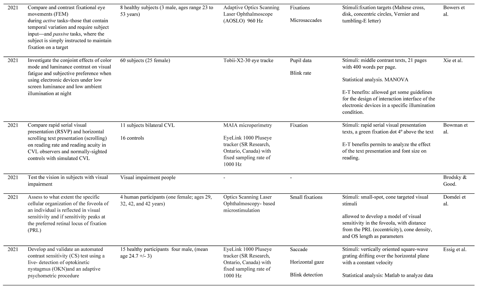
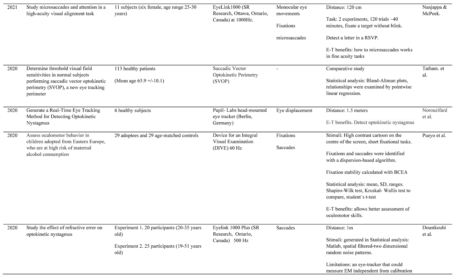
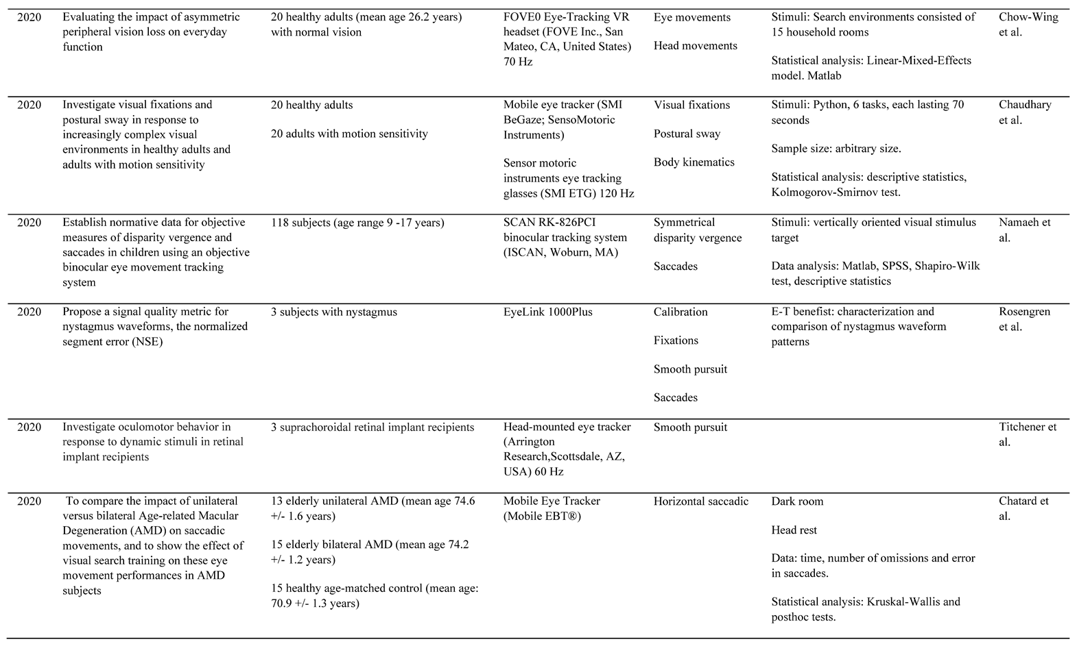
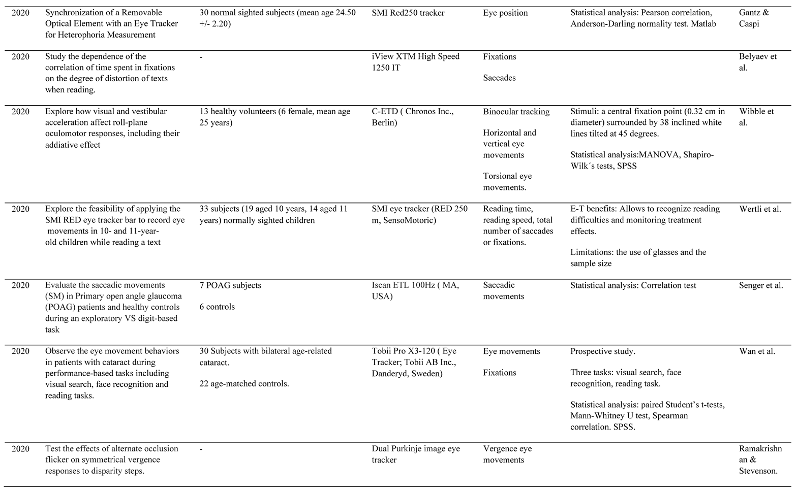
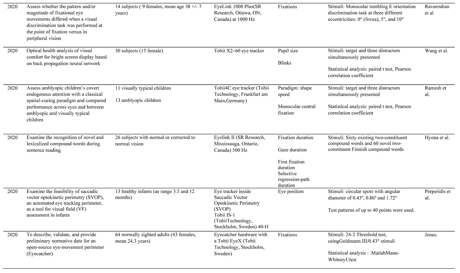
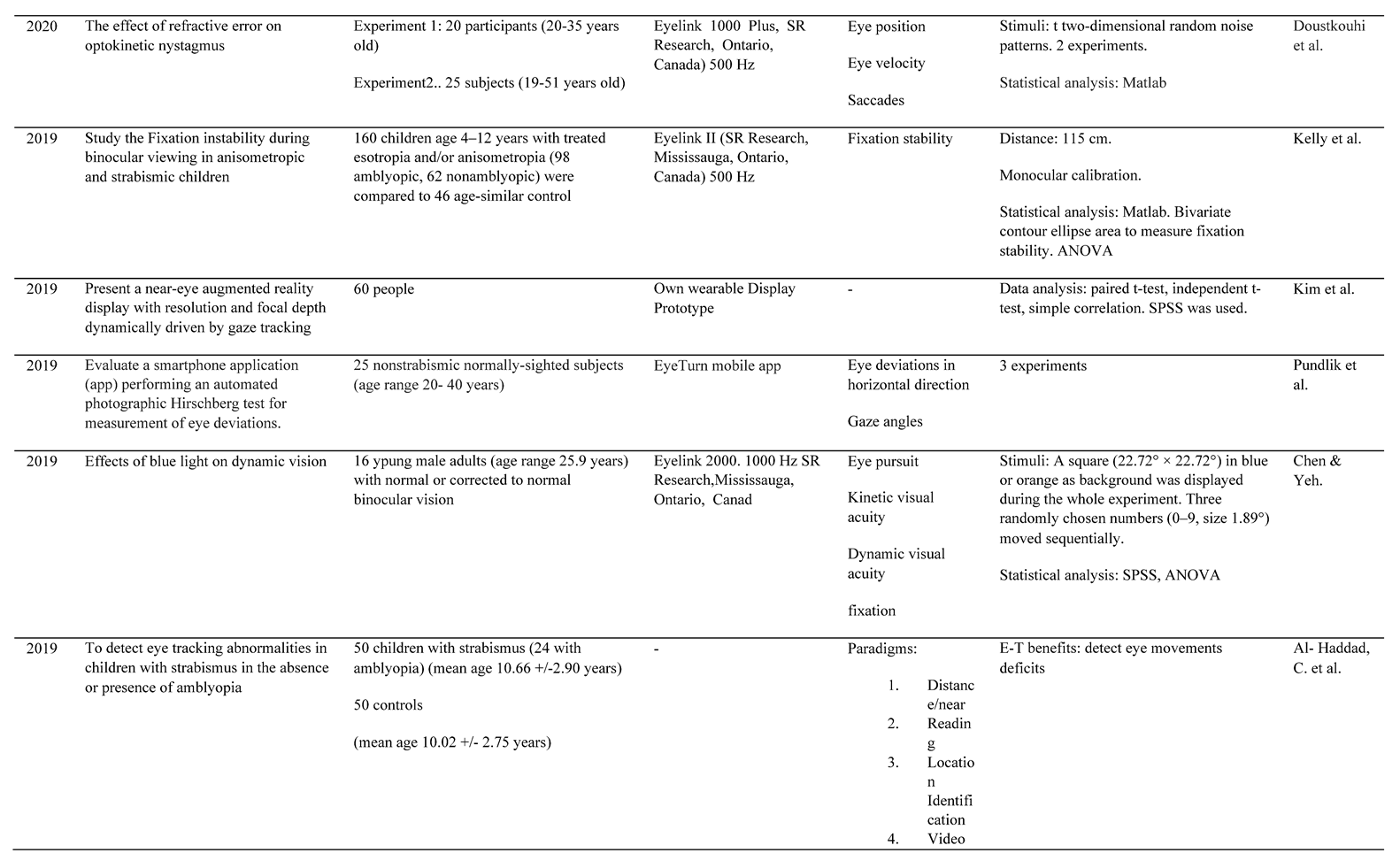
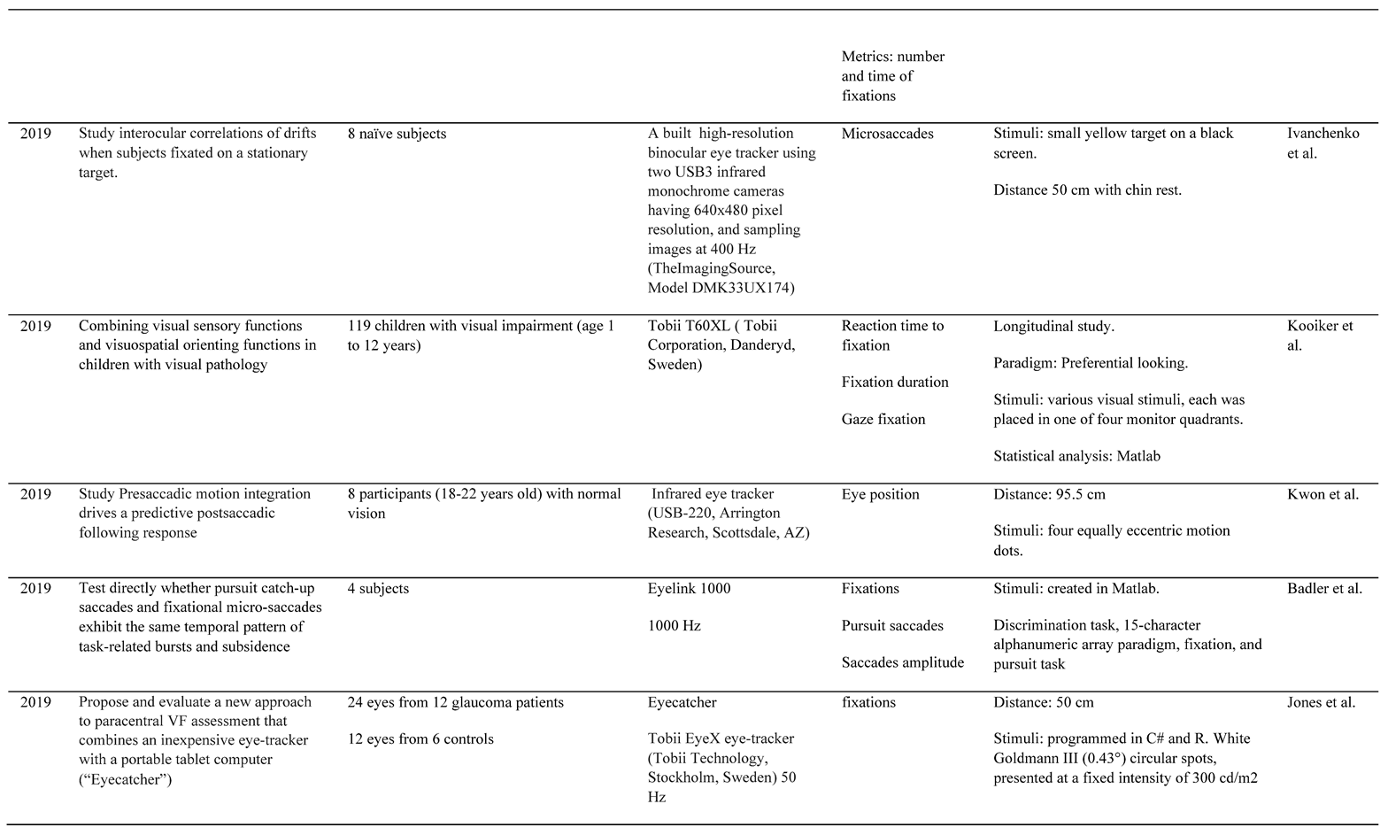
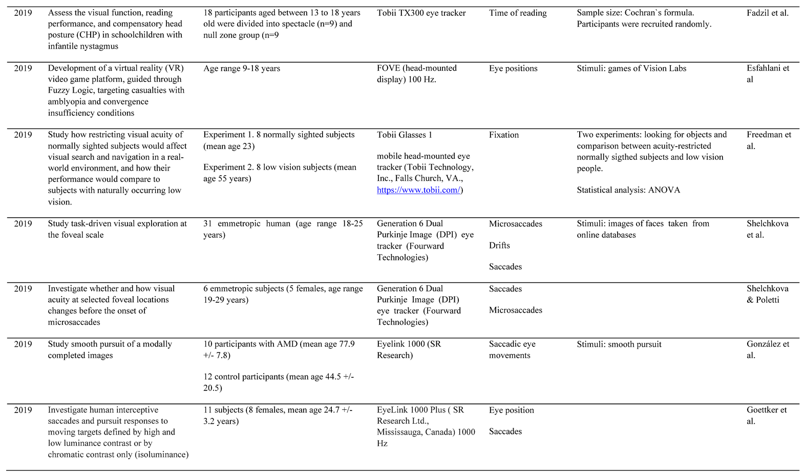
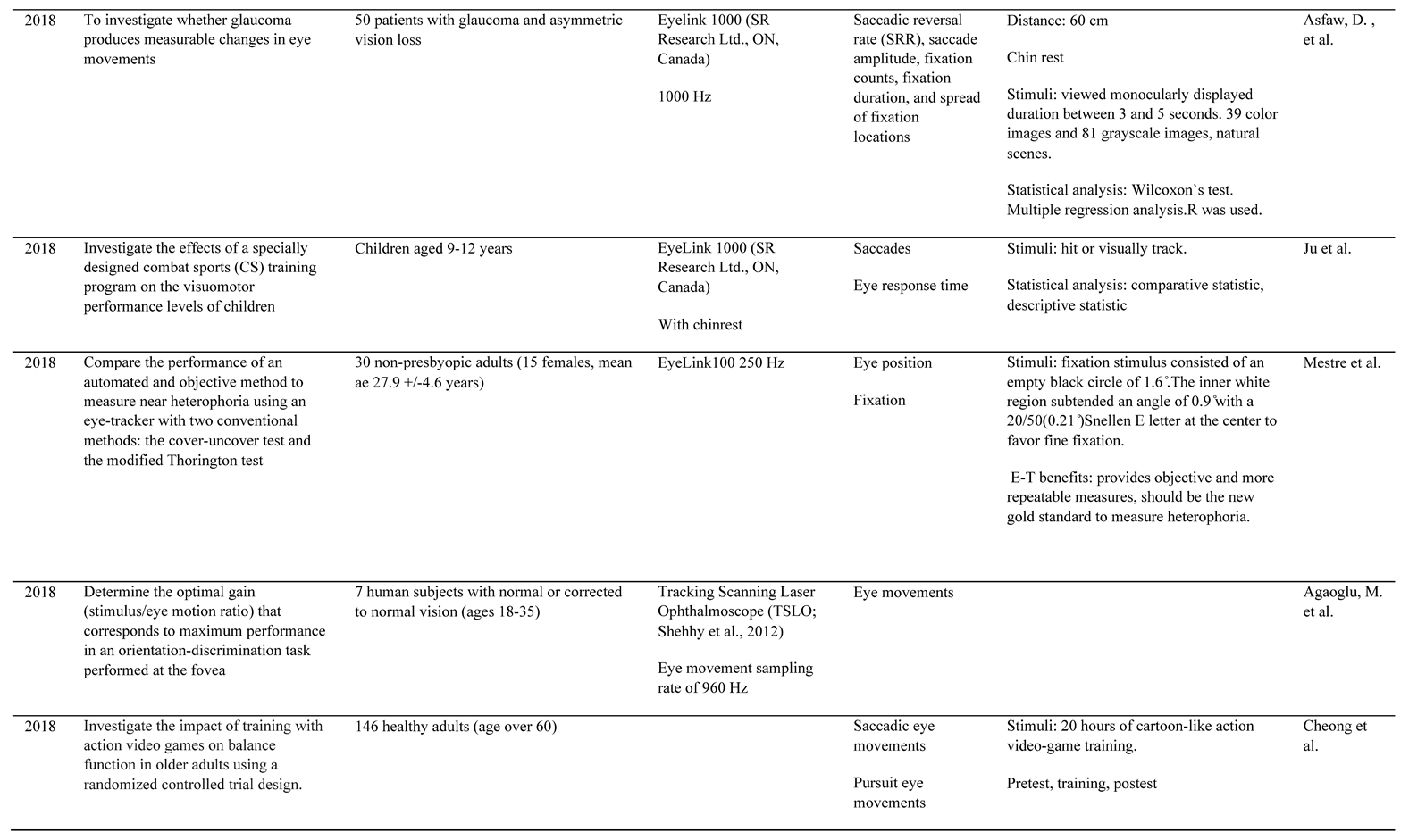
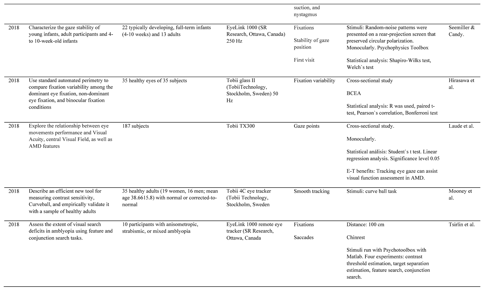
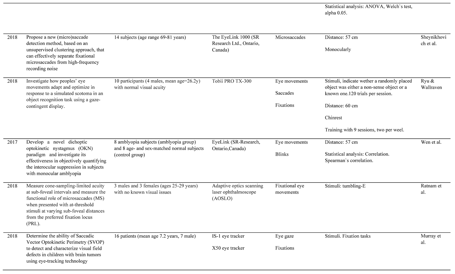
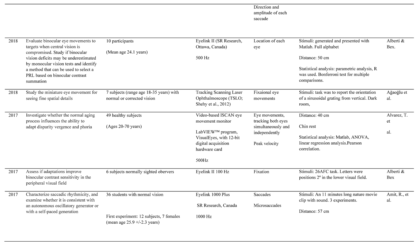
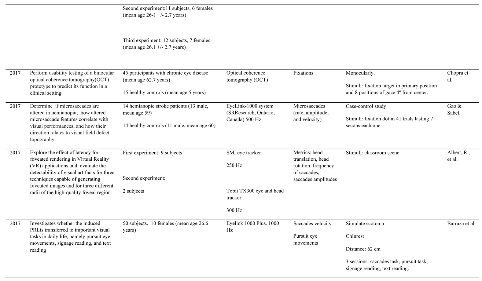
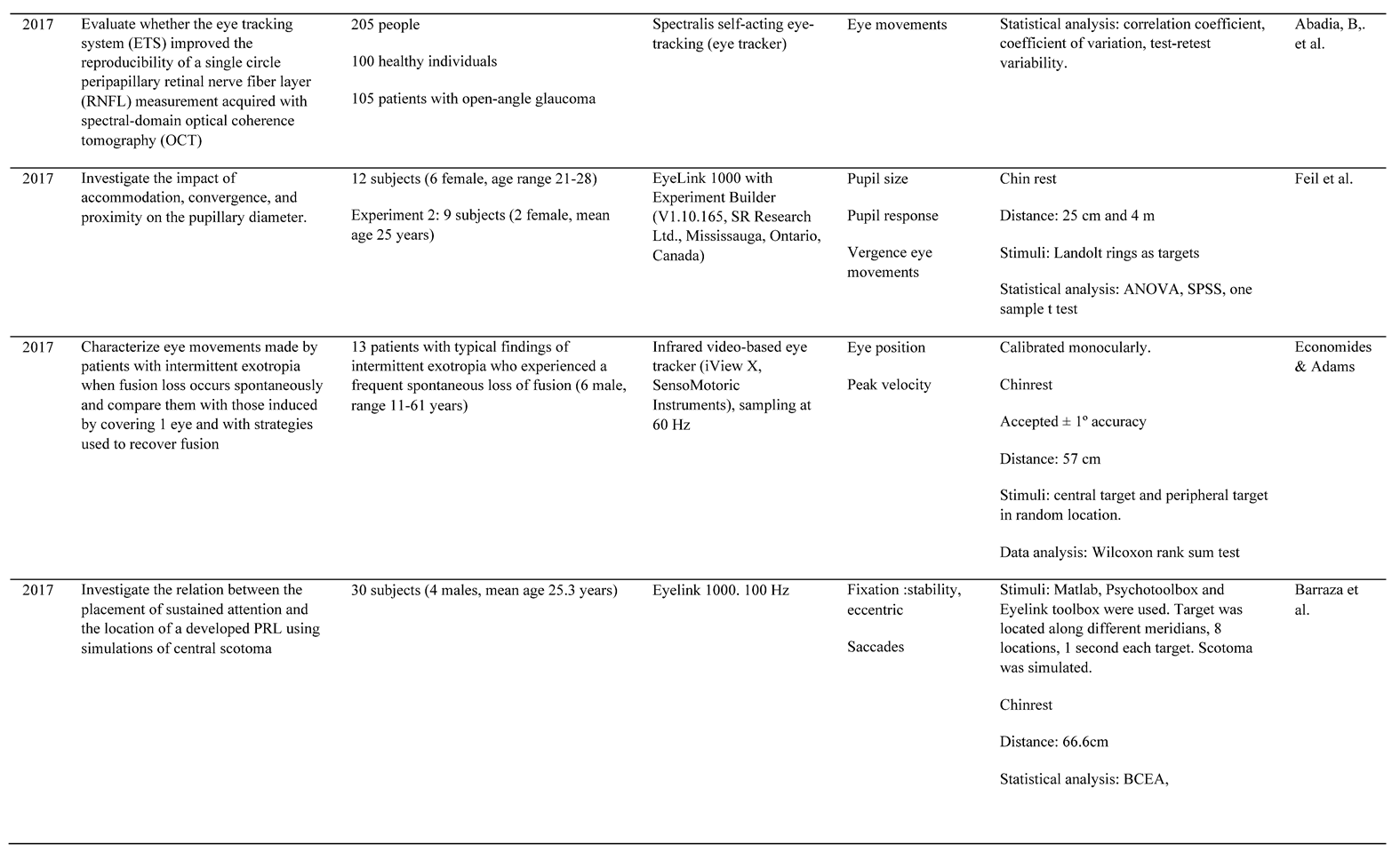
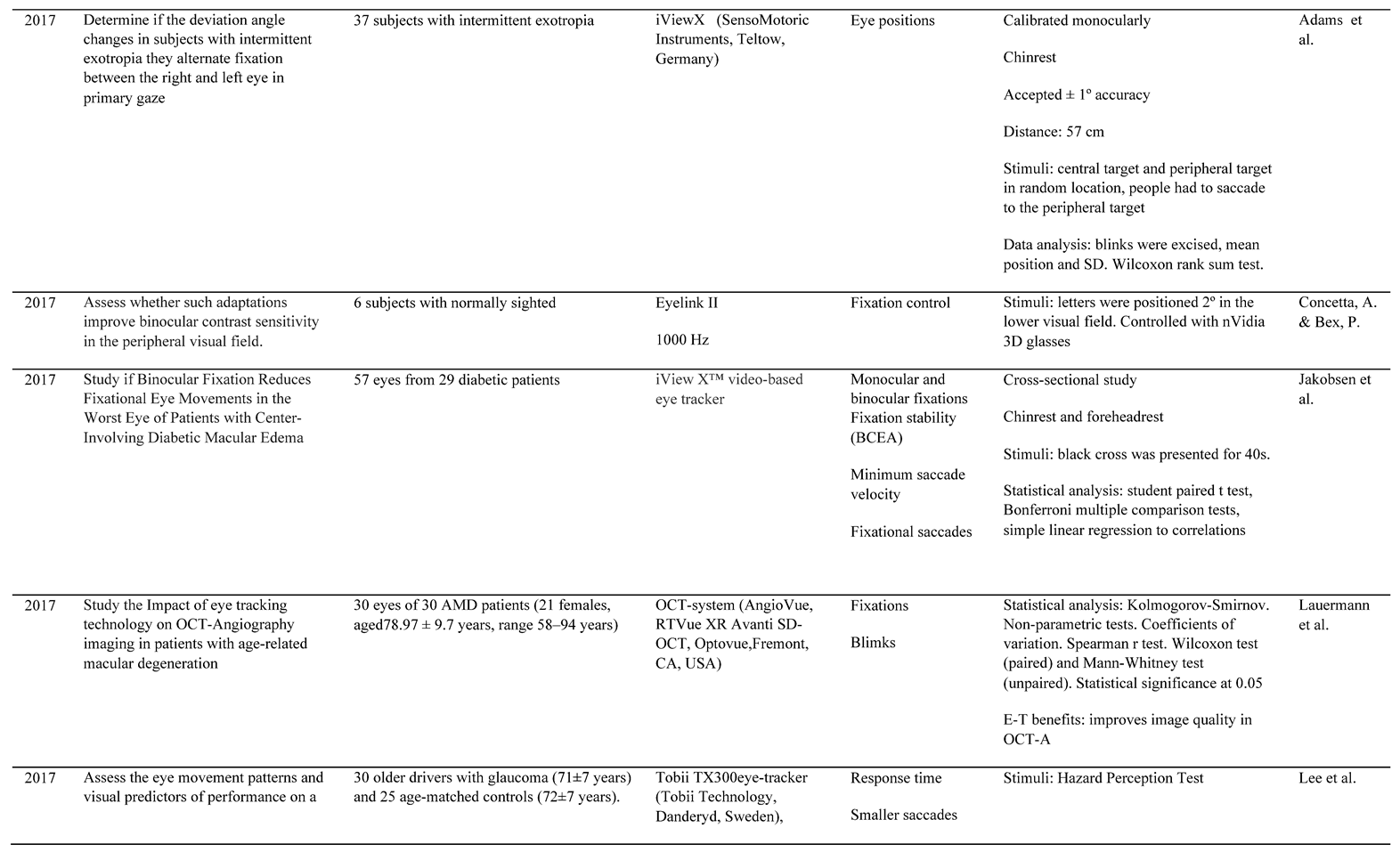
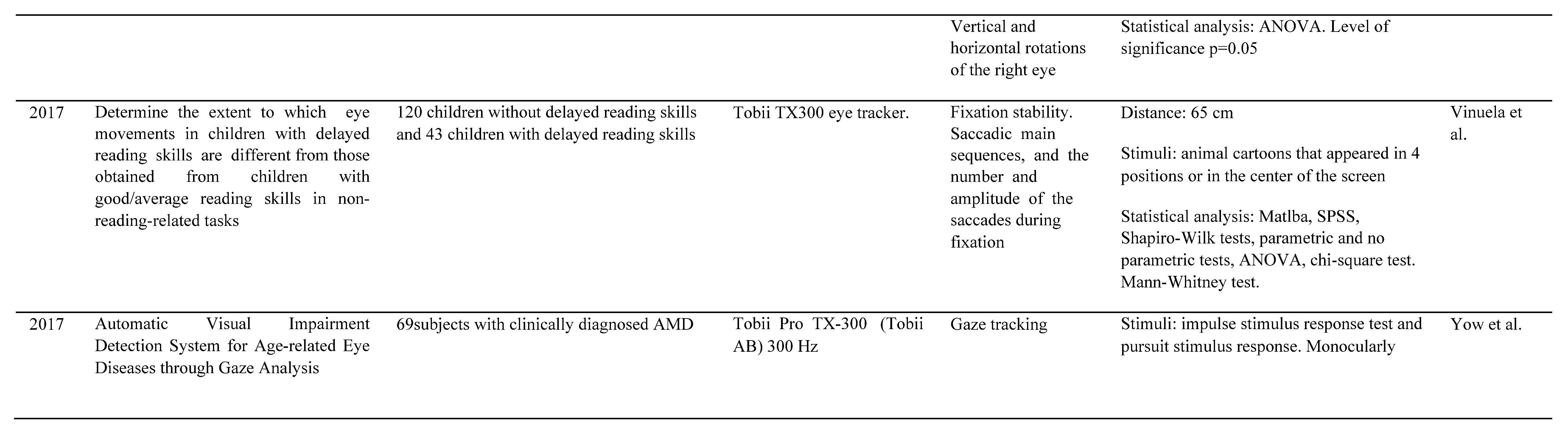
Appendix B. Relevant information of each included study


References
- Abadi, R. v., O. E. Akman, G. E. Arblaster, and R. A. Clement. 2021. Analysing nystagmus waveforms: a computational framework. Scientific Reports 11, 1. [Google Scholar] [CrossRef] [PubMed]
- Abadia, B., A. Ferreras, P. Calvo, P. Fogagnolo, M. Figus, and A. B. Pajarin. 2017. Effect of the Eye Tracking System on the Reproducibility of Measurements Obtained with Spectral-domain Optical Coherence Tomography in Glaucoma. Journal of Glaucoma 26, 7: 638–645. [Google Scholar] [CrossRef] [PubMed]
- Adams, D. L., J. R. Economides, and J. C. Horton. 2017. Incomitance and Eye Dominance in Intermittent Exotropia. Investigative Ophthalmology & Visual Science 58, 10: 4049–4055. [Google Scholar] [CrossRef]
- Aizenman, A. M., and D. M. Levi. 2021. Fixational stability as a measure for the recovery of visual function in amblyopia. Eye Tracking Research and Applications Symposium (ETRA), PartF169260. [Google Scholar] [CrossRef]
- Albert, R., A. Patney, D. Luebke, and J. Kim. 2017. Latency Requirements for Foveated Rendering in Virtual Reality. ACM Transactions on Applied Perception 14, 4. [Google Scholar] [CrossRef]
- Alberti, C. F., and P. Bex. 2017. Do oculomotor adaptations to a volume scotoma provide functional benefits for binocular vision? Investigative Ophthalmology & Visual Science 58, 8: 4695–4695. [Google Scholar]
- Al-Haddad, C., S. Hoyeck, J. Torbey, R. Houry, and R. M. N. Boustany. 2019. Eye tracking abnormalities in school-aged children with strabismus and with and without amblyopia. Journal of Pediatric Ophthalmology and Strabismus 56, 5: 297–304. [Google Scholar] [CrossRef]
- Alvarez, T. L., E. H. Kim, C. Yaramothu, and B. Granger-Donetti. 2017. The influence of age on adaptation of disparity vergence and phoria. Vision Research 133: 1–11. [Google Scholar] [CrossRef]
- Amit, R., D. Abeles, I. Bar-Gad, and S. Yuval-Greenberg. 2017. Temporal dynamics of saccades explained by a self-paced process. Scientific Reports 7, 1: 1–15. [Google Scholar] [CrossRef]
- Asfaw, D. S., P. R. Jones, V. M. Mönter, N. D. Smith, and D. P. Crabb. 2018. Does Glaucoma Alter Eye Movements When Viewing Images of Natural Scenes? A Between-Eye Study. Investigative Ophthalmology & Visual Science 59, 8: 3189–3198. [Google Scholar] [CrossRef]
- Awada, A., S. Bakhtiari, C. Legault, C. Odier, and C. C. Pack. 2022. Training with optic flow stimuli promotes recovery in cortical blindness. Restorative Neurology and Neuroscience 40, 1: 1–16. [Google Scholar] [CrossRef]
- Axelsson, I., A. Holmblad, and J. Johansson. 2019. Restoring visual capacity after stroke using an intense office-based vision therapy program: Three case reports. Clinical Case Reports 7, 4: 707–713. [Google Scholar] [CrossRef]
- Badler, J. B., S. N. J. Watamaniuk, and S. J. Heinen. 2019. A common mechanism modulates saccade timing during pursuit and fixation. Journal of Neurophysiology 122, 5: 1981–1988. [Google Scholar] [CrossRef]
- Ballae Ganeshrao, S., A. Jaleel, S. Madicharla, V. Kavya Sri, J. Zakir, C. S. Garudadri, and S. Senthil. 2021. Comparison of Saccadic Eye Movements among the High-tension Glaucoma, Primary Angle-closure Glaucoma, and Normal-tension Glaucoma. Journal of Glaucoma 30, 3: e76–e82. [Google Scholar] [CrossRef] [PubMed]
- Barraza-Bernal, M. J., I. v. Ivanov, S. Nill, K. Rifai, S. Trauzettel-Klosinski, and S. Wahl. 2017. Can positions in the visual field with high attentional capabilities be good candidates for a new preferred retinal locus? Vision Research 140: 1–12. [Google Scholar] [CrossRef] [PubMed]
- Barraza-Bernal, M. J., K. Rifai, and S. Wahl. 2017. Transfer of an induced preferred retinal locus of fixation to everyday life visual tasks. Undefined 17, 14. [Google Scholar] [CrossRef]
- Barsingerhorn, A. D., F. N. Boonstra, and J. Goossens. 2019. Saccade latencies during a preferential looking task and objective scoring of grating acuity in children with and without visual impairments. Acta Ophthalmologica 97, 6: 616–625. [Google Scholar] [CrossRef]
- Belyaev, R. v., V. I. Grachev, V. v. Kolesov, G. Y. Menshikova, A. M. Popov, and V. I. Ryabenkov. 2020. Oculomotor reactions in fixations and saccades with visual perception of information. Radioelektronika, Nanosistemy, Informacionnye Tehnologii 12, 2: 263–274. [Google Scholar] [CrossRef]
- Blignaut, P., E. J. van Rensburg, and M. Oberholzer. 2019. Visualization and quantification of eye tracking data for the evaluation of oculomotor function. Heliyon 5, 1. [Google Scholar] [CrossRef]
- Bowers, N. R., J. Gautier, S. Lin, and A. Roorda. 2021. Fixational eye movements in passive versus active sustained fixation tasks. Journal of Vision 21, 11: 16–16. [Google Scholar] [CrossRef] [PubMed]
- Bowman, B., N. C. Ross, P. J. Bex, and T. Arango. 2021. Exploration of dynamic text presentations in bilateral central vision loss. Ophthalmic and Physiological Optics 41, 6: 1183–1197. [Google Scholar] [CrossRef]
- Caspi, A., A. Roy, V. Wuyyuru, P. E. Rosendall, J. W. Harper, K. D. Katyal, M. P. Barry, G. Dagnelie, and R. J. Greenberg. 2018. Eye Movement Control in the Argus II Retinal-Prosthesis Enables Reduced Head Movement and Better Localization Precision. Investigative Ophthalmology and Visual Science 59, 2: 792–802. [Google Scholar] [CrossRef]
- Cassels, N. K., J. M. Wild, T. H. Margrain, V. Chong, and J. H. Acton. 2018. The use of microperimetry in assessing visual function in age-related macular degeneration. Syrvey of Ophthalmology 63, 1: 40–55. [Google Scholar] [CrossRef]
- Chatard, H., L. Tepenier, T. Beydoun, O. Offret, S. Salah, J.-A. Sahel, S. Mohand-Said, and M. P. Bucci. 2019. Effect of Visual Search Training on Saccades in Age-related Macular Degeneration Subjects. Current Aging Science 13, 1: 62–71. [Google Scholar] [CrossRef]
- Chaudhary, S., N. Saywell, A. Kumar, and D. Taylor. 2020. Visual Fixations and Motion Sensitivity: Protocol for an Exploratory Study. JMIR Research Protocols 9, 7. [Google Scholar] [CrossRef]
- Cheong, A. M., H.-Y. Lam, R. Li, S. Leat, and W. Tsang. 2018. Fast-paced videogame training improves balance under dynamic visual conditions in older adults. Investigative Ophthalmology & Visual Science 59, 9: 5965–5965. [Google Scholar]
- Chopra, R., P. J. Mulholland, A. M. Dubis, R. S. Anderson, and P. A. Keane. 2017. Human Factor and Usability Testing of a Binocular Optical Coherence Tomography System. Translational Vision Science & Technology 6, 4. [Google Scholar] [CrossRef]
- Chow-Wing-Bom, H., T. M. Dekker, and P. R. Jones. 2020. The worse eye revisited: Evaluating the impact of asymmetric peripheral vision loss on everyday function. Vision Research 169: 49–57. [Google Scholar] [CrossRef] [PubMed]
- Coletta, N. J., L. Walker, and F. A. Vera-Diaz. 2018. Refractive Error and Fixation Stability. Investigative Ophthalmology & Visual Science 59, 9: 1091–1091. [Google Scholar]
- di Stefano, A. 2002. World optometry: the challenges of leadership for the new millennium. Optometry (St. Louis, Mo.) 73, 6: 339–350. Available online: https://pubmed.ncbi.nlm.nih.gov/12365681/.
- Doustkouhi, S. M., P. R. K. Turnbull, and S. C. Dakin. 2020. The effect of refractive error on optokinetic nystagmus. Scientific Reports 10, 1. [Google Scholar] [CrossRef]
- Duchowski, A. 2007. Eye Tracking Methodology. In Theory and Practice. Springer-Verlag London Limited: Vol. 2. [Google Scholar]
- Economides, J. R., D. L. Adams, and J. C. Horton. 2021. Bilateral Occlusion Reduces the Ocular Deviation in Intermittent Exotropia. Investigative Ophthalmology & Visual Science 62, 1. [Google Scholar] [CrossRef]
- Engbert, R., and R. Kliegl. 2003. Microsaccades uncover the orientation of covert attention. Vision Research 43, 9: 1035–1045. [Google Scholar] [CrossRef] [PubMed]
- Essig, P., Y. Sauer, and S. Wahl. 2021. Contrast Sensitivity Testing in Healthy and Blurred Vision Conditions Using a Novel Optokinetic Nystagmus Live-Detection Method. Translational Vision Science & Technology 10, 12: 12–12. [Google Scholar] [CrossRef]
- Fadzil, N. M., Z. Mohammed, M. M. Shahimin, and N. H. Saliman. 2019. Reading Performance and Compensatory Head Posture in Infantile Nystagmus after Null Zone Training. International Journal of Environmental Research and Public Health 16, 23. [Google Scholar] [CrossRef]
- Fang, Y., C. Gill, M. Poletti, and M. Rucci. 2018. Monocular microsaccades: Do they really occur? Journal of Vision 18, 3: 18–18. [Google Scholar] [CrossRef]
- Feil, M., B. Moser, and M. Abegg. 2017. The interaction of pupil response with the vergence system. Graefe’s Archive for Clinical and Experimental Ophthalmology=Albrecht von Graefes Archiv Fur Klinische Und Experimentelle Ophthalmologie 255, 11: 2247–2253. [Google Scholar] [CrossRef]
- Freedman, A., J. Achtemeier, Y. Baek, and G. E. Legge. 2019. Gaze behavior during navigation with reduced acuity. Experimental Eye Research 183: 20–28. [Google Scholar] [CrossRef]
- Fujimoto, S., M. Iwase, and S. Matsuura. 2022. HMD Eye-Tracking Measurement of Miniature Eye Movement Toward VR Image Navigation. Lecture Notes in Computer Science (Including Subseries Lecture Notes in Artificial Intelligence and Lecture Notes in Bioinformatics), 13309 LNCS, 203–216. [Google Scholar] [CrossRef]
- Gantz, L., and A. Caspi. 2020. Synchronization of a Removable Optical Element with an Eye Tracker: Test Case for Heterophoria Measurement. Translational Vision Science & Technology 9, 7: 40–40. [Google Scholar] [CrossRef]
- Gao, Y., and B. A. Sabel. 2017. Microsaccade dysfunction and adaptation in hemianopia after stroke. Restorative Neurology and Neuroscience 35, 4: 365–376. [Google Scholar] [CrossRef]
- Garric, C., J. F. Rouland, and Q. Lenoble. 2021. Glaucoma and Computer Use: Do Contrast and Color Enhancements Improve Visual Comfort in Patients? Ophthalmology Glaucoma 4, 5: 531–540. [Google Scholar] [CrossRef]
- Giacomelli, G., A. Farini, I. Baldini, M. Raffaelli, G. Bigagli, A. Fossetti, and G. Virgili. 2020. Saccadic movements assessment in eccentric fixation: A study in patients with Stargardt disease. 31, 5, 2556–2562. [Google Scholar] [CrossRef] [PubMed]
- Goettker, A., D. I. Braun, and K. R. Gegenfurtner. 2019. Dynamic combination of position and motion information when tracking moving targets. Journal of Vision 19, 7: 2–2. [Google Scholar] [CrossRef] [PubMed]
- González, E. G., H. Liu, L. Tarita-Nistor, E. Mandelcorn, and M. Mandelcorn. 2019. Smooth pursuit of amodally completed images. Experimental Eye Research 183: 3–8. [Google Scholar] [CrossRef] [PubMed]
- Grillini, A., D. Ombelet, R. S. Soans, and F. W. Cornelissen. 2018. Towards using the spatio-temporal properties of eye movements to classify visual field defects. Eye Tracking Research and Applications Symposium (ETRA). [Google Scholar] [CrossRef]
- Hirasawa, K., K. Kobayashi, A. Shibamoto, H. Tobari, Y. Fukuda, and N. Shoji. 2018. Variability in monocular and binocular fixation during standard automated perimetry. PLoS ONE 13, 11. [Google Scholar] [CrossRef]
- Hirota, M., T. Hayashi, E. Watanabe, Y. Inoue, and A. Mizota. 2021. Automatic Recording of the Target Location During Smooth Pursuit Eye Movement Testing Using Video-Oculography and Deep Learning-Based Object Detection. Translational Vision Science & Technology 10, 6: 1–1. [Google Scholar] [CrossRef]
- Holmqvist, K., and R. Anderson. 2017. Eye tracking: A comprehensive guide to methods, paradigms and measures, Vol. 2.
- Hooge, I., K. Holmqvist, and M. Nyström. 2016. The pupil is faster than the corneal reflection (CR): Are video based pupil-CR eye trackers suitable for studying detailed dynamics of eye movements? Vision Research 128: 6–18. [Google Scholar] [CrossRef]
- Hotta, K., O. D. A. Prima, T. Imabuchi, and H. Ito. 2019. Compensatory Visual Field Training Based on a Head-Mounted Display Eye Tracker. Communications in Computer and Information Science 1032: 263–268. [Google Scholar] [CrossRef]
- Hyona, J., A. Pollatsek, M. Koski, and H. Olkoniemi. 2020. An eye-tracking study of reading long and short novel and lexicalized compound words. JOURNAL OF EYE MOVEMENT RESEARCH 13, 4. [Google Scholar] [CrossRef]
- Intoy, J., N. Mostofi, and M. Rucci. 2021. Fast and nonuniform dynamics of perisaccadic vision in the central fovea. Proceedings of the National Academy of Sciences of the United States of America 118, 37. [Google Scholar] [CrossRef]
- Ivanchenko, D., F. Schaeffel, and Z. Hafed. 2019. Microvergence fixational eye movements. Investigative Ophthalmology & Visual Science 60, 9: 520–520. [Google Scholar]
- Jakobsen, N. S., D. A. Larsen, and T. Bek. 2017. Binocular Fixation Reduces Fixational Eye Movements in the Worst Eye of Patients with Center-Involving Diabetic Macular Edema. Ophthalmic Research 58, 3: 142–149. [Google Scholar] [CrossRef]
- Jones, P. R. 2020. An Open-source Static Threshold Perimetry Test Using Remote Eye-tracking (Eyecatcher): Description, Validation, and Preliminary Normative Data. Translational Vision Science & Technology 9, 8. [Google Scholar] [CrossRef]
- Jones, P. R., N. D. Smith, W. Bi, and D. P. Crabb. 2019. Portable Perimetry Using Eye-Tracking on a Tablet Computer—A Feasibility Assessment. Translational Vision Science & Technology 8, 1. [Google Scholar] [CrossRef]
- Ju, Y. Y., Y. H. Liu, C. H. Cheng, Y. L. Lee, S. T. Chang, C. C. Sun, and H. Y. K. Cheng. 2018. Effects of combat training on visuomotor performance in children aged 9 to 12 years-an eye-tracking study. BMC Pediatrics 18, 1. [Google Scholar] [CrossRef]
- Kelly, K. R., C. S. Cheng-Patel, R. M. Jost, Y. Z. Wang, and E. E. Birch. 2019a. Fixation instability during binocular viewing in anisometropic and strabismic children. Experimental Eye Research 183: 29. [Google Scholar] [CrossRef]
- Kelly, K. R., C. S. Cheng-Patel, R. M. Jost, Y.-Z. Wang, and E. E. Birch. 2019b. Fixation instability during binocular viewing in anisometropic and strabismic children. Experimental Eye Research 183: 29–37. [Google Scholar] [CrossRef] [PubMed]
- Kim, J., Y. Lee, S. Lee, S. Kim, and S. Kwon. 2022. Implementation of Kiosk-Type System Based on Gaze Tracking for Objective Visual Function Examination. Symmetry 14, 3: 499. [Google Scholar] [CrossRef]
- König, S. D., and E. A. Buffalo. 2014. A nonparametric method for detecting fixations and saccades using cluster analysis: Removing the need for arbitrary thresholds. Journal of Neuroscience Methods 227: 121–131. [Google Scholar]
- Kooiker, M. J. G., H. J. M. Verbunt, J. van der Steen, and J. J. M. Pel. 2019. Combining visual sensory functions and visuospatial orienting functions in children with visual pathology: A longitudinal study. Brain and Development 41, 2: 135–149. [Google Scholar] [CrossRef]
- Kwon, S., M. Rolfs, and J. F. Mitchell. 2019. Presaccadic motion integration drives a predictive postsaccadic following response. Journal of Vision 19, 11: 12–12. [Google Scholar] [CrossRef]
- Laude, A., D. W. K. Wong, A. P. Yow, M. Mookiah, and T. H. Lim. 2018. Eye gaze tracking and its relationship with visual acuity, central visual field and age-related macular degeneration features. Investigative Ophthalmology & Visual Science 59, 9: 1264–1264. [Google Scholar]
- Lauermann, J. L., M. Treder, C. R. Clemens, N. Eter, and F. Alten. 2017. Impact of eye tracking technology on OCT-Angiography imaging in patients with age-related macular degeneration. Investigative Ophthalmology & Visual Science 58, 8. [Google Scholar]
- Lee, S. S. Y., A. A. Black, and J. M. Wood. 2017. Effect of glaucoma on eye movement patterns and laboratory-based hazard detection ability. PLoS ONE 12, 6. [Google Scholar] [CrossRef]
- Lee, S. S. Y., A. A. Black, and J. M. Wood. 2019. Eye Movements of Drivers with Glaucoma on a Visual Recognition Slide Test. Optometry and Vision Science 96, 7: 484–491. [Google Scholar] [CrossRef] [PubMed]
- Leonard, B. T., A. P. Kontos, G. F. Marchetti, M. Zhang, S. R. Eagle, H. M. Reecher, E. S. Bensinger, V. C. Snyder, C. L. Holland, C. K. Sheehy, and E. A. Rossi. 2021. Fixational eye movements following concussion. Journal of Vision 21, 13: 11–12. [Google Scholar] [CrossRef]
- Liu, H., Y. Xu, D. Wong, A. P. Yow, A. Laude, and T. H. Lim. 2017. Detecting impaired vision caused by AMD from gaze data. Proceedings of the Annual International Conference of the IEEE Engineering in Medicine and Biology Society, EMBS, July; pp. 3142–3145. [Google Scholar] [CrossRef]
- Liu, P., B. N. Nguyen, A. Turpin, and A. M. McKendrick. 2021. Increased Depth, Reduced Extent, and Sharpened Edges of Visual Field Defects Measured by Compass Fundus Perimeter Compared to Humphrey Field Analyzer. Translational Vision Science & Technology 10, 12. [Google Scholar] [CrossRef]
- Mankins, J. C. 1995. Technology readiness levels. White Paper. [Google Scholar]
- Mao, C., K. Go, Y. Kinoshita, K. Kashiwagi, M. Toyoura, I. Fujishiro, J. Li, and X. Mao. 2021. Different Eye Movement Behaviors Related to Artificial Visual Field Defects-A Pilot Study of Video-Based Perimetry. IEEE Access 9: 77649–77660. [Google Scholar] [CrossRef]
- Mestre, C., H. E. Bedell, F. Díaz-Doutón, J. Pujol, and J. Gautier. 2021. Characteristics of saccades during the near point of convergence test. Vision Research 187: 27–40. [Google Scholar] [CrossRef]
- Mestre, C., C. Otero, F. Díaz-Doutón, J. Gautier, and J. Pujol. 2018. An automated and objective cover test to measure heterophoria. PLOS ONE 13, 11: e0206674. [Google Scholar] [CrossRef]
- Mestre, C., C. Otero, J. Gautier, and J. Pujol. 2017. Does cover test overestimate systematically the phoria values? Investigative Ophthalmology & Visual Science 58, 8: 761–761. [Google Scholar]
- Mihara, M., A. Hayashi, K. Kakeue, and R. Tamura. 2020. Longitudinal changes in binocular coordination of smooth pursuit in patients with intermittent exotropia after strabismus surgery. Journal of AAPOS 24, 1: 20.e1–20.e7. [Google Scholar] [CrossRef]
- Mooney, S. W. J., N. M. Alam, and G. T. Prusky. 2021. Tracking-Based Interactive Assessment of Saccades, Pursuits, Visual Field, and Contrast Sensitivity in Children With Brain Injury. Frontiers in Human Neuroscience 15. [Google Scholar] [CrossRef]
- Mooney, S. W. J., N. Jeremy Hill, M. S. Tuzun, N. M. Alam, J. B. Carmel, and G. T. Prusky. 2018. Curveball: A tool for rapid measurement of contrast sensitivity based on smooth eye movements. Journal of Vision 18, 12: 7–7. [Google Scholar] [CrossRef]
- Murata, N., D. Miyamoto, T. Togano, and T. Fukuchi. 2017. Evaluating Silent Reading Performance with an Eye Tracking System in Patients with Glaucoma. PLOS ONE 12, 1. [Google Scholar] [CrossRef]
- Murray, I. C., C. Schmoll, A. Perperidis, H. M. Brash, A. D. McTrusty, L. A. Cameron, A. G. Wilkinson, A. O. Mulvihill, B. W. Fleck, and R. A. Minns. 2018. Detection and characterisation of visual field defects using Saccadic Vector Optokinetic Perimetry in children with brain tumours. Eye 32, 10: 1563. [Google Scholar] [CrossRef]
- Murray, I., A. Perperidis, L. A. Cameron, A. D. McTrusty, H. M. Brash, A. J. Tatham, P. K. Agarwal, B. W. Fleck, and R. A. Minns. 2017. Comparison of Saccadic Vector Optokinetic Perimetry and Standard Automated Perimetry in Glaucoma. Part I: Threshold Values and Repeatability. Translational Vision Science & Technology 6, 5. [Google Scholar] [CrossRef]
- Murray, J., P. Gupta, C. Dulaney, K. Garg, A. G. Shaikh, and F. F. Ghasia. 2022. Effect of Viewing Conditions on Fixation Eye Movements and Eye Alignment in Amblyopia. Investigative Ophthalmology & Visual Science 63, 2: 33–33. [Google Scholar] [CrossRef]
- Namaeh, M., M. M. Scheiman, C. Yaramothu, and T. L. Alvarez. 2020. A normative study of objective measures of disparity vergence and saccades in children 9 to 17 years old. Optometry and Vision Science 97, 6: 416–423. [Google Scholar] [CrossRef]
- Nanjappa, R., and R. M. McPeek. 2021. Microsaccades and attention in a high-acuity visual alignment task. Journal of Vision 21, 2: 6–6. [Google Scholar] [CrossRef]
- Ohlendorf, A., F. Schaeffel, and S. Wahl. 2022. Positions of the horizontal and vertical centre of rotation in eyes with different refractive errors. Ophthalmic & Physiological Optics: The Journal of the British College of Ophthalmic Opticians (Optometrists) 42, 2: 376–383. [Google Scholar] [CrossRef]
- Pel, J. J. M., A. C. Boer, and J. van der Steen. 2019. Processing speed in perceptual visual crowding. Journal of Vision 19, 3. [Google Scholar] [CrossRef]
- Perperidis, A., A. D. McTrusty, L. A. Cameron, I. C. Murray, H. M. Brash, B. W. Fleck, R. A. Minns, and A. J. Tatham. 2021. The Assessment of Visual Fields in Infants Using Saccadic Vector Optokinetic Perimetry (SVOP): A Feasibility Study. Translational Vision Science & Technology 10, 3: 14–14. [Google Scholar] [CrossRef]
- Perrin-Fievez, F., C. Lions, and M. P. Bucci. 2018. Preliminary Study: Impact of Strabismus and Surgery on Eye Movements When Children are Reading. Strabismus 26, 2: 96–104. [Google Scholar] [CrossRef]
- Pluzyczka, M. 2018. The First Hundred Years: a History of Eye Tracking as a Research Method. Applied Linguistics Papers 25, 4: 101–116. [Google Scholar] [CrossRef]
- Poletti, M., J. Intoy, and M. Rucci. 2020. Accuracy and precision of small saccades. Scientific Reports 10, 1: 1–13. [Google Scholar] [CrossRef]
- Pueyo, V., O. Castillo, I. Gonzalez, M. Ortin, T. Perez, D. Gutierrez, E. Prieto, A. Alejandre, and B. Masia. 2020. Oculomotor deficits in children adopted from Eastern Europe. Acta Paediatrica, International Journal of Paediatrics 109, 7: 1439–1444. [Google Scholar] [CrossRef]
- Punde, P. A., M. E. Jadhav, and R. R. Manza. 2017. A study of Eye Tracking Technology and its applications. Proceedings-1st International Conference on Intelligent Systems and Information Management, ICISIM 2017, January; pp. 86–90. [Google Scholar] [CrossRef]
- Rahn, U., and I. Kozak. 2021. Navigation technology/eye-tracking in ophthalmology: principles, applications and benefits—a narrative review. Annals of Eye Science 6: 6–6. [Google Scholar] [CrossRef]
- Ramakrishnan, B., and S. Stevenson. 2020. Effect of alternating flicker occlusion of different frequencies on vergence eye movements. Investigative Ophthalmology & Visual Science 61, 7: 5086–5086. [Google Scholar]
- Ramesh, P. v., M. A. Steele, and L. Kiorpes. 2020. Attention in visually typical and amblyopic children. Journal of Vision 20, 3: 1–15. [Google Scholar] [CrossRef]
- Ratnam, K., N. R. Bowers, and A. Roorda. 2018. The role of fixational eye movements in maintaining a stable fixation locus. Investigative Ophthalmology & Visual Science 59, 9: 656–656. [Google Scholar]
- Raveendran, R. N., A. K. Krishnan, B. Thompson, and R. N. Raveendran. 2020. Reduced fixation stability induced by peripheral viewing does not contribute to crowding. Journal of Vision 20, 10: 1–13. [Google Scholar] [CrossRef]
- Reinstein, D. Z., T. J. Archer, R. S. Vida, and G. I. Carp. 2018. Suction stability management in SMILE: Development of a decision tree for managing eye movements and suction loss. Journal of Refractive Surgery 34, 12: 809–816. [Google Scholar] [CrossRef] [PubMed]
- Ridder, W. H., P. Yoshinaga, G. Corner, and S. Ridder. 2017. Wilkins Reading Rates in Early and Intermediate AMD Compared to Age Matched Normal Patients. Investigative Ophthalmology & Visual Science 58, 8. [Google Scholar]
- Romano, J., and A. Schall. 2014. Eye tracking in User experience design. Elsevier Science & Technology: Available online: https://ebookcentral.proquest.com/lib/universidadcomplutense-ebooks/reader.action?docID=1651794.
- Rosengren, W., M. Nyström, B. Hammar, and M. Stridh. 2020. A robust method for calibration of eye tracking data recorded during nystagmus. Behavior Research Methods 52, 1: 36–50. [Google Scholar] [CrossRef] [PubMed]
- Ryu, H., and C. Wallraven. 2017. Out of the blue: Effects of blue-filtering lenses on EEG and eye movements during reading. Investigative Ophthalmology & Visual Science 58, 8: 866–866. [Google Scholar]
- Ryu, H., and C. Wallraven. 2018. Learning how to recognize objects with a simulated scotoma: an eye-tracking analysis. Investigative Ophthalmology & Visual Science 59, 9. [Google Scholar]
- Salvucci, D. D., and J. H. Goldberg. 2000. Identifying fixations and saccades in eye-tracking protocols. 2000 Symposium on Eye Tracking Research & Applications; pp. 71–78. [Google Scholar]
- Satgunam, P., K. Nagarajan, and G. Luo. 2021. Children with amblyopia have deficiency in searching real world images. Investigative Ophthalmology & Visual Science 62, 8: 143–143. [Google Scholar]
- Schönbach, E. M., M. A. Ibrahim, X. Kong, R. W. Strauss, B. Muñoz, D. G. Birch, J. S. Sunness, S. K. West, and H. P. N. Scholl. 2017. Metrics and Acquisition Modes for Fixation Stability as a Visual Function Biomarker. [CrossRef]
- Schweitzer, R., and M. Rolfs. 2020. An adaptive algorithm for fast and reliable online saccade detection. Behavior Research Methods 52: 1122–1139. [Google Scholar] [CrossRef]
- Seemiller, E. S., N. L. Port, and T. R. Candy. 2018. The gaze stability of 4-to 10-week-old human infants. Journal of Vision 18, 8. [Google Scholar] [CrossRef]
- Senger, C., M. A. Oliveira, C. G. de Moraes, A. Messias, J. S. Paula, Souza, and R. pantojo. 2020. Saccadic movements during an exploratory visual search task in patients with glaucomatous visual field loss. Investigative Ophthalmology & Visual Science 61, 7: 1976–1976. [Google Scholar]
- Shanidze, N., S. Heinen, and P. Verghese. 2017. Monocular and Binocular Smooth Pursuit in Central Field Loss. Vision Research 141: 181–190. [Google Scholar] [CrossRef] [PubMed]
- Shelchkova, N., C. Tang, and M. Poletti. 2019. Task-driven visual exploration at the foveal scale. Proceedings of the National Academy of Sciences of the United States of America 116, 12: 5811–5818. [Google Scholar] [CrossRef] [PubMed]
- Sheynikhovich, D., M. Bécu, C. Wu, and A. Arleo. 2018. Unsupervised detection of microsaccades in a high-noise regime. Journal of Vision 18, 6: 19–19. [Google Scholar] [CrossRef] [PubMed]
- Shivdasani, M. N., N. C. Sinclair, L. N. Gillespie, M. A. Petoe, S. A. Titchener, J. B. Fallon, T. Perera, D. Pardinas-Diaz, N. M. Barnes, and P. J. Blamey. 2017. Identification of Characters and Localization of Images Using Direct Multiple-Electrode Stimulation With a Suprachoroidal Retinal Prosthesis. Investigative Ophthalmology & Visual Science 58, 10: 3962–3974. [Google Scholar] [CrossRef]
- SR-Research. n.d. EyeLink 1000 Plus.
- SR-Research Eye Link. n.d.About Eye Tracking-Fast, Accurate, Reliable Eye Tracking. Available online: Https://Www.Sr-Research.Com/about-Eye-Tracking/.
- Taore, A., G. Lobo, P. R. Turnbull, and S. C. Dakin. 2022. Diagnosis of colour vision deficits using eye movements. Scientific Reports 12, 1: 1–14. [Google Scholar] [CrossRef]
- Tarita-Nistor, L., E. G. González, T. Brin, M. S. Mandelcorn, A. C. Scherlen, E. D. Mandelcorn, and M. J. Steinbach. 2017. Fixation Stability and Viewing Distance in Patients with AMD. Optometry and Vision Science 94, 2: 239–245. [Google Scholar] [CrossRef]
- Tatham, A. J., P. McClean, I. C. Murray, A. D. McTrusty, L. A. Cameron, A. Perperidis, H. M. Brash, B. W. Fleck, and R. A. Minns. 2020. Development of an age-corrected normative database for saccadic vector optokinetic perimetry (SVOP). Journal of Glaucoma 29, 12: 1106–1114. [Google Scholar] [CrossRef]
- Tatiyosyan, S. A., K. Rifai, and S. Wahl. 2020. Standalone cooperation-free OKN-based low vision contrast sensitivity estimation in VR-a pilot study. Restorative Neurology and Neuroscience 38, 2: 119–129. [Google Scholar] [CrossRef]
- Thomas, N., M. J. Dunn, and J. M. Woodhouse. 2022. Voluntary Flutter Presenting During Ophthalmoscopy: A Case Report. Case Reports in Ophthalmology 13: 286–291. [Google Scholar] [CrossRef]
- Thomson, D. 2017. Eye tracking and its clinical application in optometry. Optician 2017, 6: 6045–1. [Google Scholar] [CrossRef]
- Titchener, S. A., J. Kvansakul, M. N. Shivdasani, J. B. Fallon, D. A. X. Nayagam, S. B. Epp, C. E. Williams, N. Barnes, W. G. Kentler, M. Kolic, E. K. Baglin, L. N. Ayton, C. J. Abbott, C. D. Luu, P. J. Allen, and M. A. Petoe. 2020. Oculomotor Responses to Dynamic Stimuli in a 44-Channel Suprachoroidal Retinal Prosthesis. Translational Vision Science & Technology 9, 13: 31–31. [Google Scholar] [CrossRef]
- Tobii, A. B. n.d. Tobii Media Assets.
- Tsirlin, I., L. Colpa, H. C. Goltz, and A. M. F. Wong. 2018. Visual search deficits in amblyopia. Journal of Vision 18, 4: 1–16. [Google Scholar] [CrossRef]
- Vater, C., R. Kredel, and E. J. Hossner. 2017. Disentangling vision and attention in multiple-object tracking: How crowding and collisions affect gaze anchoring and dual-task performance. Journal of Vision 17, 5: 21–21. [Google Scholar] [CrossRef] [PubMed]
- Vinuela-Navarro, V., J. T. Erichsen, C. Williams, and J. M. Woodhouse. 2017. Saccades and fixations in children with delayed reading skills. Ophthalmic and Physiological Optics 37, 4: 531–541. [Google Scholar] [CrossRef]
- Wan, Y., J. Yang, X. Ren, Z. Yu, R. Zhang, and X. Li. 2020. Evaluation of eye movements and visual performance in patients with cataract. Scientific Reports 10, 1. [Google Scholar] [CrossRef]
- Wang, K., C. H. Ho, C. Tian, and Y. Zong. 2020. Optical health analysis of visual comfort for bright screen display based on back propagation neural network. Computer Methods and Programs in Biomedicine, 196. [Google Scholar] [CrossRef] [PubMed]
- Weaterton, R., S. Tan, J. Adam, H. Kaur, K. Rennie, M. Dunn, S. Ewings, M. Theodorou, D. Osborne, M. Evans, H. Lee, and J. Self. 2020. Beyond Visual Acuity: Development of a Simple Test of the Slow-To-See Phenomenon in Children with Infantile Nystagmus Syndrome. Current Eye Research, 263–270. [Google Scholar] [CrossRef]
- Wen, W., S. Wu, S. Wang, L. Zou, Y. Liu, R. Liu, P. Zhang, S. He, and H. Liu. 2018. A Novel Dichoptic Optokinetic Nystagmus Paradigm to Quantify Interocular Suppression in Monocular Amblyopia. Investigative Ophthalmology & Visual Science 59, 12: 4775–4782. [Google Scholar] [CrossRef]
- Wertli, J., A. Schötzau, S. Trauzettel-Klosinski, and A. Palmowski-Wolfe. 2020. Feasibility of Eye Movement Recordings with the SMI Tracking Bar in 10-To 11-Year-Old Children Performing a Reading Task. Klinische Monatsblatter Fur Augenheilkunde 237, 4: 510–516. [Google Scholar] [CrossRef]
- Wibble, T., J. Engström, and T. Pansell. 2020. Visual and vestibular integration express summative eye movement responses and reveal higher visual acceleration sensitivity than previously described. Investigative Ophthalmology and Visual Science 61, 5. [Google Scholar] [CrossRef]
- Wilhelmsen, G. B., M. G. Eide, and M. Felder. 2021. Assessment of eye movements and selected vision function tests in three pupils with albinism: A case study in Tanzania. 1-9 40, 2: 360–368. [Google Scholar] [CrossRef]
- Woutersen, K., A. C. Geuzebroek, A. v. van den Berg, and J. Goossens. 2020. Useful Field of View Performance in the Intact Visual Field of Hemianopia Patients. Investigative Ophthalmology & Visual Science 61, 5: 43–43. [Google Scholar] [CrossRef]
- Xie, X., F. Song, Y. Liu, S. Wang, and D. Yu. 2021. Study on the effects of display color mode and luminance contrast on visual fatigue. IEEE Access 9: 35915–35923. [Google Scholar] [CrossRef]
- Yow, A. P., D. Wong, T. H. Lim, and A. Laude. 2018. Automatic Detection of Preferred Retinal Locus (PRL) for Low Vision Rehabilitation using Oculometrics Analysis∗. Proceedings of the Annual International Conference of the IEEE Engineering in Medicine and Biology Society, EMBS, July; pp. 3954–3957. [Google Scholar] [CrossRef]
- Yow, A. P., D. Wong, H. Liu, H. Zhu, I. J.-W. Ong, A. Laude, and T. H. Lim. 2017. Automatic visual impairment detection system for age-related eye diseases through gaze analysis. 2017 39th Annual International Conference of the IEEE Engineering in Medicine and Biology Society (EMBC); pp. 2450–2453. [Google Scholar] [CrossRef]
- Zemblys, R., D. C. Niehorster, and K. Holmqvist. 2019. gazeNet: End-to-end eye-movement event detection with deep neural networks. Behavior Research Methods 51: 840–864. [Google Scholar] [CrossRef]
- Zrinscak, O., I. Grubisic, K. Skala, J. S. Herman, T. Kriz, and R. Ivekovic. 2021. Computer based eye tracker for detection of manifest strabismus. Acta Clinica Croatica 60, 4: 683–694. [Google Scholar] [CrossRef]


 |
Copyright © 2023. This article is licensed under a Creative Commons Attribution 4.0 International License.
Share and Cite
González-Vides, L.; Hernández-Verdejo, J.L.; Cañadas-Suárez, P. Eye Tracking in Optometry: A Systematic Review. J. Eye Mov. Res. 2023, 16, 1-55. https://doi.org/10.16910/jemr.16.3.3
González-Vides L, Hernández-Verdejo JL, Cañadas-Suárez P. Eye Tracking in Optometry: A Systematic Review. Journal of Eye Movement Research. 2023; 16(3):1-55. https://doi.org/10.16910/jemr.16.3.3
Chicago/Turabian StyleGonzález-Vides, Leonela, José Luis Hernández-Verdejo, and Pilar Cañadas-Suárez. 2023. "Eye Tracking in Optometry: A Systematic Review" Journal of Eye Movement Research 16, no. 3: 1-55. https://doi.org/10.16910/jemr.16.3.3
APA StyleGonzález-Vides, L., Hernández-Verdejo, J. L., & Cañadas-Suárez, P. (2023). Eye Tracking in Optometry: A Systematic Review. Journal of Eye Movement Research, 16(3), 1-55. https://doi.org/10.16910/jemr.16.3.3