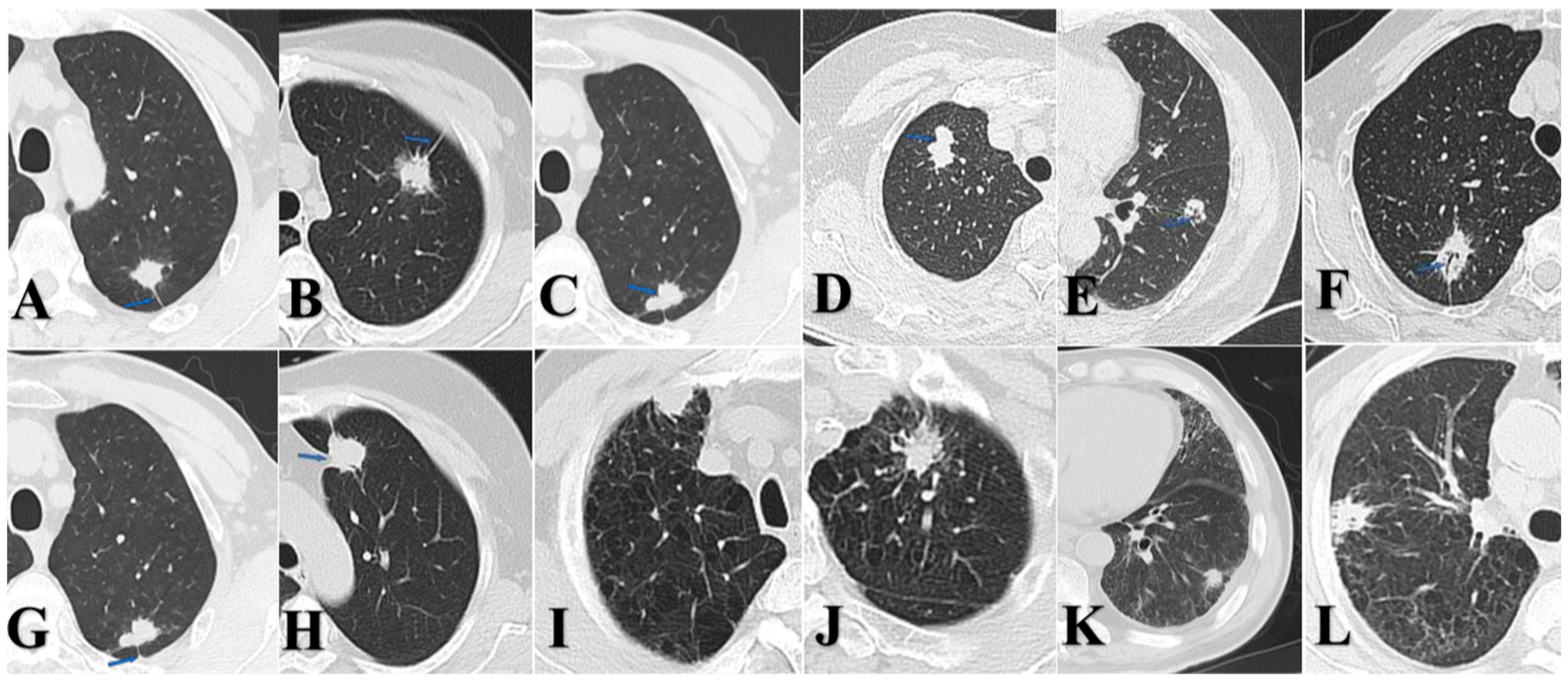
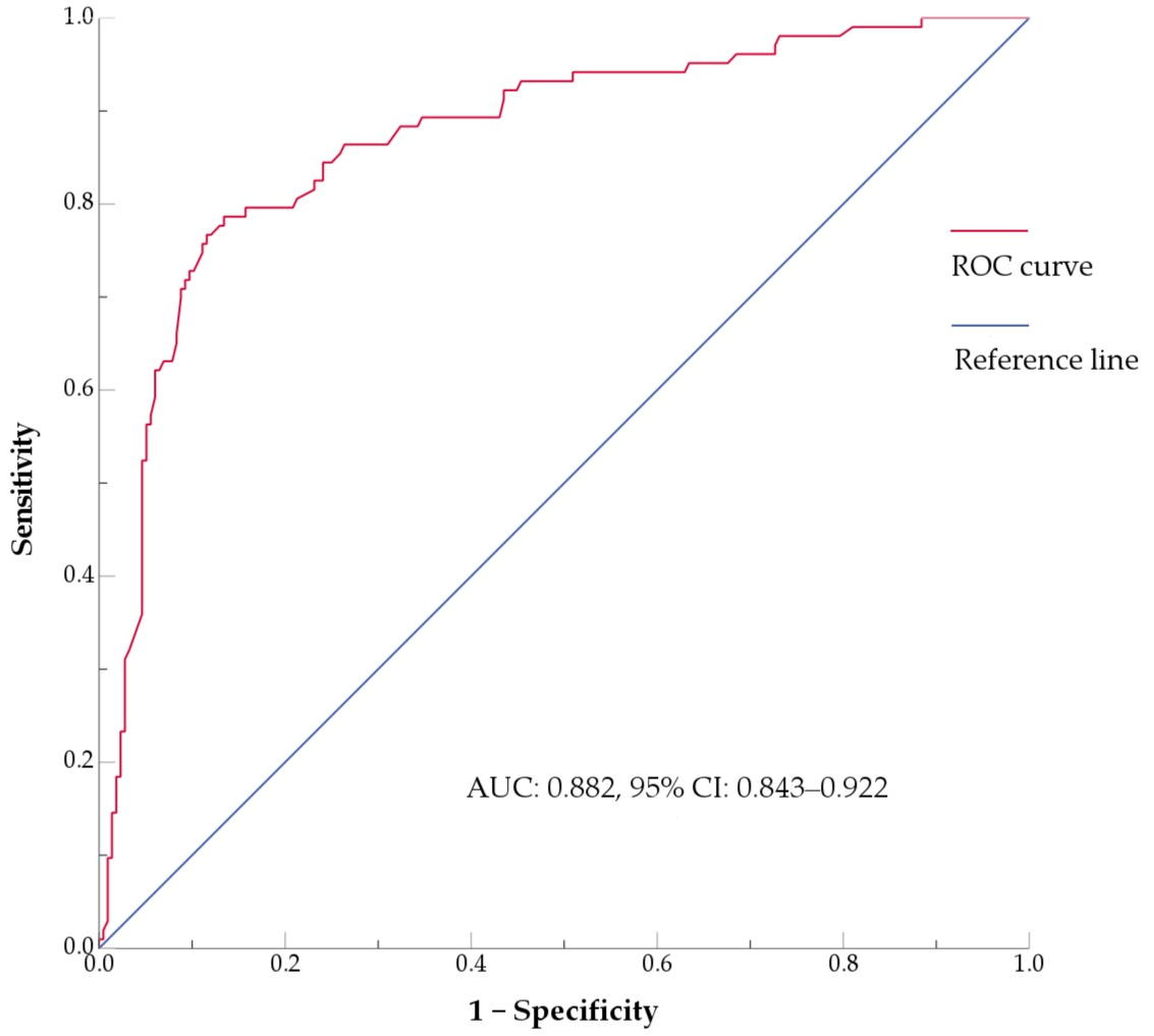
Unique Clinical Features of Imaging-Stage I Peripheral Lung Squamous Cell Carcinoma: A Retrospective Study


Simple Summary
Abstract
1. Introduction
2. Materials and Methods
2.1. Patient Cohorts
2.2. General Clinical Data
2.3. Imaging Data
2.4. Follow-Up and Endpoints
2.5. Statistical Analysis
3. Results
3.1. Patient Baseline Characteristics
3.2. General Characteristics of p-LUSC and SDNs of p-LUAD Patients
3.3. Clinicopathological Characteristics of p-LUSC and SDNs of p-LUAD Patients
3.4. Identification of Independent Prognostic Factors for RFS
4. Discussion
5. Conclusions
Author Contributions
Funding
Institutional Review Board Statement
Informed Consent Statement
Data Availability Statement
Conflicts of Interest
Abbreviations
| LUSC | lung squamous cell carcinoma |
| p-LUSC | peripheral lung squamous cell carcinoma |
| p-LUAD | peripheral lung adenocarcinoma |
| SDNs | solid nodules |
| mGGNs | mixed ground-glass nodules |
| NSCLC | non-small cell lung cancer |
| GGNs | ground-glass nodules |
| CTR | consolidation tumor ratio |
| VDT | volume doubling time |
| CEA | carcinoembryonic antigen |
| CA19-9 | carbohydrate antigen 19-9 |
| CYFRA21-1 | cytokeratin 19 fragment |
| RFS | relapse-free survival |
References
- Bray, F.; Laversanne, M.; Sung, H.; Ferlay, J.; Siegel, R.L.; Soerjomataram, I.; Jemal, A. Global cancer statistics 2022: GLOBOCAN estimates of incidence and mortality worldwide for 36 cancers in 185 countries. CA Cancer J. Clin. 2024, 74, 229–263. [Google Scholar] [CrossRef] [Scilit] [PubMed]
- Zhang, Y.; Vaccarella, S.; Morgan, E.; Li, M.; Etxeberria, J.; Chokunonga, E.; Manraj, S.S.; Kamate, B.; Omonisi, A.; Bray, F. Global variations in lung cancer incidence by histological subtype in 2020: A population-based study. Lancet Oncol. 2023, 24, 1206–1218. [Google Scholar] [CrossRef] [Scilit] [PubMed]
- Kinoshita, T.; Ohtsuka, T.; Hato, T.; Goto, T.; Kamiyama, I.; Tajima, A.; Emoto, K.; Hayashi, Y.; Kohno, M. Prognostic factors based on clinicopathological data among the patients with resected peripheral squamous cell carcinomas of the lung. J. Thorac. Oncol. 2014, 9, 1779–1787. [Google Scholar] [CrossRef] [Scilit]
- Krimsky, W.; Muganlinskaya, N.; Sarkar, S.; Vulchi, M.; Patel, P.; Rao, S.; Hammer, J.; Evans, R.; Qureshi, M.; Harley, D. The changing anatomic position of squamous cell carcinoma of the lung—A new conundrum. J. Community Hosp. Intern. Med. Perspect. 2016, 6, 33299. [Google Scholar] [CrossRef] [Scilit]
- Tane, K.; Miyoshi, T.; Samejima, J.; Aokage, K.; Ishii, G.; Tsuboi, M. Lymph node metastasis and predictive factors in clinical stage IA squamous cell carcinoma of the lung based on radiological findings. Gen. Thorac. Cardiovasc. Surg. 2022, 70, 52–58. [Google Scholar] [CrossRef] [Scilit]
- Choe, J.; Lee, S.M.; Park, S.; Choi, S.; Do, K.H.; Seo, J.B. The prognostic value of lymphovascular invasion for stage I lung adenocarcinoma based on the presence of ground-glass opacity. Eur. Radiol. 2025, 35, 2256–2264. [Google Scholar] [CrossRef] [Scilit]
- Hino, H.; Maru, N.; Utsumi, T.; Matsui, H.; Taniguchi, Y.; Saito, T.; Kouda, K.; Murakawa, T. Radiographical consolidation tumor size and preoperative clinical characteristics are significantly correlated with the postoperative survival of patients with part-solid and pure-solid adenocarcinomas: A propensity score-matched analysis. Surg. Today 2025, 55, 607–617. [Google Scholar] [CrossRef] [Scilit] [PubMed]
- Aokage, K.; Suzuki, K.; Wakabayashi, M.; Mizutani, T.; Hattori, A.; Fukuda, H.; Watanabe, S.I. Predicting pathological lymph node status in clinical stage IA peripheral lung adenocarcinoma. Eur. J. Cardiothorac. Surg. 2021, 60, 64–71. [Google Scholar] [CrossRef] [Scilit]
- Fang, C.; Xiang, Y.; Han, W. Preoperative risk factors of lymph node metastasis in clinical N0 lung adenocarcinoma of 3 cm or less in diameter. BMC Surg. 2022, 22, 153. [Google Scholar] [CrossRef] [Scilit]
- Tsutani, Y.; Murakami, S.; Miyata, Y.; Nakayama, H.; Yoshimura, M.; Okada, M. Prediction of lymph node status in clinical stage IA squamous cell carcinoma of the lung. Eur. J. Cardiothorac. Surg. 2015, 47, 1022–1026. [Google Scholar] [CrossRef] [Scilit]
- Sakurai, H.; Asamura, H.; Watanabe, S.; Suzuki, K.; Tsuchiya, R. Clinicopathologic features of peripheral squamous cell carcinoma of the lung. Ann. Thorac. Surg. 2004, 78, 222–227. [Google Scholar] [CrossRef] [Scilit]
- Honda, O.; Johkoh, T.; Sekiguchi, J.; Tomiyama, N.; Mihara, N.; Sumikawa, H.; Inoue, A.; Yanagawa, M.; Daimon, T.; Okumura, M.; et al. Doubling time of lung cancer determined using three-dimensional volumetric software: Comparison of squamous cell carcinoma and adenocarcinoma. Lung Cancer 2009, 66, 211–217. [Google Scholar] [CrossRef] [Scilit] [PubMed]
- Lin, M.W.; Huang, Y.L.; Yang, C.Y.; Kuo, S.W.; Wu, C.T.; Chang, Y.L. The Differences in Clinicopathologic and Prognostic Characteristics Between Surgically Resected Peripheral and Central Lung Squamous Cell Carcinoma. Ann. Surg. Oncol. 2019, 26, 217–229. [Google Scholar] [CrossRef] [Scilit]
- Yang, J.; Li, H.; Zheng, R.S.; Zeng, H.M.; Zhang, S.W.; Yang, Z.X.; Cao, M.M.; Chen, W.Q. Analysis of the clinical characteristics of 8081 primary lung cancer. Zhonghua Zhong Liu Za Zhi 2019, 41, 471–476. [Google Scholar] [CrossRef]
- Nakamura, H.; Nishimura, T. History, molecular features, and clinical importance of conventional serum biomarkers in lung cancer. Surg. Today 2017, 47, 1037–1059. [Google Scholar] [CrossRef] [Scilit] [PubMed]
- Ramirez-Carmona, W.; Diaz-Fabregat, B.; Yuri Yoshigae, A.; Musa de Aquino, A.; Scarano, W.R.; de Souza Castilho, A.C.; Avansini Marsicano, J.; Leal do Prado, R.; Pessan, J.P.; de Oliveira Mendes, L. Are Serum Ferritin Levels a Reliable Cancer Biomarker? A Systematic Review and Meta-Analysis. Nutr. Cancer 2022, 74, 1917–1926. [Google Scholar] [CrossRef] [Scilit]
- Fu, L.; Wang, R.; Yin, L.; Shang, X.; Zhang, R.; Zhang, P. CYFRA21-1 tests in the diagnosis of non-small cell lung cancer: A meta-analysis. Int. J. Biol. Markers 2019, 34, 251–261. [Google Scholar] [CrossRef] [Scilit]
- Yoon, E.J.; Song, S.G.; Kim, J.W.; Kim, H.C.; Kim, H.J.; Hur, Y.H.; Hong, J.H. Comprehensive CT Imaging Analysis of Primary Colorectal Squamous Cell Carcinoma: A Retrospective Study. Tomography 2024, 10, 674–685. [Google Scholar] [CrossRef] [Scilit]
- Akata, S.; Yoshimura, M.; Nishio, R.; Park, J.; Saito, K.; Uchida, O.; Ohira, T.; Kato, H.; Okada, S.; Kakizaki, D. High-resolution computed tomographic findings of small peripherally located squamous cell carcinoma. Clin. Imaging 2008, 32, 259–263. [Google Scholar] [CrossRef] [Scilit] [PubMed]
- Sakaizawa, T.; Yoshizawa, A.; Nishimura, H.; Arimura, T.; Kobayashi, N.; Ozawa, K.; Asaka, S.; Yoshida, K.; Hirai, K.; Hosaka, N. A Case of Pulmonary Squamous Cell Carcinoma Revealed Ground Glass Opacity on Computed Tomography. J. Thorac. Oncol. 2015, 10, 1229–1230. [Google Scholar] [CrossRef] [Scilit] [PubMed]
- Terada, Y.; Sato, M.; Abe, H.; Kuwano, H.; Nagayama, K.; Nitadori, J.I.; Anraku, M.; Nakajima, J. Squamous cell carcinoma of the lung showing a ground glass nodule on high-resolution computed tomography associated with pneumoconiosis: A case report. Surg. Case Rep. 2017, 3, 107. [Google Scholar] [CrossRef] [Scilit] [PubMed]
- Smith, B.M.; Schwartzman, K.; Kovacina, B.; Taylor, J.; Kasymjanova, G.; Brandao, G.; Agulnik, J.S. Lung cancer histologies associated with emphysema on computed tomography. Lung Cancer 2012, 76, 61–66. [Google Scholar] [CrossRef] [Scilit] [PubMed]
- Hubbard, R.; Venn, A.; Lewis, S.; Britton, J. Lung cancer and cryptogenic fibrosing alveolitis. A population-based cohort study. Am. J. Respir. Crit. Care Med. 2000, 161, 5–8. [Google Scholar] [CrossRef] [Scilit]
- Wilson, D.O.; Ryan, A.; Fuhrman, C.; Schuchert, M.; Shapiro, S.; Siegfried, J.M.; Weissfeld, J. Doubling times and CT screen-detected lung cancers in the Pittsburgh Lung Screening Study. Am. J. Respir. Crit. Care Med. 2012, 185, 85–89. [Google Scholar] [CrossRef] [Scilit]
- Mackintosh, J.A.; Marshall, H.M.; Yang, I.A.; Bowman, R.V.; Fong, K.M. A retrospective study of volume doubling time in surgically resected non-small cell lung cancer. Respirology 2014, 19, 755–762. [Google Scholar] [CrossRef] [Scilit]
- Ohtsuka, S.; Kato, H.; Ishikawa, R.; Watanabe, H.; Miyazaki, R.; Katsuragi, S.Y.; Yoshimura, K.; Yamada, H.; Sakai, Y.; Inoue, Y.; et al. STIL Overexpression Is Associated with Chromosomal Numerical Abnormalities in Non-Small-Cell Lung Carcinoma Through Centrosome Amplification. Curr. Oncol. 2024, 31, 7936–7949. [Google Scholar] [CrossRef] [Scilit]
- Abraham, C.G.; Ludwig, M.P.; Andrysik, Z.; Pandey, A.; Joshi, M.; Galbraith, M.D.; Sullivan, K.D.; Espinosa, J.M. ΔNp63α Suppresses TGFB2 Expression and RHOA Activity to Drive Cell Proliferation in Squamous Cell Carcinomas. Cell Rep. 2018, 24, 3224–3236. [Google Scholar] [CrossRef] [Scilit]
- Nonaka, D. A study of ΔNp63 expression in lung non-small cell carcinomas. Am. J. Surg. Pathol. 2012, 36, 895–899. [Google Scholar] [CrossRef] [Scilit] [PubMed]
- Hong, J.H.; Park, S.; Kim, H.; Goo, J.M.; Park, I.K.; Kang, C.H.; Kim, Y.T.; Yoon, S.H. Volume and Mass Doubling Time of Lung Adenocarcinoma according to WHO Histologic Classification. Korean J. Radiol. 2021, 22, 464–475. [Google Scholar] [CrossRef] [Scilit]
- Deng, H.Y.; Zeng, M.; Li, G.; Alai, G.; Luo, J.; Liu, L.X.; Zhou, Q.; Lin, Y.D. Lung Adenocarcinoma has a Higher Risk of Lymph Node Metastasis than Squamous Cell Carcinoma: A Propensity Score-Matched Analysis. World J. Surg. 2019, 43, 955–962. [Google Scholar] [CrossRef] [Scilit]
- Hung, J.J.; Jeng, W.J.; Hsu, W.H.; Chou, T.Y.; Huang, B.S.; Wu, Y.C. Predictors of death, local recurrence, and distant metastasis in completely resected pathological stage-I non-small-cell lung cancer. J. Thorac. Oncol. 2012, 7, 1115–1123. [Google Scholar] [CrossRef] [Scilit]
- Omori, T.; Aokage, K.; Nakamura, H.; Katsumata, S.; Miyoshi, T.; Sugano, M.; Kojima, M.; Fujii, S.; Kuwata, T.; Ochiai, A.; et al. Growth patterns of small peripheral squamous cell carcinoma of the lung and their impacts on pathological and biological characteristics of tumor cells. J. Cancer Res. Clin. Oncol. 2019, 145, 1773–1783. [Google Scholar] [CrossRef] [Scilit] [PubMed]
- Kadasah, S.F. Prognostic Significance of Glycolysis-Related Genes in Lung Squamous Cell Carcinoma. Int. J. Mol. Sci. 2024, 25, 1143. [Google Scholar] [CrossRef] [Scilit] [PubMed]
- Lin, P.Y.; Chang, Y.C.; Chen, H.Y.; Chen, C.H.; Tsui, H.C.; Yang, P.C. Tumor size matters differently in pulmonary adenocarcinoma and squamous cell carcinoma. Lung Cancer 2010, 67, 296–300. [Google Scholar] [CrossRef] [Scilit]
- Shoji, F.; Yamazaki, K.; Kouso, H.; Mori, R.; Takeo, S. The Impact of Pleural Lavage Cytology Both Before and After Lung Resection on Recurrence of Non-Small Cell Lung Cancer. Ann. Thorac. Surg. 2016, 101, 2141–2146. [Google Scholar] [CrossRef] [Scilit]
- Kameyama, K.; Okumura, N.; Miyaoka, E.; Asamura, H.; Yoshino, I.; Tada, H.; Fujii, Y.; Nakanishi, Y.; Eguchi, K.; Mori, M.; et al. Prognostic value of intraoperative pleural lavage cytology for non-small cell lung cancer: The influence of positive pleural lavage cytology results on T classification. J. Thorac. Cardiovasc. Surg. 2014, 148, 2659–2664. [Google Scholar] [CrossRef] [Scilit]
- Kjellberg, S.I.; Dresler, C.M.; Goldberg, M. Pleural cytologies in lung cancer without pleural effusions. Ann. Thorac. Surg. 1997, 64, 941–944. [Google Scholar] [CrossRef] [Scilit] [PubMed]
- Yun, J.K.; Kim, M.A.; Choi, C.M.; Choi, S.H.; Kim, Y.H.; Kim, D.K.; Park, S.I.; Kim, H.R. Surgical Outcomes after Pulmonary Resection for Non-Small Cell Lung Cancer with Localized Pleural Seeding First Detected during Surgery. Thorac. Cardiovasc. Surg. 2018, 66, 142–149. [Google Scholar] [CrossRef] [Scilit]
- Facciolo, F.; Cardillo, G.; Lopergolo, M.; Pallone, G.; Sera, F.; Martelli, M. Chest wall invasion in non-small cell lung carcinoma: A rationale for en bloc resection. J. Thorac. Cardiovasc. Surg. 2001, 121, 649–656. [Google Scholar] [CrossRef] [Scilit]
- Charnock, M. Primary lung tumour invading the chest wall. Ultrasound 2021, 29, 264–267. [Google Scholar] [CrossRef] [Scilit]
- Altorki, N.; Wang, X.; Kozono, D.; Watt, C.; Landrenau, R.; Wigle, D.; Port, J.; Jones, D.R.; Conti, M.; Ashrafi, A.S.; et al. Lobar or Sublobar Resection for Peripheral Stage IA Non-Small-Cell Lung Cancer. N. Engl. J. Med. 2023, 388, 489–498. [Google Scholar] [CrossRef] [Scilit]
- Saji, H.; Okada, M.; Tsuboi, M.; Nakajima, R.; Suzuki, K.; Aokage, K.; Aoki, T.; Okami, J.; Yoshino, I.; Ito, H.; et al. Segmentectomy versus lobectomy in small-sized peripheral non-small-cell lung cancer (JCOG0802/WJOG4607L): A multicentre, open-label, phase 3, randomised, controlled, non-inferiority trial. Lancet 2022, 399, 1607–1617. [Google Scholar] [CrossRef] [Scilit]
- Hattori, A.; Suzuki, K.; Takamochi, K.; Wakabayashi, M.; Sekino, Y.; Tsutani, Y.; Nakajima, R.; Aokage, K.; Saji, H.; Tsuboi, M.; et al. Segmentectomy versus lobectomy in small-sized peripheral non-small-cell lung cancer with radiologically pure-solid appearance in Japan (JCOG0802/WJOG4607L): A post-hoc supplemental analysis of a multicentre, open-label, phase 3 trial. Lancet Respir. Med. 2024, 12, 105–116. [Google Scholar] [CrossRef] [Scilit] [PubMed]
- Kamigaichi, A.; Hamada, A.; Tsuboi, M.; Yoshimura, K.; Okamoto, I.; Yamamoto, N.; Tsutani, Y. A Multi-Institutional, Randomized, Phase III Trial Comparing Anatomical Segmentectomy and Lobectomy for Clinical Stage IA3 Pure-Solid Non-Small-Cell Lung Cancer: West Japan Oncology Group Study WJOG16923L (STEP UP Trial). Clin. Lung Cancer 2024, 25, 384–388e1. [Google Scholar] [CrossRef] [Scilit] [PubMed]
- Kita, N.; Tomita, N.; Takaoka, T.; Sudo, S.; Tsuzuki, Y.; Okazaki, D.; Niwa, M.; Torii, A.; Takano, S.; Niimi, A.; et al. Comparison of Recurrence Patterns between Adenocarcinoma and Squamous Cell Carcinoma after Stereotactic Body Radiotherapy for Early-Stage Lung Cancer. Cancers 2023, 15, 887. [Google Scholar] [CrossRef] [Scilit] [PubMed]
- Ma, W.; Zeng, J.; Montoya, D.J.; Toomey, K.; Zhou, C.; Chen, S.; Liu, D.; Babich, M.; Radosevich, J.A.; Li, T. Labyrinthin Expression Is Associated with Poor Prognosis in Patients with Non-Small-Cell Lung Cancer. Cancers 2023, 15, 924. [Google Scholar] [CrossRef] [Scilit]



| Factors | Total, n = 703 | p-LUSC, n = 103 | p-LUAD, n = 600 | p Value |
|---|---|---|---|---|
| Gender | <0.001 | |||
| Females | 374 (53.2%) | 14 (13.6%) | 360 (60%) | |
| Males | 329 (46.8%) | 89 (86.4%) | 240 (40%) | |
| Age, years | <0.001 | |||
| Median (range), years | 63 (21–84) | 68 (47–84) | 62 (21–83) | |
| Tumor size | 0.0041 | |||
| 0–2.0 cm | 450 (64.0%) | 53 (51.5%) | 397 (66.2%) | |
| >2.0 to ≤3.0 cm | 253 (36.0%) | 50 (48.5%) | 203 (33.8%) | |
| Smoking history | <0.001 | |||
| Current or former | 162 (23.0%) | 57 (55.3%) | 105 (17.5%) | |
| Never | 541 (77.0%) | 46 (44.7%) | 495 (82.5%) | |
| High surgical risk | <0.001 | |||
| Yes | 165 (23.5%) | 38 (36.9%) | 113 (18.8%) | |
| No | 538 (76.5%) | 65 (63.1%) | 487 (81.2%) | |
| Lymph node metastasis | 0.31 | |||
| pN1 + pN2 | 59 (8.4%) | 6 (5.8%) | 53 (8.8%) | |
| pN0 | 644 (91.6%) | 97 (94.2%) | 547 (91.2%) | |
| Imaging CTR | <0.001 | |||
| SDNs | 319 (45.4%) | 103 (100%) | 216 (36%) | |
| mGGNs, CTR ≧ 50% | 222 (31.6%) | 0 (0%) | 222 (37%) | |
| mGGNs, CTR < 50% | 162 (23.0%) | 0 (0%) | 162 (27%) |
| Factors | Univariate Analyses | Binary Logistic Regression | |||
|---|---|---|---|---|---|
| p-LUSC, n = 103 | SDNs of p-LUAD, n = 216 | p Value | Odds Ratio (95%CI) | p Value | |
| Male (Female) a | 89 (86.4%) | 117 (54.2%) | <0.001 | 6.187 (2.866–13.354) | <0.001 |
| Median age (range), years | 68 (47–84) | 63 (34–83) | <0.001 | 1.117 (1.061–1.176) | <0.001 |
| Tumor size (0–2.0 cm) | 53 (51.5%) | 127 (58.8%) | 0.22 | - | - |
| Smoking history (No) b | 57 (55.3%) | 59 (27.3%) | <0.001 | 2.506 (1.330–4.723) | 0.004 |
| High surgical risk (No) b | 38 (36.9%) | 48 (22.2%) | 0.0058 | 1.940 (0.977–3.852) | 0.058 |
| Ferritin (ng/mL), Median (range) c, % > 322 | 31.7 (27.3–34.9) 5.8% | 31.8 (28.3–35.3) 7.4% | 0.69 d | - | - |
| CEA (ng/mL), Median (range) c, % > 5 | 2.0 (1.7–2.5) 4.9% | 2.0 (1.6–2.3) 4.2% | 0.06 d | 1.000 (0.845–1.182) | 0.996 |
| CA199 (U/mL), Median (range) c, % > 37 | 21.9 (17.6–26.8) 8.7% | 20.6 (16.3–26.0) 6.0% | 0.13 d | - | - |
| CYFRA21-1(ng/mL), Median (range) c, % > 3.3 | 2.0 (1.5–3.7) 28.2% | 1.8 (1.3–2.6) 17.6% | 0.024 d | 1.272 (0.989–1.636) | 0.061 |
| Spicular sign (No) | 52 (50.5%) | 116 (53.7%) | 0.59 | - | - |
| Lobulation sign (No) b | 76 (73.8%) | 67 (31.0%) | <0.001 | 6.879 (3.602–13.138) | <0.001 |
| Air bronchogram (No) | 32 (31.1%) | 64 (29.6%) | 0.79 | - | - |
| Pleural indentation (No) | 34 (33.0%) | 84 (38.9%) | 0.31 | - | - |
| Emphysema (No) b | 15 (14.6%) | 8 (3.7%) | <0.001 | 2.781 (0.831–9.312) | 0.097 |
| Interstitial pneumonia (No) b | 25 (24.3%) | 19 (8.8%) | <0.001 | 3.051 (1.197–7.781) | 0.019 |
| Factors | 0–2.0 cm, n = 180 | >2.0 to ≤3.0 cm, n = 139 | ||||
|---|---|---|---|---|---|---|
| p-LUSC, n = 53 | SDNs of p-LUAD, n = 127 | p Value | p-LUSC, n = 50 | SDNs of p-LUAD, n = 89 | p Value | |
| Lymph node metastasis | 0.74 | <0.001 | ||||
| Positive | 3 (5.7%) | 9 (7.1%) | 3 (6%) | 37 (41.6%) | ||
| Negative | 50 (94.3%) | 118 (92.9%) | 47 (94%) | 52 (58.4%) | ||
| Pleural invasion | 0.0036 | <0.001 | ||||
| Positive | 2 (3.8%) | 27 (21.3%) | 3 (6%) | 43 (48.3%) | ||
| Negative | 51 (96.2%) | 100 (78.7%) | 47 (94%) | 46 (51.7%) | ||
| Lymphovascular invasion | 0.45 | 0.66 | ||||
| Positive | 8 (15.1%) | 14 (11.0%) | 10 (20%) | 31 (34.8%) | ||
| Negative | 45 (84.9%) | 113 (89.0%) | 40 (80%) | 58 (65.2%) | ||
| Factors | p-LUSC, n = 103 | SDNs of p-LUAD, n = 216 | ||||
|---|---|---|---|---|---|---|
| 0–2.0 cm, n = 53 | >2.0 to ≤3.0 cm, n = 50 | p Value | 0–2.0 cm, n = 127 | >2.0 to ≤3.0 cm, n = 89 | p Value | |
| Lymph node metastasis | 0.94 | <0.001 | ||||
| Positive | 3 (5.7%) | 3 (6%) | 9 (7.1%) | 37 (41.6%) | ||
| Negative | 50 (94.3%) | 47 (94%) | 118 (92.9%) | 52 (58.4%) | ||
| Pleural invasion | 0.60 | <0.001 | ||||
| Positive | 2 (3.8%) | 3 (6%) | 27 (21.3%) | 43 (48.3%) | ||
| Negative | 51 (96.2%) | 47 (94%) | 100 (78.7%) | 46 (51.7%) | ||
| Lymphovascular invasion | 0.51 | <0.001 | ||||
| Positive | 8 (15.1%) | 10 (20%) | 14 (11.0%) | 31 (34.8%) | ||
| Negative | 45 (84.9%) | 40 (80%) | 113 (89.0%) | 58 (65.2%) | ||
| Factors (Reference) | Univariable | Multivariable | ||
|---|---|---|---|---|
| HR (95% CI) | p-Value | HR (95% CI) | p-Value | |
| Male (Female) a | 0.877 (0.448–1.715) | 0.701 | - | - |
| Age | 1.003 (0.962–1.046) | 0.882 | - | - |
| Smoker (No) b | 1.456 (0.768–2.760) | 0.250 | - | - |
| High surgical risk (No) b | 0.642 (0.283–1.459) | 0.290 | - | - |
| Size > 2.0 to ≤3.0 cm (0–2.0 cm) b | 3.507 (1.738–7.076) | <0.001 | 2.430 (1.135–5.204) | 0.022 |
| Lymph node metastasis (No) b | 3.924 (2.046–7.524) | <0.001 | 2.305 (1.083–4.903) | 0.030 |
| P-LUSC (p-LUAD) c | 0.883 (0.438–1.782) | 0.729 | - | - |
| Ferritin, ng/mL | 1.003 (1.001–1.005) | 0.003 | 1.001 (0.999–1.003) | 0.270 |
| AFP, IU/mL | 0.787 (0.522–1.187) | 0.254 | - | - |
| CEA, ng/mL | 1.115 (0.972–1.278) | 0.120 | - | - |
| CA199, U/mL | 1.003 (0.974–1.003) | 0.824 | - | - |
| NSE, ng/mL | 0.970 (0.931–1.010) | 0.142 | - | - |
| CYFRA21-1, ng/mL | 1.101 (0.871–1.391) | 0.420 | - | - |
| Spicular sign (No) b | 1.210 (0.635–2.305) | 0.563 | - | - |
| Lobulation sign (No) b | 1.114 (0.589–2.107) | 0.739 | - | - |
| Air bronchogram (No) b | 1.352 (0.699–2.614) | 0.370 | - | - |
| Pleural indentation (No) b | 0.948 (0.490–1.835) | 0.874 | - | - |
| Emphysema (No) b | 1.880 (0.734–4.817) | 0.188 | - | - |
| Interstitial pneumonia (No) b | 1.660 (0.730–3.773) | 0.227 | - | - |
| Pleural invasion (No) b | 2.262 (1.180–4.338) | 0.014 | 1.242 (0.603–2.556) | 0.557 |
| Lymphovascular invasion (No) b | 1.408 (0.684–2.899) | 0.354 | - | - |
Disclaimer/Publisher’s Note: The statements, opinions and data contained in all publications are solely those of the individual author(s) and contributor(s) and not of MDPI and/or the editor(s). MDPI and/or the editor(s) disclaim responsibility for any injury to people or property resulting from any ideas, methods, instructions or products referred to in the content. |
© 2026 by the authors. Licensee MDPI, Basel, Switzerland. This article is an open access article distributed under the terms and conditions of the Creative Commons Attribution (CC BY) license.
Share and Cite
Xiong, C.; Zhang, W.; Wang, Q.; Yin, H.; Chen, J.; Jiang, W.; Han, X. Unique Clinical Features of Imaging-Stage I Peripheral Lung Squamous Cell Carcinoma: A Retrospective Study. Curr. Oncol. 2026, 33, 47. https://doi.org/10.3390/curroncol33010047
Xiong C, Zhang W, Wang Q, Yin H, Chen J, Jiang W, Han X. Unique Clinical Features of Imaging-Stage I Peripheral Lung Squamous Cell Carcinoma: A Retrospective Study. Current Oncology. 2026; 33(1):47. https://doi.org/10.3390/curroncol33010047
Chicago/Turabian StyleXiong, Chengzhang, Wenjing Zhang, Qing Wang, Hao Yin, Jibin Chen, Wenjun Jiang, and Xu Han. 2026. "Unique Clinical Features of Imaging-Stage I Peripheral Lung Squamous Cell Carcinoma: A Retrospective Study" Current Oncology 33, no. 1: 47. https://doi.org/10.3390/curroncol33010047
APA StyleXiong, C., Zhang, W., Wang, Q., Yin, H., Chen, J., Jiang, W., & Han, X. (2026). Unique Clinical Features of Imaging-Stage I Peripheral Lung Squamous Cell Carcinoma: A Retrospective Study. Current Oncology, 33(1), 47. https://doi.org/10.3390/curroncol33010047