Successful Multidisciplinary Treatment with Laparoscopic Hepatectomy and Adjuvant Therapy for Metachronous Solitary Hepatic Metastasis after Excision of a Primary Anorectal Malignant Melanoma: A Case Report

 , , ,
, , , 
Abstract
:1. Introduction
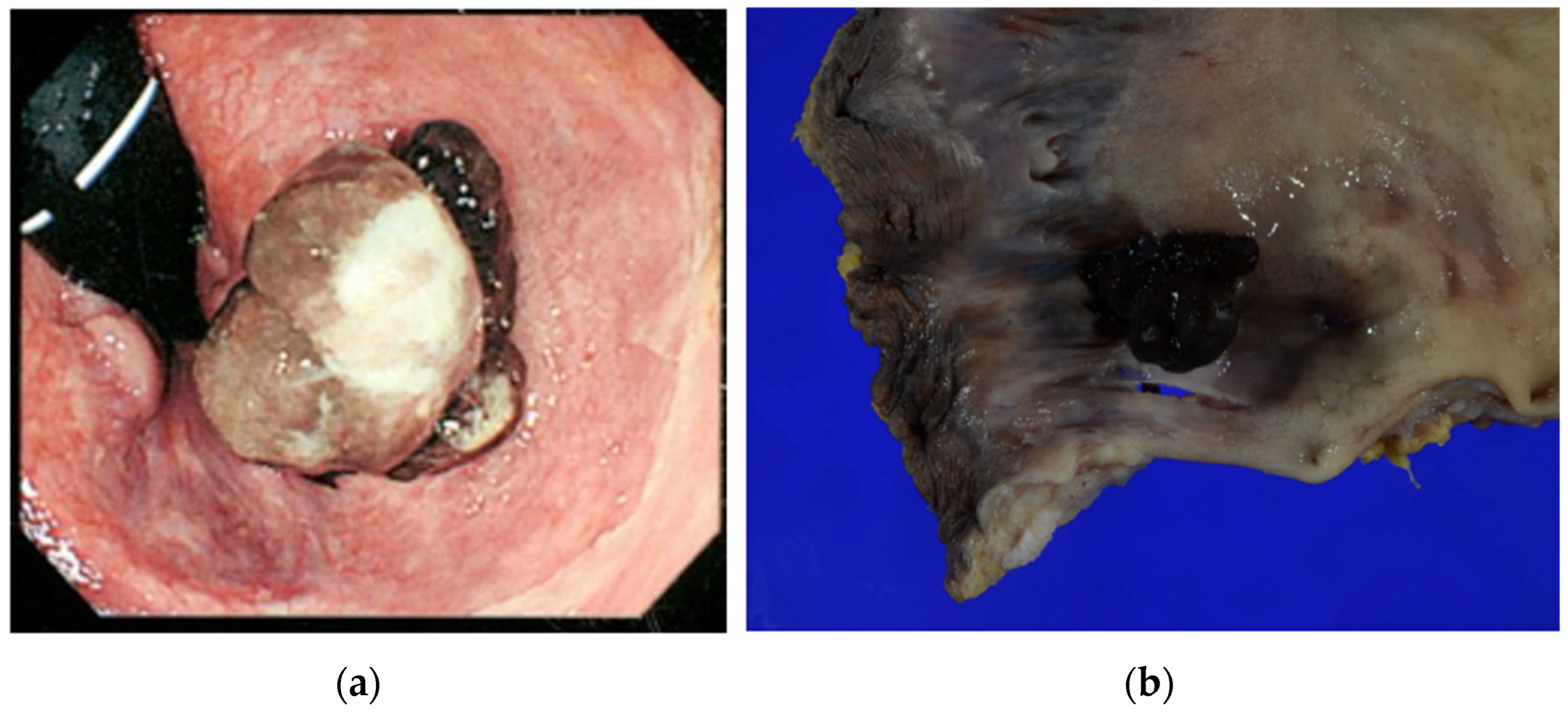
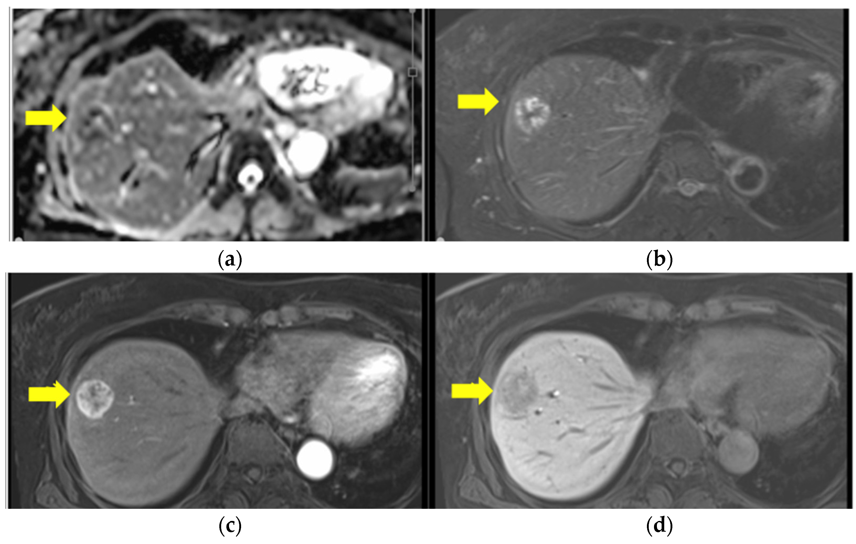
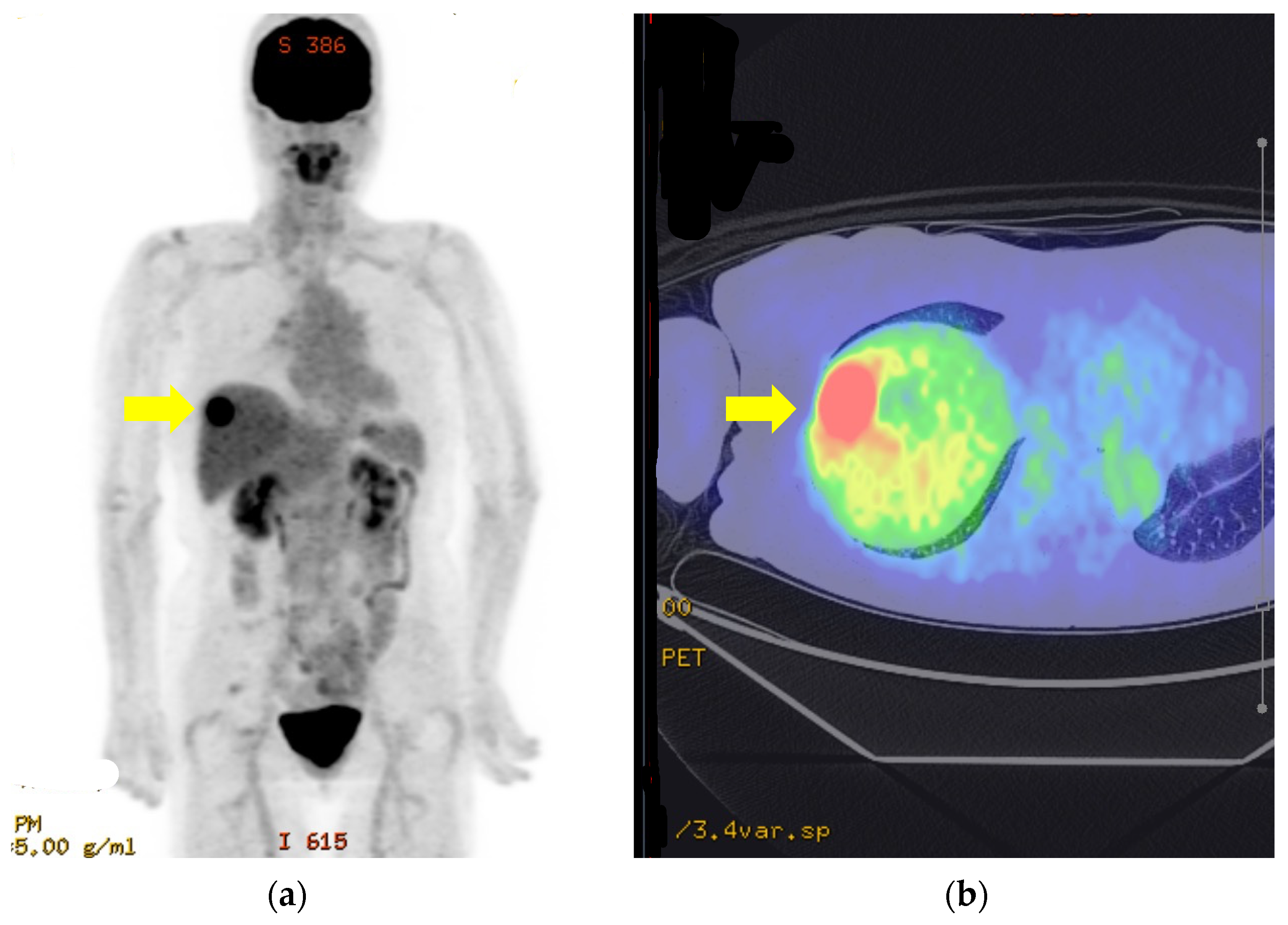
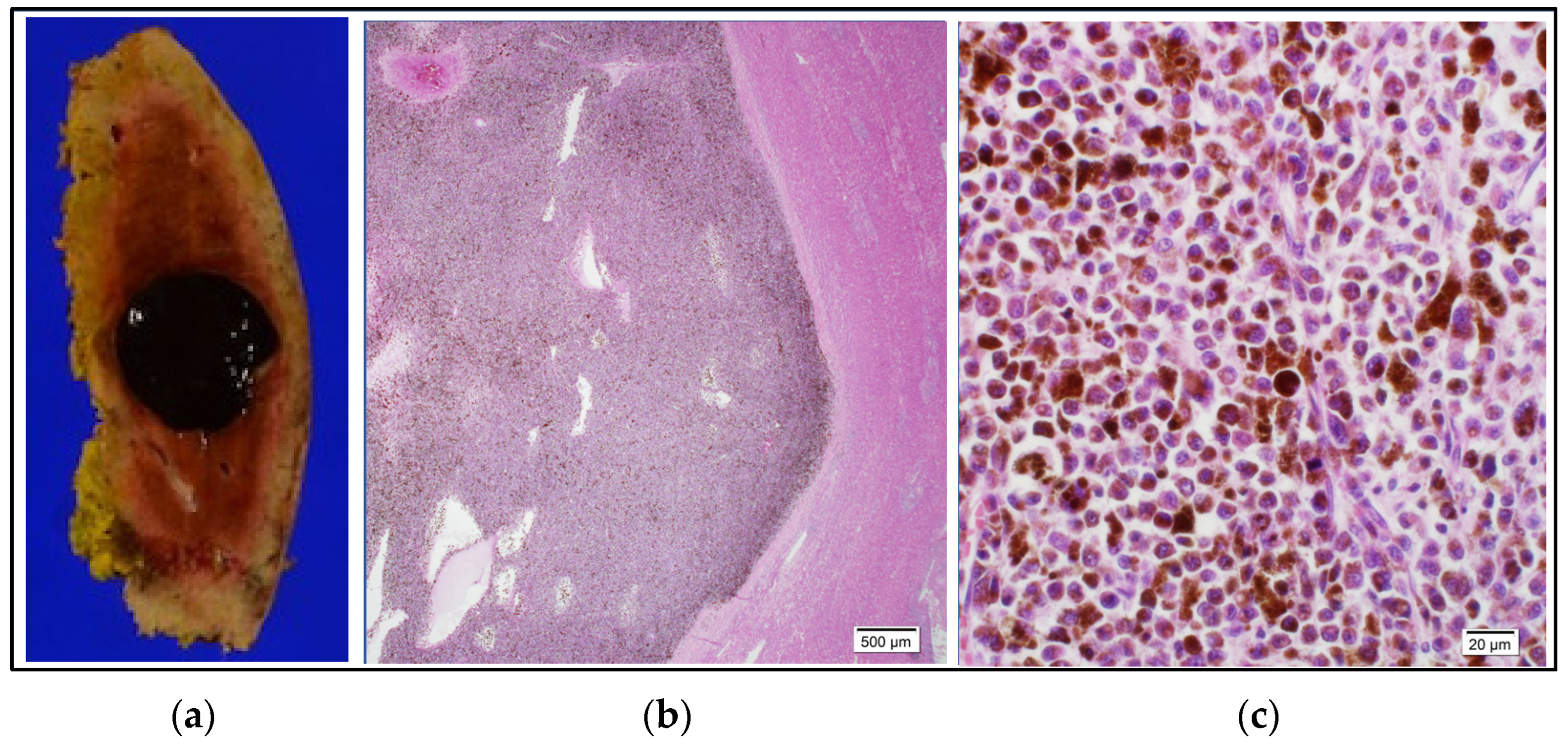
2. Case Report
3. Discussion and Conclusions
Author Contributions
Funding
Institutional Review Board Statement
Informed Consent Statement
Data Availability Statement
Acknowledgments
Conflicts of Interest
References
- Nakamura, Y.; Asai, J.; Igaki, H.; Inozume, T.; Namikawa, K.; Hayashi, A.; Fukushima, S.; Ito, T.; Imafuku, K.; Tanaka, R.; et al. Japanese Dermatological Association Guidelines: Outlines of guidelines for cutaneous melanoma 2019. J. Dermatol. 2020, 47, 89–103. [Google Scholar] [CrossRef]
- Falch, C.; Stojadinovic, A.; Hann-von-Weyhern, C.; Protic, M.; Nissan, A.; Faries, M.B.; Daumer, M.; Bilchik, A.J.; Itzhak, A.; Brücher, B.L.D.M. Anorectal malignant melanoma: Extensive 45-year review and proposal for a novel staging classification. J. Am. Coll. Surg. 2013, 217, 324–335. [Google Scholar] [CrossRef]
- Ranjith, S.; Muralee, M.; Sajeed, A.; Arun, P.M.; Cherian, K.; Nair, C.K.; Augustine, P.; Ahamed, I. Anorectal melanoma: Experience from a tertiary cancer care centre in South India. Ann. R. Coll. Surg. Engl. 2018, 100, 185–189. [Google Scholar] [CrossRef] [PubMed]
- Lian, B.; Cui, C.L.; Zhou, L.; Song, X.; Zhang, X.S.; Wu, D.; Si, L.; Chi, Z.H.; Sheng, X.N.; Mao, L.L.; et al. The natural history and patterns of metastases from mucosal melanoma: An analysis of 706 prospectively-followed patients. Ann. Oncol. 2017, 28, 868–873. [Google Scholar] [CrossRef]
- Corvino, A.; Catalano, O.; Corvino, F.; Petrillo, A. Rectal melanoma presenting as a solitary complex cystic liver lesion: Role of contrast-specific low-MI real-time ultrasound imaging. J. Ultrasound 2016, 19, 135–139. [Google Scholar] [CrossRef]
- Takahashi, M.; Morita, Y.; Hayashi, T.; Yanagibasi, S.; Sato, S.; Sasaki, S.; Takuma, K.; Okada, H. A case of laparoscopic partial hepatic S7 resection for postoperative liver metastasis of rectal malignant melanoma. Surg. Case Rep. 2021, 7, 230. [Google Scholar] [CrossRef]
- Moore, G.W.D. Recurrent melanosis of the rectum after previous removal from the verge of the anus in a sixty five-year-old patient. Lancet 1857, 72, 290–293. [Google Scholar] [CrossRef]
- Goldman, S.; Glimelius, B.; Påhlman, L. Anorectal malignant melanoma in Sweden—Report of 49 patients. Dis. Colon. Rectum 1990, 33, 874–877. [Google Scholar] [CrossRef]
- Roumen, R.M.H. Anorectal melanoma in the Netherlands: A report of 63 patients. Eur. J. Surg. Oncol. 1996, 22, 598–601. [Google Scholar] [CrossRef]
- Thibault, C.; Sagar, P.; Nivatvongs, S.; Ilstrup, D.M.; Wolff, B.G. Anorectal melanoma—An incurable disease? Dis. Colon. Rectum 1997, 40, 661–668. [Google Scholar] [CrossRef] [PubMed]
- Umemura, H.; Yamasaki, O.; Kaji, T.; Otsuka, M.; Asagoe, K.; Takata, M.; Iwatsuki, K. Usefulness of serum 5-S-cysteinyl-dopa as a biomarker for predicting prognosis and detecting relapse in patients with advanced stage malignant melanoma. J. Dermatol 2017, 44, 449–454. [Google Scholar] [CrossRef]
- Smith, H.G.; Bagwan, I.; Board, R.E.; Capper, S.; Coupland, S.E.; Glen, J.; Lalondrelle, S.; Mayberry, A.; Muneer, A.; Nugent, K.; et al. Ano-uro-genital mucosal melanoma UK national guidelines. Eur. J. Cancer 2020, 135, 22–30. [Google Scholar] [CrossRef]
- Ishizone, S.; Koide, N.; Karasawa, F.; Akita, N.; Muranaka, F.; Uhara, H.; Miyagawa, S. Surgical treatment for anorectal malignant melanoma: Report of five cases and review of 79 Japanese cases. Int. J. Color. Dis. 2008, 23, 1257–1262. [Google Scholar] [CrossRef] [PubMed]
- Bleicher, J.; Cohan, J.N.; Huang, L.C.; Peche, W.; Pickron, T.B.; Scaife, C.L.; Bowles, T.L.; Hyngstrom, J.R.; Asare, E.A. Trends in the management of anorectal melanoma: A multi-institutional retrospective study and review of the world literature. World J. Gastroenterol. 2021, 27, 267–280. [Google Scholar] [CrossRef]
- Smith, H.G.; Glen, J.; Turnbull, N.; Peach, H.; Board, R.; Payne, M.; Gore, M.; Nugent, K.; Smith, M.J.F. Less is more: A systematic review and meta-analysis of the outcomes of radical versus conservative primary resection in anorectal melanoma. Eur. J. Cancer 2020, 135, 113–120. [Google Scholar] [CrossRef] [PubMed]
- Ross, S.W.; Lauer, C.W.; Miles, W.S.; Green, J.M.; Christmas, A.B.; May, A.K.; Matthews, B.D. Maximizing the calm before the storm: Tiered surgical response plan for novel coronavirus (COVID-19). Am. Coll. Surg. 2020, 6, 1080–1091.e3. [Google Scholar] [CrossRef] [PubMed]
- Kelly, P.; Zagars, G.K.; Cormier, J.N.; Ross, M.I.; Guadagnolo, B.A. Sphincter-sparing local excision and hypofractionated radiation therapy for anorectal melanoma: A 20-Year Experience. Cancer 2011, 117, 4747–4755. [Google Scholar] [CrossRef] [PubMed]
- Höppener, D.J.; Grünhagen, D.J.; Eggermont, A.M.M.; van der Veldt, A.A.M.; Verhoef, C. An overview of liver directed locoregional therapies. Surg. Oncol. Clin. N. Am. 2021, 30, 103–123. [Google Scholar] [CrossRef]
- Wong, J.H.; Euhus, D.M.; Morton, D.L. Surgical resection for metastatic melanoma to the lung. Arch. Surg. 1988, 123, 1091–1095. [Google Scholar] [CrossRef]
- Ansai, S.I.; Umebayashi, Y.; Katsumata, N.; Kato, H.; Kadono, T.; Takai, T.; Namiki, T.; Nakagawa, M.; Soejima, T.; Koga, H.; et al. Squamous Cell Carcinoma Guidelines Committee of the Japanese Skin Cancer Society. Japanese Dermatological Association Guidelines: Outlines of Guidelines for Cutaneous Squamous Cell Carcinoma 2020. J. Dermatol. 2021, 48, e288–e311. [Google Scholar] [CrossRef] [PubMed]


Disclaimer/Publisher’s Note: The statements, opinions and data contained in all publications are solely those of the individual author(s) and contributor(s) and not of MDPI and/or the editor(s). MDPI and/or the editor(s) disclaim responsibility for any injury to people or property resulting from any ideas, methods, instructions or products referred to in the content. |
© 2023 by the authors. Licensee MDPI, Basel, Switzerland. This article is an open access article distributed under the terms and conditions of the Creative Commons Attribution (CC BY) license (https://creativecommons.org/licenses/by/4.0/).
Share and Cite
Shimazaki, R.; Hagiwara, M.; Tani, C.; Iwata, H.; Takahashi, H.; Fukuyama, M.; Matsuya, T.; Imai, K.; Yuzawa, S.; Tanino, M.; et al. Successful Multidisciplinary Treatment with Laparoscopic Hepatectomy and Adjuvant Therapy for Metachronous Solitary Hepatic Metastasis after Excision of a Primary Anorectal Malignant Melanoma: A Case Report. Curr. Oncol. 2024, 31, 203-210. https://doi.org/10.3390/curroncol31010013
Shimazaki R, Hagiwara M, Tani C, Iwata H, Takahashi H, Fukuyama M, Matsuya T, Imai K, Yuzawa S, Tanino M, et al. Successful Multidisciplinary Treatment with Laparoscopic Hepatectomy and Adjuvant Therapy for Metachronous Solitary Hepatic Metastasis after Excision of a Primary Anorectal Malignant Melanoma: A Case Report. Current Oncology. 2024; 31(1):203-210. https://doi.org/10.3390/curroncol31010013
Chicago/Turabian StyleShimazaki, Ryotaro, Masahiro Hagiwara, Chikayoshi Tani, Hiroyoshi Iwata, Hiroyuki Takahashi, Marika Fukuyama, Taisuke Matsuya, Koji Imai, Sayaka Yuzawa, Mishie Tanino, and et al. 2024. "Successful Multidisciplinary Treatment with Laparoscopic Hepatectomy and Adjuvant Therapy for Metachronous Solitary Hepatic Metastasis after Excision of a Primary Anorectal Malignant Melanoma: A Case Report" Current Oncology 31, no. 1: 203-210. https://doi.org/10.3390/curroncol31010013
APA StyleShimazaki, R., Hagiwara, M., Tani, C., Iwata, H., Takahashi, H., Fukuyama, M., Matsuya, T., Imai, K., Yuzawa, S., Tanino, M., & Yokoo, H. (2024). Successful Multidisciplinary Treatment with Laparoscopic Hepatectomy and Adjuvant Therapy for Metachronous Solitary Hepatic Metastasis after Excision of a Primary Anorectal Malignant Melanoma: A Case Report. Current Oncology, 31(1), 203-210. https://doi.org/10.3390/curroncol31010013