Next Article in Journal
Impressive Late Stent Recoil of a Drug-Eluting Resorbable Magnesium Coronary Stent
Previous Article in Journal
Fusiform Mid-LAD Aneurysm Due to Passive Arterial Wall Dilatation After Implantation of a Self-Expandable Stent
 
 
Cardiovascular Medicine is published by MDPI from Volume 28 Issue 1 (2025). Previous articles were published by another publisher in Open Access under a CC-BY (or CC-BY-NC-ND) licence, and they are hosted by MDPI on mdpi.com as a courtesy and upon agreement with Editores Medicorum Helveticorum (EMH).
Case Report

Major Right Atrial Thrombus Related to Central Venous Catheter Positioning

by
Sabine Blank
1,*,
Christoph Henzen
1,
Xavier Mueller
2 and
Thomas Syburra
2
1
Department of Medicine, Cantonal Hospital, Lucerne, Switzerland
2
Department of Cardiac Surgery, Cantonal Hospital, Lucerne, Switzerland
*
Author to whom correspondence should be addressed.
Cardiovasc. Med. 2020, 23(2), w02104; https://doi.org/10.4414/cvm.2020.02104
Submission received: 20 January 2020 / Revised: 20 February 2020 / Accepted: 20 March 2020 / Published: 20 April 2020

Abstract

We report the case of a 44-year-old woman who developed a thrombus of life-threatening size in the right atrium. The thrombus had formed around the catheter tip of her tunnelled central venous catheter, which was inserted for haemodialysis. Because of the size of the thrombus and the high risk of pulmonary embolism, surgical thrombectomy was performed. Because of an increasing incidence and usage period of tunnelled central venous catheters, the likelihood of catheter-related complications rises. Contributing factors need to be identified and reduced, especially in long-term catheter-dependent patients. One of those risk factors might be the exact position of the catheter tip.

Case description

A 44-year-old, haemodialysis-dependent Caucasian female presented for scheduled transthoracic stress echocardiography in preparation for kidney transplantation. The aetiology of her terminal renal failure was primary arterial hypertension, and she had been on haemodialysis via a transjugular tunnelled central venous catheter for 5 months. For the previous 4 weeks, she had experienced progressive shortness of breath, reaching dyspnoea of New York Heart Association (NYHA) grade II−III at the time of cardiac imaging. The echocardiography revealed a round mass in the right atrium, 5 × 6 cm in size (Figure 1), prolapsing through the tricuspid annulus into the right ventricle and resulting in a moderate tricuspid stenosis. Stress echocardiography was cancelled and the patient was admitted to our hospital for monitoring and further evaluation, to differentiate whether the mass was solely a thrombus in connection with the dialysis catheter or if there was a catheter-related infection.
Therapeutic anticoagulation using unfractionated intravenous heparin was started. Computed tomography confirmed an atrial thrombus connected to the catheter tip; furthermore, subacute segmental pulmonary emboli were detected. In the absence of established guidelines concerning treatment, surgery was chosen after interdisciplinary discussion taking into account the thrombus size, the young patient in good general condition and the risk of massive pulmonary embolism. The surgical thrombectomy of the right atrium was performed through a median sternotomy in an on-pump beating heart, with bicaval snared cannulation and standard cannulation of the aorta. The tip of the central venous catheter was found to heavily protrude onto the lateral and the caudal right atrium wall. A thrombus measuring 5 × 6 cm adhering from a wide pedicle to the corresponding portion of the atrial wall was found and was resected in one piece without difficulty. Bubble testing during intraoperative transoesophageal echocardiography (TOE) and direct intraoperative exploration did not reveal any patent interatrial communication. TOE at completion of the surgery demonstrated normal left ventricular function and complete resection of the thrombus. The catheter was removed during surgery and replaced 1 week later. Therapeutic anticoagulation was resumed postoperatively. Further examination of the thrombus tissue showed neither bacterial growth nor neoplastic cells. Haematological examination several weeks after the initial incident did not verify an underlying thrombophilic disease, so the therapeutic anticoagulation could be stopped.

Discussion

The formation of thrombi at the tip of central venous catheters is a known phenomenon caused by local intimal injury resulting over time in smooth muscle cell proliferation and the formation of thrombus between the atrial wall and the catheter [1]. Factors contributing to thrombus formation are activation of the coagulation cascade due to endothelial injury by the catheter and the movement of the heart, blood flow changes around the catheter and the patients’ hypercoagulable state in chronic kidney disease. Female gender may also promote development of thrombi under these circumstances [2].
The development of right atrial thrombi is also dependant on the location of the catheter tip. The KDOQI Guidelines on vascular access recommend positioning of the catheter tip in the right mid-atrium for maximised flow rates, yet this recommendation is much discussed among nephrologists [3]. Gilon et al. screened 55 patients for development of thrombus using TOE 1 and 8 weeks after Hickman catheter implantation; 12.5% of patients developed a thrombus, all of whom had the catheter tip placed in the right atrium. None of the patients with the catheter tip on the superior vena cava-atrium junction or in the superior vena cava itself developed a thrombus [4]. There was no difference in coagulation status or infectious complications.
Concordantly, a meta-analysis identified 71 cases of catheter-induced right atrial thrombi with the majority of the catheter tips located in the right atrium [2]. As a consequence, to avoid thrombus formation it is crucial to check the position of the catheter tip after insertion so it will allow catheter movement when the patient changes body position without chafing the atrial wall. Ideally, the position allows movement between the superior vena cava and the right atrium [3]. However, postoperative fluoroscopy or a chest radiograph are not sensitive enough to distinguish between a tip position on the vena cava-atrium junction or in the right atrium [3,4]. Therefore, some authors suggest routine transthoracic echocardiography (TTE) in patients with a haemodialysis catheter in use for more than 2 weeks [5] in order to detect thrombi that are still small and for early recognition of catheter tips protruding into the right atrium wall, which should then be repositioned as recommended in the KDOQI Guidelines. Nonetheless, visualisation of the catheter tip with TTE might not be possible in all patients, thus, requiring a TOE study in some cases [3].
In our patient, the thrombus reached life-threatening dimensions. Further studies are needed to better identify risk factors promoting thrombus formation and the pathophysiology of the growth, and also to define guidelines for prevention and treatment.

Conflicts of Interest

No financial support and no other potential conflict of interest relevant to this article was reported.

References

  1. Forauer, A.R.; Theoharis, C. Histologic changes in the human vein wall adjacent to indwelling central venous catheters. J Vasc Interv Radiol. 2003, 14, 1163–1168. [Google Scholar] [CrossRef] [PubMed]
  2. Stavroulopoulos, A.; Aresti, V.; Zounis, C. Right atrial thrombi complicating haemodialysis catheters. A meta-analysis of reported cases and a proposal of a management algorithm. Nephrol Dial Transplant. 2012, 27, 2936–2944. [Google Scholar] [CrossRef] [PubMed]
  3. Parmar, M.S. (F)utility of “routine” postprocedural chest radiograph after hemodialysis catheter (central venous catheter) insertion. J Vasc Access. 2020, 2020. [Google Scholar] [CrossRef] [PubMed]
  4. Gilon, D.; Schechter, D.; Rein, A.J.; Gimmon, Z.; Or, R.; Rozenman, Y.; et al. Right atrial thrombi are related to indwelling central venous catheter position: insights into time course and possible mechanism of formation. Am Heart J. 1998, 135, 457–462. [Google Scholar] [CrossRef] [PubMed]
  5. Bayón, J.; Martín, M.; García-Ruíz, J.M.; Rodríguez, C. “We have a tenant” a right atrial thrombus related to a central catheter. Int J Cardiovasc Imaging. 2011, 27, 5–6. [Google Scholar] [CrossRef] [PubMed]
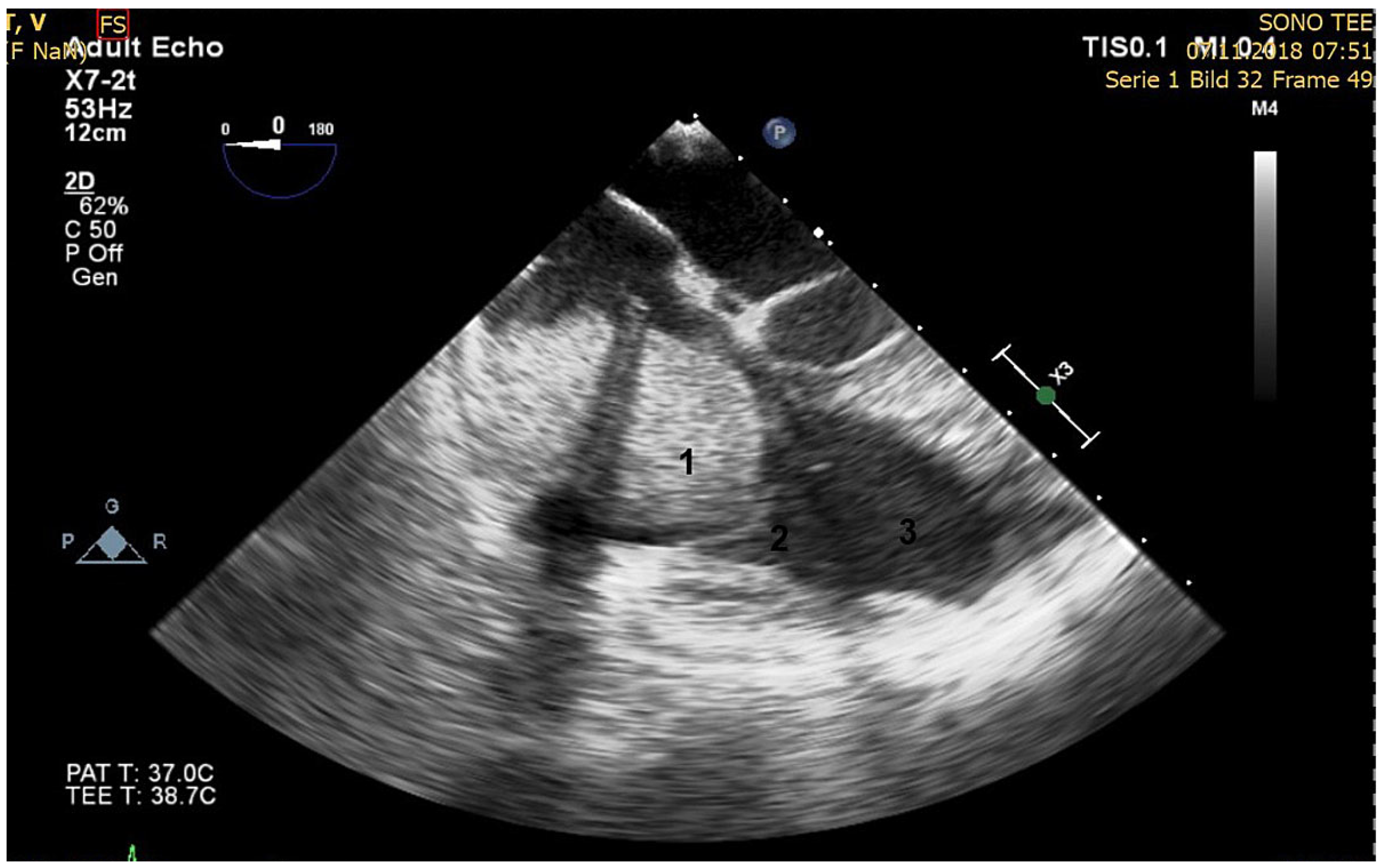
Figure 1. Preoperative transoesophageal echocardiography. 1. Thrombus in right atrium with shadow of the catheter; 2. Tricuspid valve; 3. Right ventricle.
Cardiovascmed 23 w02104 g001