Next Article in Journal
Major Right Atrial Thrombus Related to Central Venous Catheter Positioning
Previous Article in Journal
Modernes Lipid-Management
 
 
Cardiovascular Medicine is published by MDPI from Volume 28 Issue 1 (2025). Previous articles were published by another publisher in Open Access under a CC-BY (or CC-BY-NC-ND) licence, and they are hosted by MDPI on mdpi.com as a courtesy and upon agreement with Editores Medicorum Helveticorum (EMH).
Interesting Images

Fusiform Mid-LAD Aneurysm Due to Passive Arterial Wall Dilatation After Implantation of a Self-Expandable Stent

by
Alessandro Negro
1,2,*,
Luigi Biasco
3,
Giovanni Pedrazzini
4 and
Marco Moccetti
4
1
FMH Internal Medicine, FMH Cardiology, Sementina, Switzerland
2
Medical Village, Via Cantonale 38, CH-6928 Manno, Switzerland
3
Ciriè and Ivrea Hospitals, ASL TO4, Cirié, Italy
4
Cardiocentro Ticino, Lugano, Switzerland
*
Author to whom correspondence should be addressed.
Cardiovasc. Med. 2020, 23(2), w02106; https://doi.org/10.4414/cvm.2020.02106
Submission received: 20 January 2020 / Revised: 20 February 2020 / Accepted: 20 March 2020 / Published: 20 April 2020
Coronary artery aneurysm formation following percutaneous coronary intervention (PCI) with drug-eluting stents is a known, infrequent complication, causing late acquired strut malapposition, a potential trigger for in-stent thrombosis [1]. First generation durable-polymer sirolimus-eluting stents showed a significant association with coronary evaginations on optical coherence tomography (OCT), probably related to the inflammatory and durable polymer [2].
The Xposition S is a sirolimus-eluting self-apposing coronary stent with a durable polymer that, by passively adapting to variances in vessel diameter, allows for maximal stent apposition.
We herein report the case of a late acquired fusiform aneurysm of the mid-left anterior descending artery (LAD) caused by the passive arterial wall dilatation as a consequence of the implantation of aself-expandable stent.
A 61-year-old patient, with known coronary artery disease, underwent PCI with a self-expandable stent (Stentis XPO-SITION S 3.5−4.5 × 27 mm) for an anterior non-ST segment elevation myocardial infarction (NSTEMI) in January 2016. Post-implant OCT showed good apposition of the stent, with a mean vessel diameter of 3.57 mm at the distal edge and 3.67 mm at the proximal edge (Figure 1)
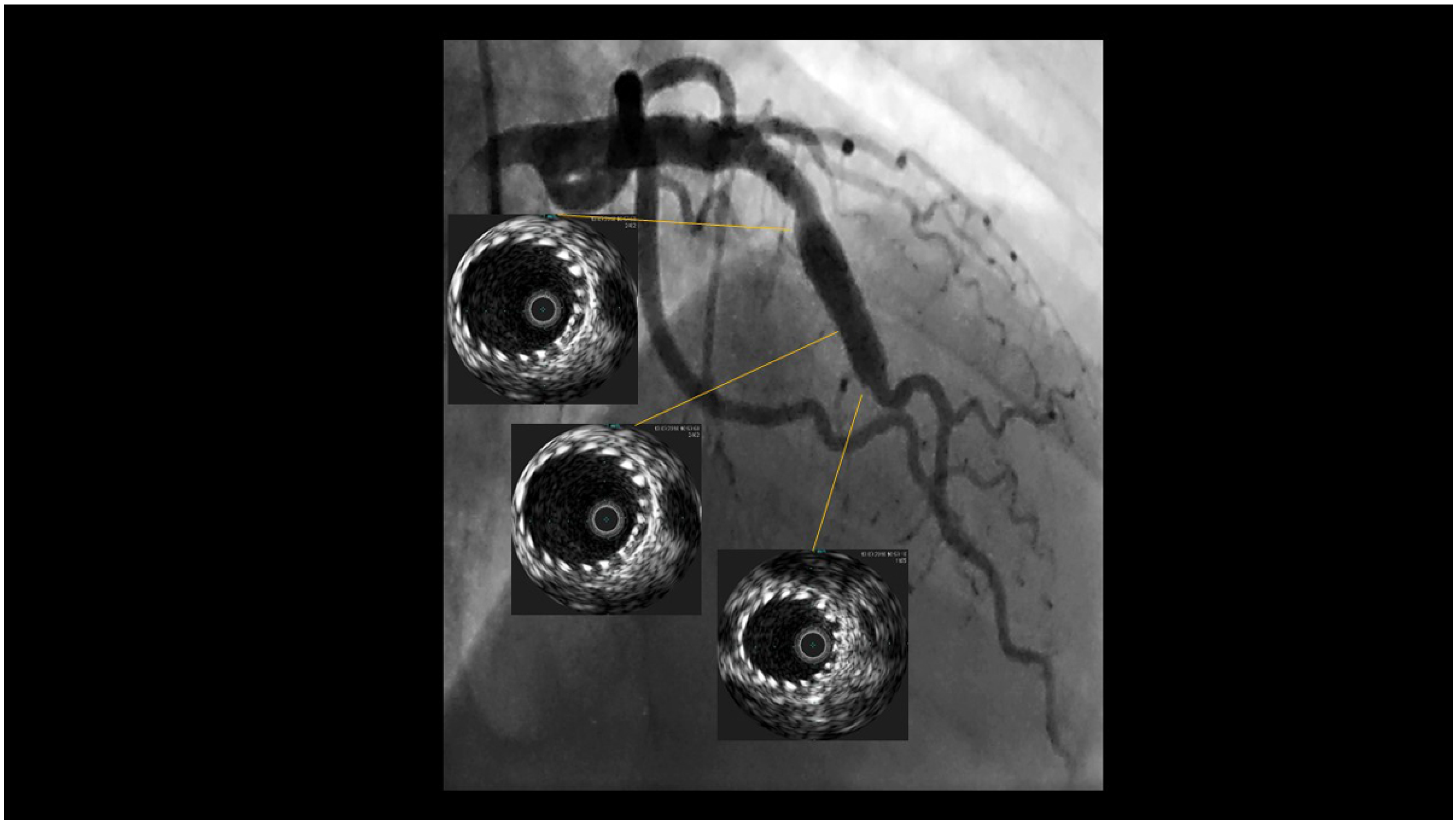
Because of recurrent angina, check-up angiography was performed in March 2018. A fusiform aneurysm of the mid-LAD was discovered. Intravascular ultrasound revealed a uniform dilatation of the vessel with a calibre of 6 mm throughout the whole stented segment, with no evidence of late acquired malapposition (Figure 2), confirming angiographic findings.
This case represents a new, previously undescribed complication after implantation of self-expandable stent, which caused passive arterial wall dilatation.

Conflicts of Interest

No financial support and no other potential conflict of interest relevant to this article was reported.

References

  1. Alfonso, F.; Pérez-Vizcayno, M.J.; Ruiz, M.; Suárez, A.; Cazares, M.; Hernández, R.; et al. Coronary aneurysms after drug-eluting stent implantation: clinical, angiographic, and intravascular ultrasound findings. J. Am. Coll. Cardiol. 2009, 53, 2053–2060. [Google Scholar] [CrossRef] [PubMed]
  2. Radu, M.D.; Räber, L.; Kalesan, B.; Muramatsu, T.; Kelbæk, H.; Heo, J.; et al. Coronary evaginations are associated with positive vessel remodelling and are nearly absent following implantation of newer-generation drug-eluting stents: an optical coherence tomography and intravascular ultrasound study. Eur. Heart J. 2014, 35, 795–807. [Google Scholar] [CrossRef] [PubMed]
Figure 1. Post-implant optical coherence tomography (January 2016): good apposition of the stent with a mean vessel diameter of 3.57 mm at the distal edge and 3.67 mm at the proximal edge.
Cardiovascmed 23 w02106 g001
Figure 2. Intravascular ultrasound (March 2018): evidence of fusiform aneurysm of the mid-left anterior descending artery, with uniform dilatation of the vessel with a calibre of 6 mm throughout the whole stented segment.
Cardiovascmed 23 w02106 g002