Article

Anti-Allergic Effect of Aqueous Extract of Coriander (Coriandrum sativum L.) Leaf in RBL-2H3 Cells and Cedar Pollinosis Model Mice

1
Department of Bioscience, Graduate School of Agriculture, Ehime University, Matsuyama 790-8566, Japan
2
Food and Health Sciences Research Center, Ehime University, Matsuyama 790-8566, Japan
3
Department of Food Science, Faculty of Bioresources and Environmental Sciences, Ishikawa Prefectural University, Nonoichi 921-8836, Japan
*
Author to whom correspondence should be addressed.
Nutraceuticals 2022, 2(3), 170-180; https://doi.org/10.3390/nutraceuticals2030013
Submission received: 22 June 2022 / Revised: 19 July 2022 / Accepted: 22 July 2022 / Published: 26 July 2022
(This article belongs to the Special Issue Functional Foods as a New Therapeutic Strategy)

Abstract

:
Coriander (Coriandrum sativum L.) is classified in the Apiaceae family and used as an herb. Coriander leaf has been reported to possess various health functions. Here, we report the anti-allergic effect of aqueous coriander leaf extract (ACLE). ACLE with 1.0 mg/mL or higher concentration significantly inhibited degranulation of RBL-2H3 cells in a concentration-dependent manner with no cytotoxicity. ACLE suppressed the increase in the intracellular Ca2+ concentration in response to antigen-specific stimulation. Immunoblot analysis demonstrated that ACLE significantly downregulates phosphorylation of phosphatidylinositol 3-kinase and tends to downregulate phosphorylation of Syk kinase in the signaling pathways activated by antigen-mediated stimulation. Oral administration of ACLE did not alter the sneezing frequency of pollinosis model mice stimulated with cedar pollen, but significantly reduced the serum IgE level. Our data show anti-allergic effects of coriander leaf in both cultured cells and pollinosis mice. These results suggest that coriander leaf has the potential to be a functional foodstuff with anti-allergy effects.

1. Introduction

An allergy means to immunize excessively against antigens invading from the outside of the body. Allergy falls into four types, among which type I allergy, including allergic rhinitis, asthma, atopic dermatitis, and hay fever, is increasing in the number of patients in recent years worldwide. The number of patients with pollinosis is approximately 30% of the population and is still increasing in Japan [1,2]. Type I allergy, also called immediate hypersensitivity or anaphylactoid type, is attributed to immunoglobulin E (IgE) [3]. Antigen-specific IgE antibodies are secreted by plasma cells and bind to FcεRI receptor on mast cells. Upon crosslinking of antigens to IgE antibody bound on FcεRI, various chemical mediators contained in granules are released from the cells. This phenomenon is called degranulation, and the secreted chemical mediators cause smooth muscle contraction, hypervascular permeability, hyperactivity, etc. Suppressing degranulation can therefore be an effective strategy for relieving allergy symptoms, and the research on the substance that alleviates allergic symptoms is considered crucially important.
Coriander (Coriandrum sativum L.), an aromatic herb commonly used for cooking in southeast Asia thanks to its unique aroma and flavor, belongs to the Apiaceae family. Coriander is well known to possess several biological activities [4,5,6,7,8]. For example, antimicrobial activities of C. sativum essential oil have been well reported [9,10,11]. C. sativum seeds exhibit antidiabetic effects in streptozotocin-induced diabetic mice [12,13], in rats fed a high-fat diet [14], and in obese–hyperglycemic–hyperlipidemic Meriones shawi rats [15]. C. sativum seeds also show anxiolytic [16] and sedative [17] activities. We then tried to find a novel biological activity of C. sativum. After screening using various cell-based assays, we found an anti-allergic effect of a water-soluble extract from leaves of C. sativum in a mast cell degranulation assay using RBL-2H3 cells, although an allergen is present in coriander [18,19]. Herein, we report a suppressive activity of an aqueous extract of coriander leaf on degranulation of RBL-2H3 cells. We also aimed to elucidate the mechanism of action of the extract. We further hypothesized that the extract is effective in a mouse model of type I allergy.

2. Materials and Methods

2.1. Reagents

Triton X-100, anti-dinitrophenyl (DNP) IgE, DNP-human serum albumin (HSA), bovine serum albumin (BSA), Trizma base, and p-coumaric acid were obtained from Sigma-Aldrich (St. Louis, MO, USA). Cedar pollen allergen (purified Cry j1 and Cry j2) was obtained from Hayashibara (Okayama, Japan). Horseradish peroxidase (HRP)-labeled anti-goat IgG antibody and goat anti-actin antibody were obtained from Santa Cruz Biotechnology (Santa Cruz, CA, USA). Anti-Syk antibody, anti-phosphorylated Syk (Tyr525/526) antibody, anti-phosphoinositide 3-kinase (PI3K) p55 antibody, anti-phosphorylated PI3K p85 (Tyr458)/p55 (Tyr199) antibody, and anti-rabbit IgG antibody labeled with HRP were purchased from Cell Signaling Technology (Danvers, MA, USA).

2.2. Sample Preparation

C. sativum L. leaf was obtained from S&B Foods Inc. (Tokyo, Japan). First, raw leaves of C. sativum L. were lyophilized and pulverized into powders. The powder was suspended in 10 mM Na phosphate buffer (pH 7.4) for 24 h and was centrifuged at 15,000× g for 20 min at 4 °C. After adjusting the pH to 7.4 and filtrating through a 0.45 µm membrane, the supernatant was used as aqueous coriander leaf extract (ACLE).

2.3. Cells and Cell Culture

Rat basophilic leukemia RBL-2H3 cells were obtained from American Type Culture Collection (Rockville, MD, USA). Dulbecco’s modified Eagle’s medium (DMEM, Sigma-Aldrich) supplemented with 100 U/mL of penicillin (Sigma-Aldrich), 100 µg/mL of streptomycin (Sigma-Aldrich), and 5% fetal bovine serum (FBS, Sigma-Aldrich) was used for propagation of RBL-2H3 cells in a monolayer culture at 37 °C under humidified 5% CO2 [20]. RBL-2H3 cells were detached using a trypsin/EDTA solution at approximately 80% confluence. RBL-2H3 cells with a viability of >90% before 15 passages were used for the experiments described below.

2.4. Mice

Female BALB/c mice were acquired from Japan SLC (Hamamatsu, Japan) and were kept at 24 ± 1 °C under a 12 h light/dark cycle. Animals freely received water and standard laboratory chow. Animal experiments were approved by the Animal Experiment Committee of Ehime University and were performed in accordance with the Guidelines of Animal Experiments of Ehime University (approval number: 08U13-1).

2.5. β-Hexosaminidase Release Assay

The assay was conducted as previously described [21] with some modifications. RBL-2H3 cells (4.0 × 10⁴ cells/well) seeded in each well of a 96-well culture plate (Corning, Corning, NY, USA) were precultured for 18 h. The cells were next sensitized with 50 ng/mL of anti-DNP IgE for 2 h and were subsequently treated with the indicated concentration of ACLE for 10 min. Degranulation was then induced with 50 ng/mL of DNP-HSA for 30 min. Following the collection of the medium from each well, the cells were sonicated on ice. After preincubating the collected medium and cell lysate for 5 min at 37 °C, the β-hexosaminidase substrate (p-nitrophenyl 2-acetamido-2-deoxy-β-d-glucopyranoside) was added and incubated at 37 °C for 25 min. After terminating the enzymatic reaction, the absorbance (405 nm) was measured using an SH-8000Lab microplate reader (Corona Electric, Hitachinaka, Japan). β-Hexosaminidase release rate (%) was calculated as follows:
100 × [(Asupernatant − Ablank of supernatant)/{(Asupernatant − Ablank of supernatant) + (Acell lysate − Ablank of cell lysate)}]
where “A” is the absorbance of each well.

2.6. Cell Viability

Cytotoxicity of ACLE to RBL-2H3 cells was assessed by WST-8 assay described previously [22]. RBL-2H3 cells were seeded at 4.0 × 10⁴ cells/well into each well of a 96-well culture plate (Corning). After treatment with 0, 1.0, 2.0, and 4.0 mg/mL of ACLE for 10 min, the cells were incubated with a WST-8 reagent for 15 min at 37 °C, and the absorbance (450 nm) was measured using a Model 550 microplate reader (Bio-Rad Laboratories, Hercules, CA, USA). The absorbance of wells with the cells treated with ACLE was compared to the absorbance of wells with the cells untreated. The assay was conducted in triplicate.

2.7. Monitoring of Intracellular Ca2+ Concentration ([Ca2+]i)

[Ca2+]i was monitored using a fluorescent calcium indicator Fluo 3-AM (Dojindo Laboratories, Mashiki, Japan) as described previously [23]. RBL-2H3 cells were seeded and sensitized as described in Section 2.5. The cells were next incubated in the loading buffer containing Fluo 3-AM for 1 h. After washing with phosphate-buffered saline (PBS, pH 7.4), the cells were incubated in the recording buffer containing ACLE (4.0 mg/mL) for 10 min at 37 °C. The cells were subsequently stimulated with DNP-HSA at a final concentration of 2.5 µg/mL, and the fluorescence intensity was immediately monitored using the SH-8000Lab microplate reader (excitation wavelength: 480 nm; emission wavelength: 530 nm).

2.8. Immunoblot Analysis

Immunoblot analysis was conducted as described previously [24]. RBL-2H3 cells seeded at 5.0 × 10⁵ cells/dish in 3.5 cm culture dishes (Corning) were precultured for 18 h. After sensitizing with 50 ng/mL of anti-DNP IgE for 2 h, the cells were subsequently treated with 980 µL of modified Tyrode’s buffer containing 4.0 mg/mL of ACLE for 10 min. The cells were next stimulated with DNP-HSA at a final concentration of 50 ng/mL for 5 min. After the added reagents were removed, the cells were lysed and centrifuged at 12,000× g for 15 min at 4 °C. Proteins were then separated by sodium dodecyl sulfate-polyacrylamide gel electrophoresis and were transferred onto a polyvinylidene fluoride membrane (GE Healthcare, Buckinghamshire, UK). After blocking with 20 mM Tris-buffered saline (TBS, pH 7.6) containing 5% skim milk for 1 h at room temperature, the membrane was reacted with anti-actin antibody (1:200 dilution), anti-Syk antibody (1:1000 dilution), anti-phosphorylated Syk antibody (1:1000 dilution), anti-PI3K p55 antibody (1:1000 dilution), or anti-phosphorylated PI3K p85/p55 antibody (1:1000 dilution) as a primary antibody at 4 °C overnight. After washing with TBS containing 0.1% Tween 20 thrice, the membrane was reacted with HRP-labeled anti-rabbit IgG antibody (1:2000 dilution) or HRP-labeled anti-goat IgG antibody (1:2000 dilution) as a secondary antibody for 1 h at room temperature. After washing the membrane with TBS containing 0.05% Tween 20 thrice, the blot was developed with ImmunoStar LD chemiluminescence detection reagent (Fujifilm Wako Pure Chemical, Osaka, Japan), and bands were visualized using a ChemiDoc XRS system (Bio-Rad Laboratories).

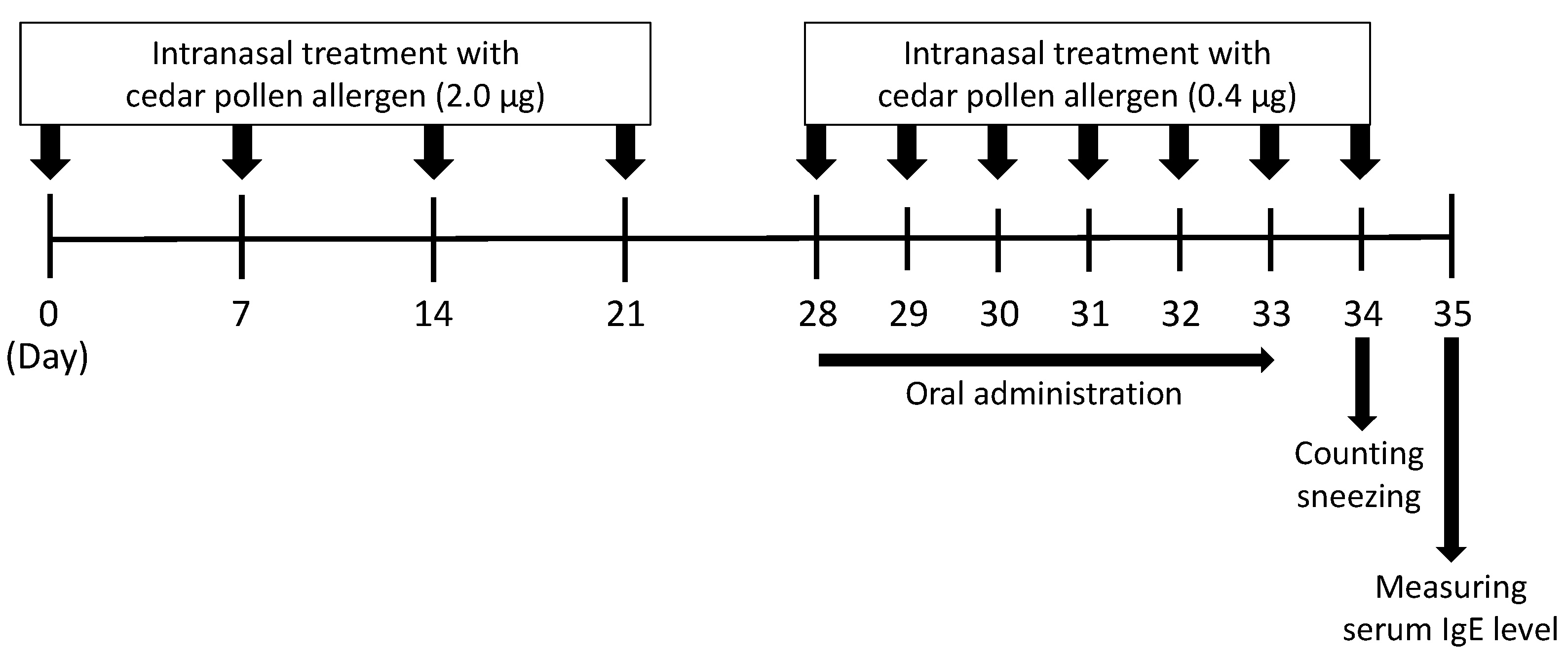
2.9. A Mouse Model of Japanese Cedar Pollinosis

Japanese cedar pollinosis model mice were developed as described previously [25,26]. Following an adaptation period for 1 week, 6-week-old female BALB/c mice were randomly assigned to two groups as follows: the control group (9 mice) and the ACLE group (8 mice). The mice were intranasally sensitized with 10 μL of PBS containing 2.0 μg of purified cedar pollen allergen on days 0, 7, 14, and 21, as shown in Figure 1. The mice were next intranasally challenged with 10 μL of PBS containing 0.4 μg of cedar pollen allergen for seven consecutive days from day 28 to 34. The mice in the ACLE group were orally administered 20 µL of ACLE at 50 mg/kg/day for six consecutive days from day 28 to 33, while the mice in the control group were with 20 µL of the vehicle alone. On day 34, all the mice were intranasally challenged with 0.4 μg of cedar pollen allergen. Sneezing frequency was counted for 15 min immediately after the final nasal treatment. On day 35, serum was obtained to determine their serum IgE levels.

2.10. Enzyme-Linked Immunosorbent Assay (ELISA)

Serum IgE levels were measured by in-house-developed ELISA as previously de-scribed [27]. The absorbance (415 nm) was measured using the SH-8000Lab microplate reader after adding 1.5% oxalic acid (100 µL/well) to terminate the enzymatic reaction.

2.11. High-Performance Liquid Chromatography (HPLC) Analysis

Reversed-phase HPLC analysis of ACLE was performed on a LaChrom Elite HPLC system (Hitachi, Tokyo, Japan) with an XBridge C18 column (100 × 4.6 mm, 3.5 μm, Waters, Milford, MA, USA). A linear gradient elution program was applied as follows: 0–6 min (20% acetonitrile + 0.1% formic acid); 6–9 min (20–95% acetonitrile + 0.1% formic acid); 9–12 min (95% acetonitrile + 0.1% formic acid). The flow rate was maintained at 1.0 mL/min, and the temperature of the column was set at 35 °C. The chromatogram was monitored at a wavelength of 220 nm.

2.12. Statistical Analysis

Data obtained are shown as mean ± SEM. Statistical analysis was conducted using GraphPad Prism version 7.02 (GraphPad Software, La Jolla, CA, USA). Mann-Whitney U test or Dunnett’s test was used to assess the statistical significance of the treatments against control. Values of * p < 0.05, ** p < 0.01 and *** p < 0.001 were considered statistically significant.

3. Results and Discussion

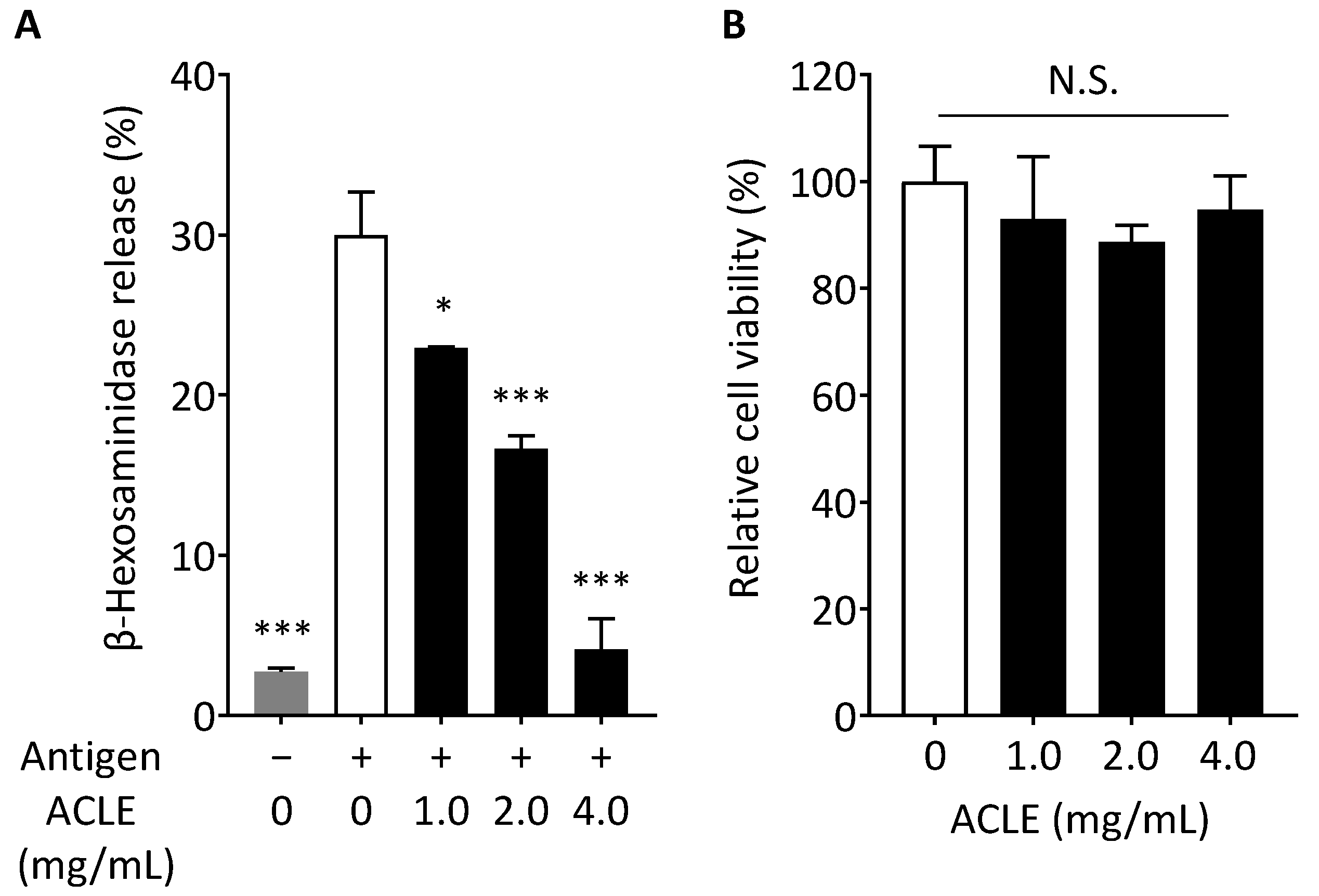
3.1. Effect of ACLE on Degranulation of RBL-2H3 Cells

We first investigated whether ACLE possesses a suppressive effect on degranulation. RBL-2H3 cells sensitized with anti-DNP IgE were treated with the indicated concentration of ACLE, and degranulation was induced with the antigen. Secreted β-hexosaminidase was utilized as a degranulation marker. ACLE with 1.0 mg/mL or higher concentration significantly inhibited the degranulation in a concentration-dependent manner, as shown in Figure 2A. This result indicates that water-soluble, bioactive substance exist in coriander leaf, which exhibits anti-allergic activity by suppressing degranulation. We next examined by the WST-8 assay whether ACLE affects the viability of RBL-2H3 cells. As shown in Figure 2B, the viability of RBL-2H3 cells was unaffected by ACLE with the concentration tested. These data indicated that ACLE suppresses the degranulation of RBL-2H3 cells with no cytotoxicity.

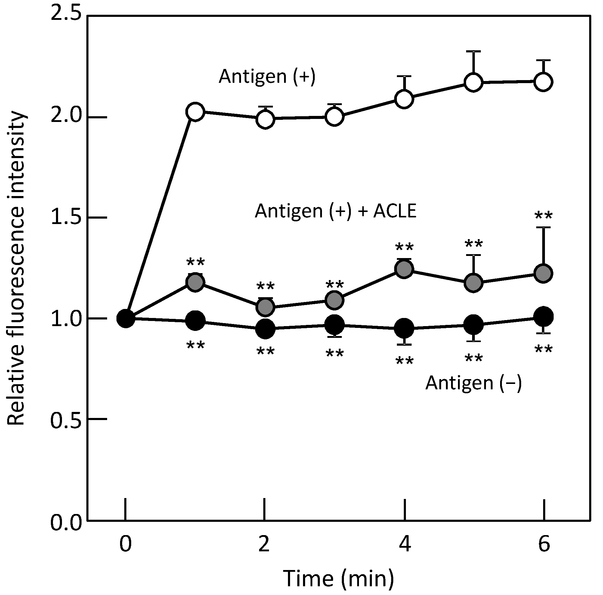
3.2. Effect of ACLE on the Elevation of [Ca2+]i Induced by Antigen

Degranulation occurs upon the antigen binding to IgE on mast cells, thereby initiating intracellular signal transduction and increasing Ca2+ concentration in the cell. The elevation of intracellular concentration of Ca2+, the major second messenger in intracellular signaling, is the critical process in mast cell degranulation. The effect of ACLE on the elevation of intracellular calcium concentration was hence evaluated using Fluo 3-AM. Relative fluorescence intensity of Fluo 3 reflects the intracellular Ca2+ concentration. RBL-2H3 cells sensitized with anti-DNP IgE antibody were treated with ACLE (4.0 mg/mL), and [Ca2+]i was monitored for 6 min. [Ca2+]i rapidly rose upon stimulation with antigen, as shown in Figure 3. However, the increase in intracellular calcium ion concentration was significantly suppressed in the presence of 4.0 mg/mL of ACLE. It was supposed from this result that ACLE inhibits degranulation by downregulating the intracellular signaling pathway leading to the elevation of [Ca2+]i.

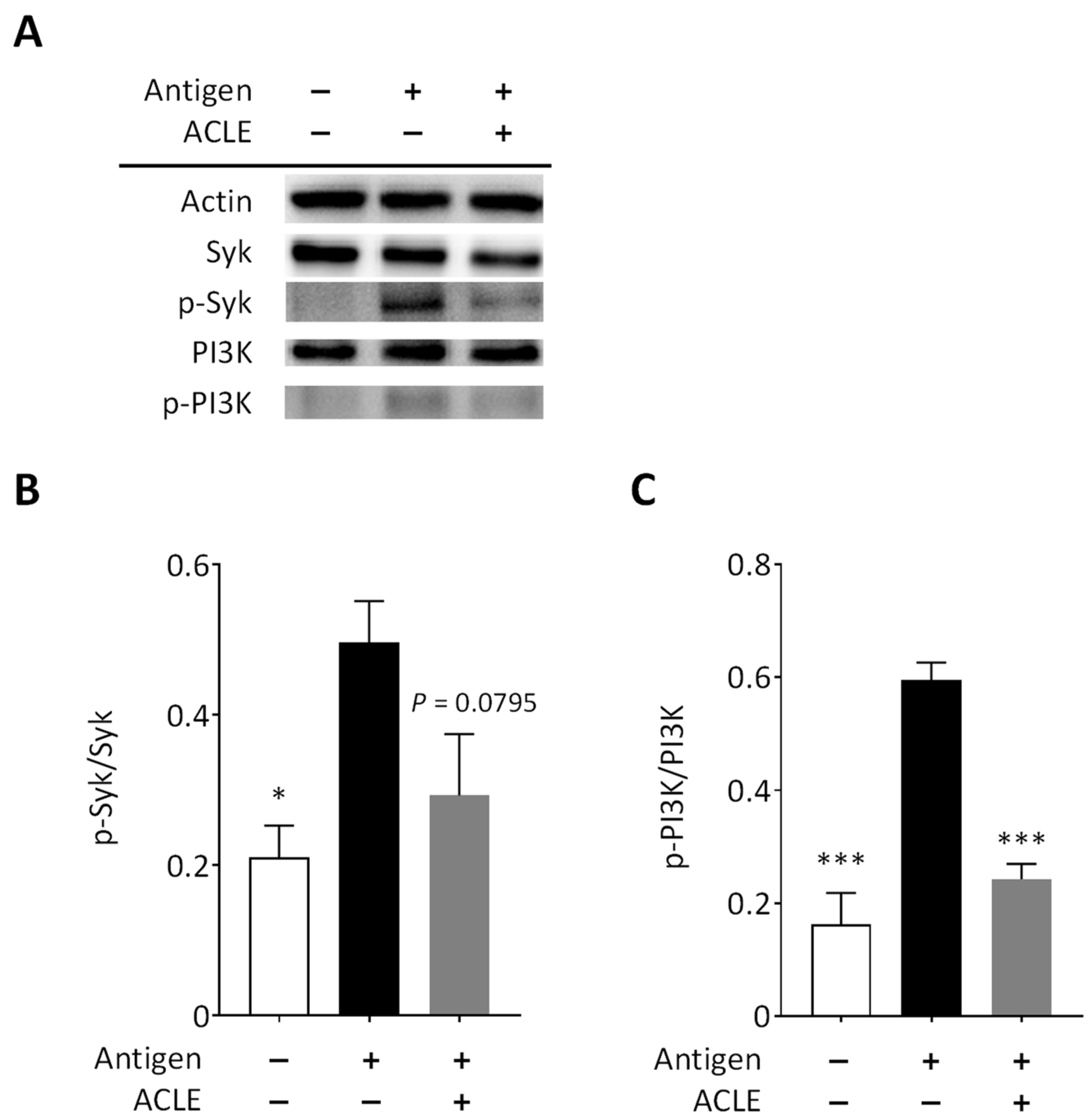
3.3. Effect of ACLE on Intracellular Signaling Pathways Leading to Degranulation

The onset of the signaling pathways resulting in degranulation is regulated by phos-phorylation of Syk kinase located close to FcεRI, accompanied by phosphorylation of downstream signaling factors, leading to the increase in intracellular calcium ion concentration. We examined by immunoblot analysis how ACLE affects the signaling pathways related to degranulation (Figure 4A). As shown in Figure 4B, treating cells with ACLE tended to decrease the phosphorylation of Syk kinase (p = 0.0795 vs. control), and phosphorylation of PI3K was significantly inhibited by ACLE treatment (Figure 4C).
There are two pathways for mast cell degranulation: calcium-dependent and calci-um-independent pathways [28,29]. The calcium-dependent pathway is activated by the tyrosine kinase Lyn located proximal to FcεRI, thereby activating another tyrosine kinase Syk [30]. Phosphorylated Syk activates a signaling cascade including phospholipase Cγ. As a result, Ca2+ inflow into the interior of cells through the Ca2+ channel is induced, which causes degranulation by increased [Ca2+]i. On the other hand, tyrosine kinase Fyn is activated by cross-linking of antigen to IgE on FcεRI, which results in the activation of the calcium-independent pathway [31]. Phosphorylation of PI3K leads to microtubule reorganization that causes degranulation by migrating granules [32]. In the degranulation process, the calcium-dependent pathway plays a crucial role, while the calcium-independent pathway has been reported to be given priority under weak stimulation when cells were stimulated with a low concentration of antigen [33]. Our results indicated that downregulated phosphorylation of Syk kinase and PI3K is a key process for the suppressive activity of ACLE on degranulation. As PI3K is located downstream of Syk kinase, downregulated phosphorylation of PI3K, as shown in Figure 4C, might be attributed to reduced phosphorylation of Syk. Our data suggested that an inhibitory effect of ACLE on the calcium-dependent pathway causes the reduced elevation of [Ca2+]i induced with antigen (Figure 3), resulting in suppressed degranulation.
PI3K plays a crucial role in certain tumor progression [34,35]. Because ACLE inhibits phosphorylation of PI3K, it might exhibit antitumor activity. Indeed, the C. sativum leaf has been reported to possess antitumor effect [36,37]. Syk is expressed in various cells and is a potential target for treating several diseases, such as liver fibrosis [38], acute myeloid leukemia [39], and autoimmune diseases [40]; ACLE might therefore be effective in attenuating the symptoms of these diseases by inhibiting phosphorylation of Syk. In addition, because a Syk inhibitor can attenuate allergen-induced symptoms in patients with allergic rhinitis exposed to pollens [41], we further investigated whether ACLE is capable of reducing allergic symptoms in pollinosis mice.

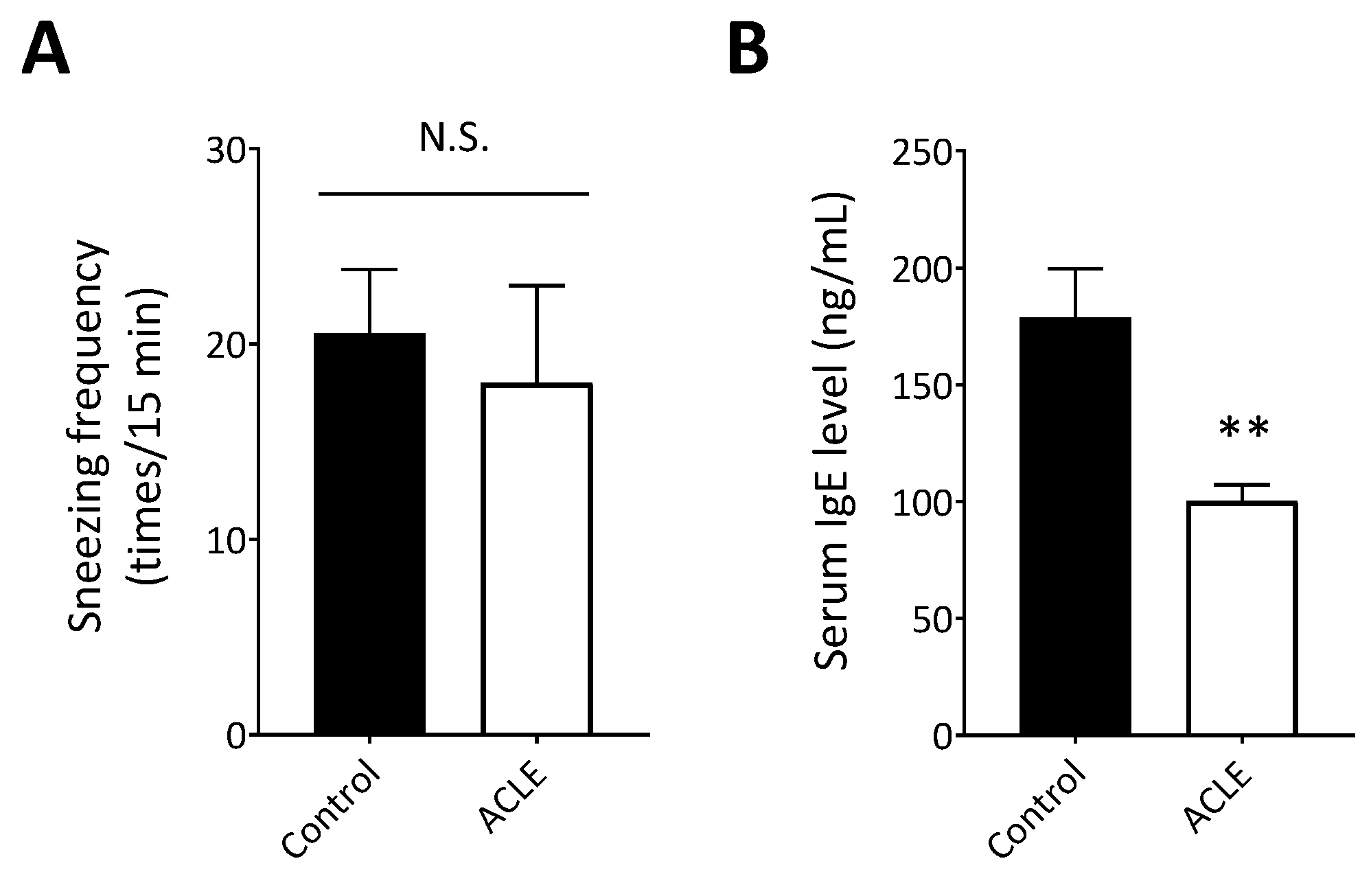
3.4. Effect of ACLE on a Mouse Model of Japanese Cedar Pollinosis

Cedar pollinosis mice were used to investigate the in vivo effect of ACLE. Mice were sensitized with Cry j1 and Cry j2, the main allergens of Japanese cedar pollen. Cedar pollinosis was induced by intranasal treatment of cedar pollen allergen 11 times for 5 weeks. The ACLE group was orally administered ACLE for the last six days. Oral administration of ACLE unaffected the body weight of mice (data not shown), indicating that ACLE is not toxic to mice. The sneezing frequency was counted for 15 min on day 34. As a result, ACLE administration did not alter sneezing frequency (Figure 5A). This result indicated that the water-soluble, bioactive ingredient in ACLE seems to be inefficiently absorbed into the mouse body or to be readily metabolized after the absorption into the body. ACLE thus did not alleviate the allergic symptom.
On day 35, sera were collected to measure the serum IgE level by ELISA. Unexpectedly, the serum IgE level of the ACLE group was significantly reduced compared with that of the control group (Figure 5B). This result suggested that ACLE could suppress the IgE production as well as mast cell degranulation. This result also implied that ACLE might possess the potential to prevent sensitization with an allergen by inhibiting IgE production. The bioavailability of ACLE in mice is now under investigation to elucidate why ACLE did not mitigate the allergic symptom but decreased IgE levels in pollinosis mice. In addition, the mechanism of ACLE underlying the decrease in IgE production is also under exploration. It might be possible that ACLE affects lymphocytes, such as T lymphocytes and B lymphocytes, to inhibit the class switching to IgE antibody.

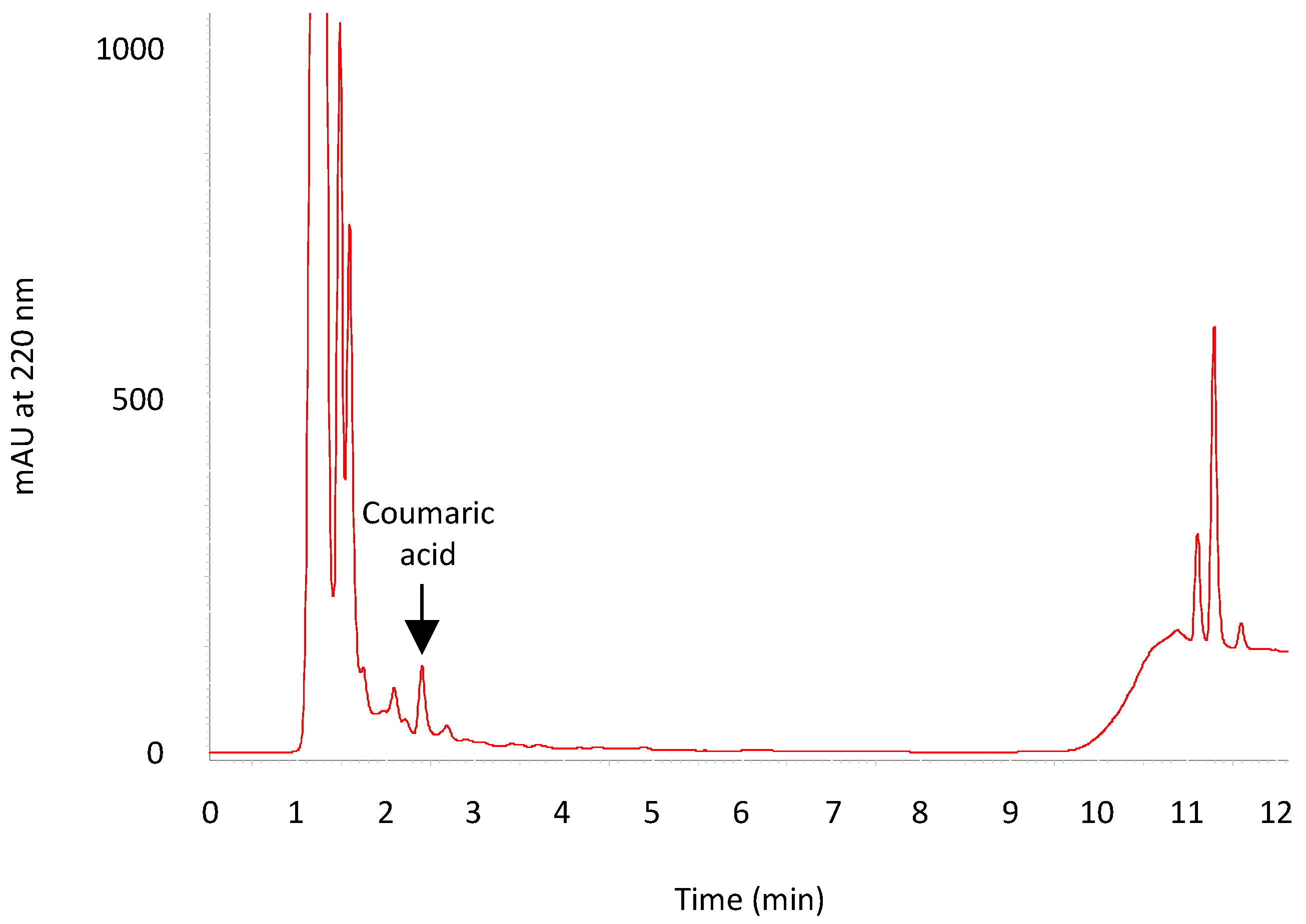
3.5. HPLC Analysis of ACLE

Finally, a reversed-phase HPLC analysis of ACLE was conducted. The chemical composition of essential oils of C. sativum has been well reported, and various secondary metabolites in aerial part of C. sativum, such as flavonoids and coumarins, have been also identified [42,43]; however, papers reporting the chemical composition of water-soluble molecules in C. sativum is limited [44], although several papers have reported estimated total phenolic contents.
ACLE (20 µL) was subjected to an XBridge C18 column. Figure 6 shows an HPLC chromatogram of ACLE. The result showed that coumaric acid was present at 6.8 µg/mL in ACLE. Other peaks have not been identified yet. Zeković et al. [45] identified ferulic acid and sinapic acid, in addition to coumaric acid, in an aqueous coriander seeds extract; however, ferulic acid and sinapic acid were not found in ACLE in this study. This seems to result from the differences in the part of the plant used for extraction (seeds vs. leaves) and in the extraction solvent (water vs. sodium phosphate buffer.) Chen et al. [46] have reported that coumaric acid exhibits a suppressive effect on the degranulation of RBL-2H3 cells. Coumaric acid, a secondary metabolite of this plant, thus might be partially attributed to the suppressive effect of ACLE on degranulation of RBL-2H3 cells; however, the coumaric acid concentration (6.8 µg/mL) found in ACLE seems too low to exhibit the suppressive effect of ACLE by itself, as shown in Figure 2A. We thus assume that there is another bioactive ingredient in ACLE in addition to coumaric acid. Identification of the peaks shown in Figure 6 is now under investigation to explore the water-soluble bioactives contained in coriander leaf.

4. Conclusions

ACLE inhibited degranulation of RBL-2H3 cells with no cytotoxicity by suppressing the elevation of intracellular Ca2+ concentration resulting from downregulated phosphorylation of the intracellular signal transduction pathways. In addition, oral administration of ACLE significantly decreased the serum IgE level in pollinosis model mice. These findings suggest that coriander leaf possesses the potential to be a functional foodstuff with anti-allergic effects.

Author Contributions

Conceptualization, T.S.; data curation, Y.K., K.N., M.I., S.N. and T.S.; investigation, Y.K. and K.N.; supervision, K.N. and T.S.; writing—original draft, Y.K. and K.N.; writing—review and editing, K.N. and T.S. All authors have read and agreed to the published version of the manuscript.

Funding

This research received no external funding.

Institutional Review Board Statement

The animal study protocol was conducted in accordance with the Guidelines of Animal Experiments of Ehime University and approved by the Animal Experiment Committee of Ehime University (approval number: 08U13-1).

Informed Consent Statement

Not applicable.

Data Availability Statement

The data that support the findings in this study are available from the corresponding author upon reasonable request.

Acknowledgments

Animal experiments were conducted at the Division of Genetic Research of the Advanced Research Support Center (ADRES), Ehime University.

Conflicts of Interest

Ehime University has filed a patent application related to a new anti-allergic agent, and T.S. is an inventor of the patent application. The remaining authors declare no conflict of interest.

References

  1. Yamada, T.; Saito, H.; Fujieda, S. Present state of Japanese cedar pollinosis: The national affliction. J. Allergy Clin. Immunol. 2014, 133, 632–639. [Google Scholar] [CrossRef] [PubMed]
  2. Nishihata, S.; Murata, T.; Inoue, S.; Okubo, K.; Sahashi, N.; Takahashi, H.; Hirooka, J.; Hoshiyama, Y.; Murayama, K.; Mezawa, A.; et al. Prevalence of Japanese cedar pollinosis in Tokyo: A survey conducted by the Tokyo Metropolitan Government. Clin. Exp. Allergy Rev. 2010, 10, 8–11. [Google Scholar] [CrossRef]
  3. Galli, S.J.; Tsai, M.; Piliponsky, A.M. The development of allergic inflammation. Nature 2008, 454, 445–454. [Google Scholar] [CrossRef] [PubMed] [Green Version]
  4. Laribi, B.; Kouki, K.; M’Hamdi, M.; Bettaieb, T. Coriander (Coriandrum sativum L.) and its bioactive constituents. Fitoterapia 2015, 103, 9–26. [Google Scholar] [CrossRef] [PubMed]
  5. Sahib, N.G.; Anwar, F.; Gilani, A.H.; Hamid, A.A.; Saari, N.; Alkharfy, K.M. Coriander (Coriandrum sativum L.): A potential source of high-value components for functional foods and nutraceuticals—A review. Phytother. Res. 2013, 27, 1439–1456. [Google Scholar] [CrossRef] [PubMed] [Green Version]
  6. Wei, J.N.; Liu, Z.H.; Zhao, Y.P.; Zhao, L.L.; Xue, T.K.; Lan, Q.K. Phytochemical and bioactive profile of Coriandrum sativum L. Food Chem. 2019, 286, 260–267. [Google Scholar] [CrossRef]
  7. Prachayasittikul, V.; Prachayasittikul, S.; Ruchirawat, S.; Prachayasittikul, V. Coriander (Coriandrum sativum): A promising functional food toward the well-being. Food Res. Int. 2018, 105, 305–323. [Google Scholar] [CrossRef]
  8. Sobhani, Z.; Mohtashami, L.; Amiri, M.S.; Ramezani, M.; Emami, S.A.; Simal-Gandara, J. Ethnobotanical and phytochemical aspects of the edible herb Coriandrum sativum L. J. Food Sci. 2022, 87, 1386–1422. [Google Scholar] [CrossRef]
  9. Matasyoh, J.C.; Maiyo, Z.C.; Ngure, R.M.; Chepkorir, R. Chemical composition and antimicrobial activity of the essential oil of Coriandrum sativum. Food Chem. 2009, 113, 526–529. [Google Scholar] [CrossRef]
  10. Begnami, A.F.; Duarte, M.C.T.; Furletti, V.; Rehder, V.L.G. Antimicrobial potential of Coriandrum sativum L. against different Candida species in vitro. Food Chem. 2010, 118, 74–77. [Google Scholar] [CrossRef]
  11. Silva, F.; Domingues, F.C. Antimicrobial activity of coriander oil and its effectiveness as food preservative. Crit. Rev. Food Sci. Nutr. 2017, 57, 35–47. [Google Scholar] [CrossRef] [PubMed]
  12. Gray, A.M.; Flatt, P.R. Insulin-releasing and insulin-like activity of the traditional anti-diabetic plant Coriandrum sativum (coriander). Br. J. Nutr. 1999, 81, 203–209. [Google Scholar] [CrossRef] [PubMed] [Green Version]
  13. Eidi, M.; Eidi, A.; Saeidi, A.; Molanaei, S.; Sadeghipour, A.; Bahar, M.; Bahar, K. Effect of coriander seed (Coriandrum sativum L.) ethanol extract on insulin release from pancreatic beta cells in streptozotocin-induced diabetic rats. Phytother. Res. 2009, 23, 404–406. [Google Scholar] [CrossRef] [PubMed]
  14. Chithra, V.; Leelamma, S. Coriandrum sativum—Mechanism of hypoglycemic action. Food Chem. 1999, 67, 229–231. [Google Scholar] [CrossRef]
  15. Aissaoui, A.; Zizi, S.; Israili, Z.H.; Lyoussi, B. Hypoglycemic and hypolipidemic effects of Coriandrum sativum L. in Meriones shawi rats. J. Ethnopharmacol. 2011, 137, 652–661. [Google Scholar] [CrossRef]
  16. Emamghoreishi, M.; Khasaki, M.; Aazam, M.F. Coriandrum sativum: Evaluation of its anxiolytic effect in the elevated plus-maze. J. Ethnopharmacol. 2005, 96, 365–370. [Google Scholar] [CrossRef]
  17. Gastón, M.S.; Cid, M.P.; Vázquez, A.M.; Decarlini, M.F.; Demmel, G.I.; Rossi, L.I.; Aimar, M.L.; Salvatierra, N.A. Sedative effect of central administration of Coriandrum sativum essential oil and its major component linalool in neonatal chicks. Pharm. Biol. 2016, 54, 1954–1961. [Google Scholar] [CrossRef] [Green Version]
  18. Suhonen, R.; Keskinen, H.; Björkstén, F.; Vaheri, E.; Zitting, A. Allergy to coriander a case report. Allergy 1979, 34, 327–330. [Google Scholar] [CrossRef]
  19. Jensen-Jarolim, E.; Leitner, A.; Hirschwehr, R.; Kraet, D.; Wüthrich, B.; Scheiner, O.; Graf, J.; Ebner, C. Characterization of allergens in Apiaceae spices: Anise, fennel, coriander and cumin. Clin. Exp. Allergy 1997, 27, 1299–1306. [Google Scholar] [CrossRef]
  20. Morita, Y.; Siraganian, R.P. Inhibition of IgE-mediated histamine release from rat basophilic leukemia cells and rat mast cells by inhibitors of transmethylation. J. Immunol. 1981, 127, 1339–1344. [Google Scholar]
  21. Kim, I.H.; Kanayama, Y.; Nishiwaki, H.; Sugahara, T.; Nishi, K. Structure-activity relationships of fish oil derivatives with antiallergic activity in vitro and in vivo. J. Med. Chem. 2019, 62, 9576–9592. [Google Scholar] [CrossRef]
  22. Nugrahini, A.D.; Ishida, M.; Nakagawa, T.; Nishi, K.; Sugahara, T. Anti-degranulation activity of caffeine: In vitro and in vivo study. J. Funct. Foods 2019, 60, 103422. [Google Scholar] [CrossRef]
  23. Nugrahini, A.D.; Ishida, M.; Nakagawa, T.; Nishi, K.; Sugahara, T. Trigonelline: An alkaloid with anti-degranulation properties. Mol. Immunol. 2020, 118, 201–209. [Google Scholar] [CrossRef]
  24. Hada, M.; Nishi, K.; Ishida, M.; Onda, H.; Nishimoto, S.; Sugahara, T. Inhibitory effect of aqueous extract of Cuminum cyminum L. seed on degranulation of RBL-2H3 cells and passive cutaneous anaphylaxis reaction in mice. Cytotechnology 2019, 71, 599–609. [Google Scholar] [CrossRef]
  25. Nomiya, R.; Okano, M.; Fujiwara, T.; Maeda, M.; Kimura, Y.; Kino, K.; Yokoyama, M.; Hirai, H.; Nagata, K.; Hara, T.; et al. CRTH2 plays an essential role in the pathophysiology of Cry j 1-induced pollinosis in mice. J. Immunol. 2008, 180, 5680–5688. [Google Scholar] [CrossRef] [Green Version]
  26. Nishi, K.; Kanayama, Y.; Kim, I.H.; Nakata, A.; Nishiwaki, H.; Sugahara, T. Docosahexaenoyl ethanolamide mitigates IgE-mediated allergic reactions by inhibiting mast cell degranulation and regulating allergy-related immune cells. Sci. Rep. 2019, 9, 16213. [Google Scholar] [CrossRef]
  27. Kondo, M.; Nishi, K.; Sugahara, T. Ishizuchi dark tea suppresses IgE-mediated degranulation of RBL-2H3 cells and nasal rubbing behavior of pollinosis in mice. J. Funct. Foods 2015, 14, 659–669. [Google Scholar] [CrossRef]
  28. Nishida, K.; Yamasaki, S.; Ito, Y.; Kabu, K.; Hattori, K.; Tezuka, T.; Nishizumi, H.; Kitamura, D.; Goitsuka, R.; Geha, R.S.; et al. FcεRI-mediated mast cell degranulation requires calcium-independent microtubule-dependent translocation of granules to the plasma membrane. J. Cell Biol. 2005, 170, 115–126. [Google Scholar] [CrossRef]
  29. Kopeć, A.; Panaszek, B.; Fal, A.M. Intracellular signaling pathways in IgE-dependent mast cell activation. Arch. Immunol. Ther. Exp. 2006, 54, 393–401. [Google Scholar] [CrossRef]
  30. Siraganian, R.P.; Zhang, J.; Suzuki, K.; Sada, K. Protein tyrosine kinase Syk in mast cell signaling. Mol. Immunol. 2002, 38, 1229–1233. [Google Scholar] [CrossRef]
  31. Parravicini, V.; Gadina, M.; Kovarova, M.; Odom, S.; Gonzalez-Espinosa, C.; Furumoto, Y.; Saitoh, S.; Samelson, L.E.; O’Shea, J.J.; Rivera, J. Fyn kinase initiates complementary signals required for IgE-dependent mast cell degranulation. Nat. Immunol. 2002, 3, 741–748. [Google Scholar] [CrossRef]
  32. Ménasché, G.; Longé, C.; Bratti, M.; Blank, U. Cytoskeletal transport, reorganization, and fusion regulation in mast cell-stimulus secretion coupling. Front. Cell Dev. Biol. 2021, 9, 652077. [Google Scholar] [CrossRef]
  33. Furumoto, Y.; Gonzalez-Espinosa, C.; Gomez, G.; Kovarova, M.; Odom, S.; Parravicini, V.; Ryan, J.J.; Rivera, J. Rethinking the role of Src family protein tyrosine kinases in the allergic response. Immunol. Res. 2004, 30, 241–253. [Google Scholar] [CrossRef]
  34. Goncalves, M.D.; Hopkins, B.D.; Cantley, L.C. Phosphatidylinositol 3-kinase, growth disorders, and Cancer. N. Engl. J. Med. 2018, 379, 2052–2062. [Google Scholar] [CrossRef]
  35. Alzahrani, A.S. PI3K/Akt/mTOR inhibitors in cancer: At the bench and bedside. Semin. Cancer Biol. 2019, 59, 125–132. [Google Scholar] [CrossRef]
  36. Cortés-Eslava, J.; Gómez-Arroyo, S.; Villalobos-Pietrini, R.; Espinosa-Aguirre, J.J. Antimutagenicity of coriander (Coriandrum sativum) juice on the mutagenesis produced by plant metabolites of aromatic amines. Toxicol. Lett. 2004, 153, 283–292. [Google Scholar] [CrossRef]
  37. Huang, H.; Nakamura, T.; Yasuzawa, T.; Ueshima, S. Effects of Coriandrum sativum on migration and invasion abilities of cancer cells. J. Nutr. Sci. Vitaminol. 2020, 66, 468–477. [Google Scholar] [CrossRef]
  38. Qu, C.; Zheng, D.; Li, S.; Liu, Y.; Lidofsky, A.; Holmes, J.A.; Chen, J.; He, L.; Wei, L.; Liao, Y.; et al. Tyrosine kinase SYK is a potential therapeutic target for liver fibrosis. Hepatology 2018, 68, 1125–1139. [Google Scholar] [CrossRef] [Green Version]
  39. Bartaula-Brevik, S.; Lindstad Brattås, M.K.; Tvedt, T.H.A.; Reikvam, H.; Bruserud, Ø. Splenic tyrosine kinase (SYK) inhibitors and their possible use in acute myeloid leukemia. Expert Opin. Investig. Drugs 2018, 27, 377–387. [Google Scholar] [CrossRef] [PubMed]
  40. Tang, S.; Yu, Q.; Ding, C. Investigational spleen tyrosine kinase (SYK) inhibitors for the treatment of autoimmune diseases. Expert Opin. Investig. Drugs 2022, 31, 291–303. [Google Scholar] [CrossRef]
  41. Denyer, J.; Patel, V. Syk kinase inhibitors in allergic diseases. Drug News Perspect. 2009, 22, 146–150. [Google Scholar] [CrossRef]
  42. Burdock, G.A.; Carabin, I.G. Safety assessment of coriander (Coriandrum sativum L.) essential oil as a food ingredient. Food Chem. Toxicol. 2009, 47, 22–34. [Google Scholar] [CrossRef]
  43. Shoko, T.; Manhivi, V.E.; Mtlhako, M.; Sivakumar, D. Changes in functional compounds, volatiles, and antioxidant properties of culinary herb coriander leaves (Coriandrum sativum) stored under red and blue LED light for different storage times. Front. Nutr. 2022, 9, 856484. [Google Scholar] [CrossRef]
  44. Ishikawa, T.; Kondo, K.; Kitajima, J. Water-soluble constituents of coriander. Chem. Pharm. Bull. 2003, 51, 32–39. [Google Scholar] [CrossRef] [Green Version]
  45. Zeković, Z.; Kaplan, M.; Pavlić, B.; Olgun, E.O.; Vladić, J.; Canlı, O.; Vidović, S. Chemical characterization of polyphenols and volatile fraction of coriander (Coriandrum sativum L.) extracts obtained by subcritical water extraction. Ind. Crops Prod. 2016, 87, 54–63. [Google Scholar] [CrossRef]
  46. Chen, H.J.; Shih, C.K.; Hsu, H.Y.; Chiang, W. Mast cell-dependent allergic responses are inhibited by ethanolic extract of adlay (Coix lachryma-jobi L. var. ma-yuen Stapf) testa. J. Agric. Food Chem. 2010, 58, 2596–2601. [Google Scholar] [CrossRef]
Figure 1. Schedule of the in vivo experiment using Japanese cedar pollinosis mice.
Nutraceuticals 02 00013 g001
Figure 2. Effects of aqueous coriander leaf extract (ACLE) on degranulation and viability of RBL-2H3 cells. (A) Effect of ACLE on the degranulation. (B) Effect of ACLE on the cell viability. Data are represented as mean ± SEM (n = 3). * p < 0.05, *** p < 0.001 against control (an open bar) by Dunnett’s test. N.S. indicates not significant.
Nutraceuticals 02 00013 g002
Figure 3. Effect of aqueous coriander leaf extract (ACLE) on the elevation in intracellular Ca2+ concentration in RBL-2H3 cells. Relative fluorescence intensity of Fluo 3 reflects intracellular Ca2+ concentration. RBL-2H3 cells were treated with ACLE (4.0 mg/mL). Open circle, the cells not treated with ACLE but stimulated with antigen (control); gray circle, the cells treated with ACLE and stimulated with antigen; closed circle, the cells not treated with ACLE nor stimulated with antigen. Data are shown as mean ± SEM (n = 3). ** p < 0.01 against control by Dunnett’s test.
Nutraceuticals 02 00013 g003
Figure 4. Effect of aqueous coriander leaf extract (ACLE) on the intracellular signaling molecules participating in the degranulation of RBL-2H3 cells. p-Syk and p-PI3K represent phosphorylated Syk and phosphorylated phosphoinositide 3-kinase (PI3K), respectively. (A) A representative blot from four independent experiments. (B) The ratio of phosphorylated protein to the whole protein of Syk. (C) The ratio of phosphorylated protein to the whole protein of PI3K. Data are shown as mean ± SEM (n = 4). * p < 0.05, *** p < 0.001 against control (a closed bar) by Dunnett’s test.
Nutraceuticals 02 00013 g004
Figure 5. Effect of aqueous coriander leaf extract (ACLE) on pollinosis model mice. BALB/c mice were assigned randomly to two groups: the control group (9 mice) and the ACLE group (8 mice). (A) Sneezing frequency was counted for 15 min following the final challenge with pollen allergen. (B) Serum IgE level in each group. Data are shown as mean ± SEM. ** p < 0.01 against control by Mann-Whitney U test. N.S. indicates not significant.
Nutraceuticals 02 00013 g005
Figure 6. Reversed-phase high-performance liquid chromatography fingerprint of aqueous coriander leaf extract. A linear gradient elution program was as follows: 0–6 min (20% CH3CN + 0.1% formic acid); 6–9 min (20–95% CH3CN + 0.1% formic acid); 9–12 min (95% CH3CN + 0.1% formic acid). The chromatogram was monitored at 220 nm. The retention time of coumaric acid was 2.4 min.
Nutraceuticals 02 00013 g006
Publisher’s Note: MDPI stays neutral with regard to jurisdictional claims in published maps and institutional affiliations.

