Article

Immunological Effects of an Add-On Physical Exercise Therapy in Depressed Adolescents and Its Interplay with Depression Severity

1
Department of Child and Adolescent Psychiatry Psychosomatic and Psychotherapy, University Hospital of Cologne, 50937 Cologne, Germany
2
Institute of Medical Statistics and Computational Biology (IMSB), University of Cologne, 50937 Cologne, Germany
3
Department for Molecular and Cellular Sports Medicine, German Sport University Cologne, 50933 Cologne, Germany
4
Department of Psychiatry and Psychotherapy, Medical Faculty Heinrich-Heine University, 40225 Düsseldorf, Germany
5
Institute of Movement and Neuroscience, German Sport University Cologne, 50933 Cologne, Germany
6
Children’s Hospital, University Hospital of Cologne & UniReha, University Hospital of Cologne, 50933 Cologne, Germany
7
Department of Child and Adolescent Psychiatry, Psychotherapy and Child Neurology, Gemeinschaftskrankenhaus Herdecke & Chair of Child and Adolescent Psychiatry, Witten/Herdecke University, 58313 Witten/Herdecke, Germany
*
Author to whom correspondence should be addressed.
Int. J. Environ. Res. Public Health 2021, 18(12), 6527; https://doi.org/10.3390/ijerph18126527
Submission received: 8 May 2021 / Revised: 26 May 2021 / Accepted: 27 May 2021 / Published: 17 June 2021
(This article belongs to the Collection Physical Activity and Adolescent Students' Health)

Abstract

:
Background: Pro-inflammatory cytokines (PICs) have gained attention in the pathophysiology and treatment of depressive disorders. At the same time, the therapeutic effect of physical activity seems to work via immunomodulatory pathways. The interventional study “Mood Vibes” analyzed the influence of exercise on depression severity (primary endpoint) in depressive adolescents; the influence of PICs on the clinical outcome was analyzed as a secondary endpoint. Methods: Clinically diagnosed depressed adolescents (N = 64; 28.1% male; mean age = 15.9; mean BMI = 24.6) were included and participated either in Whole Body Vibration (WBV) (n = 21) or bicycle ergometer training (n = 20) in addition to treatment-as-usual (TAU). Patients in the control treatment group received TAU only (n = 23). The PICs (interleukin-6—IL-6 and tumor necrosis factor-α—TNF-α) were analyzed before intervention, after 6 weeks of training (t1), and 8 weeks post-intervention (t2). The effects of the treatment on depression severity were rated by self-rating “Depression Inventory for Children and Adolescents” (DIKJ). Results: Basal IL-6 decreased in all groups from t0 to t1, but it increased again in WBV and controls at t2. TNF-α diminished in ergometer and controls from baseline to t1. PIC levels showed no correlation with depression severity at baseline. The influence on DIKJ scores over time was significant for IL-6 in the WBV group (p = 0.008). Sex had an impact on TNF-α (p < 0.001), with higher concentrations in male patients. Higher body mass index was associated with higher IL-6 concentrations over all measurement points (p < 0.001). Conclusions: The positive effects of an intensive add-on exercise therapy on adolescent depression seem to be partly influenced by immunomodulation. A small sample size and non-randomized controls are limitations of this study.

1. Introduction

Adolescent depression with prevalence rates of between 3 and 6% increases the risk of chronicity in adulthood and leads frequently to major impairments in psychosocial functioning and scholastic performances [1,2]. The increased risk for suicide in depressed adolescents enhances the urge for adequate treatment [2]. In most clinical guidelines, cognitive behavioral therapy (CBT) and pharmacotherapy with selective serotonin re-uptake inhibitors (SSRI) as fluoxetine, or the combination of both, are established as evidence-based treatment options [3,4]. However, as psychotherapy can be sometimes inaccessible or stigmatizing, and as a clear benefit of pharmacotherapy for these ages was recently questioned, the search for alternative therapy options is rising [5,6]. There is a growing interest in physical activity as an alternative treatment option not only for adults but also for adolescents, [7,8,9]. Trials in juvenile depression are still rare but promising [8,10]. However, the underlying mechanisms of action are still far from being understood. One of the more recently explored influencing factors is the pro-inflammatory cytokines (PICs) in adult depression [11,12,13]. However, the particularities in the pathophysiology and treatment of adolescent depression, contingent to the neurodevelopmental and hormonal changes, raise the need for specific research in this clientele [14,15,16]. The role of PICs as biomarkers for depression and as response-predictors to antidepressant treatment is particularly interesting in adolescents, where antidepressant treatment is often unsatisfactory [17]. The yield of psychoneuroimmunology could be to find additional treatment options. Especially, a more profound understanding of the role of cytokines in exercise treatment could open further options of tailored therapies in adolescent MDD [18]. Until now, only a few studies analyze juvenile depression from an immune perspective, and to our knowledge, our study is the first to explore the effects of exercise treatment in MDD on cytokines and depression outcomes [13,19,20,21].

1.1. Neuroimmunology in Pathophysiology and Treatment of Depression

Initial research on the link between the immune system and depression dates from the 1990s, when Smith and Maes developed the “macrophage theory” [22,23]. Later, Dantzer et al. depicted how the activation of the peripheral immune system in somatic diseases influences the brain and leads to symptoms of depression [15]. PICs, including interleukin-1α and ß (IL-1 α and ß), TNF-α, and IL-6, were shown to act on the brain, causing behavioral symptoms of sickness.
Even though the blood–brain barrier protects from a multitude of pathologic agents, microglial cells can produce PICs in the brain directly. In parallel, brain cells contain receptors for immune mediators. A neural and a humoral pathway are supposed to transmit peripheral infection to the central nervous system (CNS) [15]. The authors underline the resemblance of depressive symptoms and cytokine-induced sickness behavior in animal models. They suggest that depression may be a maladaptive reaction to PICs in individuals with enhanced vulnerability [15,24]. More recent reviews resume that IL-1 ß, IL-6, TNF-α, and C-reactive protein (CRP) are the most consistently identified biomarkers of inflammation in depression [11,18,25]. Some studies examine the influence of antidepressants on PICs and investigate if cytokine levels could serve as predictor to antidepressant-drug response [11,25,26]. Studies on psychotherapy and reduction of PICs are scarce and inconclusive [11]. Del Grande da Silva et al. report in a small sample a decrease of depression scores, IL-6 and TNF-α only for psychodynamic therapy, not for CBT, whereas Moreira et al. found a cytokine decrease for CBT and not narrative therapy [27].
In the early years of immune therapies in medical conditions (e.g., interferon therapy), the occurrence of depressive symptoms was discovered. Conversely, blocking TNF-α or other cytokines was reported to reduce depressive behavior [28,29]. Therefore, if depression could be seen as a dysregulation of immunological mechanisms, medicaments blocking PICs could play a part in the treatment [12]. Studies focusing on anti-inflammatory treatment in MDD are still sparse. Only a sub-category of depressed patients seems to present increased inflammation, predisposing to mental and physical diseases [12,17,30,31]. In addition to pharmacological strategies, alternative treatments, such as physical activity, are discussed to have anti-inflammatory effects [32].

1.2. Immunomodulatory Effects of Exercise in the Treatment of Depression

Studies focusing on the neuroimmunological effects of physical exercise are increasing in number [13,19,21,33]. Some authors suggest that physical activity increases some PICs acutely (e.g., interleukin (IL-10 and IL-6)), but it sustainably decreases them in a second step. Hence, exercise could help to restore the imbalance of pro- and anti-inflammatory cytokines [13,19]. Some studies show that regular physical activity has positive immunomodulatory and antidepressant effects, which could help to reduce the use of antidepressants and present positive secondary results (metabolic and psychosocial) [21,34,35,36,37,38].
The mechanisms of how physical exercise could influence immune function, neurotransmitter levels, and depression are complex. Starting with the contracting skeletal muscles, physical activity leads to acute elevations of IL-6, which in a second step generates the synthesis of IL-10 and the inhibition of TNF-α [39,40,41]. Regular exercise is supposed to reduce basal IL-6 as a training adaption [37,42,43,44,45]. Eyre et al. conclude in their review that the positive neuro-immune effects of physical activity may occur centrally and peripherally, including IL-10, IL-6, and many more CNS-specific immune factors [19]. Philipps and Fahimi, in their extensive analysis, found that the muscle-derived protein peroxisome proliferator-activated receptor C coactivator-1α (PGC-1α) plays an important role. This factor was found to control pro-inflammatory gene expression in muscle and seems to be enhanced through physical activity [13]. Additionally, intensity of physical exercise seems to play an important role: high-intensity training seems to enhance the pro-inflammatory state, whilst moderate training seems to be beneficial and lower it [13,21]. The findings of Lavebratt et al. deduced an impact of exercise in a decrease of IL-6, which is associated with depression [20]. Schuch et al., in their review on acute and chronic biomarker responses to exercise, found no consistent association between inflammation, exercise, and depression [37], whereas other authors found an influence of physical exercise on the acquired immunity or the kynurenine pathway [20]. In summary, studies are abundant and detailed but not homogeneous [13,19,39,42,46,47,48].

1.3. Neuroimmunology in Depressed Adolescents

The amount of scientific literature about the role of inflammation in the pathophysiology and treatment of children and adolescent depression is still small. It shows both similarities and differences to adults [14,49,50,51,52]. Brambilla et al. reported no higher Il-1ß or TNF-α levels in eleven depressive participants aged 8 to 14, compared to controls [53]. Gabbay et al. suggested an imbalance of T1 and T2 helper cells (Th1/Th2), and Gariup et al. found IL-1ß, IL6, IL8, and IL-10 significantly elevated in depressive patients aged 12 to 19 [54,55]. Pallavi et al. reported high levels of IL-2 and IL-6, but IL-6 only in females [56], whereas Henje Blom et al. found higher levels of IL-6 only in non-medicated patients (no-SSRI) [57]. The differences to adult major depression (MDD) could come from hormonal and neurodevelopmental changes during adolescence [49]. Compared with adults, adolescents show more frequently intense and volatile emotions [58]. Adolescent-specific hormonal changes, pubertal maturation, and social learning seem to also affect the neural mechanisms of affective behavior. The neuronal reorganization of the adolescent brain includes pruning, synaptic reorganization, and neurogenesis [59]. This specific hormonal development and neuronal plasticity during adolescence contributes to the specificities of adolescent depression compared to adult MDD [60]. As suggested in the study by Henje Blom, antidepressant treatment with SSRIs could have an anti-inflammatory effect [57,61]. Other authors found increased cytokines in SSRI non-responding children and discussed it as a biomarker for non-response [62]. In a recent meta-analysis, d’Acunto et al. resume that the state of research on inflammatory changes in depressed children and adolescents is still limited and that no secure conclusions can be drawn out of the existing data. Only five studies could be included into the meta-analysis, where they found TNF-α increased in participants with depression compared to controls [18].

1.4. Exercise-Induced Neuroimmune-Modulation in Depressed Adolescents

Exercise treatment appears to be a promising strategy not only for adults but also for depressed adolescents, as recently outlined in two reviews [8,63]. However, to the best of our knowledge, studies that examine the underlying anti-inflammatory mechanisms are missing to date. In our interventional study “Mood Vibes”, we focused on the two most implicated PICs in adult studies: IL-6 and TNF-α. The hypothesis was that higher depression scores would show higher PIC-levels. Furthermore, we hypothesized that physical activity would decrease IL-6 and TNF-α more strongly than TAU, resulting in a stronger decrease of the depression severity.
We expected that WBV, a more muscle strengthening training, would have superior effects compared to ergometer cycling. We based our hypothesis on the findings of Pedersen et al. and expected that the contracting muscles (more in WBV than in ergometer) would acutely increase IL-6 during the trainings, and in this way, inhibit TNF-α production [39,40,41]. We expected that after 6 weeks of regular training, basal IL-6 levels would decrease in the sense of a training adaption, as reported by Fischer et al. and Lira and colleagues. They found that training adaptation could result in lower basal IL-6 at rest as well as after exercise [42,43,44,45]. WBV treatment was chosen hypothesizing that it would be more accessible for depressed adolescents lacking drive but have comparable antidepressant effects compared to an ergometer [10].

2. Materials and Methods

2.1. Study Design

The semi-randomized longitudinal study “Mood Vibes” was conducted over two years, from July 2013 to July 2015, in the inpatient department of child and adolescent psychiatry of the University Hospital of Cologne. The patients allocated to the intervention groups had to perform physical activity (randomly assigned to either WBV or ergometer training) during 6 weeks adjuvant to treatment-as-usual (TAU) three to five days a week for 30 min, respectively. The patients allocated to the non-randomized control group received TAU only.
The study was designed as a feasibility study. Sample size was calculated for the parent study with primary endpoint depression severity that was measured using the German “Depression Inventory for Children and Adolescents” (DIKJ). Based on existing studies on adults, a moderate to large effect (Cohen’s d = 0.8) was expected. Alpha was set at 5%. Test power (1–β) was set to 70%. Participants DIKJ scores at baseline were planned to be included in the model as covariate and a correlation between baseline and post-treatment scores of r = 0.5 was assumed. Ten percent drop-outs were expected, leading to a sample size of 18 per group. The adolescents willing to participate in the measurements but not in the physical activity intervention were recruited for the TAU group. Detailed information about the study design can be found in the scheme in Table 1, in Wunram et al., 2018, and in the Supplementary Material to this article [10].
Laboratory measurements were taken before intervention (t0), after 6 weeks (t1), and after week 14 (t2). Psychological measurements were captured additionally after week 26 (t3) (S1). Blinding was achieved for rating of the non self-report questionnaires and physical examinations. The therapists involved in treatment on the ward were blinded for the allocation to the intervention groups. The study protocol was approved by the University of Cologne Ethics Committee and registered in the German Clinical Trials Register (DRKS00005120). External monitoring according to protocol and regulatory requirements was executed at the initiation and close-out visit.

2.2. Participants

Inpatients aged 13–18 were included consecutively. Fulfillment of DSM-IV/-5 and ICD-10 criteria of non-psychotic MDD was assessed by clinician rating with the Structured Clinical Interview for DSM-IV Axis I Disorders, German version (SKID-I) [64,65,66]. For inclusion in the study, patients needed a baseline score in the German “Depression Inventory for Children and Adolescents” (DIKJ) of at least 18 raw points, cut-off for a clinically moderate depression [67]. Normal intelligence, (i.e., IQ > 70) based on prior testing by Kaufman Assessment Battery for Children (K-ABC) [68] or Wechsler Intelligence Scale for Children (WISC) [69,70]) and German language were required. Sport aptitude according to the official guidelines of the Society of Pediatric Sports Medicine and a spiroergometry with electrocardiography were done before starting physical activity [71]. Both sexes were included. Patients were naïve for long-term medication; i.e., they did not receive medication before inclusion in the study. Comorbidities were allowed as long as not being part of the following exclusion criteria: schizophrenia, other psychotic disorders (current or in medical history), psychotic depression, bipolar I and II disorder, severe borderline personality disorder (defined as a score of ≥ 30 in the Borderline Symptom List-scale of Bohus et al. [72]), pervasive developmental disorder, current substance abuse, malignant diseases, BMI < 16 kg/m2, contraindications to physical activity. For cytokine measurements, patients had to be free of acute infections and chronic immunological diseases. Infection status was monitored by self-report. If infection was suspected, clinical assessment (including temperature, blood-drawing, and urine status) was performed immediately. Blood drawing for the study was not performed as early as two days after remission. Urine testing for substance abuse was done if self-report or medical history indicated a probability. In this case, we conducted additional urine testing before inclusion into the study and then (if indicated) once weekly. After written consent (adolescents and parents/legal guardians) to participate in the study, randomization to WBV and ergometer (1:1 ratio, permuted blocks of varying length) was implemented based on sealed, opaque envelopes by the Institute of Medical Statistics and Computational Biology (IMSB). The randomization sequence was generated using SPSS Statistics software (random number seed).

2.3. Depression Inventory for Children and Adolescents (DIKJ)

The self-report questionnaire DIKJ, conceived on the basis of the Children’s Depression Inventory (CDI), was used as primary outcome measure for depression severity [73]. It conforms to the criteria of MDD of DSM-IV. In clinically conspicuous children and adolescents, the internal consistency lies at α ≥ 0.91 [67]. As suggested by Stiensmeier et al., a raw score of 18 as cut-off for clinically moderate depression was set as criteria for inclusion [67].

2.4. Structured Clinical Interview for DSM-IV (SKID-I)

The Structured Clinical Interview for DSM-IV, German version, serves to determine and diagnose psychic disorders based on definitions of Axis I disorders within the DSM-IV (2000). It is lasting approximately 60 min [66]. The SKID-I was used to establish categorical diagnosis of depression without severity rating. Moreover, it was used to assess potential comorbid disorders.

2.5. Physical Examinations

The following physical examinations were done before intervention (t0), after 6 weeks (t1), and after 8 weeks (t2): spiroergometry (using systems ZAN®, Blue Cherry and ergometer-cycle ergoline®) with ECG (AMEDTEC ECG pro®), including measurement of VO2max (maximal aerobic capacity), VE (respiratory minute volume), RER (respiratory quotient), HR (heart rate), assessing the maximal watts per kilogram; anthropometric measurements (height, weight, BMI, using clinical standard stadiometer and digital electronic scales; mechanography assessing peak jump force (PJF), and peak jump power (PJP), with the Leonardo® Jumping Platform (Novotec GmbH, Pforzheim, Germany) (Table S6). The German Spiroergometry guidelines were applied for Spiroergometry [74]. Mechanography was done according to Novotec® handbook instructions [75,76].

2.6. Serum Analytics

Blood samples (2 × 5 mL) were collected (after an overnight fast and bed resting) from peripheral blood by venipuncture from the ratio humeral venous plexus into Vacutainer® gel-serum tubes. Patients had to be healthy (no infection symptoms allowed). The tubes were transported directly into the central laboratory of the University Hospital of Cologne. One serum sample was separated into 2 aliquots of 500 µL, centrifuged immediately at 4 °C and 4.000/min for 10 min, and then stored at −70 °C until latter analysis for TNF-α. All frozen samples were assayed together to avoid variations in measurements. TNF-α was analyzed in the laboratory of the German Sports University (DSHS) with Human TNF-α Immunoassay Quantikine HS ELISA of R&D Systems [77]. For intra-assay precision, CV(%) was reported between 4.4% and 5.3%; for inter-assay precision, CV(%) was reported between 6.8% and 8.7% [77]. Measuring was conducted twice; the mean was used for statistical analysis. The reliability coefficient between the two measurements was 0.865 (Person’s correlation). Serum samples for IL-6 were analyzed directly by the central laboratory of the University Hospital of Cologne. For IL-6, the modular E-module of Roche Diagnotics Cobas E801 was used (electrochemiluminescence-immunoassay, Elecsys IL-6-method) [78,79]. Quality control was maintained via the laboratory’s standard procedures. For intra-assay precision, CV(%) was reported between 1.1% and 14.4%; for inter-assay precision, CV(%) was reported between 1.8% and 17.4% [79].

2.7. Training Procedures

Both trainings were conducted daily from 5 to 6 p.m. in the facilities of the rehabilitation and physiotherapy facilities of the University Hospital of Cologne. Trainings were always supervised by study staff controlling for correct execution. The group size differed constantly as inclusion was consecutively. The total number of trainings had to be 18 sessions in 6 weeks. Participants with fewer sessions were counted as intention-to-treat (ITT).

2.8. Whole Body Vibration

WBV training was executed on the Galileo® training device (model Advanced Plus) from Novotec Medical GmbH, Germany. The training improves muscle power and coordination of the legs, the hip, and partly of the trunk. It has only a small effect on the cardiovascular system. The plate stimulates a movement pattern similar to human gait. The training principle is based on the activation of proprioceptive spinal circuits. The number of stretch reflex contractions per second depends on the frequency chosen. It is used in rehabilitation and training in different areas [80,81,82,83,84]. With amplitude and frequency, the training load can be continuously varied. Frequency was fixed at 20 Hz with an amplitude of 2 mm. The duration of the 6 exercises on the plate was of two minutes each for the first twelve days, then three minutes. An equal pausing time, necessary for recovery, took place between the stimulations. Muscle contractions equaled a walking of 15,840 steps in the first 12, and 23,760 steps in the following sessions. The 6 standardized exercises, comprised contractions of arms and shoulders, rotation of the trunk, varieties of leg positions, and squats (Table S2).

2.9. Ergometer Training

The ergometer training was conducted on stationary cycles from the firm Ergosana®. From the maximal performance results of the previous spiroergometry, we calculated a 30 min interval training in cooperation with the German Sport University Cologne (DSHS) (Table S2). The training staff supervised the fulfillment of each participant’s protocol.

2.10. Treatment as Usual

Subjects in the TAU control condition followed only the common inpatient therapies of the Department of Child and Adolescent Psychiatry of the University Hospital of Cologne. Common therapies comprise of individual and group psychotherapy, exercise, art, and music therapy. As the study’s physical activity intervention was adjuvant, the intervention groups received TAU likewise. The quantity of TAU therapies for every subject (controls and interventions) was documented daily and then summarized for the study period (Table S3).

2.11. Statistical Analysis

Analyses were carried out according to the (modified) intention-to-treat (mITT) approach, i.e., including all randomized patients with at least two valid assessments, having participated in at least one training session. Subjects, who started medication during the study that was administered for longer than 3 weeks were also considered ITT. Patient data were summarized using count (percentage), mean ± standard deviation (SD), or median (interquartile range (IQR)), contingent on distributional characteristics. IL-6 concentrations below the quantification limit (2 pg/mL) were replaced with 0.1 pg/mL in “IL-6 min”, 1.9 pg/mL in “IL-6 max”, and 1.0 pg/mL in “IL-6 med” (replacement models). Normality of empirical distributions was formally evaluated by the Shapiro–Wilk test (at 5% significance level). A mixed model for repeated measures (MMRM) approach was taken for the endpoints, TNF-α and IL-6, with fixed effects for treatment group, time, sex, age and the interaction treatment group*time (type III sums of squares, ARH1 covariance structure over time). Pairwise contrasts of estimated marginal means with corresponding standard errors (SE) were computed and reported. Potentially confounding variables were evaluated in a stepwise manner (inclusion and exclusion from the model equation, e.g., covariates such as BMI, sex, medication, number of trainings, total therapy times, additional sports). Other endpoints (DIKJ) were analyzed along the same lines. All reported p-values are two-sided and considered statistically significant if ≤5%. All calculations were performed using SPSS Statistics 25 (IBM Corp., Armonk, NY, USA).

3. Results

3.1. Subjects Included

Sixty-four patients met criteria for study eligibility. Forty-one participants were randomized to the interventions (WBV = 21; ergometer = 20); 23 were included in the TAU control condition.

3.2. Drop-Outs and Intention to Treat

Drop-outs were defined as subjects that did not participate in t1 (second assessment of measurements). The subjects with two valid measurements, not meeting the 18 training sessions (n = 1), as well as the subjects who in the course of the study received medication for longer than 3 weeks, were analyzed as ITT (n = 6 from control, n = 1 from WBV and n = 1 Ergometer). Twelve subjects of 64 participants dropped out prior to t1 (WBV = 3, ergometer = 3, control = 6). The total number of participants at t2 was 48 (ergometer = 16, WBV = 18, control = 14). One participant did not give the blood sample. Some questionnaires, within all conditions, could not be included in the analysis because they were either not completed or not handed in.

3.3. Demographic and Clinical Characteristics

Eighteen boys (28.1%) participated in the study (Table 2). Mean age was 15.88 ± 1.1 years, and the average IQ assessed by WISC IV was 100.12 ± 11.94. BMI was with percentiles 68.3 ± 31.9 borderline between normal and overweight. The most frequent comorbidities in the sample were anxiety (n = 14 (21.9%)) and somatoform disorders (n = 8 (12.5%)), without significant group differences (Table 2). DIKJ mean scores, IL-6, and TNF-α levels at baseline did not differ significantly between groups (Table 2). p-values were deduced from one-way ANOVA (analysis of variance) for quantitative variables and Chi-square test and Fisher’s exact test were used for qualitative variables. Patients were supposed to be medication naïve. Nevertheless, those who received medication (Pro Re Nata, PRN) with psychotropic effects in the course of the study for longer than 3 weeks were treated as ITT. Introduced in the mixed model analysis medication showed no statistically significant influence on DIKJ- or BDI-II scores, IL-6, or TNF-α. The therapies comprising TAU were quantified for each patient of every therapy form offered in the ward. Total psychotherapy in minutes during weeks 1–6 did not differ significantly (p = 0.954, mean of entire sample = 311 ± 173, Table S3). The medium length of stay was 68 ± 33 days for the entire sample. The total therapy time, number of trainings, and additional sports therapy included in the mixed model analysis did not show a significant influence on the dependent variables DIKJ, BDI-II, IL-6, or TNF-α.

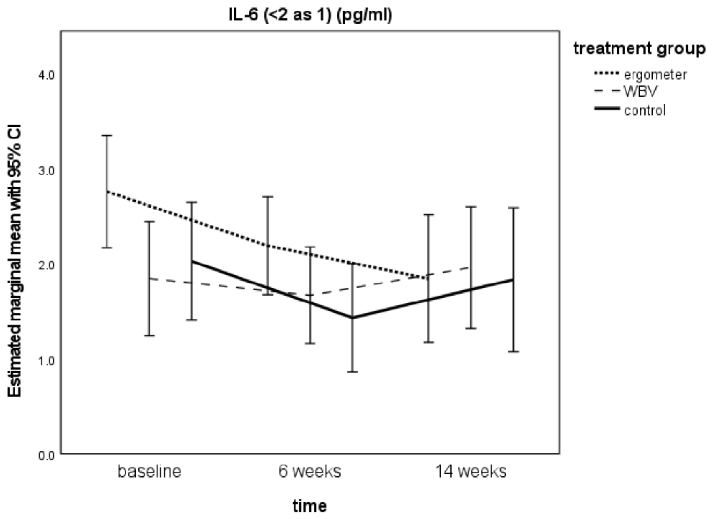
3.4. IL-6 Levels from Baseline to t2

For the model IL-6 med (all values reported in pg/mL), IL-6 decreased in all groups from t0 to t1, but it increased again in WBV and controls, whereas it got lower again in ergometer for t2 (Ergometer: t0 = 2.76 ± 0.3, t1 = 2.19 ± 0.26, t2 = 1.84± 0.33; WBV: t0 = 1.85 ± 0.3; t1 = 1.67 ± 0.25; t2 = 1.96 ± 0.32; Control t0 = 2.03 ± 0.31; t1 = 1.43 ± 0.28; t2 = 1.83 ± 0.38). This observation was found for all three calculation models (Table 3).
The changes over time were significant for ergometer when applying the IL-6 med- and IL-6 min models (Table S4). For WBV and controls, changes over time were not statistically significant (Table S4).

3.5. TNF-α Levels from Baseline to t2

TNF-α mean scores (all values reported in pg/mL) decreased during intervention time in ergometer (t0 = 1.81 ±0.16; t1 = 1.68 ± 0.18) but increased again for t2 (1.85 ± 0.2). The same could be observed in controls (t0 = 1.92 ± 0.18; t1 = 1.73 ± 0.2, t2 = 1.79 ± 0.25). In WBV, TNF-α increased from t0 (1.67 ± 0.17) to t1 (1.81 ± 0.19) and decreased for t2 (1.74 ± 0.19). Time and group differences (pairwise comparisons) were not significant (Table S4, Figure S2).

3.6. Influence of Covariates (Age, Sex, BMI, Number of Trainings, Total Therapy Times, Medication) on TNF-α and IL-6

Sex was significant for TNF-α (p < 0.001) in all measurement points (Figure S3). TNF-α levels of males were in the mean 0.823 pg/mL higher than in females (p < 0.001). Over time, the mean TNF-α of male patients was 2.145 (t0); 2.165 (t1); 2.291 (t2). For female patients, the mean TNF-α was 1.428 (t0); 1.296 (t1); 1.312 (t3) (Table S5).
BMI was significant in all IL-6 models (p < 0.001). In the example of IL-6 med, 1 BMI point increased IL-6 for 0.9 pg/mL. The influence of medication (PRN or SSRI for > 3 weeks) showed a statistically significant effect on IL-6 in all IL-6-models, increasing levels for 0.84 pg/mL for PRN and 0.11 for SSRI and 0.57 pg/mL for combined medication. The total therapy time in 6 weeks showed a trend influence in IL-6 med and IL-6 min models (IL-6 med p = 0.082; IL-6 min p = 0.099) (Table 4, for estimates, see Tables S7–S10).

3.7. Influence of IL-6 Levels on TNF-α and Vice Versa

There were no significant correlations between the levels of IL-6 and TNF-α (Table 5).

3.8. Influence of IL-6 and TNF-α on DIKJ Scores Over Time

For the entire sample, IL-6 and TNF-α showed no correlation to depression scores in DIKJ at baseline (Table 5). In the longitudinal analysis, IL-6 levels (in all calculation models) showed significant effects on DIKJ only for WBV (IL-6 med p = 0.008; IL-6 max p = 0.013; IL-6 min p = 0.009) but not for controls. There were no significant effects of TNF-α on DIKJ scores over time (Table 6).

3.9. Adverse Events

There were no Adverse Events or Serious Adverse Events reported for the interventions. One subject suffered a metacarpals fracture due to self-injury but continued trainings. Another suffered a skin infection but also continued exercising. One subject was moved to the closed ward because of elevated suicidality for one night, which was not related to the trainings, and they could also continue exercising.

4. Discussion

In the clinical study “Mood Vibes”, two vigorous exercise interventions (WBV and ergometer cycling) were applied in comparison to TAU in the treatment of adolescent depression. In both intervention groups, patients improved earlier and more strongly measured by DIKJ (Wunram et al. 2018: Figure S1, [10]). In the present analyses, we investigated if cytokine levels influenced depression severity and mediated treatment success.
Regarding the entire sample in our results, we could not find a significant correlation between depression scores in DIKJ and TNF-α or IL-6-levels at baseline. Therefore, our findings cannot replicate the elevated PIC levels reflecting a chronic low-grade inflammation in MDD found in adult literature [15,16,85] and in some lower extent also in children and adolescent studies [18]. However, one has to take into account that in our study, we lack healthy controls. In a recent meta-analysis on adult trials, Kohler et al. included 82 studies with around 3000 patients. They stated that taken together, peripheral levels of IL-6 and TNF-α (among others) were elevated in MDD patients compared to healthy controls [26]. Our results do not support this. It is important to consider that in adolescents, trials are scarcer and more divergent. The five studies included in the meta-analysis of d’Acunto, focusing on PICs in children and adolescents diagnosed with MDD, supported only a trend for higher levels of peripheral TNF-α. In contrast, levels of other cytokines included in the meta-analysis (among others IL-6) were not significantly different in adolescent patients with MDD (although Gabbay et al. found a trend for IL-6 elevation compared to controls). However, the authors state that only two studies in children and adolescents were available for IL-6, and as such, the results should be interpreted with caution. Not included in the meta-analysis of d’Acunto is a clinical study by Pérez-Sanchez et al. that found significantly elevated TNF-α and IL-6 in 22 adolescents with MDD compared to healthy controls.
In our study, we did not include healthy controls, and the low elevation levels of PICs did not correlate with depression severity. Therefore, our findings are not in line with the aforementioned literature but concordant to the study of Byrne et al., who likewise did not find elevated PICs in depressed adolescents. They concluded that maybe inflammatory markers are not associated with MDD in young ages because being young could be a protective factor against low-grade chronic inflammation in MDD [86].
On the other hand, it would have been interesting to analyze whether PIC levels in our sample would have correlated with specific symptoms or sub-dimensions of depression (e.g., fatigue, somatic or cognitive deficits) rather than with the total depression scores. Some studies in adults found associations to sub-dimensions instead of total scores [87,88], although it is still discussed controversially if e.g., Beck’s Depression Inventory (BDI-II) can be divided in specific subscales or should be used only as a unidimensional scale. Additionally, for the applied DIKJ, no factor structure in the sense of cognitive versus somatic sub-dimension exists [67,89]. Nevertheless, this could be a point to consider for future studies.
With regard to our second hypothesis that exercise treatment of MDD would result in a stronger decrease of PIC levels than TAU, our results are inconsistent. A continuous decrease could only be found for IL-6 in the ergometer group after weeks 6 and 14 (Figure 1). The WBV group showed less decrease than TAU. For TNF-α, no continuous decrease was seen (Table 3). This is contrary to adult studies, which at least for antidepressant treatment (but more inconsistent for psychotherapy) could demonstrate an anti-inflammatory effect [11,25]. In two studies with children and adolescents, antidepressant drugs significantly reduced TNF-α without significant changes in IL-6 (Amitai et al.), whereas SSRI-treatment decreased TNF-α and IL-6 in the clinical trial of Pérez-Sánchez et al. [62,90].
As in our intervention, we have applied a vigorous physical activity treatment as add-on with patients who were supposed to be medication-naïve, we should compare our findings not to drug-treatment but to exercise-treatment studies. In adults, there is some body of literature supporting a decrease of PICs by exercise therapy in MDD. Lavebratt et al. found in 116 adults with mild-to-moderate depression who participated in a physical activity intervention 3 times/week for 12 weeks that the vigorous intensity group showed an increase in IL-6; whereas, low and moderate intensities showed a decrease in IL-6 over time. A higher decrease in IL-6 levels in this study was associated with a stronger reduction of depression scores [20]. Fischer et al. suggested also a reduction of basal IL-6 and decreased IL-6-response to exercise in the sense of a training adaptation [42]. Our findings of decreased basal IL-6 levels in the intervention groups from t0 to t1 could express such a training adaptation progress. However, we found the same for the control group, which did not participate in the add-on physical activity interventions. Therefore, the reduction in IL-6 cannot be interpreted only as training adaptation. It can be discussed if the reduction of IL-6 could be the result of the TAU psychotherapeutic treatment. Further research in this direction would be helpful. The basal IL-6 reduction was persistent and significant over time only for ergometer cycling. In WBV and controls, levels increased again for t2 after ending the exercise intervention.
With regard to an anti-depressive effect of the IL-6-decrease over the 14 weeks, only WBV was significant (Table 6). As IL-6 decreased more strongly in ergometer, the anti-depressant effect of the treatments cannot be deduced only by the immunological factors.
Concerning TNF-α, Lira et al. showed in mice that after 8 weeks of aerobic training, TNF-α had decreased in skeletal muscle depending on the fiber composition [43]. In a human sample of 61 university students, Paolucci et al. could demonstrate that exercise of moderate intensity could lower TNF-α and by this decrease depressive mood [21]. Petersen et al. and Smart and colleagues also commented that regular exercise reduced TNF-α levels [44]. However, Schuch et al. concluded in their meta-analysis that regular exercise did not affect TNF-α levels [37]. Our findings show a decrease of TNF-α only for ergometer and controls during the intervention time, whereas the WBV group increased in this time. Therefore, physical activity did only show an anti-inflammatory effect for endurance training. Nevertheless, also here, the question remains as to why the control group showed an equal response. Again, it could be interpreted as being the effect of the psychotherapeutic treatment. However, the increase of TNF-α in the WBV group during the intervention time contradicts an anti-inflammatory effect of this exercise intervention. In addition, as we commented before, no significant interactions between changes in TNF-α and depression severity could be found.
Relating to sex differences, the study of Pallavi et al. found higher IL-6 levels in females, whereas TNFα was not affected by sex. This was the contrary in our sample, where we found higher TNF-α in males compared to females, whereas IL-6 was not affected by sex but by BMI. With respect to the influence of BMI, we would have expected a significant effect on both cytokines, given the existing literature in obese adults [91]. Therefore, our results are only partly in concordance: we found the influences of BMI only on IL-6. Finally, it is also contradictory to existing data that in our study, medication (patients then counted as ITT) resulted in increased levels of IL-6 (for PRN with neuroleptics stronger than SSRIs alone). However, in the study of Henje Blom or Pérez-Sanchez, lower IL-6 scores (and TNF-α) were found for medicated patients [57,90]. Concordantly, Amitai et al. had found a decrease of TNF-α in SSRI-treated children and adolescents [62].

5. Conclusions

To our knowledge, this is the first study about the interplay between physical activity treatments, cytokines, and depression in adolescent inpatients. The body of literature focusing on the role of cytokines in adolescent depression is already scarce, and there is growing but not yet sufficient research on exercise interventions in the treatment of adolescent depression. Nevertheless, our results have to be interpreted with caution. The PIC levels were not the primary endpoint of the study; therefore, the study was underpowered for these parameters. Furthermore, the sample size was small, and we did not have a healthy control in our design. Moreover, the non-randomization of the control group represents a major danger for a selection bias. In addition, for IL-6, we had to apply statistical replacements for results of IL-6 < 2 pg/mL. Statistical replacement strategy reduced standard errors and introduced bias, affecting reproducibility and effect sizes. Therefore, more exact assay methods for values < 2 ng/mL are a need for future studies. In addition, a broader immune panel would have allowed a deeper insight in further neuroimmunological mechanisms Another interesting point would have been to analyze the association of PIC levels and sub-dimensions of depression, rather than with the total depression scores. As further limitations, nutrition was not taken into account, and our training protocols were based on the prior spiroergometry but not monitored via heart rate. Therefore, we can only presume that it was a vigorous training.
Some of the differences we found in our sample compared to adult patients could result from neurodevelopmental or hormonal changes, as commented by d’Acunto et al. [18]. A more profound understanding of the role of cytokines in adolescent MDD and its response to exercise treatment could open further options of tailored therapies for a sub-group of individuals who show this immune pattern. However, to date, we can only conclude with the words of Brambilla et al. that in adolescent depression exists a cytokine pathology “of obscure etiopathogenetical significance” [53]. Further research with bigger samples and this focus will be needed.

Supplementary Materials

The following are available online at https://www.mdpi.com/article/10.3390/ijerph18126527/s1, Figure S1–S3 and Table S1–S10 (see E-Supplement).

Author Contributions

H.L.W.: author; M.O.: exercise counseling, statistical counseling; S.H. statistics and statistical counseling; S.N.: content supervision; N.G.: content supervision; M.K.K.: content supervision, statistic counseling; W.B.: IL-6 and TNF-alpha analytics; E.S.: IL-6 and TNF-alpha counseling; S.B.: adolescent psychiatric supervision; O.F.: adolescent psychiatric supervision. All authors have read and agreed to the published version of the manuscript.

Funding

This research was funded by: HLW received funding from Walter and Marga Boll Foundation (210-07.2-11, Kerpen, Germany (https://www.bollstiftung.de accessed on 8 June 2021).

Institutional Review Board Statement

The study was conducted according to the guidelines of the Declaration of Helsinki, and approved by the Ethics Committee) of the University of Cologne on 23rd October 2011 and registered at the German Clinical Trials Register (DRKS00005120; Universal Trial Number (UTN): U1111-1145-1854).

Informed Consent Statement

Informed written consent was obtained from all subjects involved in the study.

Data Availability Statement

Data supporting reported results are property of the University Hospital of Cologne. They can be made available on demand after legal review, please contact for further information the corresponding author.

Acknowledgments

This study could only be conducted with financial support of the Marga and Walter Boll Foundation, Cologne, as well as the work of the entire “Mood Vibes” doctoral students team (in alphabetic order): Fabian Abuhsin, Ralf Beccard, Luisa Belke, Sarah Damsch, Nils Grote, Michael Holder, Franziska Jänicke, Franziska Reinhard, Andrea Steffen, Till Thimme, Maxi Volk, Alischa Ziemendorff. This manuscript contains parts of a master and doctoral thesis by NG. We are very grateful for the final native speaker proof-reading by Steven Dublin.

Conflicts of Interest

All authors declare that the research was conducted in the absence of any commercial or financial relationships that could be construed as a potential conflict of interest. All authors declare having materially participated in the research and/or article preparation. All authors have approved the final article.

Abbreviations

BMIbody mass index
CNScentral nervous system
CRPC-reactive protein
DIKJDepression Inventory for Children and Adolescents
DSHSSport University Cologne
IL-6interleukin-6
IMSBInformatics and Epidemiology of the University of Cologne
ITTintention-to-treat
K-ABCKaufman Assessment Battery for Children
MDDmajor depressive disorder
MMRMmixed model for repeated measures
PICsPro-inflammatory cytokines
PRNpro re nata
SKID-IStructured Clinical Interview for DSM-IV Axis I Disorders, German version
SSRIselective serotonin reuptake inhibitor
TAUtreatment-as-usual
TNF-αtumor necrosis factor-α
WBVWhole Body Vibration
WISCWechsler Intelligence Scale for Children

References

  1. McLeod, J.D.; Uemura, R.; Rohrman, S. Adolescent mental health, behavior problems, and academic achievement. J. Health Soc. Behav. 2012, 53, 482–497. [Google Scholar] [CrossRef] [PubMed] [Green Version]
  2. WHO. Mental Health: New Understanding, New Hope; The World Health Report; World Health Organization: Geneva, Switzerland, 2001. [Google Scholar]
  3. Emslie, G.J. The psychopharmacology of adolescent depression. J. Child Adolesc. Psychopharmacol. 2012, 22, 2–4. [Google Scholar] [CrossRef]
  4. Dolle, K.; Schulte-Korne, G. The treatment of depressive disorders in children and adolescents. Dtsch. Arztebl. Int. 2013, 110, 854–860. [Google Scholar] [CrossRef] [Green Version]
  5. Cipriani, A.; Zhou, X.; Del Giovane, C.; Hetrick, S.E.; Qin, B.; Whittington, C.; Coghill, D.; Zhang, Y.; Hazell, P.; Leucht, S.; et al. Comparative efficacy and tolerability of antidepressants for major depressive disorder in children and adolescents: A network meta-analysis. Lancet 2016, 388, 881–890. [Google Scholar] [CrossRef]
  6. Dolle, K.; Schulte-Korne, G. [Complementary treatment methods for depression in children and adolescents]. Praxis der Kinderpsychologie und Kinderpsychiatrie 2014, 63, 237–263. [Google Scholar] [CrossRef] [PubMed]
  7. Cooney, G.M.; Dwan, K.; Greig, C.A.; Lawlor, D.A.; Rimer, J.; Waugh, F.R.; McMurdo, M.; Mead, G.E. Exercise for depression. Cochrane Database Syst. Rev. 2013. [Google Scholar] [CrossRef] [PubMed]
  8. Oberste, M.; Medele, M.; Javelle, F.; Lioba Wunram, H.; Walter, D.; Bloch, W.; Bender, S.; Fricke, O.; Joisten, N.; Walzik, D.; et al. Physical Activity for the Treatment of Adolescent Depression: A Systematic Review and Meta-Analysis. Front. Physiol. 2020, 11, 185. [Google Scholar] [CrossRef]
  9. Larun, L.; Nordheim, L.V.; Ekeland, E.; Hagen, K.B.; Heian, F. Exercise in prevention and treatment of anxiety and depression among children and young people (Review). Cochrane Database Syst. Rev. 2006. [Google Scholar] [CrossRef]
  10. Wunram, H.L.; Hamacher, S.; Hellmich, M.; Volk, M.; Janicke, F.; Reinhard, F.; Bloch, W.; Zimmer, P.; Graf, C.; Schonau, E.; et al. Whole body vibration added to treatment as usual is effective in adolescents with depression: A partly randomized, three-armed clinical trial in inpatients. Eur. Child Adolesc. Psychiatry 2018, 27, 645–662. [Google Scholar] [CrossRef]
  11. Himmerich, H.; Patsalos, O.; Lichtblau, N.; Ibrahim, M.A.A.; Dalton, B. Cytokine Research in Depression: Principles, Challenges, and Open Questions. Front. Psychiatry 2019, 10, 30. [Google Scholar] [CrossRef] [Green Version]
  12. Shariq, A.S.; Brietzke, E.; Rosenblat, J.D.; Barendra, V.; Pan, Z.; McIntyre, R.S. Targeting cytokines in reduction of depressive symptoms: A comprehensive review. Prog. Neuropsychopharmacol. Biol. Psychiatry 2018, 83, 86–91. [Google Scholar] [CrossRef]
  13. Phillips, C.; Fahimi, A. Immune and Neuroprotective Effects of Physical Activity on the Brain in Depression. Front. Neurosci. 2018, 12, 498. [Google Scholar] [CrossRef] [PubMed] [Green Version]
  14. Herpertz-Dahlmann, B.; Buhren, K.; Remschmidt, H. Growing up is hard: Mental disorders in adolescence. Dtsch. Arztebl. Int. 2013, 110, 432–439. [Google Scholar] [CrossRef] [PubMed] [Green Version]
  15. Dantzer, R.; O’Connor, J.C.; Freund, G.G.; Johnson, R.W.; Kelley, K.W. From inflammation to sickness and depression: When the immune system subjugates the brain. Nat. Rev. Neurosci. 2008, 9, 46–56. [Google Scholar] [CrossRef] [PubMed] [Green Version]
  16. Dowlati, Y.; Herrmann, N.; Swardfager, W.; Liu, H.; Sham, L.; Reim, E.K.; Lanctot, K.L. A meta-analysis of cytokines in major depression. Biol. Psychiatry 2010, 67, 446–457. [Google Scholar] [CrossRef]
  17. Pariante, C.M. Why are depressed patients inflamed? A reflection on 20 years of research on depression, glucocorticoid resistance and inflammation. Eur. Neuropsychopharmacol. 2017, 27, 554–559. [Google Scholar] [CrossRef] [Green Version]
  18. D’Acunto, G.; Nageye, F.; Zhang, J.; Masi, G.; Cortese, S. Inflammatory Cytokines in Children and Adolescents with Depressive Disorders: A Systematic Review and Meta-Analysis. J. Child Adolesc. Psychopharmacol. 2019, 29, 362–369. [Google Scholar] [CrossRef] [PubMed]
  19. Eyre, H.A.; Papps, E.; Baune, B.T. Treating depression and depression-like behavior with physical activity: An immune perspective. Front. Psychiatry 2013, 4, 3. [Google Scholar] [CrossRef] [Green Version]
  20. Lavebratt, C.; Herring, M.P.; Liu, J.J.; Wei, Y.B.; Bossoli, D.; Hallgren, M.; Forsell, Y. Interleukin-6 and depressive symptom severity in response to physical exercise. Psychiatry Res. 2017, 252, 270–276. [Google Scholar] [CrossRef] [PubMed]
  21. Paolucci, E.M.; Loukov, D.; Bowdish, D.M.E.; Heisz, J.J. Exercise reduces depression and inflammation but intensity matters. Biol. Psychol. 2018, 133, 79–84. [Google Scholar] [CrossRef]
  22. Smith, R.S. The macrophage theory of depression. Med. Hypotheses 1991, 35, 298–306. [Google Scholar] [CrossRef]
  23. Maes, M. Evidence for an immune response in major depression: A review and hypothesis. Prog. Neuropsychopharmacol. Biol. Psychiatry 1995, 19, 11–38. [Google Scholar] [CrossRef]
  24. Capuron, L.; Ravaud, A.; Dantzer, R. Early depressive symptoms in cancer patients receiving interleukin 2 and/or interferon alfa-2b therapy. J. Clin. Oncol. 2000, 18, 2143–2151. [Google Scholar] [CrossRef] [PubMed]
  25. Lopresti, A.L.; Maker, G.L.; Hood, S.D.; Drummond, P.D. A review of peripheral biomarkers in major depression: The potential of inflammatory and oxidative stress biomarkers. Prog. Neuropsychopharmacol. Biol. Psychiatry 2014, 48, 102–111. [Google Scholar] [CrossRef] [Green Version]
  26. Gadad, B.S.; Jha, M.K.; Czysz, A.; Furman, J.L.; Mayes, T.L.; Emslie, M.P.; Trivedi, M.H. Peripheral biomarkers of major depression and antidepressant treatment response: Current knowledge and future outlooks. J. Affect. Disord. 2018, 233, 3–14. [Google Scholar] [CrossRef]
  27. Kohler, C.A.; Freitas, T.H.; Maes, M.; de Andrade, N.Q.; Liu, C.S.; Fernandes, B.S.; Stubbs, B.; Solmi, M.; Veronese, N.; Herrmann, N.; et al. Peripheral cytokine and chemokine alterations in depression: A meta-analysis of 82 studies. Acta Psychiatr. Scand. 2017, 135, 373–387. [Google Scholar] [CrossRef] [PubMed]
  28. Del Grande da Silva, G.; Wiener, C.D.; Barbosa, L.P.; Goncalves Araujo, J.M.; Molina, M.L.; San Martin, P.; Oses, J.P.; Jansen, K.; Dias de Mattos Souza, L.; Azevedo da Silva, R. Pro-inflammatory cytokines and psychotherapy in depression: Results from a randomized clinical trial. J. Psychiatr. Res. 2016, 75, 57–64. [Google Scholar] [CrossRef]
  29. Miller, A.H.; Raison, C.L. The role of inflammation in depression: From evolutionary imperative to modern treatment target. Nat. Rev. Immunol. 2016, 16, 22–34. [Google Scholar] [CrossRef] [Green Version]
  30. Karson, A.; Demirtas, T.; Bayramgurler, D.; Balci, F.; Utkan, T. Chronic administration of infliximab (TNF-alpha inhibitor) decreases depression and anxiety-like behaviour in rat model of chronic mild stress. Basic Clin. Pharmacol. Toxicol. 2013, 112, 335–340. [Google Scholar] [CrossRef]
  31. Danese, A.; Pariante, C.M.; Caspi, A.; Taylor, A.; Poulton, R. Childhood maltreatment predicts adult inflammation in a life-course study. Proc. Natl. Acad. Sci. USA 2007, 104, 1319–1324. [Google Scholar] [CrossRef] [Green Version]
  32. Baumeister, D.; Akhtar, R.; Ciufolini, S.; Pariante, C.M.; Mondelli, V. Childhood trauma and adulthood inflammation: A meta-analysis of peripheral C-reactive protein, interleukin-6 and tumour necrosis factor-alpha. Mol. Psychiatry 2016, 21, 642–649. [Google Scholar] [CrossRef] [PubMed] [Green Version]
  33. Baumeister, D.; Ciufolini, S.; Mondelli, V. Effects of psychotropic drugs on inflammation: Consequence or mediator of therapeutic effects in psychiatric treatment? Psychopharmacology 2016, 233, 1575–1589. [Google Scholar] [CrossRef] [PubMed]
  34. Dishman, R.K.; Berthoud, H.R.; Booth, F.W.; Cotman, C.W.; Edgerton, V.R.; Fleshner, M.R.; Gandevia, S.C.; Gomez-Pinilla, F.; Greenwood, B.N.; Hillman, C.H.; et al. Neurobiology of exercise. Obesity 2006, 14, 345–356. [Google Scholar] [CrossRef]
  35. Mead, G.E.; Morley, W.; Campbell, P.; Greig, C.A.; McMurdo, M.; Lawlor, D.A. Exercise for depression. Cochrane Database Syst. Rev. 2009. [Google Scholar] [CrossRef] [Green Version]
  36. Rimer, J.; Dwan, K.; Lawlor, D.A.; Greig, C.A.; McMurdo, M.; Morley, W.; Mead, G.E. Exercise for depression. Cochrane Database Syst. Rev. 2012. [Google Scholar] [CrossRef]
  37. Walsh, N.P.; Gleeson, M.; Shephard, R.J.; Gleeson, M.; Woods, J.A.; Bishop, N.C.; Fleshner, M.; Green, C.; Pedersen, B.K.; Hoffman-Goetz, L.; et al. Position statement. Part one: Immune function and exercise. Exerc. Immunol. Rev. 2011, 17, 6–63. [Google Scholar]
  38. Schuch, F.B.; Deslandes, A.C.; Stubbs, B.; Gosmann, N.P.; Silva, C.T.; Fleck, M.P. Neurobiological effects of exercise on major depressive disorder: A systematic review. Neurosci. Biobehav. Rev. 2016, 61, 1–11. [Google Scholar] [CrossRef]
  39. Deslandes, A.; Moraes, H.; Ferreira, C.; Veiga, H.; Silveira, H.; Mouta, R.; Pompeu, F.A.; Coutinho, E.S.; Laks, J. Exercise and mental health: Many reasons to move. Neuropsychobiology 2009, 59, 191–198. [Google Scholar] [CrossRef]
  40. Pedersen, B.K.; Steensberg, A.; Schjerling, P. Muscle-derived interleukin-6: Possible biological effects. J. Physiol. 2001, 536, 329–337. [Google Scholar] [CrossRef] [PubMed]
  41. Apostolopoulos, V.; Borkoles, E.; Polman, R.; Stojanovska, L. Physical and immunological aspects of exercise in chronic diseases. Immunotherapy 2014, 6, 1145–1157. [Google Scholar] [CrossRef]
  42. Starkie, R.; Ostrowski, S.R.; Jauffred, S.; Febbraio, M.; Pedersen, B.K. Exercise and IL-6 infusion inhibit endotoxin-induced TNF-alpha production in humans. FASEB J. 2003, 17, 884–886. [Google Scholar] [CrossRef]
  43. Fischer, C.P. Interleukin-6 in acute exercise and training: What is the biological relevance? Exerc. Immunol. Rev. 2006, 12, 6–33. [Google Scholar]
  44. Lira, F.S.; Koyama, C.H.; Yamashita, A.S.; Rosa, J.C.; Zanchi, N.E.; Batista, M.L., Jr.; Seelaender, M.C. Chronic exercise decreases cytokine production in healthy rat skeletal muscle. Cell Biochem. Funct. 2009, 27, 458–461. [Google Scholar] [CrossRef]
  45. Petersen, A.M.; Pedersen, B.K. The anti-inflammatory effect of exercise. J. Appl. Physiol. 2005, 98, 1154–1162. [Google Scholar] [CrossRef] [PubMed] [Green Version]
  46. Smart, N.A.; Larsen, A.I.; Le Maitre, J.P.; Ferraz, A.S. Effect of exercise training on interleukin-6, tumour necrosis factor alpha and functional capacity in heart failure. Cardiol. Res. Pract. 2011, 2011, 532620. [Google Scholar] [CrossRef] [Green Version]
  47. Jolly, M.; Verbeke, W. Immune System Function and its Relation to Depression: How Exercise can Alter the Immune SystemDepression Dynamics. J. Depress. Anxiety 2018, 7, 4. [Google Scholar] [CrossRef]
  48. Paula Martins, R.; Lim, C.K.; Ghisoni, K.; Staats, A.; Dallagnol, K.; Solano, A.; Guillemin, G.; Aguiar, A.S., Jr.; Latini, A. Treating depression with exercise: The inflammasome inhibition perspective. J. Syst. Integr. Neurosci. 2016, 3, 1–9. [Google Scholar] [CrossRef] [Green Version]
  49. Euteneuer, F.; Dannehl, K.; Del Rey, A.; Engler, H.; Schedlowski, M.; Rief, W. Immunological effects of behavioral activation with exercise in major depression: An exploratory randomized controlled trial. Transl. Psychiatry 2017, 7, e1132. [Google Scholar] [CrossRef] [PubMed] [Green Version]
  50. Mills, N.T.; Scott, J.G.; Wray, N.R.; Cohen-Woods, S.; Baune, B.T. Research review: The role of cytokines in depression in adolescents: A systematic review. J. Child Psychol. Psychiatry 2013, 54, 816–835. [Google Scholar] [CrossRef] [PubMed] [Green Version]
  51. Angold, A.; Costello, E.J. Puberty and depression. Child Adolesc. Psychiatr. Clin. N. Am. 2006, 15, 919–937, ix. [Google Scholar] [CrossRef]
  52. Brotman, M.A.; Schmajuk, M.; Rich, B.A.; Dickstein, D.P.; Guyer, A.E.; Costello, E.J.; Egger, H.L.; Angold, A.; Pine, D.S.; Leibenluft, E. Prevalence, clinical correlates, and longitudinal course of severe mood dysregulation in children. Biol. Psychiatry 2006, 60, 991–997. [Google Scholar] [CrossRef]
  53. Brooks, S.J.; Kutcher, S. Diagnosis and measurement of adolescent depression: A review of commonly utilized instruments. J. Child Adolesc. Psychopharmacol. 2001, 11, 341–376. [Google Scholar] [CrossRef]
  54. Brambilla, F.; Monteleone, P.; Maj, M. Interleukin-1beta and tumor necrosis factor-alpha in children with major depressive disorder or dysthymia. J. Affect. Disord. 2004, 78, 273–277. [Google Scholar] [CrossRef]
  55. Gabbay, V.; Klein, R.G.; Alonso, C.M.; Babb, J.S.; Nishawala, M.; De Jesus, G.; Hirsch, G.S.; Hottinger-Blanc, P.M.; Gonzalez, C.J. Immune system dysregulation in adolescent major depressive disorder. J. Affect. Disord. 2009, 115, 177–182. [Google Scholar] [CrossRef] [PubMed] [Green Version]
  56. Gariup, M.; Gonzalez, A.; Lazaro, L.; Torres, F.; Serra-Pages, C.; Morer, A. IL-8 and the innate immunity as biomarkers in acute child and adolescent psychopathology. Psychoneuroendocrinology 2015, 62, 233–242. [Google Scholar] [CrossRef]
  57. Pallavi, P.; Sagar, R.; Mehta, M.; Sharma, S.; Subramanium, A.; Shamshi, F.; Sengupta, U.; Pandey, R.M.; Mukhopadhyay, A.K. Serum cytokines and anxiety in adolescent depression patients: Gender effect. Psychiatry Res. 2015, 229, 374–380. [Google Scholar] [CrossRef] [PubMed]
  58. Henje Blom, E.; Lekander, M.; Ingvar, M.; Asberg, M.; Mobarrez, F.; Serlachius, E. Pro-inflammatory cytokines are elevated in adolescent females with emotional disorders not treated with SSRIs. J. Affect. Disord. 2012, 136, 716–723. [Google Scholar] [CrossRef]
  59. Guyer, A.E.; Silk, J.S.; Nelson, E.E. The neurobiology of the emotional adolescent: From the inside out. Neurosci. Biobehav. Rev. 2016, 70, 74–85. [Google Scholar] [CrossRef] [PubMed] [Green Version]
  60. Schulz, K.M.; Sisk, C.L. The organizing actions of adolescent gonadal steroid hormones on brain and behavioral development. Neurosci. Biobehav. Rev. 2016, 70, 148–158. [Google Scholar] [CrossRef] [PubMed] [Green Version]
  61. Sisk, C.L. Hormone-dependent adolescent organization of socio-sexual behaviors in mammals. Curr. Opin. Neurobiol. 2016, 38, 63–68. [Google Scholar] [CrossRef] [PubMed] [Green Version]
  62. Hannestad, J.; DellaGioia, N.; Bloch, M. The effect of antidepressant medication treatment on serum levels of inflammatory cytokines: A meta-analysis. Neuropsychopharmacology 2011, 36, 2452–2459. [Google Scholar] [CrossRef]
  63. Amitai, M.; Taler, M.; Carmel, M.; Michaelovsky, E.; Eilat, T.; Yablonski, M.; Orpaz, N.; Chen, A.; Apter, A.; Weizman, A.; et al. The Relationship Between Plasma Cytokine Levels and Response to Selective Serotonin Reuptake Inhibitor Treatment in Children and Adolescents with Depression and/or Anxiety Disorders. J. Child Adolesc. Psychopharmacol. 2016, 26, 727–732. [Google Scholar] [CrossRef]
  64. Carter, T.; Morres, I.D.; Meade, O.; Callaghan, P. The Effect of Exercise on Depressive Symptoms in Adolescents: A Systematic Review and Meta-Analysis. J. Am. Acad. Child Adolesc. Psychiatry 2016, 55, 580–590. [Google Scholar] [CrossRef]
  65. American Psychiatric Association (APA). Diagnostic and Statistical Manual of Mental Disorders: DSM-V; American Psychiatric Association: Washington, DC, USA, 2013. [Google Scholar]
  66. WHO. ICD-10 Classification of Mental and Behavioural Disorders. Available online: www.who.int/classifications/icd/en/GRNBOOK.pdf (accessed on 29 June 2016).
  67. Wittchen, H.; Zaudig, T. Strukturiertes Klinisches Interview für DSM-IV; Hogrefe: Göttingen, Germany, 1997. [Google Scholar]
  68. Stiensmeier-Pelster, J.; Schürmann, M.; Duda, K. DIKJ Depressionsinventar für Kinder und Jugendliche, 2nd ed.; Hogrefe: Göttingen, Germany, 1989. [Google Scholar]
  69. Melchers, P.; Preuß, U. Kaufman Assessment Battery for Children, 8th ed.; Pearson Assessment: Frankfurt, Germany, 2009. [Google Scholar]
  70. Petermann, F.; Daseking, M. HAWIK-IV, 3rd ed.; Huber Verlag: Bern, Germany, 2010. [Google Scholar]
  71. Petermann, F.; Daseking, M. WISC-IV; Pearson Assessment: Frankfurt am Main, Germany, 2011. [Google Scholar]
  72. Deutsche Gesellschaft für Sportmedizin (DGSP). S1-Leitlinie Vorsorgeuntersuchung im Sport. Available online: http://www.dgsp.de/_downloads/allgemein/leitlinie_vorsorgeuntersuchung_4.10.2007-1-19.pdf (accessed on 29 June 2016).
  73. Bohus, M.; Limberger, M.F.; Frank, U.; Chapman, A.L.; Kuhler, T.; Stieglitz, R.D. Psychometric properties of the Borderline Symptom List (BSL). Psychopathology 2007, 40, 126–132. [Google Scholar] [CrossRef] [PubMed]
  74. Kovacs, M. The Children’s Depression, Inventory (CDI). Psychopharmacol. Bull. 1985, 21, 995–998. [Google Scholar]
  75. Trappe, H.J.; Löllgen, H. Leitlinien zur Ergometrie (Deutsche Ges. für Kardiologie). Zeitschrift für Kardiologie 2000, 89, 821–837. [Google Scholar]
  76. Veilleux, L.N.; Rauch, F. Reproducibility of jumping mechanography in healthy children and adults. J. Musculoskelet. Neuronal Interact. 2010, 10, 256–266. [Google Scholar]
  77. Novotec. Leonardo Mechanography Ground Reaction Force Platform (GRFP): Getting Started v4.1; Novotec Medical GmbH: Pforzheim, Germany, 2006. [Google Scholar]
  78. R&D Systems, Inc. Quantikine ELISA Human TNF-α Immunoassay. Available online: https://resources.rndsystems.com/pdfs/datasheets/dta00c.pdf?v=20210526&_ga=2.26931572.893794815.1622046480-926488157.1622046480 (accessed on 26 May 2021).
  79. Roche. Diagnostics. Steckbrief IL 6 Analysekit. Available online: http://www.uk-koeln.de/institute/kchemie/Diagnostik/Parameter/Testkits/Roche/IL6.pdf (accessed on 22 November 2019).
  80. Roche. Elecsys IL-6 Cobas. Available online: http://www.unsere-uniklinik.de/institute/kchemie/Zentrallabor/Diagnostik/Parameter/Testkits/Roche/IL-6.pdf (accessed on 26 May 2021).
  81. Semler, O.; Fricke, O.; Vezyroglou, K.; Stark, C.; Stabrey, A.; Schoenau, E. Results of a prospective pilot trial on mobility after whole body vibration in children and adolescents with osteogenesis imperfecta. Clin. Rehabilit. 2008, 22, 387–394. [Google Scholar] [CrossRef]
  82. Rauch, F.; Sievanen, H.; Boonen, S.; Cardinale, M.; Degens, H.; Felsenberg, D.; Roth, J.; Schoenau, E.; Verschueren, S.; Rittweger, J. Reporting whole-body vibration intervention studies: Recommendations of the International Society of Musculoskeletal and Neuronal Interactions. J. Musculoskelet. Neuronal Interact. 2010, 10, 193–198. [Google Scholar]
  83. Gibbons, M.W. Effect of Whole Body Vibration on Sterotypy of Young Children with Autism; Utah State University: Logan, UT, USA, 2011. [Google Scholar]
  84. Semler, O.; Fricke, O.; Vezyroglou, K.; Stark, C.; Schoenau, E. Preliminary results on the mobility after whole body vibration in immobilized children and adolescents. J. Musculoskelet. Neuronal Interact. 2007, 7, 77–81. [Google Scholar]
  85. Stark, C.; Nikopoulou-Smyrni, P.; Stabrey, A.; Semler, O.; Schoenau, E. Effect of a new physiotherapy concept on bone mineral density, muscle force and gross motor function in children with bilateral cerebral palsy. J. Musculoskelet. Neuronal Interact. 2010, 10, 151–158. [Google Scholar] [PubMed]
  86. Bhattacharya, A.; Derecki, N.C.; Lovenberg, T.W.; Drevets, W.C. Role of neuro-immunological factors in the pathophysiology of mood disorders. Psychopharmacology 2016, 233, 1623–1636. [Google Scholar] [CrossRef]
  87. Byrne, M.L.; O’Brien-Simpson, N.M.; Reynolds, E.C.; Walsh, K.A.; Laughton, K.; Waloszek, J.M.; Woods, M.J.; Trinder, J.; Allen, N.B. Acute phase protein and cytokine levels in serum and saliva: A comparison of detectable levels and correlations in a depressed and healthy adolescent sample. Brain Behav. Immun. 2013, 34, 164–175. [Google Scholar] [CrossRef]
  88. Pedraz-Petrozzi, B.; Neumann, E.; Sammer, G. Pro-inflammatory markers and fatigue in patients with depression: A case-control study. Sci. Rep. 2020, 10, 9494. [Google Scholar] [CrossRef]
  89. Duivis, H.E.; Vogelzangs, N.; Kupper, N.; de Jonge, P.; Penninx, B.W. Differential association of somatic and cognitive symptoms of depression and anxiety with inflammation: Findings from the Netherlands Study of Depression and Anxiety (NESDA). Psychoneuroendocrinology 2013, 38, 1573–1585. [Google Scholar] [CrossRef] [PubMed] [Green Version]
  90. McElroy, E.; Casey, P.; Adamson, G.; Filippopoulos, P.; Shevlin, M. A comprehensive analysis of the factor structure of the Beck Depression Inventory-II in a sample of outpatients with adjustment disorder and depressive episode. Ir. J. Psychol. Med. 2018, 35, 53–61. [Google Scholar] [CrossRef]
  91. Perez-Sanchez, G.; Becerril-Villanueva, E.; Arreola, R.; Martinez-Levy, G.; Hernandez-Gutierrez, M.E.; Velasco-Velasquez, M.A.; Alvarez-Herrera, S.; Cruz-Fuentes, C.; Palacios, L.; de la Pena, F.; et al. Inflammatory Profiles in Depressed Adolescents Treated with Fluoxetine: An 8-Week Follow-up Open Study. Mediat. Inflamm. 2018, 2018, 4074051. [Google Scholar] [CrossRef] [PubMed] [Green Version]
Figure 1. IL-6 differences between groups over time (mixed model analysis).
Ijerph 18 06527 g001
Table 1. Schematic representation of study procedures.
ScreeningRandomization
Inpatient treatment
Major depression (SKID I)
DIKJ > 18 raw points
No exclusion criteria
Meeting inclusion criteria
Ergometer training + TAU
WBV training + TAU

Non-randomized controls (TAU)
3 Measure PointsLaboratory and physical measurements at t0, t1, t2Psychological Parameters at t0, t1, t2
t0= inclusion
t1 = after 6 weeks intervention
t2 = 8 weeks after t1 (no further intervention)
Serum analytics: IL-6, TNF-α
Physical measurements: BMI; Spiroergometry, Jump mechanography; Calipermetry
Clinical interview (SKID I)
Depression questionnaire DIKJ
Table 2. Demographic and clinical characteristics at t0 and t1.
Data at t0Total
N = 64
Ergometer
N = 20
WBV
N = 21
Controls
N = 23
p-Value
Sex ♂18(28.1)8 (40)6 (28.6)4(17.4)0.268
Age15.9±1.116.1±1.215.9±1.215.7±1.10.531
BMI24.6±6.226 ±7.624.7±5.923.3±5.00.361
IQ100.1±11.9 (4)100.4±8.4 (1)100.4±14.1 (1)99.6±13.0 (2)0.968
DIKJ score27.6±6.427.0±6.226.9±6.228.8±6.90.560
IL-6 (med)2.31±1.33 (5)2.80±1.471.95±1.12.16±1.300.108
TNF-α1.64±0.68 (16)1.70±0.7 (3)1.55±0.66 (5)1.68±0.73 (8)0.787
Data at t1
Length of stay68±3380±3660±2064±390.123
Dropouts12(19)3(4.8)3(4.7)6(9.3)0.565
Medication
None47(73.4)14(70)18(85.7)15(65.2)0.224
PRN or SSRI < 3 weeks9(14.1)5(25)2(9.5)2(8.7)0.224
> 3 weeks8(12.5)1(5)1(4.8)6(26.1)0.224
Number of Trainings 23.5±2.47 (3)22.1±3.7 (3) 0.690
Additional Sports/minutes476±567298±415 (13)650±456(15)480±0 (22)0.013
Total therapy time/minutes1077±419(13)1149±419 (4)979±381 (3)1113±463 (6)0.698
Quantitative variables are summarized as mean ± SD (missing); p from one-way ANOVA; qualitative variables as n (%); p from Chi square test or Fisher’s exact test; PRN = Pro Re Nata; IL-6 med = scores <2 put at 1; See: Wunram et al., Eur Child Adolesc Psychiatry. 2018 May; 27(5): 645–662.
Table 3. Means and standard error (SE) of IL-6 and TNF-α for all treatment groups over time, estimates of mixed model analysis.
Treatment GroupTimeIL-6 MedSECI
Upper B.–Lower B.
p-ValueIL-6 MaxSECI
Upper B.-Lower B.
p-ValueIL-6 MinSECI
Upper B.–Lower B.
p-ValueTNF-αSECI
Upper B.–Lower B.
p-Value
ErgometerBaseline2.760 b0.2952.016–3.5470.0872.913 b0.2422.306–3.5030.2632.608 b0.3651.678–3.6770.0461.805 c0.1620.756–2.5120.516
6 Weeks2.190 b0.2561.485–2.8570.2882.560 b0.1902.045–3.0760.3161.824 b0.3430.884–2.7480.2751.681 c0.1840.621–2.4130.664
14 Weeks1.843 b0.3330.922–2.6380.0262.313 b0.2441.655–2.8880.0651.371 b0.4340.159–2.4630.0171.848 c0.2000.705–2.5020.896
WBVBaseline1.845 b0.3001.314–2.7910.5352.250 b0.2451.768–2.9220.6471.443 b0.3710.796–2.7270.5061.669 c0.1670.769–2.3560.420
6 Weeks1.668 b0.2531.216–2.4690.4172.119 b0.1871.745–2.6760.1161.217 b0.3390.628–2.3510.7601.811 c0.1850.906–2.5070.703
14 Weeks1.960 b0.3181.335–2.9010.3342.553 b0.2332.070–3.1930.3721.368 b0.4160.563–2.6740.7871.739 c0.1970.827–2.4420.746
ControlBaseline2.026 b0.3101.766–3.8160.3612.460 b0.2531.973–3.5500.2371.596 b0.3851.390–4.0790.1211.925 c0.1820.515–3.9740.112
6 Weeks1.433 b0.2831.300–3.2020.3921.998 b0.2111.685–3.1050.2020.885 b0.3780.760–3.3240.3331.727 c0.2040.198–3.6760.597
14 Weeks1.833 b0.3761.606–3.7950.4602.403 b0.2761.992–3.5890.9341.277 b0.4901.062–3.9890.7231.791 c0.2510.305–3.8220.500
b Covariates appearing in the model are evaluated at the following values: Age = 15.44; c Covariates appearing in the model are evaluated at the following values: Age = 15.48; IL-6 and TNF-α values reported in pg/mL; CI = Confidence interval; SE = Standard error; B = Bound.
Table 4. Influences of covariates on IL-6 and TNF-α fixed effects of mixed model analysis.
SourceIL6 Med
p-Value
IL-6 Max
p-Value
IL-6 Min
p-Value
TNF-α
p-Value
Intercept 0.712 0.051 0.668 0.099
Treatment Group 0.396 0.485 0.406 0.914
Time 0.093 0.246 0.053 0.668
Treatment Group * Time 0.379 0.256 0.446 0.536
Sex 0.674 0.835 0.462 <0.001
Age 0.782 0.294 0.877 0.236
Medication 0.020 0.024 0.031 0.406
BMI <0.001 <0.001 <0.001 0.125
Number_Trainings 0.418 0.847 0.322 0.772
Total therapy time week 1–6 0.082 0.108 0.099 0.625
Total therapy time week 7–14 0.449 0.453 0.489 0.653
Table 5. Correlations of IL-6 and TNF-α and DIKJ at t0, t1, t2, and whole sample.
IL6 Med t0IL6 Med t1IL6 Med t2TNF Alpha t0TNF Alpha t1TNF Alpha t2DIKJ Raw Score t0DIKJ Raw Score t1DIKJ Raw Score t2
IL6 med t0Pearson Correlation10.361 *0.1930.0100.2270.086−0.1030.026−0.256
p-value 0.0130.2100.9460.1250.5870.4380.8610.111
IL6 med t1Pearson Correlation 10.312 *−0.0510.1150.467 **0.1170.049−0.088
p-value 0.0350.7330.4380.0020.4180.7340.586
IL6 med t2Pearson Correlation 1−0.098−0.0860.1060.1450.171−0.117
p-value 0.5280.5760.4980.3310.2510.462
TNF alpha t0Pearson Correlation 10.497 **0.427 **−0.182−0.243−0.241
p-value <0.0010.0050.2150.0960.139
TNF alpha t1Pearson Correlation 10.564 **0.033−0.042−0.031
p-value <0.0010.8230.7770.847
TNF alpha t2Pearson Correlation 1−0.054−0.139−0.194
p-value 0.7290.3740.243
DIKJ raw score t0Pearson Correlation 10.725 **0.510 **
p-value <0.001<0.001
DIKJ raw score t1Pearson Correlation 10.707 **
p-value <0.001
DIKJ raw score t2Pearson Correlation 1
p-value
* Correlation is significant at the 0.05 level (2-tailed) in bold; ** Correlation is significant at the 0.01 level (2-tailed) in bold.
Table 6. Influences of IL-6 and TNF-α on DIKJ in ergometer, WBV, and controls (fixed effects mixed model analysis).
ErgometerWBVControls
p-Valuep-Valuep-Valuep-Valuep-Valuep-Valuep-Valuep-Valuep-Valuep-Valuep-Valuep-Value
Time 0.005 0.006 0.0050.0160.0130.0220.0100.078 0.022 0.024 0.022 0.102
Gender 0.034 0.034 0.0360.0820.2840.2880.3090.988 0.070 0.102 0.058 0.175
Age 0.547 0.515 0.5610.7200.9870.7160.8720.864 0.037 0.042 0.031 0.157
IL-6 med 0.175 0.008 0.610
IL-6 max 0.276 0.013 0.982
IL-6 min 0.130 0.009 0.430
TNF-α 0.835 0.182 0.364
p-values represent the influence of Il-6 and TNF-α on depression scores for each treatment group over time calculated with separate MMRM models; dependent variable: DIKJ raw score.
Publisher’s Note: MDPI stays neutral with regard to jurisdictional claims in published maps and institutional affiliations.

