
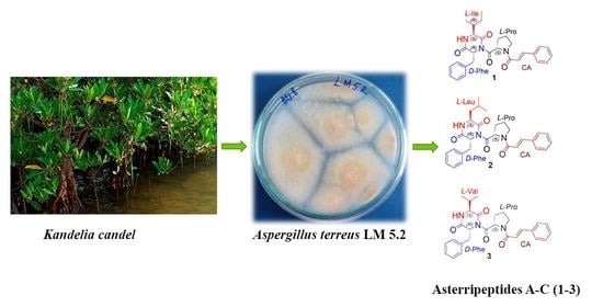
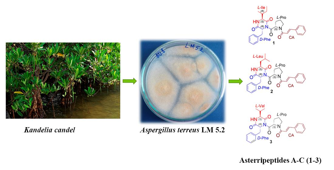
New Tripeptide Derivatives Asterripeptides A–C from Vietnamese Mangrove-Derived Fungus Aspergillus terreus LM.5.2

 ,
,  , ,
, , 
Abstract
:
1. Introduction
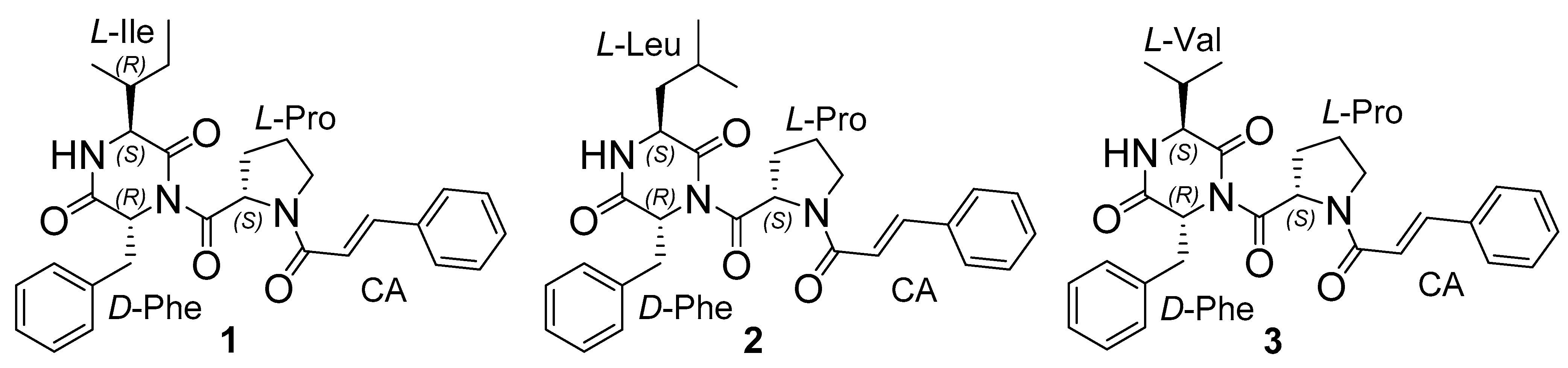
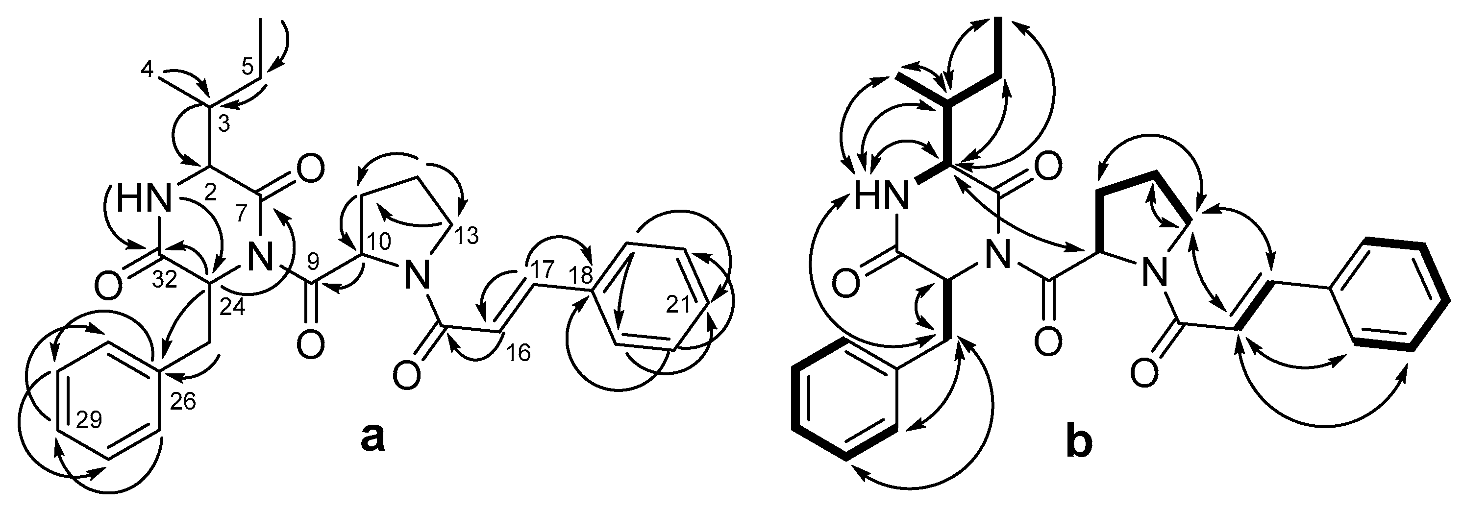
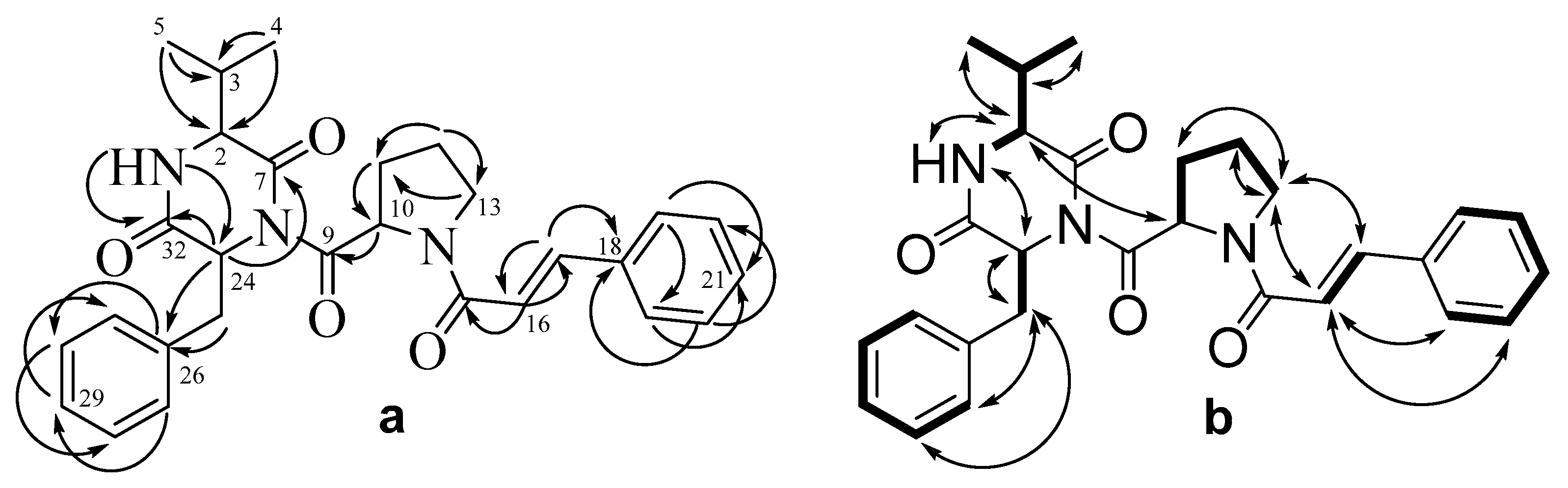
2. Results
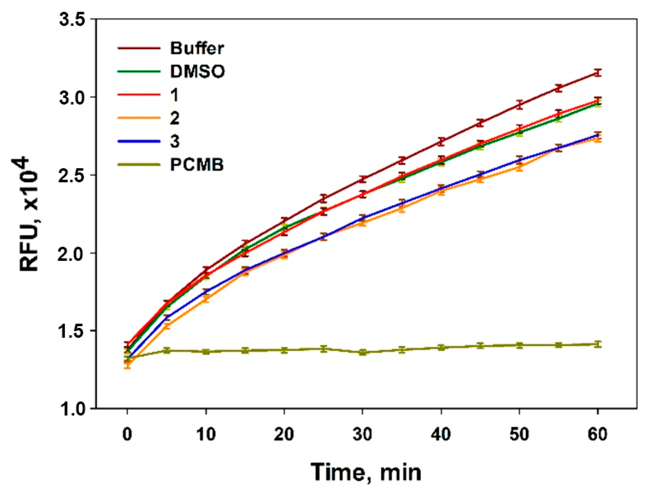
Bioassays
3. Materials and Methods
3.1. General Experimental Procedures
3.2. Fungal Strain
3.3. Cultivation of Fungus
3.4. Extraction and Isolation
3.5. Stereo Configuration Analysis of Amino Acids in Compounds 1–3
3.6. The Effect of Compounds 1–3 on Sortase A Enzymatic Activity
3.7. Cell Lines and Culture Conditions
3.8. In Vitro MTT-Based Cytotoxicity Assay
3.9. Statistical Data Evaluation
Supplementary Materials
Author Contributions
Funding
Institutional Review Board Statement
Informed Consent Statement
Data Availability Statement
Acknowledgments
Conflicts of Interest
References
- Varga, J.; Samson, R.A. Aspergillus in the Genomic Era; Wageningen Academic Publishers: Wageningen, The Netherlands, 2008; pp. 1–334. [Google Scholar]
- Uras, I.S.; Ebada, S.S.; Korinek, M.; Albohy, A.; Abdulrazik, B.S.; Wang, Y.-H.; Chen, B.-H.; Horng, J.-T.; Lin, W.; Hwang, T.-L.; et al. Anti-inflammatory, antiallergic, and COVID-19 main protease (Mpro) inhibitory activities of butenolides from a marine-derived fungus Aspergillus terreus. Molecules 2021, 26, 3354. [Google Scholar] [CrossRef] [PubMed]
- Inamori, Y.; Kato, Y.; Kubo, M.; Kamiki, T.; Takemoto, T.; Nomoto, K. Studies on metabolites produced by Aspergillus terreus var. aureus. I. Chemical structures and antimicrobial activities of metabolites isolated from culture broth. Chem. Pharm. Bull. 1983, 31, 4543–4548. [Google Scholar] [CrossRef] [PubMed]
- Li, H.L.; Li, X.M.; Yang, S.Q.; Meng, L.H.; Li, X.; Wang, B.G. Prenylated phenol and benzofuran derivatives from Aspergillus terreus EN-539, an endophytic fungus derived from marine red alga Laurencia okamurai. Mar. Drugs 2019, 17, 605. [Google Scholar] [CrossRef]
- Liu, W.; Xu, W.; Zeng, X.; Cheng, Z.; Li, Q.; Li, Y. Aspterrics A and B, new sesquiterpenes from deep sea-derived fungus Aspergillus terreus YPGA10. Rec. Nat. Prod. 2020, 14, 18–22. [Google Scholar] [CrossRef]
- Tsuda, Y.; Kaneda, M.; Tada, A.; Nitta, K.; Yamamoto, Y.; Iitaka, Y. Aspterric acid, a new sesquiterpenoid of the carotane group, a metabolite from Aspergillus terreus IFO-6123. X-ray crystal and molecular structure of its p-bromobenzoate. J. Chem. Soc. Chem. Commun. 1978, 4, 160–161. [Google Scholar] [CrossRef]
- Cane, D.E.; Whittle, Y.G.; Liang, T.C. Sesquiterpene biosynthesis: The biosynthesis of quadrone and terrecyclic acid. Bioorg. Chem. 1986, 14, 417–428. [Google Scholar] [CrossRef]
- Ling, K.H.; Yang, C.K.; Peng, F.T. Territrems, tremorgenic mycotoxins of Aspergillus terreus. Appl. Environ. Microbiol. 1979, 37, 355–357. [Google Scholar] [CrossRef] [PubMed]
- Arai, K.; Yamamoto, Y. Metabolic products of Aspergillus terreus X: Biosynthesis of asterriquinones. Chem. Pharm. Bull. 1990, 38, 2929–2932. [Google Scholar] [CrossRef]
- Huang, X.; Men, P.; Tang, S.; Lu, X. Aspergillus terreus as an industrial filamentous fungus for pharmaceutical biotechnology. Curr. Opin. Biotechnol. 2021, 69, 273–280. [Google Scholar] [CrossRef] [PubMed]
- Khaldi, N.; Seifuddin, F.T.; Turner, G.; Haft, D.; Nierman, W.C.; Wolfe, K.H.; Fedorova, N.D. SMURF: Genomic mapping of fungal secondary metabolite clusters. Fungal Genet. Biol. 2010, 47, 736–741. [Google Scholar] [CrossRef]
- Gao, H.; Guo, W.; Wang, Q.; Zhang, L.; Zhu, M.; Zhu, T.; Gu, Q.; Wang, W.; Li, D. Aspulvinones from a mangrove rhizosphere soil-derived fungus Aspergillus terreus Gwq-48 with anti-influenza A viral (H1N1) activity. Bioorg. Med. Chem. Lett. 2013, 23, 1776–1778. [Google Scholar] [CrossRef] [PubMed]
- Liu, M.; He, Y.; Shen, L.; Anbari, W.H.A.; Li, H.; Wang, J.; Qi, C.; Hu, Z.; Zhang, Y. Asperteramide A, An unusual N-Phenyl-carbamic acid methyl ester trimer isolated from the coral-derived fungus Aspergillus terreus. Eur. J. Org. Chem. 2019, 2019, 2928–2932. [Google Scholar] [CrossRef]
- He, F.; Bao, J.; Zhang, X.-Y.; Tu, Z.-C.; Shi, Y.-M.; Qi, S.-H. Asperterrestide A, A cytotoxic cyclic tetrapeptide from the marine-derived fungus Aspergillus terreus SCSGAF0162. J. Nat. Prod. 2013, 76, 1182–1186. [Google Scholar] [CrossRef]
- Chaiyosang, B.; Kanokmedhakul, K.; Boonmak, J.; Youngme, S.; Kukongviriyapan, V.; Soytong, K.; Kanokmedhakul, S. A new lumazine peptide penilumamide e from the fungus Aspergillus terreus. Nat. Prod. Res. 2016, 30, 1017–1024. [Google Scholar] [CrossRef]
- Agrawal, S.; Acharya, D.; Adholeya, A.; Barrow, C.J.; Deshmukh, S.K. Nonribosomal peptides from marine microbes and their antimicrobial and anticancer potential. Front. Pharmacol. 2017, 8, 828. [Google Scholar] [CrossRef]
- Sieber, S.A.; Marahiel, M.A. Learning from Nature’s drug factories: Nonribosomal synthesis of macrocyclic peptides. J. Bacteriol. 2003, 185, 7036–7043. [Google Scholar] [CrossRef]
- Daniel, J.F.D.S.; Rodrigues Filho, E. Peptaibols of trichoderma. Nat. Prod. Rep. 2007, 24, 1128–1141. [Google Scholar] [CrossRef]
- Youssef, F.S.; Ashour, M.L.; Singab, A.N.B.; Wink, M. A comprehensive review of bioactive peptides from marine fungi and their biological significance. Mar. Drugs 2019, 17, 559. [Google Scholar] [CrossRef]
- Girich, E.V.; Yurchenko, A.N.; Smetanina, O.F.; Trinh, P.T.; Ngoc, N.T.; Pivkin, M.V.; Popov, R.S.; Pislyagin, E.A.; Menchinskaya, E.S.; Chingizova, E.A.; et al. Neuroprotective metabolites from vietnamese marine derived fungi of Aspergillus and Penicillium genera. Mar. Drugs 2020, 18, 608. [Google Scholar] [CrossRef]
- Fujii, K.; Shimoya, T.; Ikai, Y.; Oka, H.; Harada, K.-I. Further application of advanced Marfey’s method for determination of absolute configuration of primary amino compound. Tetrahedron Lett. 1998, 39, 2579–2582. [Google Scholar] [CrossRef]
- De, P.; Baltas, M.; Bedos-Belval, F. Cinnamic acid derivatives as anticancer agents—A review. Curr. Med. Chem. 2011, 18, 1672–1703. [Google Scholar] [CrossRef] [PubMed]
- Lafay, S.; Gil-Izquierdo, A. Bioavailability of phenolic acids. Phytochem. Rev. 2008, 7, 301–311. [Google Scholar] [CrossRef]
- Hahlbrock, K.; Scheel, D. Physiology and molecular-biology of phenylpropanoid metabolism. Annu. Rev. Plant Physiol. Plant Molec. Biol. 1989, 40, 347–369. [Google Scholar] [CrossRef]
- Seshime, Y.; Praveen, R.J.; Fujii, I.; Kitamoto, K. Genomic evidences for the existence of a phenylpropanoid metabolic pathway in Aspergillus oryzae. Biochem. Biophys. Res. Commun. 2005, 337, 747–751. [Google Scholar] [CrossRef] [PubMed]
- Guo, Y.; Cai, S.; Gu, G.; Guo, Z.; Long, Z. Recent progress in the development of sortase A inhibitors as novel anti-bacterial virulence agents. RSC Adv. 2015, 5, 49880–49889. [Google Scholar] [CrossRef]
- Rentero Rebollo, I.; McCallin, S.; Bertoldo, D.; Entenza, J.M.; Moreillon, P.; Heinis, C. Development of potent and selective S. aureus sortase A inhibitors based on peptide macrocycles. ACS Med. Chem. Lett. 2016, 7, 606–611. [Google Scholar] [CrossRef]
- Chingizova, E.A.; Menchinskaya, E.S.; Chingizov, A.R.; Pislyagin, E.A.; Girich, E.V.; Yurchenko, A.N.; Guzhova, I.V.; Mikhailov, V.V.; Aminin, D.L.; Yurchenko, E.A. Marine fungal cerebroside flavuside B protects HaCaT keratinocytes against Staphylococcus aureus induced damage. Mar. Drugs 2021, 19, 553. [Google Scholar] [CrossRef] [PubMed]


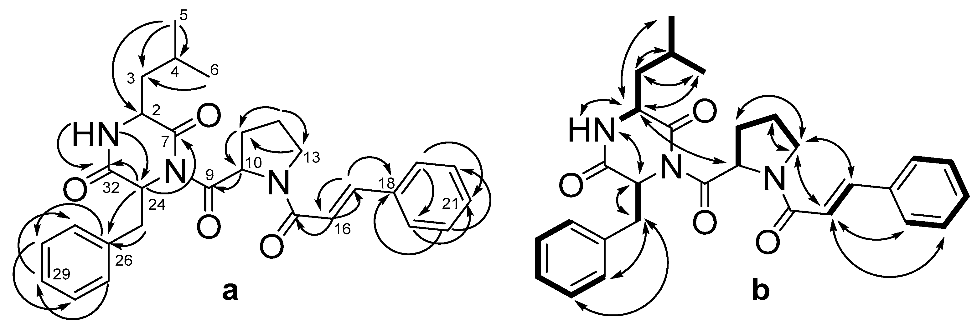
| Pos. | δC, Mult | δH (J in Hz) | HMBC | COSY | ROESY | |
|---|---|---|---|---|---|---|
| Ile | 1 | (NH) | 5.57, brs | 2, 7, 24, 32 | 2 | 2, 24 |
| 2 | 59.4, CH | 2.54, d (2.7) | 3, 4, 5, 7 | 3 | 5, 6 | |
| 3 | 38.2, CH | 1.97, m | 4, 5, 7 | 2, 4, 5 | 1, 4, 6 | |
| 4 | 15.7, CH3 | 0.89, d (7.2) | 2, 3, 5 | 3 | 1, 3 | |
| 5 | 24.1, CH2 | 1.24, m 1.40, m | 2, 3, 4, 6, 10 2, 3, 4, 6 | 2 | ||
| 6 | 12.1, CH3 | 0.85, t (7.4) | 3, 5 | 5a, 5b | 2, 3 | |
| 7 | 170.1, C | |||||
| Pro | 9 | 174.7, C | ||||
| 10 | 61.7, CH | 5.10, dd (8.9, 4.1) | 9, 11, 12, 13 * | 11 | 2, 11 | |
| 11 | 29.7, CH2 | 2.14, m 2.50, m | 9, 10, 12, 13 9, 10, 12, 13 | 10, 12 10, 12 | 13 | |
| 12 | 24.4, CH2 | 2.09, m 2.17, m | 10, 11, 13 10, 11, 13 | 11, 13 11, 13 | 13 | |
| 13 | 47.6, CH2 | 3.79, dt (9.2, 7.1) 3.90, m | 11, 12 11, 12 | 12 12 | 11, 12, 16, 17 | |
| CA | 15 | 164.6, C | ||||
| 16 | 117.9, CH | 6.73, d (15.5) | 15, 17, 18 | 17 | 13, 22, 23 | |
| 17 | 142.8, CH | 7.66, d (15.6) | 16, 18, 19, 23 | 16 | 13 | |
| 18 | 135.1, C | |||||
| 19 | 127.9, CH | 7.51 | 17, 21, 23 | 20 | ||
| 20 | 128.8, CH | 7.36, m | 18, 22 | 19, 21 | ||
| 21 | 129.7, CH | 7.35, m | 19, 23 | 20, 22 | ||
| 22 | 128.8, CH | 7.36, m | 18, 20 | 21, 23 | ||
| 23 | 127.9, CH | 7.51 | 17, 19, 21 | 22 | ||
| Phe | 24 | 58.2, CH | 5.22, t (4.5) | 7, 9 *, 25, 26, 32 | 25 | 25 |
| 25 | 38.5, CH2 | 3.28, dd (14.0, 4.2) 3.33, dd (14.0, 4.9) | 24, 26, 27, 31, 32 24, 26, 27, 31, 32 | 24 24 | 11, 24, 26, 27 | |
| 26 | 135.0, C | |||||
| 27 | 130.5, CH | 7.10, m | 25, 29, 31 | 28 | 25 | |
| 28 | 128.7, CH | 7.29, m | 26, 30 | 27, 29 | 25 | |
| 29 | 127.8, CH | 7.30, m | 27, 31 | 28, 30 | ||
| 30 | 128.7, CH | 7.29, m | 26, 28 | 29, 31 | ||
| 31 | 130.5, CH | 7.10, m | 25, 27, 29 | 30 | ||
| 32 | 168.2, C |
| Pos. | δC, Mult | δH (J in Hz) | HMBC | COSY | ROESY | |
|---|---|---|---|---|---|---|
| Leu | 1 | (NH) | 5.62, brs | 2, 7, 24, 32 | 2 | 2, 24 |
| 2 | 52.6, CH | 2.48, dd (8.8, 3.8) | 3, 4, 5, 7 | 3 | 5, 6 | |
| 3 | 40.7, CH2 | 1.67, m 1.52, m | 2, 3, 4, 5, 6, 7, 32 | 2, 4, 5 | 1, 4, 6 | |
| 4 | 24.4, CH | 2.07, m | 2, 3, 5, 6 | 2 | ||
| 5 | 23.1, CH3 | 0.88, d (6.2) | 2, 3, 6 | 3 | 1, 3 | |
| 6 | 20.7, CH3 | 0.70, d (6.3) | 3, 5 | 5a, 5b | 2, 3 | |
| 7 | 170.8, C | |||||
| Pro | 9 | 175.0, C | ||||
| 10 | 61.8, CH | 5.17, dd (8.6, 3.6) | 9, 13 * | 11 | 2, 11 | |
| 11 | 29.7, CH2 | 2.12, m 2.48, dd (8.8, 3.8) | 9, 10, 12, 13 9, 10, 12, 13 | 10, 12 10, 12 | 13 | |
| 12 | 29.6, CH2 | 2.11, m 1.26, m | 10, 11, 13 10, 11, 13 | 11, 13 11, 13 | 13 | |
| 13 | 47.6, CH2 | 3.93, m 3.79, dd (16.3, 7.3) | 11, 12 11, 12 | 12 12 | 11, 12, 16, 17 | |
| CA | 15 | 164.6, C | ||||
| 16 | 117.8, CH | 6.73, d (15.4) | 15, 17, 18 | 17 | 13, 22, 23 | |
| 17 | 142.9, CH | 7.66, d (15.5) | 16, 18, 19, 23 | 16 | 13 | |
| 18 | 135.1, C | |||||
| 19 | 127.9, CH | 7.52, d (2.3) | 17, 21, 23 | 20 | ||
| 20 | 128.7, CH | 7.29, m | 18, 22 | 19, 21 | ||
| 21 | 129.8, CH | 7.36, m | 19, 23 | 20, 22 | ||
| 22 | 128.7, CH | 7.29, m | 18, 20 | 21, 23 | ||
| 23 | 127.9, CH | 7.52, d (2.3) | 17, 19, 21 | 22 | ||
| Phe | 24 | 58.6, CH | 5.23, t (4.5) | 7, 9, 25, 26, 32 | 25 | 25 |
| 25 | 38.4, CH2 | 3.28, dd (14.0, 4.9) 3.35, dd (14.0, 5.0) | 24, 26, 27, 31, 32 24, 26, 27, 31, 32 | 24 24 | 11, 24, 26, 27 | |
| 26 | 135.2, C | |||||
| 27 | 130.4, CH | 7.13, d (1.8) | 25, 29, 31 | 28 | 25 | |
| 28 | 128.8, CH | 7.29, m | 26, 30 | 27, 29 | 25 | |
| 29 | 127.7, CH | 7.36, m | 27, 31 | 28, 30 | ||
| 30 | 128.8, CH | 7.29, m | 26, 28 | 29, 31 | ||
| 31 | 130.4, CH | 7.13, d (1.8) | 25, 27, 29 | 30 | ||
| 32 | 168.1, C |
| Pos. | δC, Mult | δH (J in Hz) | HMBC | COSY | ROESY | |
|---|---|---|---|---|---|---|
| Val | 1 | (NH) | 5.67, brs | 2, 8, 23, 31 | 2 | 2, 24 |
| 2 | 58.0, CH | 2.60, d (2.7) | 3, 4, 5, 7 | 3 | 5 | |
| 3 | 31.7, CH | 2.31, m | 2, 3, 4, 5, 7, 31 | 2, 4, 5 | 1, 4 | |
| 4 | 15.8, CH3 | 0.93, d (6.8) | 2, 3, 5 | 3 | 1, 3 | |
| 5 | 18.9, CH3 | 0.90, d (7.1) | 2, 3, 4 | 2 | ||
| 7 | 170.2, C | |||||
| Pro | 9 | 174.6, C | ||||
| 10 | 61.6, CH | 5.11, dd (8.6, 3.7) | 9, 11, 12 | 11 | 2, 11 | |
| 11 | 29.7, CH2 | 2.14, m 2.48, m | 9, 10, 12 9, 12 | 10, 12 10, 12 | 13 | |
| 12 | 24.4, CH2 | 2.18, m 2.08, m | 10, 13 10, 13 | 12b 12a, 12b | ||
| 13 | 47.6, CH2 | 3.79, m 3.94, m | 11, 12 11, 12 | 10, 11b, 12a, 12b 10, 11b, 12a, 12b | 15, 11b 15, 11a | |
| 15 | 164.6, C | |||||
| CA | 16 | 117.8, CH | 6.72, d (15.5) | 15, 17, 18, 19/23 | 17 | 13a, 13b, 19/23, 20/22 |
| 17 | 142.9, CH | 7.66, d (15.6) | 15, 16, 18, 19/23 | 16 | 13a, 13b | |
| 18 | 135.1, C | |||||
| 19 | 127.9, CH | 7.50, d (6.9) | 21, 23 | 20 | 16 | |
| 20 | 128.8, CH | 7.35, brd (1.4) | 17, 21 | 19, 21 | 16 | |
| 21 | 129.7, CH | 7.34, m | 19, 23 | 20, 22 | ||
| 22 | 128.8, CH | 7.35, brd (1.4) | 18, 20 | 21, 23 | 16 | |
| 23 | 127.9, CH | 7.51, d (7.8) | 17, 19, 21 | 16 | ||
| Phe | 24 | 59.8, CH | 5.22, t (4.5) | 7, 9, 25, 26, 32 | ||
| 25 | 38.6, CH2 | 3.28, dd (14.0, 5.0) 3.34, dd (14.0, 4.0) | 25, 27/31, 32 25, 27/31, 32 | 29 29 | 27/31, 28 27/31, 28 | |
| 26 | 134.9, C | |||||
| 27 | 130.6, CH | 7.10, d (1.7) | 25, 27, 29 | 10, 24 | ||
| 28 | 128.7, CH | 7.29, m | 26, 29, 31 | |||
| 29 | 127.9, CH | 7.29, m | 27, 31 | |||
| 30 | 128.7, CH | 7.29, m | 26, 27, 29 | |||
| 31 | 130.6, CH | 7.10, d (1.7) | 25, 27, 29 | 10, 24 | ||
| 32 | 168.2, C |
| Compound | IC50, µM | |||
|---|---|---|---|---|
| MCF-7 | DLD-1 | PC-3 | H9c2 | |
| 1 | 96.8 ± 7.0 | 87.7 ± 5.3 | 64.6 ± 2.4 | 76.7 ± 5.2 |
| 2 | >100 | >100 | 75.5 ± 1.9 | 104.1 ± 3.3 |
| 3 | 96.6 ± 1.5 | 84.9 ± 7.4 | 58.3 ± 3.2 | 87.6 ± 4.5 |
Publisher’s Note: MDPI stays neutral with regard to jurisdictional claims in published maps and institutional affiliations. |
© 2022 by the authors. Licensee MDPI, Basel, Switzerland. This article is an open access article distributed under the terms and conditions of the Creative Commons Attribution (CC BY) license (https://creativecommons.org/licenses/by/4.0/).
Share and Cite
Girich, E.V.; Rasin, A.B.; Popov, R.S.; Yurchenko, E.A.; Chingizova, E.A.; Trinh, P.T.H.; Ngoc, N.T.D.; Pivkin, M.V.; Zhuravleva, O.I.; Yurchenko, A.N. New Tripeptide Derivatives Asterripeptides A–C from Vietnamese Mangrove-Derived Fungus Aspergillus terreus LM.5.2. Mar. Drugs 2022, 20, 77. https://doi.org/10.3390/md20010077
Girich EV, Rasin AB, Popov RS, Yurchenko EA, Chingizova EA, Trinh PTH, Ngoc NTD, Pivkin MV, Zhuravleva OI, Yurchenko AN. New Tripeptide Derivatives Asterripeptides A–C from Vietnamese Mangrove-Derived Fungus Aspergillus terreus LM.5.2. Marine Drugs. 2022; 20(1):77. https://doi.org/10.3390/md20010077
Chicago/Turabian StyleGirich, Elena V., Anton B. Rasin, Roman S. Popov, Ekaterina A. Yurchenko, Ekaterina A. Chingizova, Phan Thi Hoai Trinh, Ngo Thi Duy Ngoc, Mikhail V. Pivkin, Olesya I. Zhuravleva, and Anton N. Yurchenko. 2022. "New Tripeptide Derivatives Asterripeptides A–C from Vietnamese Mangrove-Derived Fungus Aspergillus terreus LM.5.2" Marine Drugs 20, no. 1: 77. https://doi.org/10.3390/md20010077
APA StyleGirich, E. V., Rasin, A. B., Popov, R. S., Yurchenko, E. A., Chingizova, E. A., Trinh, P. T. H., Ngoc, N. T. D., Pivkin, M. V., Zhuravleva, O. I., & Yurchenko, A. N. (2022). New Tripeptide Derivatives Asterripeptides A–C from Vietnamese Mangrove-Derived Fungus Aspergillus terreus LM.5.2. Marine Drugs, 20(1), 77. https://doi.org/10.3390/md20010077