Article

Solid Matrix-Supported Supercritical CO2 Enhances Extraction of γ-Linolenic Acid from the Cyanobacterium Arthrospira (Spirulina) platensis and Bioactivity Evaluation of the Molecule in Zebrafish

1
Center for Microalgal Biotechnology and Biofuels, Institute of Hydrobiology, Chinese Academy of Sciences, Wuhan 430072, China
2
State Key Laboratory of Freshwater Ecology and Biotechnology, Institute of Hydrobiology, Chinese Academy of Sciences, Wuhan 430072, China
3
Laboratory for Marine Biology and Biotechnology, Qingdao National Laboratory for Marine Science and Technology, Qingdao 266237, China
4
Key Laboratory for Algal Biology, Institute of Hydrobiology, Chinese Academy of Sciences, Wuhan 430072, China
5
College of Life Sciences, University of Chinese Academy of Sciences, Beijing 100049, China
6
College of Advanced Agricultural Sciences, University of Chinese Academy of Sciences, The Innovative Academy of Seed Design, Chinese Academy of Sciences, Beijing 100049, China
7
Beijing Key Laboratory of Algae Biomass, Microalgae Biotechnology Center, SDIC Biotech Investment Co., LTD., State Development & Investment Corp., Beijing 100142, China
*
Authors to whom correspondence should be addressed.
Mar. Drugs 2019, 17(4), 203; https://doi.org/10.3390/md17040203
Submission received: 20 February 2019 / Revised: 21 March 2019 / Accepted: 26 March 2019 / Published: 30 March 2019
(This article belongs to the Special Issue Compounds from Cyanobacteria II)

Abstract

:
Marine cyanobacteria represent a large untapped source of functional glycolipids enriched with polyunsaturated fatty acids (PUFAs) for human health. However, advanced methods for scalable isolation of diverse species containing high-purity PUFA-rich glycolipids will have to be developed and their possible pharmaceutical and nutraceutical functions identified. This paper introduces a novel solid matrix-supported supercritical CO2 extraction method for scalable isolation of the PUFA γ-linolenic acid (GLA)-enriched glycolipids from the cyanobacterium Arthrospira (Spirulina) platensis, which has been the most widely used among microalgae in the nutraceutical and pharmaceutical industries. Of various porous materials studied, diatomite was the best to facilitate extraction of GLA-rich glycolipids, resulting in an extraction efficiency of 98%. Gamma-linolenic acid made up 35% of total fatty acids (TFAs) in the extracts, which was considerably greater than that obtained with ethanol (26%), Bligh and Dyer (24%), and in situ transesterification (24%) methods, respectively. Lipidomics analysis revealed that GLA was exclusively associated with galactolipids. Pharmaceutical functions of GLA-rich galactolipids were investigated on a zebrafish caudal fin regeneration model. The results suggested that GLA extracted from A. platensis possessed anti-oxidative, anti-inflammatory, and anti-allergic activities, which acted in a concerted manner to promote post-injury regeneration of zebrafish.

Graphical Abstract

1. Introduction

Gamma-linolenic acid (GLA) is an omega-6 polyunsaturated fatty acid (PUFA) that has shown multiple nutraceutical and pharmaceutical functions. Gamma-linolenic acid is a potential precursor in the biosynthesis of arachidonic acid (ARA), which possesses a wide range of biological activities related to its conversion to 20-carbon long-chain polyunsaturated fatty acid (C20 LC-PUFA), dihomo-gamma-linolenic acid (DGLA) [1]. Supplementing dietary GLA can increase serum DGLA, as well as serum ARA levels [2]. Also, GLA has been used to treat various medical conditions like rheumatoid arthritis [3,4], eczema [5], premenstrual syndrome [6,7], and reduction of nerve pain in people with diabetic neuropathy [8].
Many marine cyanobacteria contain considerable amounts of GLA in the form of glycolipids [9,10,11]. For example, the cyanobacterium Arthrospira (Spirulina) platensis can produce ca. 2% of GLA on a dry weight basis [12]. Arthrospira platensis has been commercially produced in large open raceway ponds using seawater [13,14] or alkaline water as a culture medium [15]. As GLA accounted for as high as 30% of total fatty acids (TFAs) in A. platensis cells [16], and α-linolenic acid (the isomer of GLA) was absent in TFAs [15], isolation, and purification of GLA-containing glycolipids from A. platensis could be cost-effective. In cyanobacteria glycolipids, 80 mol% are galactolipids [17], which contain the various fatty acid acyl groups (e.g., GLA) that are important for their functions. Although marine cyanobacteria represent an untapped natural source of glycolipids for pharmaceutical and nutraceutical applications, knowledge aboutcyanobacteria-derived glycolipids is rather limited. Arthrospira platensis has been subjected to the most extensive and intensive studies with regard to the health benefits of its biologically active compounds [14,18]. It may serve as an ideal model system to better understand the function of glycolipids in general and marine cyanobacteria in particular [19,20,21].
Extraction of lipids from A. platensis biomass is a crucial step toward isolation and purification of GLA [22,23,24]. Bligh and Dyer and Folch extraction methods are commonly applied to extraction of lipids from numerous microalgae including A. platensis under laboratory conditions [25]. Considering that GLA is produced for human consumption, the extraction processes require the use of solvents safe for human health and the environment. However, chloroform and methanol used in the conventional approaches can be toxic to both workers and consumers. Therefore, searching for more bio-safe and cost-effective solvents is imperative for extraction of bioproducts, particularly for human consumption.
Supercritical fluid extraction is an advanced technique that has recently been applied for extraction of biologically active products from microalgae under laboratory conditions [26,27,28,29,30]. The main supercritical solvent used is CO2, which is accessible, environmentally friendly, and generally recognized as safe by the U.S. Food and Drug Administration [31]. Carbon dioxide is easily removed from extractants and gives solvent-free end-products. Despite numerous advantages, supercritical CO2 (SC-CO2) has a main drawback, i.e., low polarity of CO2 that may lead to low extraction efficiency. Aiming at this challenge, a co-solvent ethanol was used as a polar modifier to alter the polarity of the supercritical fluid and enhance its solvating power towards targeted products. Previous studies addressed the effect of operational variables (e.g., temperature, pressure, CO2, and co-solvent consumption) on extraction efficiency of GLA [27,28,29,30,32,33,34], but the extraction efficiency remained relatively low (25–78%).
Previously, many nutraceutical studies of A. platensis were conducted with whole biomass or crude extracts of A. platensis [35,36]. To better explore the therapeutic potentials of A. platensis, functions of the refined bioproducts (e.g., GLA) from A. platensis biomass need to be demonstrated by employing an advanced toolbox. The functions of crude GLA extracted from A. platensis were studied using various cell line models [37,38]. To bridge the gap between the in vitro cellular level studies and nutraceutical or therapeutic applications, appropriate animal models and in vivo studies are required. Zebrafish have become an effective and popular model organism for drug screening, drug efficacy evaluation, and dissecting molecular mechanisms of drug action, as approximately 82% of human disease-related genes have at least one zebrafish orthologue [39]. The zebrafish larval caudal fin regeneration model is a well-established system for studying the roles of the immune system in organ injury and regeneration [40], and thus can be utilized as an in vivo model to investigate the bioactivities of compounds of interest as immunomodulators.
This work aimed at improving extraction efficiency of GLA from A. platensis by introducing a porous solid material to the supercritical fluid extraction (SFE) method. We hypothesized that prior to SFE treatment, microalgal biomass pre-mixed with the right type of porous solid materials could increase the surface area of algal biomass accessible to a solvent (CO2 alone or with ethanol), which facilitates penetration of the solvent into algal cells, and thus results in an enhanced extraction efficiency of GLA-enriched lipids. To test our hypothesis, we pre-mixed algal powders with several porous solid materials (i.e., molecular sieve, macroporous resin, and diatomite), individually, with different weight ratios. Total lipids, TFAs, and GLA extraction efficiencies were measured and compared with the control experiments that used algal powders only. Bioactivities of GLA obtained in this study were evaluated using a zebrafish caudal fin regeneration model.

2. Results and Discussion

2.1. Quantitative Analysis of Total Lipids and Fatty Acid Composition of A. platensis

Four methods were assessed for extraction of total lipids and GLA from A. platensis. They were (1) in situ transesterification followed by gas chromatography-mass spectrometry (GC-MS) analysis; (2) the Bligh and Dyer method; (3) extraction by ethanol with the aid of mechanical stirring; and (4) solid matrix-supported supercritical CO2 (SMSSC-CO2). The lipid content of A. platensis was 11.52 ± 0.11% of dry cellular weight (DCW), as determined by the Bligh and Dyer method. A slightly lower lipid content of 10.22 ± 0.17% DCW was obtained by extraction of algal biomass with ethanol. The SMSSC-CO2 method yielded the lowest amount of lipids (9.36 ± 0.30% DCW), but resulted in the greatest amounts of GLA on per TFAs and per total lipids basis (Table 1). In situ transesterification, followed by GC-MS analysis, showed that the TFAs content was 5.25 ± 0.07% DCW, and consisted of C16:0 (45.82% of TFAs), C16:1 (5.78%), C18:0 (0.64%), C18:1trans (1.41%), C18:1cis (0.22%), C18:2trans (0.15%), C18:2cis (21.99%), and C18:3n6 (GLA, 23.98%). Gamma-linolenic acid accounted for up to 1.26 ± 0.04% DCW and 1.18 ± 0.03% DCW for in situ transesterification and Bligh and Dyer methods, respectively (Table 1). Thus, the Bligh and Dyer method was selected to evaluate the GLA recovery in following studies.

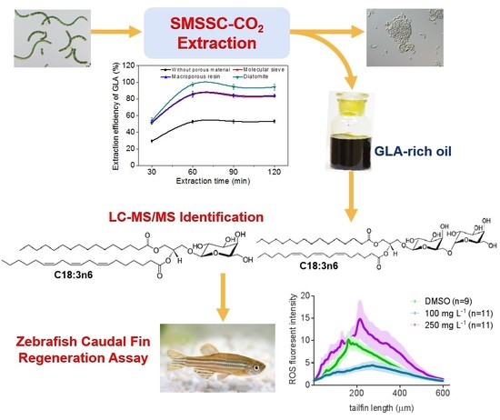
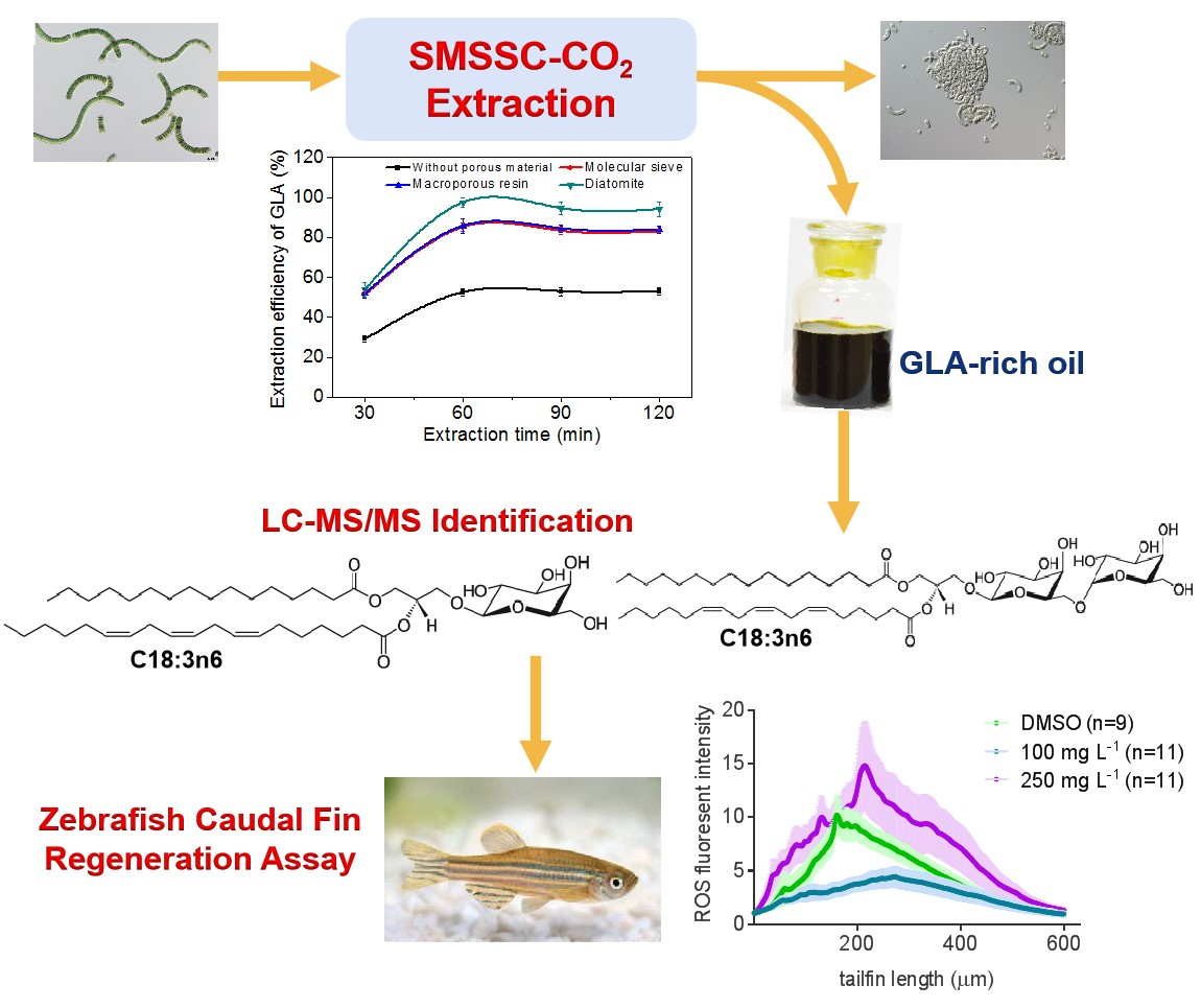
2.2. Effect of Incorporated Solid Support on GLA Extraction Efficiency

Figure 1 shows that the introduction of a solid material (e.g., molecular sieve, macroporous resin, or diatomite) remarkably increased the extraction efficiencies of total lipids, TFAs, and GLA, when compared to the control without addition of such materials. It is believed that mixing algal powder with porous solids may increase bulk volume of algal biomass, therefore reducing the dead volume that exists between algal biomass and the internal boundary of the extraction vessel (particularly under supercritical state), thereby increasing the contact area between solvents and the algal-porous solid bed. Moreover, good dispersion of algal cells with porous solid materials may create a highly effective volume between algal cells and solvents, which is dependent on a desirable mixed-weight ratio of the porous solid material to algal biomass (Figure S1).
Among the three solid support materials tested, diatomite resulted in the highest extraction efficiencies of 81%, 83%, and 98%, for total lipids, TFAs, and GLA, respectively. These results were in large contrast to the control experiments (without porous materials) where the extraction efficiencies of total lipids, TFAs, and GLA were 46%, 47%, and 53%, respectively (Figure 1). Under the supercritical conditions, algal powder loaded inside the reaction vessel was compressed and clogged (mainly due to high pressure) to form a dense bed rather than a porous structure, which reduced the penetration (by diffusion and dispersion) of the solvents (CO2 and ethanol) into the cells, and thus decreased mass transfer of the solvents into individual algal cells. The improved extraction efficiencies that resulted from pre-mixing of algal biomass with solid porous materials may be contributable to increased porosity, and thus the surface-to-volume ratio of algal biomass by the porous materials, which in turn facilitated the solvents (liquid CO2 alone or with ethanol) penetrating into algal cells. The porous materials also enhanced the repartition of biomass matter leading to a more intimate contact between the supercritical solvents (CO2 + ethanol) and microalgal cells, leading to a higher extraction kinetics. Moreover, the recovery of GLA was much greater than that of total lipids and TFAs. The shape of the curves show that the porous materials play a role in the solubility of some compounds towards supercritical CO2 since the extract composition illustrates a better selectivity, particularly for GLA. That means GLA-rich lipid molecules were more readily extractable than non-GLA containing ones under the given conditions.
As diatomite was superior to the other two porous materials, it was used for further study. Mixing of diatomite and biomass at a ratio of 1:2 (w/w) was the optimal for extraction of total lipids and GLA, resulting in the extraction efficiencies of 81% and 98% for total lipids and GLA, respectively. Smaller or greater (e.g., 0, 1:4, 1:3, 1:2, 1:1, and 2:1) than the optimal ratio resulted in reduced extraction efficiencies of total lipids and TFAs, as well as the recovery of GLA (Figure 2A). This may be attributable to increased dense biomass patches in the extraction vessel when the amount of diatomite was relatively too small, which prevents the penetration of solvents into algal cells. When porous solid materials were over-loaded, it resulted in the reduction of effective volume and contact area of algal cells and solvents.
Furthermore, the moisture content of diatomite was also found to significantly affect lipid extraction efficiency. The extraction efficiency of GLA increased from 52% to the maximum value of 98%, when the water content of diatomite increased from 0 to 60 wt.%. However, further increasing the water content to 80 wt.% resulted in decreased extraction efficiency to 65% (Figure 2B). It was speculated that soaking diatomite (containing 60 wt.% water) may have altered the polarity of the solvent mixture, i.e., CO2–ethanol–water, which facilitated the extraction of less-polar lipids galactolipids containing predominant amounts of GLA in the cells [29]. However, further increasing the water content to 80 wt.% might have brought about the formation of water film on the surface of the solid support and algal biomass, which reduced the mass transfer of ethanol and liquid CO2 into algal cells, and thus reduced overall lipid extraction. The extraction of GLA followed the similar trends as total lipids and TFAs over the investigated range of the water content of diatomite (Figure 2B).
When the SMSSC-CO2 approach was applied, the ratio of GLA in extracted TFAs increased to 35%, which was comparable with the record figure of 35.5% in the previous studies that employed direct transesterification on the freeze-dried material (Table 2). The recovery of GLA (based on the Bligh and Dyer method) by SMSSC-CO2 was virtually complete, which reached as high as 98%. Although glass wool [27,28], sea sand [30], and glass microspheres [32] were used to partially fill up the extraction vessel in order to eliminate the dead volume, and the recovery of lipids and GLA from these treatments was not examined in detail. On the other hand, these materials mixed into the A. platensis biomass will be introducing more problems, such that algal residues can be hardly separated from the dispersing agent after extraction. Compared with the microspheres and sea sand used in previous studies, diatomite offers an additional benefit of being safe for livestock. Diatomite represent a popular feed additive for livestock to ameliorate nutrient deficiency and illness problems [41]. Also, after lipid extraction, residues enriched with protein and carbohydrate can be applied to the animal feed industry [42,43].

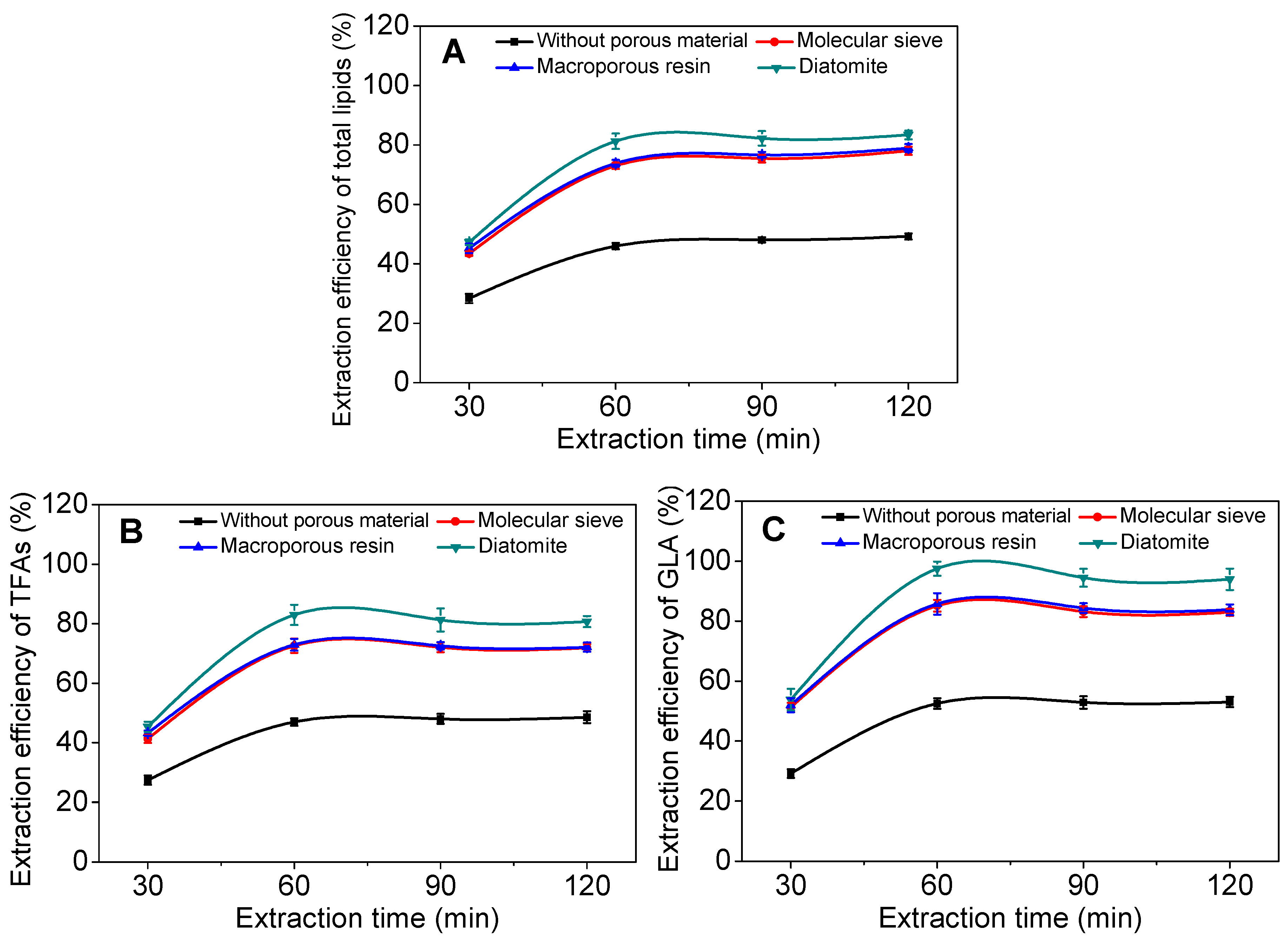
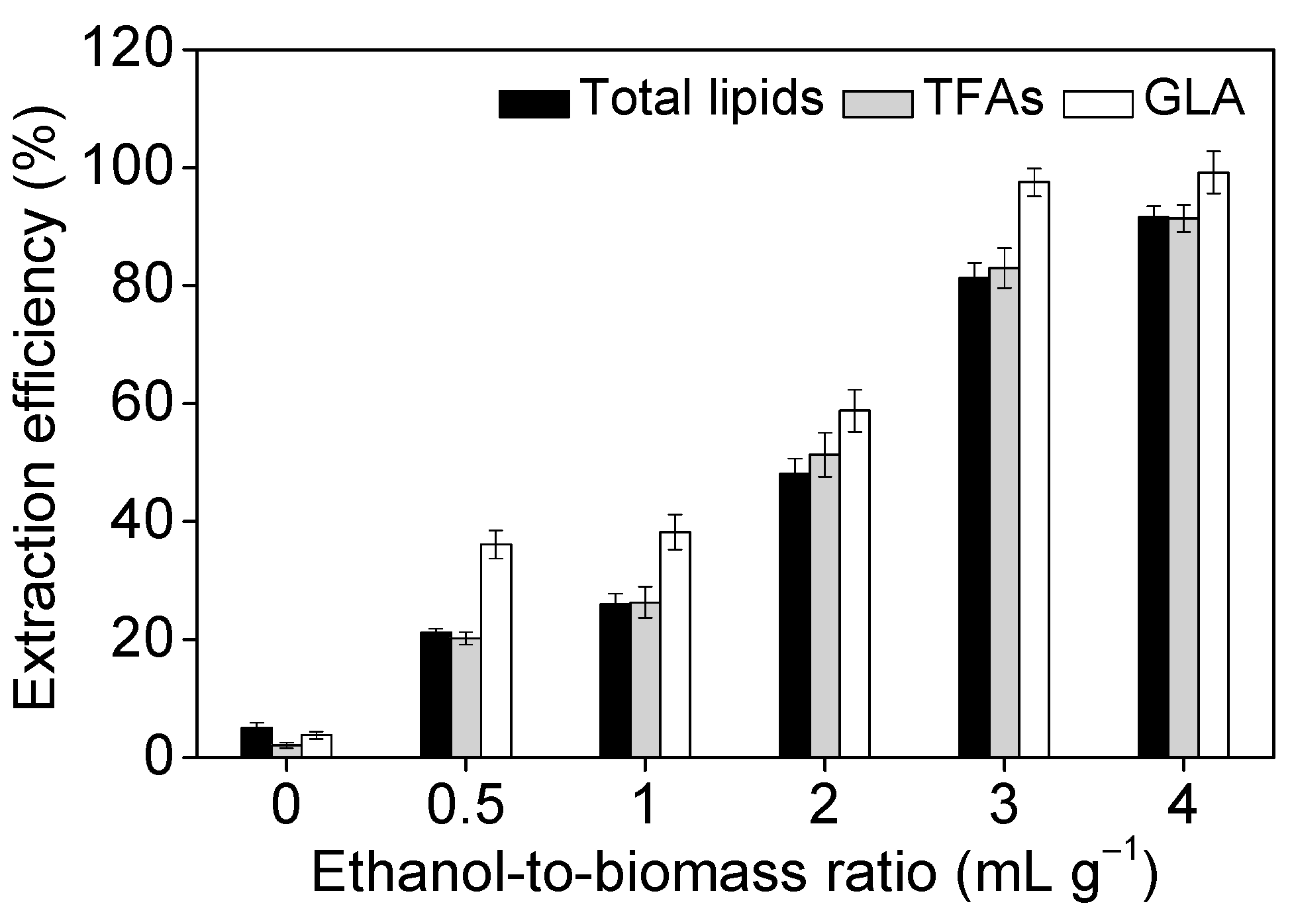
2.3. Effect of Operational Parameters on GLA Extraction Efficiency

As presented in Figure 3, there were almost no polar lipids recovered from the algal biomass after supercritical CO2 treatment in the absence of ethanol as the co-solvent, but the extraction efficiency of total lipids and GLA increased gradually from 5% and 4% to 92% and 99%, respectively, when ethanol was introduced to the biomass with a ratio of 3:1 (v/w). Our results were similar to those reported by Sajilata et al. [27] and Mendes et al. [29], in which introducing ethanol as a co-solvent increased the partitioning of GLA into the galactolipids fraction. Supercritical CO2 alone can extract 100% of neutral lipids contained in microalgae provided the biomass is previously dried and the extraction pressure is higher than 30 MPa. It is attributable to increasing the polarity of the CO2–ethanol mixture relative to CO2 only, thereby improving the extractability of algal biomass and accelerating partitioning of polar lipids (including GLA) into the solvent phase [44]. The extraction efficiencies of total lipids and TFAs at the ethanol-to-biomass ratio of 3:1 were 81% and 83%, respectively, and yet greater extraction efficiencies of total lipids and TFAs were 92% and 91%, respectively, when the ethanol-to-biomass ratio increase further to 4:1. As far as GLA was concerned, however, the extraction efficiency at the ethanol-to-biomass ratios of 3:1 and 4:1 was the same, i.e., 98 ± 1.2%. From an economic point of view, the ethanol-to-biomass ratio of 3:1 is sufficient to extract more than 97% of GLA from A. platensis.
Ethanol as an ideal solvent used to extract total lipids could provide a reasonable partition co-efficiency, forcing the solute to migrate to the solvent phase [45]. Principally, hydrophobic lipids (e.g., neutral lipids) are preferably partitioned into the non-polar solvent phase, whereas polar lipids are not so easy to dissolve in non-polar solvents due to their binding with the biomass matrix [46]. Therefore, co-solvents are applied to break the linkages between the polar lipids and biomass, leading to the increased solubility of the polar lipids. Both ethanol and liquid CO2 are generally recognized as safe solvents and easy to be removed from GLA, total lipids have a high partition coefficient in the ethanol.
Figure 4A showed that the maximum recovery of GLA was obtained at the extraction temperature of 40 °C, which was remarkably higher than that at 30 °C. The extraction efficiency obtained at 30 °C was low because the solvent was not under supercritical conditions. However, further increasing temperatures to 50 °C and 60 °C resulted in a noticeable decrease in the efficiency. Depending on the pressure, the decrease of lipid yields from 40 °C up to 60 °C may be due to a retrograde solubility phenomenon. The optimal temperature identified in this study was the same as that reported by Sajilata et al. [27]. It is believed that a higher temperature than the optimum may increase the ethanol evaporation rate, which leads to the diffusion of ethanol from the liquid phase into the gas phase, resulting in reduced lipid extraction efficiency, in particular of GLA-containing molecules (Figure 4A). Moreover, the supercritical CO2 extraction method is operated at low temperature using a non-oxidant medium, hence it is particularly suitable for the extraction and separation of heat-sensitive and easily oxidized substances (particularly bioactive compounds) [47]. Data presented in Figure 4B showed that the extraction efficiency of GLA increased dramatically when the pressure increased from 28 MPa to 34 MPa, and yet greater extraction efficiencies of 98% and 99% were obtained at 41 MPa and 48 MPa, respectively. It is obvious that lipid yields are improved when pressure increases due to a higher density lead to a solubility increase. However, where energy consumption was concerned, the pressure was set at 41 MPa for further investigation. Note that the optimal pressure level determined in this study is similar to that applied for supercritical CO2-based extraction of astaxanthin from the green microalga Haematococcus pluvialis [48].
Lipid extraction efficiency is time-dependent, when the extraction duration increased from 30 min (15 min for main extraction and 15 min for draining) to 60 min (30 min for main extraction and 30 min for draining), the extraction efficiency of GLA increased from 54% to 98% (Figure 4C). Further extending extraction time from 90 min (30 min for main extraction and 60 min for draining) to 120 min (30 min for main extraction and 90 min for draining) resulted in gradual decrease of extraction kinetics, and the extraction efficiency of GLA reached a plateau. The same trend was observed for total lipids and TFAs (Figure 4C). The optimal extraction time of 60 min was shorter than that described in the literature, which ranges from 3 h to 9 h [49]. The extraction efficiency of GLA gradually increased from 71% to 98% when the consumption of CO2 increased from 0.8 to 2.0 mL g−1 min−1 (Figure 4D). It is likely due to enhanced elution of extracted lipids from biomass residues by passing greater amounts of the main solvent, i.e., liquid CO2 through both the extraction and collection vessels. Taken together, the key process parameters of extraction temperature, pressure, time, and CO2-to-mass ratio under investigation of GLA yields were established as 40 °C, 41 MPa, 60 min, and 2.0 mL g−1 min−1, respectively. SMSSC-CO2 at a temperature moderately above room temperature (40 °C), using CO2 as a green solvent, avoided the decomposition of heat-sensitive and easily oxidized substances to ensure the high quality and stability of products. Therefore, supercritical fluid extraction has more potential compared with other solvents extraction methods, as it is a fast and efficient unconventional extraction method developed for extraction of compounds of interest from solid matrices [50].

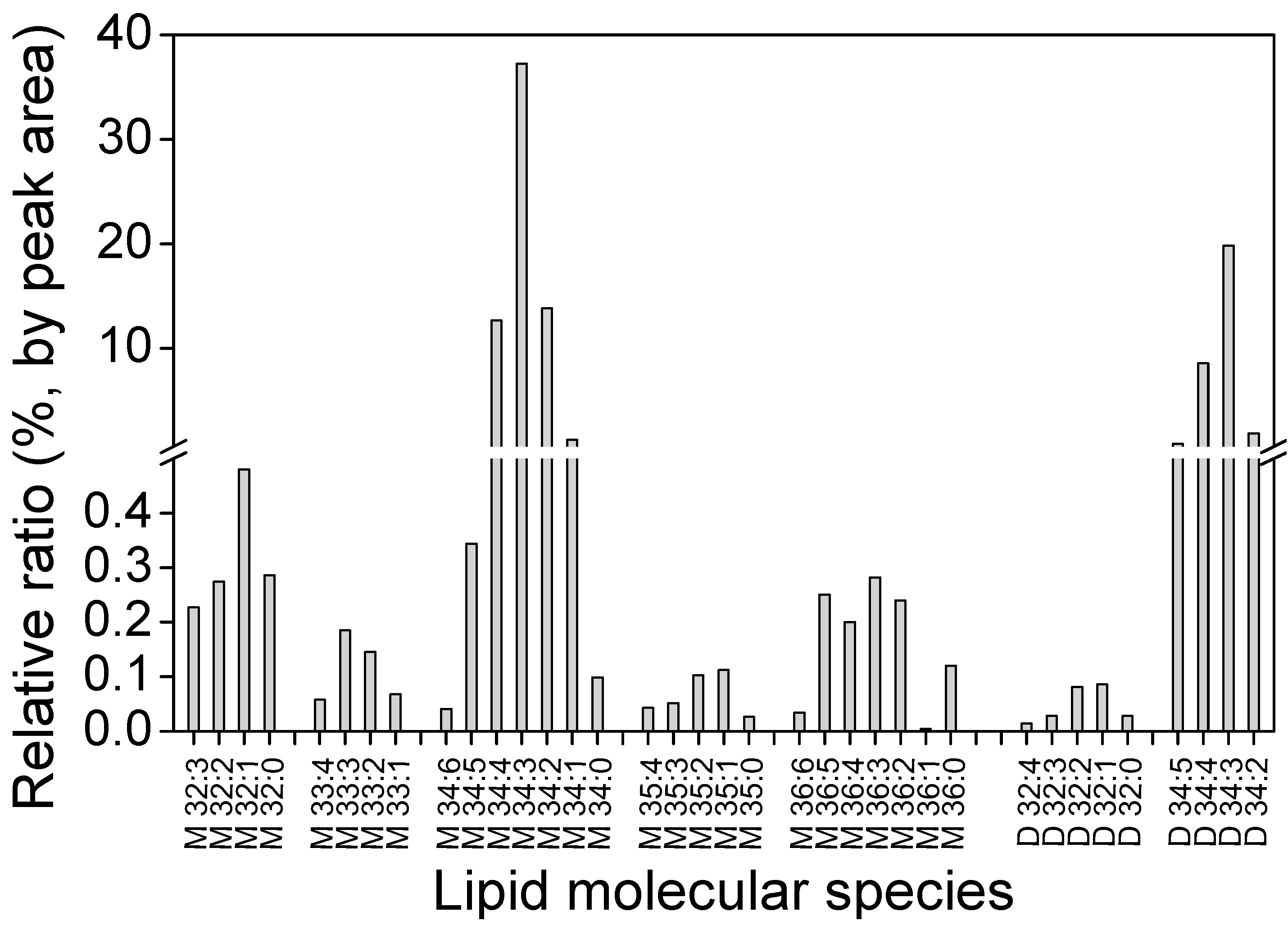
2.4. Distribution of GLA in Different Fractions

After supercritical extraction, total lipids were partitioned into three fractions, i.e., (1) lipid extracts obtained from the collection vessel; (2) lipids from the bottom of the kettle and the surface of sample basket eluted by ethanol, followed by filtering through a micro-pore bag; and (3) lipids remaining inside the residual biomass. As shown in Table 3, GLA was mainly present in the extracted lipids, accounting for 94.31% of the total GLA. The fraction eluted by ethanol from the bottom of the kettle and the surface of the sample basket accounted for 3.25% of the total GLA, whereas GLA in the remaining residual biomass was 2.44%, which is an inevitable loss and cannot be extracted using the current technique and process. Combining the lipid fractions 1 and 2 resulted in an extraction efficiency of 97.56% (94.31% + 3.25%), which was the highest reported thus far in the literature. Note that TFAs (fatty acid methyl esters equivalent) in the residual biomass was 19.35% (Table 3), which was mainly composed of fatty acids like C16:0, C16:1, and C18:0, suggesting that GLA-containing molecules were relatively more extractable than many other lipid molecules in A. platensis cells. Considering that biomass residues after lipid extraction may contain large amounts of proteins and carbohydrates along with valuable trace elements, the exploitation of algal biomass residues as animal feed additives or bio-fertilizers may deserve attention.
Major cyanobacterial lipids are monogalactosyldiacylglycerol (MGDG), digalactosyldiacylglycerol (DGDG), sulfoquinovosyl diacylglycerol (SQDG), and phosphatidylglycerol (PG) [10]. The GLA-containing galactolipid extracts obtained from supercritical CO2 extraction of A. platensis were analyzed using liquid chromatography-tandem mass spectroscopy (LC-MS/MS) in the positive mode with an electrospray ionization (ESI) source. Electrospray ionization is a soft ionization technique that accomplishes the transfer of ions from solution to a gas phase [51]. Prior separation with liquid chromatography can facilitate detection of analytes in complex biological samples [52]. A total of 36 molecular species of galactolipids were identified by tandem MS, and the profiles and composition of each molecular lipid species of MGDG and DGDG in lipid extracts are shown in Figure 5. The dominant lipid molecules of A. platensis galactolipids and phospholipids. The bioactivity of polar lipids depends on the content and structure of fatty acryl groups, namely because ω-6 are essential for human health. Many glycolipids and phospholipids have been described to alleviate senescence, improve cognitive functions, and treat inflammatory diseases [53].
In this study, we uncovered that GLA was chiefly distributed in galactolipids in the form of a fatty acyl group (C18:3) among several classes of determined glycerolipids. According to the fragmentation patterns, 27 MGDG and 9 DGDG molecular species were identified in A. platensis. There are 17 species of galactolipids containing GLA (C18:3), which accounted for 47% of extracted lipids. It was revealed that the single most abundant fatty acyl chain in galactolipids is GLA (C18:3). Table 4 shows that GLA-containing MGDGs are mainly M34:4 (16:1–18:3) and M34:3 (16:0–18:3), making up 53% of total MGDG (Figure 5). The GLA-containing DGDG were mainly D34:5 (16:2–18:3), D34:4 (16:1–18:3), and D34:3 (16:0–18:3), which accounted for 29% of total DGDG. In addition, a trace amount of PG-containing GLA was detected, which was identified as PG 16:0–18:3. Otherwise, no other phospholipids contained GLA. The presence of the above bioactive fatty acyl groups demonstrated new types of lipids in A. platensis with respect to the configuration of lipids.

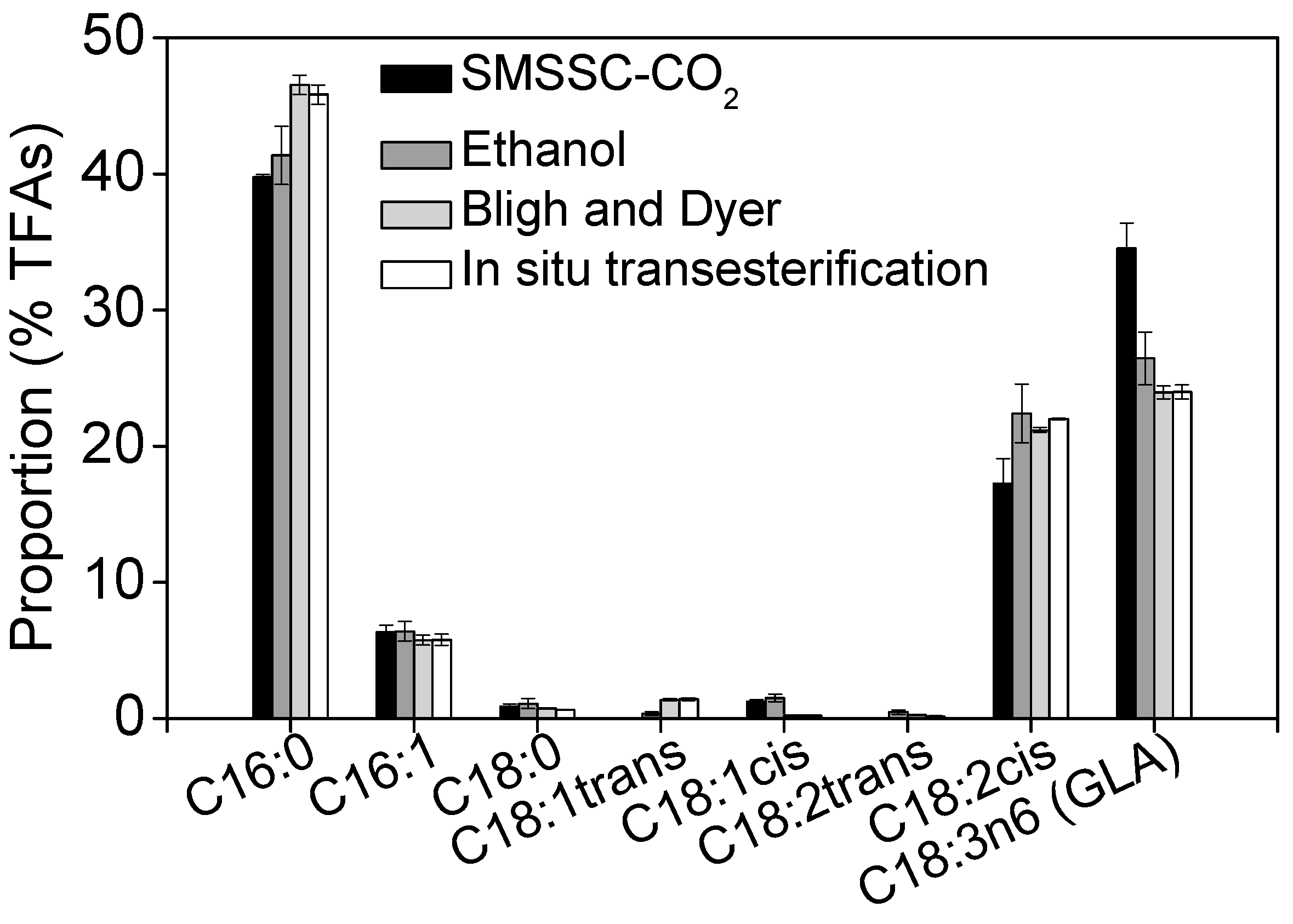
2.5. Recovery of GLA Using Different Extraction Methods

In order to obtain lipid extracts with high concentrations of GLA, four extraction procedures were compared, i.e., SMSSC-CO2, ethanol (extraction with ethanol under mechanical stirring), Bligh and Dyer, and in situ transesterification method (Table 1 and Figure 6). Although the yields of total lipids and fatty acids obtained with the SMSSC-CO2 approach were lower by 23% and 38% than those obtained using Bligh and Dyer and in situ transesterification methods, respectively, the yield of GLA using the former method (SMSSC-CO2) was 1.16% DCW, which was comparable to that of the latter two methods (1.18% DCW and 1.26% DCW for Bligh and Dyer and in situ transesterification methods, respectively) (Table 1). Moreover, the data illustrated in Figure 6 revealed that the SMSSC-CO2 resulted in remarkably greater content of GLA (34.52%) as compared to that (23.95–26.44%) obtained by the other three approaches, and was 67% greater than that reported by Gunstone [54]. It was demonstrated that SMSSC-CO2 had greater extraction selectivity for GLA than the other three methods tested.
On the other hand, ethanol, Bligh and Dyer, and in situ transesterification methods were found to exert greater extraction selectivity for palmitic acid (C16:0) and linoleic acid (C18:2cis) (Figure 6 and Figure S2). A possible reason is that the porous solid materials used in SMSSC-CO2 might have served as an absorbent to absorb impurities such as saturated fatty acids and pigments, making the downstream purification of crude GLA-containing lipids easier incidentally. This finding indicated that SMSSC-CO2 is a promising technology particularly for GLA extraction from A. platensis biomass, due to its greater extraction selectivity for GLA, and its use of green solvents including CO2 and ethanol. It is considered more environmentally friendly compared to the Bligh and Dyer method that involves harmful solvents including chloroform and methanol. The technology developed in this study can not only guide for designing a large-scale GLA extraction process, but also be applicable for extraction of low-polarity lipophilic compounds, like polyphenols, polysaccharides, carotenoids, and polyunsaturated fatty acids from marine microalgae or higher plants [55].

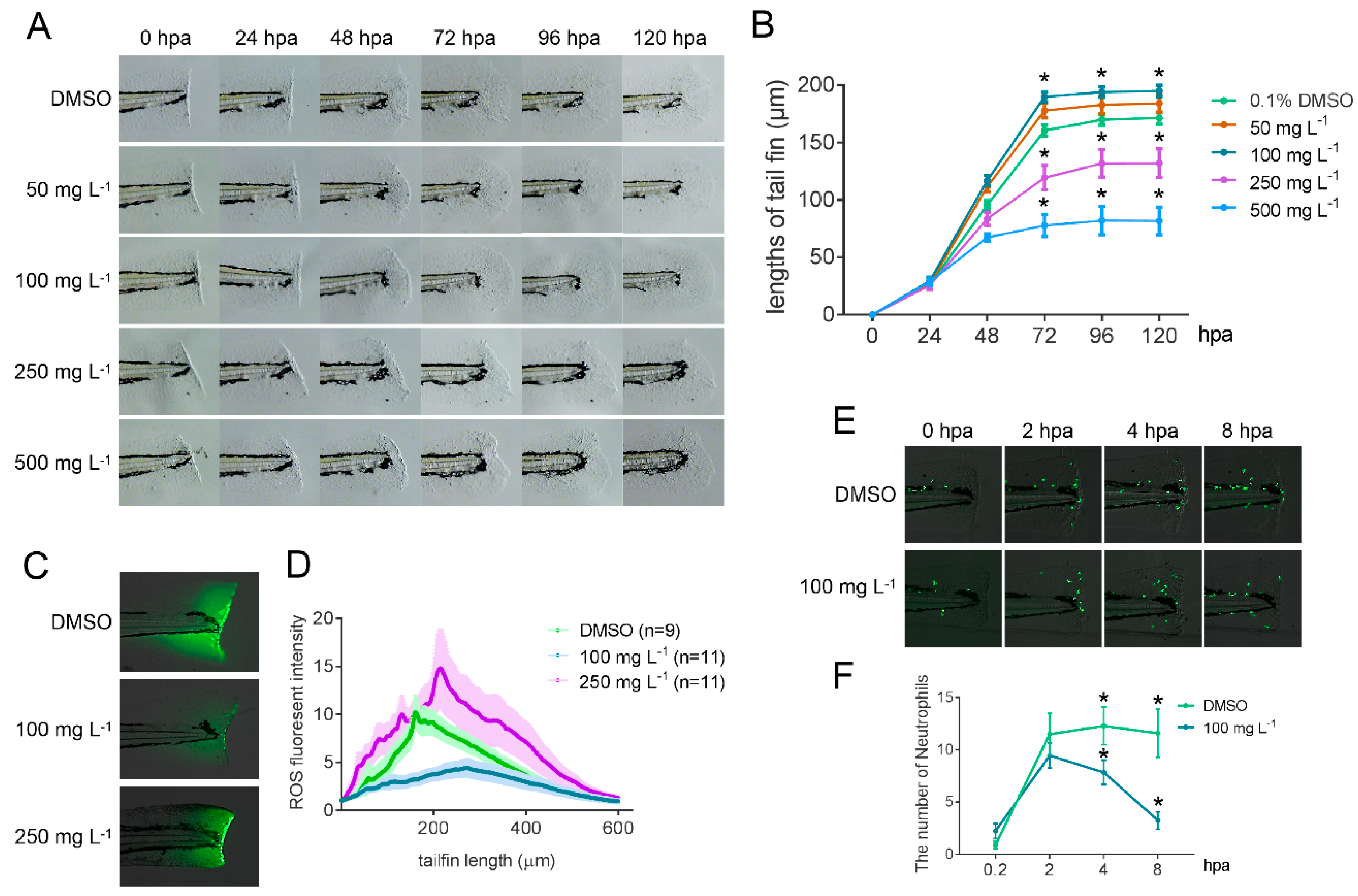
2.6. Bioactivity Test of GLA Using a Zebrafish Caudal Fin Regeneration Model

Gamma-linolenic acid extracted from A. platensis biomass was used for bioactivity test in a zebrafish caudal fin regeneration assay. The caudal fin of wild-type AB zebrafish at 54-h post fertilization (hpf) were amputated, and the amputated zebrafish were raised individually in the different concentrations (i.e., 50, 100, 250, 500 mg L−1) of GLA extracts. Compared to the dimethyl sulfoxide (DMSO) control, the addition of 50 and 100 mg L−1 of GLA accelerated the regeneration of the wound tissue obviously, but a higher concentration than 250 mg L−1 of GLA decelerated the regeneration (Figure 7A,B). In order to reveal the mechanism underlying this phenomenon, a reactive oxygen species (ROS) assay and a neutrophil attraction assay were performed. The results showed that 100 mg L−1 of GLA exhibited strong oxygen free radical scavenging activity, but 250 mg L−1 of GLA did not (Figure 7C,D). Furthermore, both recruitment at the very beginning after amputation and subsequent clearance of neutrophil cells were more efficient in the zebrafish cultured in 100 mg L−1 of GLA when compared with the controls (Figure 7E,F). These results indicate that a proper concentration of GLA improved anti-oxidative capability of zebrafish. Meanwhile, it could also modulate the immune system of zebrafish to better respond to the external injury in a more efficient manner. All these results suggest that GLA has multiple-bioactivities, including anti-oxidation, anti-inflammation, and anti-allergy, which led to the accelerated regeneration of the wound in zebrafish.
Gamma-linolenic acid has been demonstrated to be effective in treating various disorders such as rheumatoid arthritis [3], diabetes, multiple trauma, and premenstrual syndrome [7]. Several studies have shown that different esterified forms of polyunsaturated fatty acids exhibited different levels of bioavailability and effectiveness of their bioactivities. For example, docosahexaenoic acid-phosphatidylcholine (DHA-PC) produced a more significant improvement in brain development, cognitive performance and emotional well-being than docosahexaenoic acid-triglyceride (DHA-TG) [56,57,58]. Moreover, DHA-PC exhibited better efficacy than the recombination of DHA-TG with egg PC or α-glycerylphosphorylcholine in various mice tissues [59]. We suspect that A. platensis-derived GLA, which is mostly present in the galactolipid molecules, may combine or surpass the bioactivities of galactolipids without GLA or GLA alone.

3. Materials and Methods

3.1. Materials and Reagents

Arthrospira platensis powders (5–6% moisture) were obtained from C.B.N. Spirulina Bio-engineering Inc. (Dongtai, Jiangsu Province, China). The standards of 37 fatty acid methyl esters (37 FAMEs, C4–C24, Sigma–Aldrich #18919-1AMP), tridecanoic acid (C13) standard (Sigma–Aldrich #91988-5G), pentadecane (C15) (Sigma–Aldrich #76509-5ML) were purchased from Sigma Chemical Co. (St Louis, MO, USA). Monogalactosyldiacylglycerol (MGDG), digalactosyldiacylglycerol (DGDG), and sulfoquinovosyl diacylglycerol (SQDG) were purchased from Avanti Polar Lipids, Inc. (Alabaster, USA). The physicochemical properties of molecular sieve (Nanda Synthetic, Jiangyin, China), HP 20 macroporous resin (SEPDEX, Zhengzhou, China), and diatomite (Thermo ScientificTM, Waltham, MA, USA) are listed in Table 5. Thin-layer chromatography silica gel 60 was purchased from Merck KGaA (Darmstadt, Germany). All extraction reagents were of analytical grade and the other reagents were of HPLC grade. Hydrochloric acid (36–38%) was purchased from Sinopharm Chemical Reagent Co. (Beijing, China). Wild-type of the AB strain and Tg(mpx:EGFP) transgenic zebrafish (Cat. ID: CZ13) [60] were purchased, maintained, and raised at the China Zebrafish Resource Center (CZRC, http://zfish.cn), Wuhan, China.

3.2. Methods

3.2.1. Lipid Extraction

Total lipids of A. platensis powders were extracted and determined using a modified Bligh and Dyer method that was described in Laurens et al. [61]. Briefly, 100 mg of A. platensis powder was manually ground into fine powder in liquid nitrogen and transferred to a labeled glass vial. Ten milliliters of chloroform and methanol (2:1, v/v) mixture as extraction solvent was added into the vial and the mixture was kept stirring at 130 rpm for 1 h in a thermal incubator at a constant temperature of 30 °C. Then, 2.5 mL of 0.7% (w/v) potassium chloride was added into the vial and vortexed for 30 s, followed by centrifugation at 1000 g for 5 min at 20 °C to remove crude proteins. The chloroform phase containing crude lipids was manually collected and transferred into a pre-weighed vial. The extraction procedure was repeated once with addition of 4 mL chloroform and methanol (2:1, v/v) to the aliquot and vortexed for 30 s at room temperature. The chloroform phases from the two extractions were combined into a pre-weighed vial. The solvents in the vial were evaporated with the aid of nitrogen gas (NDK200-2, Lang Yue Instrument Manufacturing Co., Ltd., Changzhou, China) and remaining lipid extracts were lyophilized (LABCONCO®, Kansas City, USA) overnight. Lipids in the vial were weighed and total lipids were gravimetrically calculated on the basis of dry algal biomass. The extracted lipids were stored under −20 °C for fatty acids analysis. Error bars represent the standard deviation (n = 3).

3.2.2. Determination of Fatty Acid Methyl Esters (FAMEs)

Fatty acid profiles in the algal biomass were determined by the protocol described by Van Wychen et al. [62] with slight modifications. In brief, 10 mg of algal biomass sample was added into a glass vial (Agilent Technologies, CA, USA) containing 300 µL hydrochloric acid and methanol mixtures (5%, v/v), 200 µL chloroform and methanol (2:1, v/v) solvents in the presence of 25 µL of tridecanoic acid (10 mg mL−1). Tridecanoic acid, an odd-chain fatty acid that does not naturally occur in algae, is transesterified with the sample to quantify the total FAMEs content on GC and thus can be used as a surrogate/recovery standard. The transesterification reaction took place at 85 °C for 1 h. The resulting FAMEs were extracted with 1 mL hexane (in the presence of 5 µL pentadecane as an internal standard) and analyzed by GC-MS for determination of fatty acid composition and content (further details are described in Section 3.3.1). Error bars represent the standard deviation (n = 3).

3.2.3. Lipids and GLA Extraction with Ethanol under Mechanical Stirring

Ten grams of dried biomass were extracted with ethanol (liquid-to-solid ratio of 1:10, w/v) at 50 °C for 2 h under stirring in a round glass bottle. The mixture was centrifuged and supernatant was manually collected, followed by evaporation using a vacuum rotary evaporator (Rotavapor R-300, BÜCHI Labortechnik AG, Postfach, Switzerland) at a pressure of 0.015 MPa and a temperature of 45 °C. Total lipids were obtained by lyophilization (LABCONCO®, Kansas City, U.S.). The profile and content of fatty acids were analyzed with GC-MS (as described in Section 3.2.2 and Section 3.3.1). The extraction was repeated twice.

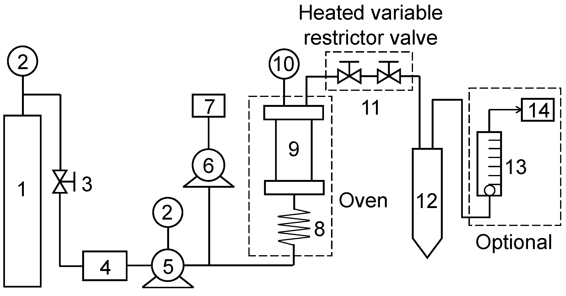
3.2.4. Supercritical Fluid Extraction

Supercritical carbon dioxide (SC-CO2) extraction of lipids and GLA from A. platensis biomass was conducted in an SFT-110XW SFE apparatus (Supercritical Fluid Technologies, Inc., Delaware, USA) (Figure 8) using carbon dioxide (purity 99.99%) (Chengweifeng Technologies Inc., Beijing, China) as solvent. For each experiment, 10 g of spray-dried biomass with a particle size of 0.2 mm was subjected to a sample basket and loaded into a 100 mL extraction vessel. Briefly, liquid CO2 supplied from a gas cylinder (1) was cooled by ethanol (CO2-to-ethanol ratio = 20:1, v/v) to −5 °C before being pressurized to the desired pressure and passed through an extraction vessel (9), which contained dry A. platensis powder and support materials (solid form) mixed at different ratios. Extracted lipids were regularly collected at determined time intervals into a collection vessel (12), and were cooled to room temperature after the extractor was depressurized to the atmospheric pressure. Crude lipids bound onto the internal surface of the extraction vessel and residual biomass were recovered by washing with ethanol, and then mixed with the above extracted lipids. Total lipids were obtained by evaporation of solvent using a rotary evaporator and were quantified gravimetrically. Composition of fatty acids was analyzed using GC-MS (see Section 3.2.2 and Section 3.3.1).
To optimize the operation conditions for GLA yield, the lipids were extracted with variables that could affect GLA extraction efficiency, including solid support types (molecular sieve, macroporous resin, diatomite) (Table 5), solid-to-biomass ratio (0, 1:4, 1:3, 1:2, 1:1 and 2:1 w/w), moisture content of solid support (0, 33, 50, 60, 67 and 80 wt.%), ethanol-to-biomass ratio (0, 0.5:1, 1:1, 2:1, 3:1, and 4:1 v/w), extraction temperature (30, 40, 50, and 60 °C), extraction pressure (28, 34, 41, and 48 MPa), extraction time range from 30 min (15 min for main extraction and 15 min for draining), 60 min (30 min for main extraction and 30 min for draining), 90 min (30 min for main extraction and 60 min for draining), and 120 min (30 min for main extraction and 90 min for draining), and CO2-to-mass ratio (0.8, 1.2, 1.6, and 2.0 mL g−1 min−1).

3.2.5. Determination of Yields of Total Lipids and GLA

Total lipids yield (YieldTotal lipids), GLA yield (YieldGLA), and extraction efficiencies (or recoveries) of total lipids, TFAs, and GLA were calculated by using the following equations:
Yield Total   lipids   ( %   DCW ) = m Total   lipids   ( g ) m Biomass   ( DCW )   ( g ) × 100
Yield   GLA ( %   DCW ) = m GLA   ( g ) m Biomass   ( DCW )   ( g ) × 100
Extraction   efficiency   or   recovery   ( % ) = m Total   lipids ,   TFAs   or   GLA   measured   via   SC CO 2   ( g ) m Total   lipids ,   TFAs   or   GLA   measured   via   Bligh   and   Dyer   method   ( g ) × 100

3.2.6. Zebrafish Caudal Fin Regeneration Assay

Wild-type of AB strain and transgenic zebrafish were purchased from, maintained and raised at the China Zebrafish Resource Center (CZRC, http://zfish.cn),Wuhan, China. Briefly, zebrafish were maintained at 28.5 °C with a 14/10 photoperiod of light/dark cycle and fed brine shrimp three times a day. All procedures relating to the care and use of fish were approved by the ethics committee from the Institute of Hydrobiology, Chinese Academy of Sciences on 28 May 2018 (Approval Protocol No. IHB20180528). The investigation conforms to the Guide for the Care and Use of Laboratory Animals published by the US National Institutes of Health.
The caudal fins of wild-type AB zebrafish were amputated at 54 hpf with a scalpel under a stereomicroscope (Olympus). The cutting site was at the tip of the caudal notochord along the vertical axis of the length. Amputated larval fish were raised individually in a 24-well plate with 2 mL different concentrations of GLA solution as indicated in the Results section and Figure 7A. Gamma-linolenic acid were diluted in DMSO (50 mg mL−1) as stock solution. The stock was further diluted with 0.3 × Danieau buffer, which 17.4 mM NaCl, 0.21 mM KCl, 0.12 mM MgSO4, 0.18 mM Ca(NO3)2, 1.5 mM 4-(2-hydroxyethyl)-1-piperazineethanesulfonic acid buffer (HEPES, pH 7.6), to make the working solution. Twelve larvae were treated with one concentration of working solution. For taking pictures, each larva was first anesthetized with 0.015% Tricaine (Sigma Cat. No: A-5040) in 0.3 × Danieau buffer and then mounted with 2% methylcellulose. The image was taken under a stereomicroscope (AF205, Leica). Images of every larva were taken at 0-, 12-, 24-, 48-, 72-, and 120-h post amputation (hpa). Embryos were recovered in 0.3 × Danieau buffer and put back into freshly prepared GLA solution after imaging.
For detection of ROS, 48-hpf wild-type larval fish were cultured in 2 mL of 0.1% DMSO, 50, 100, or 250 mg L−1 GLA in a 6-well plate with 10 larvae per well for 24 h. Then, the treated larvae were anesthetized with 0.015% Tricaine and their tails were wounded as previously described [40]. After tail injury, 10 larvae of one group were incubated in 1 mL of working solution of the ROS detection kit in the dark for 10 min (ENZO, Cat. No: ENZ-51042-K500), washed 3 times with HBSS, and then mounted with 2% methylcellulose for imaging. All images were taken under a Leica DMI 6000 microscope using a 10×/numerical aperture 0.3 objective with the same parameters. For measurement of fluorescence intensity of ROS, 8-bit images were imported into Fiji software and converted into 32-bit [63]. A rectangle tool was used to select the wound region and applied to the “ROI” manager which can be reused for other images. The function of the “Plot Profile” was used to measure the intensity of the selected region.
The Tg(mpx:EGFP) transgenic zebrafish was used in the live image analysis of neutrophil attraction assay. Forty-eight hpf larval fish were cultured in 10 mL of 0.1% DMSO or 100 mg L−1 GLA in a 6-well plate with 10 larvae per well for 24 h. Then, the treated larvae were anesthetized with 0.015% Tricaine and their tails were wounded as previously described [40]. After tail injury, the larval fish were embedded in 1% low-melting agarose and covered with 10 mL 0.3 × Danieau buffer in a 60-mm Petri dish at 28.5 °C. Images of each larva were taken at 20 min, 2, 4, and 8 h after tail injury under a Leica DMI-6000 microscope using a 10×/NA 0.3 objective. Images were imported into Fiji software [63] and the neutrophils at the wound site were counted.

3.3. Analytical Methods

3.3.1. Fatty Acid Analysis

An Agilent 7890B+5977A GC/MS (Agilent Technologies Inc., USA) was used to determine the composition of FAMEs. The capillary column was HP-88 (60 m × 0.25 mm × 0.2 µm). Initial temperature of the oven was 50 °C and maintained for 2 min, heated at a rate of 25 °C min−1 to 175 °C, and maintained for 5 min, then heated again at a rate of 7 °C min−1 to 210 °C and maintained for 2 min, and finally heated at a rate of 2 °C min−1 to 230 °C and maintained for 1 min. The injector temperature was kept at 250 °C in split (20:1) mode for an injection volume of 1 µL. The auxiliary heater, electron ionization (EI) source, and MS Quadrupole temperature were 250 °C, 230 °C, and 150 °C, respectively. Helium was used as the carrying gas at 1 mL min−1.

3.3.2. Polar Lipids Analysis

Qualitative analysis of galactolipids (including MGDG and DGDG) and phospholipids was performed with a Waters TQ-S triple quadruple electrospray ionization mass spectrometer (ESI-MS/MS) equipped with a Waters ACQUITY ultra-performance liquid chromatography (Waters, USA). Lipid extracts (30 mg) were dissolved in 2 mL chloroform and methanol (1:1, v/v) solutions, then filtered with a 0.22-μm nylon membrane to remove insoluble debris. The samples were diluted by 1000-fold progressively chloroform and methanol (1:1, v/v) before instrumental analysis [64,65].

4. Conclusions

A porous solid material supported supercritical-CO2 method was successfully developed for extraction of GLA-enriched glycolipids from the biotechnologically important cyanobacterium A. platensis. Under the optimal conditions, which included the solvent-to-solid ratio of 3:1 (v/w), diatomite-to-biomass ratio of 1:2 (w/w), CO2-to-mass ratio of 2.0 mL g−1 min−1, temperature of 40 °C, pressure of 41 MPa, and time of 60 min, the extraction efficiency of GLA can reach 98%, corresponding to 1.16% DCW. Gamma-linolenic acid made up 34.52% of TFAs, which was significantly greater than 26.44%, 23.95%, and 23.98% obtained with ethanol extraction in aid of mechanical stirring, Bligh and Dyer, and in situ transesterification methods, respectively. Moreover, SMSSC-CO2 using CO2 as a solvent and ethanol as a co-solvent at a low temperature (e.g., 40 °C) may better preserve heat-sensitive bioactive compounds in the cells, while at the same time be safer for production operators and the environment. Lipidomics analysis revealed that GLA was exclusively associated with galactolipids in A. platensis. Furthermore, we utilized a zebrafish model to demonstrate the bioactivities of A. platensis GLA as an anti-oxidant and immune system regulator. These findings highlight the application potentials of these new forms of GLA in nutraceutical and pharmaceutical industries.

Supplementary Materials

The following are available online at https://www.mdpi.com/1660-3397/17/4/203/s1, Figure S1: Comparison of the states of mixing porous materials with algal powders and the microscopic images of residues (Scale bar 50 μm), Figure S2: The chromatogram of fatty acids obtained from four extraction methods.

Author Contributions

Q.H., D.H., X.Y., and Y.S. designed the research. X.Y. performed extraction experiments and analyzed the data. Y.L. contributed to the lipidomics analysis. D.Y. and Y.L. performed the bioactivities assay. X.Y., L.Y., D.Y., D.H., Y.S. and Q.H. wrote the article.

Funding

This study was partially funded by the State Development & Investment Corporation (SDIC), State Key Laboratory of Freshwater Ecology and Biotechnology, Institute of Hydrobiology, Chinese Academy of Sciences (2016FBZ06 and 2016FBZ03), One Hundred Talents Program of Chinese Academy of Sciences (Y623031Z01), and the Beijing Key Laboratory of Algae Biomass (SDICMBC-00003).

Acknowledgments

The authors thank Shaokun Tang at the Key Laboratory for Green Chemical Technology of the Ministry of Education, Tianjin University, for technical guidance of supercritical fluid extraction. Thanks to Zhengbao Yu and Yijun Xue from the State Development & Investment Corporation for their constructive advice on the experimental operations. Thanks to Danni Yuan and Mingyang Ma from the Institute of Hydrobiology, Chinese Academy of Sciences, for providing guidelines and revisions of the manuscript format. Thank you to Kuoyu Li from the China Zebrafish Resource Center (CZRC) for taking care of the zebrafish.

Conflicts of Interest

The authors declare no conflict of interest.

References

  1. Xiao, Y.; Gu, Y.; Purwaha, P.; Ni, K.; Law, B.; Mallik, S.; Qian, S.Y. Characterization of free radicals formed from COX-catalyzed DGLA peroxidation. Free Radic. Biol. Med. 2011, 50, 1163–1170. [Google Scholar] [CrossRef]
  2. Johnson, M.M.; Swan, D.D.; Surette, M.E.; Stegner, J.; Chilton, T.; Fonteh, A.N.; Chilton, F.H. Dietary supplementation with γ-linolenic acid alters fatty acid content and eicosanoid production in healthy humans. J. Nutr. 1997, 127, 1435–1444. [Google Scholar] [CrossRef]
  3. Zurier, R.B.; Rossetti, R.G.; Jacobson, E.W.; DeMarco, D.M.; Liu, N.Y.; Temming, J.E.; White, B.M.; Laposata, M. Gamma linolenic acid treatment of rheumatoid arthritis. A randomized, placebo-controlled trial. Arthritis Rheumatol. 1996, 39, 1808–1817. [Google Scholar] [CrossRef]
  4. Kast, R.E. Borage oil reduction of rheumatoid arthritis activity may be mediated by increased cAMP that suppresses tumor necrosis factor-alpha. Int. Immunopharmacol. 2001, 1, 2197–2199. [Google Scholar] [CrossRef]
  5. Worm, M.; Henz, B.M. Novel unconventional therapeutic approaches to atopic eczema. Dermatology 2000, 201, 191–195. [Google Scholar] [CrossRef] [PubMed]
  6. Ponnala, S.; Rao, K.P.; Chaudhury, J.R.; Ahmed, J.; Rama Rao, B.; Kanjilal, S.; Hasan, Q.; Das, U.N. Effect of polyunsaturated fatty acids on diphenyl hydantoin-induced genetic damage in vitro and in vivo. Prostaglandins Leukot. Essent. Fatty Acids 2009, 80, 43–50. [Google Scholar] [CrossRef]
  7. Bugnariu, S. Beneficial effect of gamma-linolenic acid-enriched diet therapeutic properties of Oenothera biennis oil (evening primrose oil). Farmacia 1996, 44, 3–4. [Google Scholar]
  8. Wanasundara, U.N.; Wanasundara, J.P.D.; Akoh, C.C. γ-Linolenic acid: Purification and functionality. In Handbook of Functional Lipids; Akoh, C.C., Ed.; CRC Press, Taylor & Francis Group: New York, NY, USA, 2006; pp. 47–75. [Google Scholar]
  9. Vargas, M.A.; Rodríguez, H.; Moreno, J.; Olivares, H.; Campo, J.A.D.; Rivas, J.; Guerrero, M.G. Biochemical Composition and Fatty Acid Content of Filamentous Nitrogen-Fixing Cyanobacteria. J. Phycol. 1998, 34, 812–817. [Google Scholar] [CrossRef]
  10. Maslova, I.P.; Mouradyan, E.A.; Lapina, S.S.; Klyachko-Gurvich, G.L.; Los, D.A. Lipid Fatty Acid Composition and Thermophilicity of Cyanobacteria. Russ. J. Plant Physiol. 2004, 51, 353–360. [Google Scholar] [CrossRef]
  11. Yang, D.; Nam, S.; Hwang, S.-J.; An, K.-G.; Park, Y.-S.; Shin, K.-H.; Park, S. Fatty acid biomarkers to verify cyanobacteria feeding abilities of herbivorous consumers. J. Freshw. Ecol. 2015, 31, 77–91. [Google Scholar] [CrossRef]
  12. Roughan, P.G. Spirulina: A Source of Dietary Gamma-Linolenic Acid? J. Sci. Food Agric. 1989, 47, 85–93. [Google Scholar] [CrossRef]
  13. Leema, J.T.; Kirubagaran, R.; Vinithkumar, N.V.; Dheenan, P.S.; Karthikayulu, S. High value pigment production from Arthrospira (Spirulina) platensis cultured in seawater. Bioresour. Technol. 2010, 101, 9221–9227. [Google Scholar] [CrossRef]
  14. Raposo, M.F.; de Morais, R.M.; Bernardo de Morais, A.M. Bioactivity and applications of sulphated polysaccharides from marine microalgae. Mar. Drugs 2013, 11, 233–252. [Google Scholar] [CrossRef] [PubMed]
  15. Belay, A. Biology and Industrial Production of Arthrospira (Spirulina). In Handbook of Microalgal Culture, 2nd ed.; Richmond, A., Hu, Q., Eds.; Applied Phycology and Biotechnology, Wiley: New York, NY, USA, 2013; pp. 339–358. [Google Scholar]
  16. Cohen, Z.; Reungjitchachawali, M.; Siangdung, W.; Tanticharoen, M. Production and partial purification of γ-linolenic acid and some pigments from Spirulina platensis. J. Appl. Phycol. 1993, 5, 109–115. [Google Scholar] [CrossRef]
  17. Apdila, E.T.; Awai, K. Configuration of the sugar head of glycolipids in thylakoid membranes. Genes Genet. Syst. 2017, 92, 235–242. [Google Scholar] [CrossRef] [PubMed] [Green Version]
  18. Sommella, E.; Conte, G.M.; Salviati, E.; Pepe, G.; Bertamino, A.; Ostacolo, C.; Sansone, F.; Prete, F.D.; Aquino, R.P.; Campiglia, P. Fast Profiling of Natural Pigments in Different Spirulina (Arthrospira platensis) Dietary Supplements by DI-FT-ICR and Evaluation of their Antioxidant Potential by Pre-Column DPPH-UHPLC Assay. Molecules 2018, 23, 1152. [Google Scholar] [CrossRef]
  19. Hao, S.; Li, S.; Wang, J.; Zhao, L.; Yan, Y.; Cao, Q.; Wu, T.; Liu, L.; Wang, C. Transcriptome Analysis of Phycocyanin-Mediated Inhibitory Functions on Non-Small Cell Lung Cancer A549 Cell Growth. Mar. Drugs 2018, 16, 511. [Google Scholar] [CrossRef]
  20. Martinez-Samano, J.; Torres-Montes de Oca, A.; Luqueno-Bocardo, O.I.; Torres-Duran, P.V.; Juarez-Oropeza, M.A. Spirulina maxima Decreases Endothelial Damage and Oxidative Stress Indicators in Patients with Systemic Arterial Hypertension: Results from Exploratory Controlled Clinical Trial. Mar. Drugs 2018, 16, 496. [Google Scholar] [CrossRef]
  21. Hernandez-Lepe, M.A.; Lopez-Diaz, J.A.; Juarez-Oropeza, M.A.; Hernandez-Torres, R.P.; Wall-Medrano, A.; Ramos-Jimenez, A. Effect of Arthrospira (Spirulina) maxima Supplementation and a Systematic Physical Exercise Program on the Body Composition and Cardiorespiratory Fitness of Overweight or Obese Subjects: A Double-Blind, Randomized, and Crossover Controlled Trial. Mar. Drugs 2018, 16, 364. [Google Scholar] [CrossRef] [PubMed]
  22. Mendiola, J.A.; Jaime, L.; Santoyo, S.; Reglero, G.; Cifuentes, A.; Ibañez, E.; Señoráns, F.J. Screening of functional compounds in supercritical fluid extracts from Spirulina platensis. Food Chem. 2007, 102, 1357–1367. [Google Scholar] [CrossRef]
  23. Mallikarjun Gouda, K.G.; Udaya Sankar, K.; Sarada, R.; Ravishankar, G.A. Supercritical CO2 extraction of functional compounds from Spirulina and their biological activity. J. Food Sci. Technol. 2015, 52, 3627–3633. [Google Scholar]
  24. Michalak, I.; Chojnacka, K.; Saeid, A. Dietary Feed Supplements and Cosmetics Formulated with Supercritical CO2 Algal Extracts. Molecules 2017, 22, 66. [Google Scholar] [CrossRef]
  25. Axelsson, M.; Gentili, F. A single-step method for rapid extraction of total lipids from green microalgae. PLoS ONE 2014, 9, e89643. [Google Scholar] [CrossRef]
  26. Sajilata, M.G.; Singhal, R.S.; Kamat, M.Y. Fractionation of lipids and purification of γ-linolenic acid (GLA) from Spirulina platensis. Food Chem. 2008, 109, 580–586. [Google Scholar] [CrossRef]
  27. Sajilata, M.G.; Singhal, R.S.; Kamat, M.Y. Supercritical CO2 extraction of γ-linolenic acid (GLA) from Spirulina platensis ARM 740 using response surface methodology. J. Food Eng. 2008, 84, 321–326. [Google Scholar] [CrossRef]
  28. Mendes, R.L.; Nobre, B.P.; Cardoso, M.T.; Pereira, A.P.; Palavra, A.F. Supercritical carbon dioxide extraction of compounds with pharmaceutical importance from microalgae. Inorg. Chim. Acta 2003, 356, 328–334. [Google Scholar] [CrossRef]
  29. Mendes, R.L.; Reis, A.D.; Palavra, A.F. Supercritical CO2 extraction of γ-linolenic acid and other lipids from Arthrospira (Spirulina) maxima: Comparison with organic solvent extraction. Food Chem. 2006, 99, 57–63. [Google Scholar] [CrossRef]
  30. Golmakani, M.-T.; Mendiola, J.A.; Rezaei, K.; Ibáñez, E. Expanded ethanol with CO2 and pressurized ethyl lactate to obtain fractions enriched in γ-Linolenic Acid from Arthrospira platensis (Spirulina). J. Supercrit. Fluids 2012, 62, 109–115. [Google Scholar] [CrossRef]
  31. Herrero, M.; Mendiola, J.A.; Cifuentes, A.; Ibanez, E. Supercritical fluid extraction: Recent advances and applications. J. Chromatogr. A 2010, 1217, 2495–2511. [Google Scholar] [CrossRef] [Green Version]
  32. Andrich, G.; Zinnai, A.; Nesti, U.; Venturi, F. Supercritical fluid extraction of oil from microalga Spirulina (arthrospira) platensis. Acta Aliment. 2006, 35, 195–203. [Google Scholar] [CrossRef]
  33. Hu, Q.H. Supercritical Carbon Dioxide Extraction of Spirulina platensis Component and Removing the Strench. J. Agric. Food Chem. 1999, 47, 2705–2706. [Google Scholar]
  34. Hsueh, Y.C.; Wang, B.J.; Yu, Z.R.; Wang, C.C.; Koo, M. Optimization of a Continuous Preparation Method of Arthrospira platensis γ-linolenic Acid by Supercritical Carbon Dioxide Technology using Response Surface Methodology. Sains Malays. 2015, 44, 1739–1744. [Google Scholar]
  35. de la Jara, A.; Ruano-Rodriguez, C.; Polifrone, M.; Assunçao, P.; Brito-Casillas, Y.; Wägner, A.M.; Serra-Majem, L. Impact of dietary Arthrospira (Spirulina) biomass consumption on human health: Main health targets and systematic review. J. Appl. Phycol. 2018, 30, 2403–2423. [Google Scholar] [CrossRef]
  36. Lee, J.J.; Kim, K.B.; Heo, J.; Cho, D.H.; Kim, H.S.; Han, S.H.; Ahn, K.J.; An, I.S.; An, S.; Bae, S. Protective effect of Arthrospira platensis extracts against ultraviolet B-induced cellular senescence through inhibition of DNA damage and matrix metalloproteinase-1 expression in human dermal fibroblasts. J. Photochem. Photobiol. B 2017, 173, 196–203. [Google Scholar] [CrossRef]
  37. Wang, F.; Miao, M.; Chen, B.; Wang, R.; Sun, B.; Ren, D.; Lu, J. Antineoplastic activity of γ-linolenic acid extract from Spirulina platensis on HepG2 cells and its inhibition effect on platelet aggregation. Food Agric. Immunol. 2014, 26, 97–108. [Google Scholar] [CrossRef]
  38. Esquivel-Hernandez, D.A.; Rodriguez-Rodriguez, J.; Cuellar-Bermudez, S.P.; Garcia-Perez, J.S.; Mancera-Andrade, E.I.; Nunez-Echevarria, J.E.; Ontiveros-Valencia, A.; Rostro-Alanis, M.; Garcia-Garcia, R.M.; Torres, J.A.; et al. Effect of Supercritical Carbon Dioxide Extraction Parameters on the Biological Activities and Metabolites Present in Extracts from Arthrospira platensis. Mar. Drugs 2017, 15, 174. [Google Scholar] [CrossRef] [PubMed]
  39. Howe, K.; Clark, M.D.; Torroja, C.F.; Torrance, J.; Berthelot, C.; Muffato, M.; Collins, J.E.; Humphray, S.; McLaren, K.; Matthews, L.; et al. The zebrafish reference genome sequence and its relationship to the human genome. Nature 2013, 496, 498–503. [Google Scholar] [CrossRef] [Green Version]
  40. Li, L.; Yan, B.; Shi, Y.Q.; Zhang, W.Q.; Wen, Z.L. Live imaging reveals differing roles of macrophages and neutrophils during zebrafish tail fin regeneration. J. Biol. Chem. 2012, 287, 25353–25360. [Google Scholar] [CrossRef]
  41. Limpitlaw, U.G. Ingestion of Earth materials for health by humans and animals. Int. Geol. Rev. 2010, 52, 726–744. [Google Scholar] [CrossRef]
  42. Holman, B.W.B.; Malau-Aduli, A.E.O. Spirulina as a livestock supplement and animal feed. J. Anim. Physiol. Anim. Nutr. 2013, 97, 615–623. [Google Scholar] [CrossRef]
  43. Singh, J.; Gu, S. Commercialization potential of microalgae for biofuels production. Renew. Sustain. Energy Rev. 2010, 14, 2596–2610. [Google Scholar] [CrossRef]
  44. Pan, J.-L.; Wang, H.-M.; Chen, C.-Y.; Chang, J.-S. Extraction of astaxanthin from Haematococcus pluvialis by supercritical carbon dioxide fluid with ethanol modifier. Eng. Life Sci. 2012, 12, 638–647. [Google Scholar] [CrossRef]
  45. Dong, T.; Knoshaug, E.P.; Pienkos, P.T.; Laurens, L.M.L. Lipid recovery from wet oleaginous microbial biomass for biofuel production: A critical review. Appl. Energy 2016, 177, 879–895. [Google Scholar] [CrossRef] [Green Version]
  46. Grima, E.M.; González, M.J.I.; Giménez, A.G. Solvent Extraction for Microalgae Lipids. In Algae for Biofuels and Energy; Borowitzka, M.A., Moheimani, N.R., Eds.; Springer: Dordrecht, The Netherlands, 2013; pp. 187–205. [Google Scholar]
  47. Esquivel-Hernandez, D.A.; Lopez, V.H.; Rodriguez-Rodriguez, J.; Aleman-Nava, G.S.; Cuellar-Bermudez, S.P.; Rostro-Alanis, M.; Parra-Saldivar, R. Supercritical Carbon Dioxide and Microwave-Assisted Extraction of Functional Lipophilic Compounds from Arthrospira platensis. Int. J. Mol. Sci. 2016, 17, 658. [Google Scholar] [CrossRef]
  48. Thana, P.; Machmudah, S.; Goto, M.; Sasaki, M.; Pavasant, P.; Shotipruk, A. Response surface methodology to supercritical carbon dioxide extraction of astaxanthin from Haematococcus pluvialis. Bioresour. Technol. 2008, 99, 3110–3115. [Google Scholar] [CrossRef] [PubMed]
  49. Crampon, C.; Nikitine, C.; Zaier, M.; Lépine, O.; Tanzi, C.D.; Vian, M.A.; Chemat, F.; Badens, E. Oil extraction from enriched Spirulina platensis microalgae using supercritical carbon dioxide. J. Supercrit. Fluids 2017, 119, 289–296. [Google Scholar] [CrossRef]
  50. Kaufmann, B.; Christen, P. Recent extraction techniques for natural products: Microwave-assisted extraction and pressurised solvent extraction. Phytochem. Anal. 2002, 13, 105–113. [Google Scholar] [CrossRef]
  51. Fenn, J.B.; Mann, M.; Meng, C.K.; Wong, S.F.; Whitehouse, C.M. Electrospray ionization for mass spectrometry of large biomolecules. Science 1989, 246, 64–71. [Google Scholar] [CrossRef] [PubMed] [Green Version]
  52. Han, X.; Gross, R.W. Global analyses of cellular lipidomes directly from crude extracts of biological samples by ESI mass spectrometry: A bridge to lipidomics. J. Lipid Res. 2003, 44, 1071–1079. [Google Scholar] [CrossRef]
  53. Marcus, D.M.; Schwarting, G.A. Immunochemical Properties of Glycolipids and Phospholipids. Adv. Immunol. 1976, 23, 203–240. [Google Scholar]
  54. Gunstone, F.D. Gamma linolenic acid-occurrence and physical and chemical properties. Prog. Lipid Res. 1992, 31, 145–161. [Google Scholar] [CrossRef]
  55. Cikos, A.M.; Jokic, S.; Subaric, D.; Jerkovic, I. Overview on the Application of Modern Methods for the Extraction of Bioactive Compounds from Marine Macroalgae. Mar. Drugs 2018, 16, 348. [Google Scholar] [CrossRef]
  56. He, C.; Qu, X.; Cui, L.; Wang, J.; Kang, J.X. Improved spatial learning performance of fat-1 mice is associated with enhanced neurogenesis and neuritogenesis by docosahexaenoic acid. Proc. Natl. Acad. Sci. USA 2009, 106, 11370–11375. [Google Scholar] [CrossRef] [PubMed] [Green Version]
  57. Ramprasath, V.R.; Eyal, I.; Zchut, S.; Jones, P.J. Enhanced increase of omega-3 index in healthy individuals with response to 4-week n-3 fatty acid supplementation from krill oil versus fish oil. Lipids Health Dis. 2013, 12, 178. [Google Scholar] [CrossRef] [PubMed]
  58. Liu, L.; Bartke, N.; Van Daele, H.; Lawrence, P.; Qin, X.; Park, H.G.; Kothapalli, K.; Windust, A.; Bindels, J.; Wang, Z.; et al. Higher efficacy of dietary DHA provided as a phospholipid than as a triglyceride for brain DHA accretion in neonatal piglets. J. Lipid Res. 2014, 55, 531–539. [Google Scholar] [CrossRef] [Green Version]
  59. Wu, F.; Wang, D.D.; Wen, M.; Che, H.X.; Xue, C.H.; Yanagita, T.; Zhang, T.T.; Wang, Y.M. Comparative analyses of DHA-Phosphatidylcholine and recombination of DHA-Triglyceride with Egg-Phosphatidylcholine or Glycerylphosphorylcholine on DHA repletion in n-3 deficient mice. Lipids Health Dis. 2017, 16, 234. [Google Scholar] [CrossRef] [PubMed] [Green Version]
  60. Renshaw, S.A.; Loynes, C.A.; Trushell, D.M.; Elworthy, S.; Ingham, P.W.; Whyte, M.K. A transgenic zebrafish model of neutrophilic inflammation. Blood 2006, 108, 3976–3978. [Google Scholar] [CrossRef] [PubMed] [Green Version]
  61. Laurens, L.M.; Dempster, T.A.; Jones, H.D.; Wolfrum, E.J.; Van Wychen, S.; McAllister, J.S.; Rencenberger, M.; Parchert, K.J.; Gloe, L.M. Algal biomass constituent analysis: Method uncertainties and investigation of the underlying measuring chemistries. Anal. Chem. 2012, 84, 1879–1887. [Google Scholar] [CrossRef]
  62. Van Wychen, S.; Ramirez, K.; Laurens, L.M.L. Determination of Total Lipids as Fatty Acid Methyl Esters (FAME) by in Situ Transesterification Laboratory Analytical Procedure (LAP); NREL/TP-5100-60958; National Renewable Energy Laboratory: Golden, CO, USA, 2016.
  63. Schindelin, J.; Arganda-Carreras, I.; Frise, E.; Kaynig, V.; Longair, M.; Pietzsch, T.; Preibisch, S.; Rueden, C.; Saalfeld, S.; Schmid, B.; et al. Fiji: An open-source platform for biological-image analysis. Nat. Methods 2012, 9, 676–682. [Google Scholar] [CrossRef]
  64. Yoon, K.; Han, D.; Li, Y.; Sommerfeld, M.; Hu, Q. Phospholipid: Diacylglycerol acyltransferase is a multifunctional enzyme involved in membrane lipid turnover and degradation while synthesizing triacylglycerol in the unicellular green microalga Chlamydomonas reinhardtii. Plant Cell 2012, 24, 3708–3724. [Google Scholar] [CrossRef]
  65. Xu, J.; Chen, D.; Yan, X.; Chen, J.; Zhou, C. Global characterization of the photosynthetic glycerolipids from a marine diatom Stephanodiscus sp. by ultra performance liquid chromatography coupled with electrospray ionization-quadrupole-time of flight mass spectrometry. Anal. Chim. Acta 2010, 663, 60–68. [Google Scholar] [CrossRef]
Figure 1. Effect of different solid supporting materials on extraction efficiencies of total lipids (A), total fatty acids (TFAs) (B), and γ-linolenic acid (GLA) (C) over extraction time. Experiments were performed under the following conditions: solid materials-to-algal biomass ratio, 1:2 (w/w); water content of solid materials, 60 wt.%; ethanol-to-biomass ratio, 3:1 (v/w); temperature, 40 °C; pressure, 41 MPa; CO2-to-mass ratio, 2.0 mL g−1 min−1. Error bars represent the standard deviation (n = 3).
Marinedrugs 17 00203 g001
Figure 2. Effect of diatomite-to-biomass ratio (A) and moisture content of diatomite (B) on extraction efficiencies of total lipids, total fatty acids (TFAs), and γ-linolenic acid (GLA). Experiments were performed under the following conditions: ethanol-to-biomass ratio, 3:1 (v/w); temperature, 40 °C; pressure, 41 MPa; extraction time, 60 min (static extraction for 30 min + dynamic 30 min); CO2-to-mass ratio, 2.0 mL g−1 min−1. Error bars represent the standard deviation (n = 3).
Marinedrugs 17 00203 g002
Figure 3. Effect of ethanol-to-biomass ratio on extraction efficiencies of total lipids, total fatty acids (TFAs), and γ-linolenic acid (GLA) in the presence of diatomite. Experiments were performed under the following conditions: diatomite-to-biomass ratio, 1:2 (w/w); water content of solid materials, 60 wt.%; temperature, 40 °C; pressure, 41 MPa; extraction time, 60 min (static extraction for 30 min + dynamic 30 min); CO2-to-mass ratio, 2 mL g−1 min−1. Error bars represent the standard deviation (n = 3).
Marinedrugs 17 00203 g003
Figure 4. Effect of temperature (A), pressure (B), time (C), and CO2-to-mass ratio (D) on extraction efficiencies of total lipids, total fatty acids (TFAs), and γ-linolenic acid (GLA) by solid matrix-supported supercritical CO2 (SMSSC-CO2) extraction. Experiments were performed under the following conditions: diatomite-to-biomass ratio, 1:2 (w/w); water content of solid materials, 60 wt.%; ethanol-to-biomass ratio, 3:1 (v/w); and (A) pressure = 41 MPa, extraction time = 60 min (static extraction for 30 min + dynamic 30 min), CO2-to-mass ratio = 2.0 mL g−1 min−1; (B) temperature = 40 °C; extraction time = 60 min (static extraction for 30 min + dynamic 30 min), CO2-to-mass ratio = 2.0 mL g−1 min−1; (C) temperature = 40 °C, pressure = 41 MPa, CO2-to-mass ratio = 2.0 mL g−1 min−1; (D) temperature = 40 °C, pressure = 41 MPa, extraction time = 60 min (static extraction for 30 min + dynamic 30 min). Error bars represent the standard deviation (n = 3).
Marinedrugs 17 00203 g004
Figure 5. Relative ratio of MGDG and DGDG and molecular species profiles of MGDG and DGDG in lipid extracts of A. platensis. M, MGDG; D, DGDG.
Marinedrugs 17 00203 g005
Figure 6. Fatty acid profiles obtained with different extraction methods. Error bars represent the standard deviation (n = 3).
Marinedrugs 17 00203 g006
Figure 7. Bioactivities of GLA tested on a zebrafish caudal fin regeneration model. (A–B) Effects of 50, 100, 250, and 500 mg L−1 of GLA on zebrafish caudal fin regeneration. Proper concentrations (50 and 100 mg L−1) of GLA promoted the regeneration of early fin primordia of zebrafish larvae, whereas higher concentrations (250 and 500 mg L−1) did not. (C–D) Reactive oxygen species (ROS) test of wounded zebrafish treated with DMSO, 100 and 250 mg L−1 of GLA. Proper concentration (100 mg L−1) of GLA reduced oxidative stress at the wound region, whereas higher concentration (250 mg L−1) did not. (E–F) Neutrophil attraction of wounded zebrafish caudal fin treated with DMSO and 100 mg L−1 of GLA. Proper concentration (100 mg L−1) of GLA reduced the number of neutrophils at the wound region revealing the anti-inflammatory effect of this fatty acid. Asterisks (*) indicate statistically significant differences compared with the DMSO (t-test, p < 0.05).
Marinedrugs 17 00203 g007
Figure 8. Single vessel configuration of the supercritical CO2 extraction apparatus: 1, liquid CO2 cylinder; 2, manometers; 3, check valve; 4, CO2 cooler; 5, 6, high-pressure pumps for pumping CO2 and co-solvent; 7, co-solvent addition; 8, preheat coil; 9, extraction vessel; 10, thermometer; 11, static/dynamic valve; 12, collection vial; 13, flow meter gas CO2; 14, CO2 vent.
Marinedrugs 17 00203 g008
Table 1. Comparative analysis of total lipids, total fatty acids (TFAs), and γ-linolenic acid (GLA) from Arthrospira platensis by four extraction methods (means ± standard deviation).
MethodsTotal Lipids Yield
(% DCW)
Total Lipids Recovery (%)GLA Yield (% DCW)GLA Recovery (%)GLA
(% Total Lipids)
TFAs Yield (% DCW)GLA
(% TFAs)
In situ TNANA1.26 ± 0.04100NA5.25 ± 0.0723.98 ± 0.52
B & D11.52 ± 0.111001.18 ±0.0310010.334.97 ± 0.1123.95 ± 0.47
Ethanol10.22 ± 0.1788.721.13 ± 0.0694.96 a11.064.27 ± 0.1626.44 ± 1.94
SMSSC-CO29.36 ± 0.3081.251.16 ± 0.0797.58 a12.393.80 ± 0.1634.52 ± 1.88
In situ T: in situ transesterification; B & D: Bligh and Dyer method; Ethanol: extraction with ethanol; SMSSC-CO2: solid matrix-supported supercritical CO2; NA: not available; a Based on the data of the Bligh and Dyer method.
Table 2. Supercritical CO2 extraction of total lipids and GLA contents of several Arthrospira (Spirulina) species and strains.
StrainsBiomass FormsSolventPressure (MPa)Temp. (°C)Time (min)CO2-to-Mass RatioTotal Lipids
(% DCW)
GLA
(% DCW)
GLA
(% TFAs)
GLA Recovery of B & D (or in situ T) (%)Reference(s)
Arthrospira platensis10 g spray-dried powder (0.2 mm) (with porous materials)CO2 + ethanol4140602 mL g−1 min−19.41.1634.598 (92)This study
Spirulina platensis16 g freeze-dried (with glass wool), crushing (0.25 mm)CO2 + 13.7 mL ethanol404060700 mL min−18.60.6415.8100 (78)[27]
Arthrospira maxima5 g freeze-dried (with glass wool), crushing (0.2 mm)CO2 + 10 mol% ethanol3560NA2 g min−1NA0.4435.545[28,29]
Arthrospira platensis2 g biomass (with 4 g sea sand)50% CO2 + ethanol304090NA6.70.906.5–11.725[30]
Spirulina platensis80 g freeze-dried (with 100 g glass microspheres), crushing (≤0.37 mm)CO270559010 kg h−17.80.6718.5–21.1NA[32]
Spirulina platensisDried powderCO2404024024 kg h−17.20.2923.2NA[33]
Arthrospira platensisDried powderCO23060NA3 mL min−1NA0.04NANA[34]
B & D: Bligh and Dyer method; In situ T: in situ transesterification; NA: not available.
Table 3. Total lipids and GLA recovery estimated from different distribution compositions after SMSSC-CO2 extraction in duplicate (means ± standard deviation).
SamplesTotal Lipids Yield (% DCW)TFAs Yield
(% DCW)
Percentage (%)GLA Yield (% DCW)Percentage (%)
Extraction vessel a9.36 ± 0.303.80 ± 0.1672.801.16 ± 0.0794.31
Washing b1.10 ± 0.040.41 ± 0.017.850.04 ± 0.003.25
Biomass residues cND1.01 ± 0.0219.350.03 ± 0.002.44
Total10.465.221001.23100
a Collected in the collection vial; b Washing bottom of kettle and surface of sample basket with ethanol; c Lipids remaining in residual biomass were extracted via in situ transesterification method; ND: not determined.
Table 4. The molecular species of monogalactosyldiacylglycerol (MGDG) and digalactosyldiacylglycerol (DGDG) in A. platensis lipid extracts.
LipidsMGDG Acyl ChainLipidsMGDG Acyl Chain
32:416:3–16:1; 16:4–16:0; 14:1–18:3; 14:2–18:233:516:2–17:3
32:316:3–16:0; 16:2–16:1; 14:0–18:333:416:1–17:3; 16:0–17:4
32:216:2–16:0; 16:1–16:1; 14:0–18:233:316:0–17:3
32:116:1–16:033:216:0–17:2
32:016:0–16:033:116:0–17:1
34:516:2–18:3; 16:1–18:435:516:1–19:4; 17:2–18:3
34:416:1–18:335:416:1–19:3; 17:1–18:3
34:316:0–18:335:316:1–19:2; 16:0–19:3
34:216:0–18:235:216:0–19:2; 16:1–19:1
34:116:0–18:135:116:0–19:1
34:016:0–18:035:016:0–19:0
36:618:3–18:336:216:0–20:2; 16:1–20:1; 18:2–18:0; 18:1–18:1
36:518:2–18:336:116:0–20:1; 16:1–20:0; 18:1–18:0
36:418:3–18:1; 18:2–18:2; 16:0–20:4; 16:1–20:336:016:0–20:0
36:316:0–20:3; 16:1–20:2; 18:3–18:0; 18:2–18:1
LipidsDGDG acyl chainLipidsDGDG acyl chain
34:516:2–18:332:3NA
34:416:1–18:332:214:2–18:0; 15:2–17:0; 13:2–19:0
34:316:0–18:332:114:1–18:0; 13:1–19:0
34:216:0–18:232:0NA
NA: not available.
Table 5. Physicochemical properties of porous materials.
MaterialsMolecular Sieve aMacroporous Resin
(HP 20) a
Diatomite a
Chemical composition3/4CaO·1/4Na2O·Al2O3·
2SiO2·9/2H2O
Polystyrene benzeneSiO2 (>85%)
Particle size (µm)2.5420–840 (20–40 mesh)590 (30 mesh)
Unit surface area (m2 g−1)300–1000500−60040–65
Pore size (nm)2–5046NA
Moisture content (%)<245–600.11
Density (g cm−3)0.641.05–1.090.47
Functional groupsOHOH/H+OH
Hydrophobic/hydrophilicHydrophilicHydrophilicHydrophilic
a Data were obtained from manufacturers; NA: not available.

