Article

Exposure to Fine Particulate Matter (PM2.5) and Heavy Metals During the Second Trimester of Pregnancy Increases the Risk of Preeclampsia and Eclampsia: An Analysis of National Health Insurance Claims Data from South Korea

1
Department of Anesthesiology and Pain Medicine, Eulji University Uijeongbu Eulji Medical Center, Eulji University School of Medicine, Uijeongbu 11759, Republic of Korea
2
Department of Anesthesiology and Pain Medicine, Korea University Ansan Hospital, Korea University College of Medicine, Ansan 15355, Republic of Korea
3
Department of Anesthesiology and Pain Medicine, Chosun University, Chosun University Hospital, Gwangju 61453, Republic of Korea
4
Department of Anesthesiology and Pain Medicine, College of Medicine, Korea University Anam Hospital, Seoul 02841, Republic of Korea
*
Author to whom correspondence should be addressed.
These authors contributed equally to this work.
Medicina 2025, 61(7), 1146; https://doi.org/10.3390/medicina61071146
Submission received: 16 May 2025 / Revised: 5 June 2025 / Accepted: 11 June 2025 / Published: 25 June 2025
(This article belongs to the Section Obstetrics and Gynecology)

Abstract

Background and Objectives: Air pollutants have been shown to affect hypertensive disorders and placental hypoxia due to vasoconstriction, inflammation, and oxidative stress. The objective of this study was to evaluate whether high levels of maternal exposure to heavy metals during the second trimester of pregnancy are associated with an increased risk of preeclampsia and eclampsia, using national health insurance claim data from South Korea. Methods: Data on mothers and their newborns from 2016 to 2020, provided by the National Health Insurance Service, were used (n = 1,274,671). Exposure data for ambient air pollutants (PM2.5, CO, SO2, NO2, and O3) and heavy metals (Pb, Cd, Cr, Cu, Mn, Fe, Ni, and As) during the second trimester of pregnancy were retrieved from the Korea Environment Corporation. Atmospheric condition data based on the mother’s registration area were matched. A logistic regression model was adjusted for maternal age, infant sex, season of conception, and household income. Results: In total, 16,920 cases of preeclampsia and 592 cases of eclampsia were identified. In the multivariate model, copper exposure remained significantly associated with an increased risk of preeclampsia (odds ratio: 1.011; 95% confidence interval: 1.001–1.023), and higher ozone exposure during pregnancy was associated with an elevated risk of eclampsia. Conclusions: Increased copper exposure during the second trimester of pregnancy was associated with a high incidence of preeclampsia.

1. Introduction

Preeclampsia is a hypertensive disorder of pregnancy, accounting for approximately 45% of diagnoses of hypertensive disorders during pregnancy [1]. Preeclampsia is a complex syndrome affecting approximately 3–5% of all pregnant women [2,3]. It typically presents in pregnant women after 20 weeks of gestation with elevated maternal blood pressure or proteinuria [4]. Preeclampsia is often accompanied by dysfunction in multiple organs, including the liver, kidneys, circulatory system, and placenta [5,6]. It may lead to thrombocytopenia and hemolysis, potentially progressing to HELLP syndrome. Nervous system manifestations encountered are headache, blurred vision, scotomata, and hyperreflexia. Although uncommon, posterior reversible encephalopathy syndrome may accompany severe preeclampsia and is associated with a poor prognosis if diagnosis and treatment are delayed [7]. It is a pregnancy-associated complication for which there is no effective treatment other than delivery, and it can potentially lead to life-threatening complications for both the mother and fetus.
Although the exact cause of preeclampsia remains unclear, previous research suggests that it is multifactorial, including obstetric, clinical, and sociodemographic factors [8,9,10]. Recent studies show that environmental factors, including air pollution, may play a more important role in the occurrence of this hypertensive disorder of pregnancy than previously thought. Air pollution is mainly caused by urbanization, vehicle emissions, and industrial activities, including the release of NO2, SO2, and PM [11,12]. Heavy metals are typically bound to particulate matter from these sources [13]. Air pollution may contribute to hypertensive disorders and placental hypoxia through vasoconstriction, inflammation, and oxidative stress [14,15]. Abnormal placentation is considered an important factor in the development of hypertensive disorders [16]. The pathophysiology of preeclampsia is most commonly explained using a two-stage model. The first stage involves decreased placental blood supply due to inadequate trophoblastic invasion of the maternal spiral arteries. The second stage is characterized by the release of biological factors from the ischemic placenta, causing endothelial injury and leading to acute maternal syndrome and systemic multiple organ failure [17,18].
The association between air pollution exposure and hypertensive disorders during pregnancy has been investigated by many studies, but the findings are not consistent [19,20,21].
This study aims to clarify the effect of air pollutants on hypertensive disorders of pregnancy by analyzing retrospective data from the National Health Insurance (NHI) Service in Korea and comparing the incidence of preeclampsia and eclampsia according to the air pollutant exposure.

2. Materials and Methods

Background and Objectives: Air pollutants have been shown to affect hypertensive disorders and placental hypoxia due to vasoconstriction, inflammation, and oxidative stress. The objective of this study was to evaluate whether high levels of maternal exposure to PM2.5 and heavy metals during the second trimester of pregnancy are associated with an increased risk of preeclampsia and eclampsia, using national health insurance claims data from South Korea. Methods: Data on mothers and their newborns from 2016 to 2020, provided by the National Health Insurance Service, were used (n = 1,274,671). Exposure data for ambient air pollutants (PM2.5, CO, SO2, NO2, and O3) and heavy metals (Pb, Cd, Cr, Cu, Mn, Fe, Ni, and As) during the second trimester of pregnancy were retrieved from the Korea Environment Corporation. The Korea Environment Corporation is a government agency of Korea and launched a website (https://www.airkorea.or.kr, last accessed on 18 April 2022) in 2005 to provide nationwide data on outdoor air quality. This agency operates over 500 measuring stations across Korea. Measurements of air pollutants are reported as a monthly average.
This study used air pollution monitoring data provided by the Korea Environment Corporation from 16 regions in South Korea on the monthly average concentrations of air pollutants from 2016 to 2020. The atmospheric condition data during the mothers’ respective pregnancy periods were matched based on their registration area and pregnancy periods, obtained from the National Health Insurance Service.
In another study that analyzed the same area, outdoor sources were the predominant contributors to particle exposure compared to indoor sources, because the daily integrated particle exposures were three times higher from outdoor than from indoor sources [22]. Moreover, pregnant women engaged in the highest levels of various types of physical activity during second trimester [23]. Therefore, we used atmospheric condition data without adjusting for indoor exposure.
A logistic regression model was adjusted for maternal age, infant sex, season of conception, and household income. Results: In total, 16,920 cases of preeclampsia and 592 cases of eclampsia were identified. In the multivariate model, copper exposure remained significantly associated with an increased risk of preeclampsia (odds ratio: 1.011; 95% confidence interval: 1.001–1.023), and higher ozone exposure during pregnancy was associated with an elevated risk of eclampsia. Conclusions: Increased copper exposure during the second trimester of pregnancy was associated with a high incidence of preeclampsia, whereas increased ozone exposure during the second trimester of pregnancy was associated with a high incidence of eclampsia. Measured data on ambient air pollutants (PM10, PM2.5, SO2, NO2, O3, and CO) and heavy metals (Pb, Cd, Cr, Cu, Mn, Fe, Ni, and As) in South Korea from January 2016 to December 2020 were extracted from the Korea Environment Corporation (https://www.airkorea.or.kr/eng, last accessed on 18 April 2022). Atmospheric conditions were matched to mothers and their newborns based on the mother’s NHI registration area. Air pollutant data measured during pregnancy were matched to the mother’s health insurance claim registration. The gestational period was divided into three stages: the first 1–3 months were defined as stage 1, 4–7 months as stage 2, and 8–10 months as stage 3. Household incomes were categorized, with the lowest 40% being defined as low-income and the highest 5% as high-income.

Statistical Analysis

Data are presented as the mean ± standard deviation and the number (%) of patients. Confounding variables and key characteristics of the groups with and without preeclampsia and eclampsia were analyzed using an independent t-test for continuous variables and Fisher’s exact test or the chi-square test for categorical variables.
Logistic regression analysis was conducted to assess the association between exposure to air pollutants and heavy metals during the second trimester of pregnancy and the risks of preeclampsia and eclampsia. A single-pollutant model was applied to each pollutant, and univariate analyses were conducted to identify the associations.
Variables included in the multivariate model were selected based on their clinical relevance to preeclampsia and eclampsia, as well as their statistical significance in the univariate analysis. The model was adjusted for maternal age, infant sex, gestational age, and household income. Adjusted odds ratios (ORs) and 95% confidence intervals (CIs) for preeclampsia and eclampsia were calculated to evaluate the effects of second-trimester exposure to selected air pollutants and heavy metals.
All statistical analyses were performed using SAS® version 9.4 (SAS Institute Inc., Cary, NC, USA), with statistical significance set at a p-value of less than 0.05.

3. Results

From January 2016 to December 2020, data on 1,274,671 mothers and their newborns in South Korea were retrieved through an electronic search of NHI claim data. After excluding 213,595 cases due to incomplete medical records, a total of 1,061,076 mother–newborn pairs were included in the final analysis (Figure 1). The study population was analyzed to evaluate the effects of fine dust and air pollutant exposure during pregnancy on the incidence of preeclampsia and eclampsia.
Table 1 presents the demographic and clinical characteristics of the study participants, comparing individuals with preeclampsia and eclampsia to those without these conditions. Statistically significant differences (p < 0.05) were observed across several categories, including maternal age, household income, infant sex, and pre-existing health conditions. The rates of pre-existing hypertension, gestational hypertension, and gestational diabetes were significantly higher among participants with preeclampsia or eclampsia compared to those in the general study population.
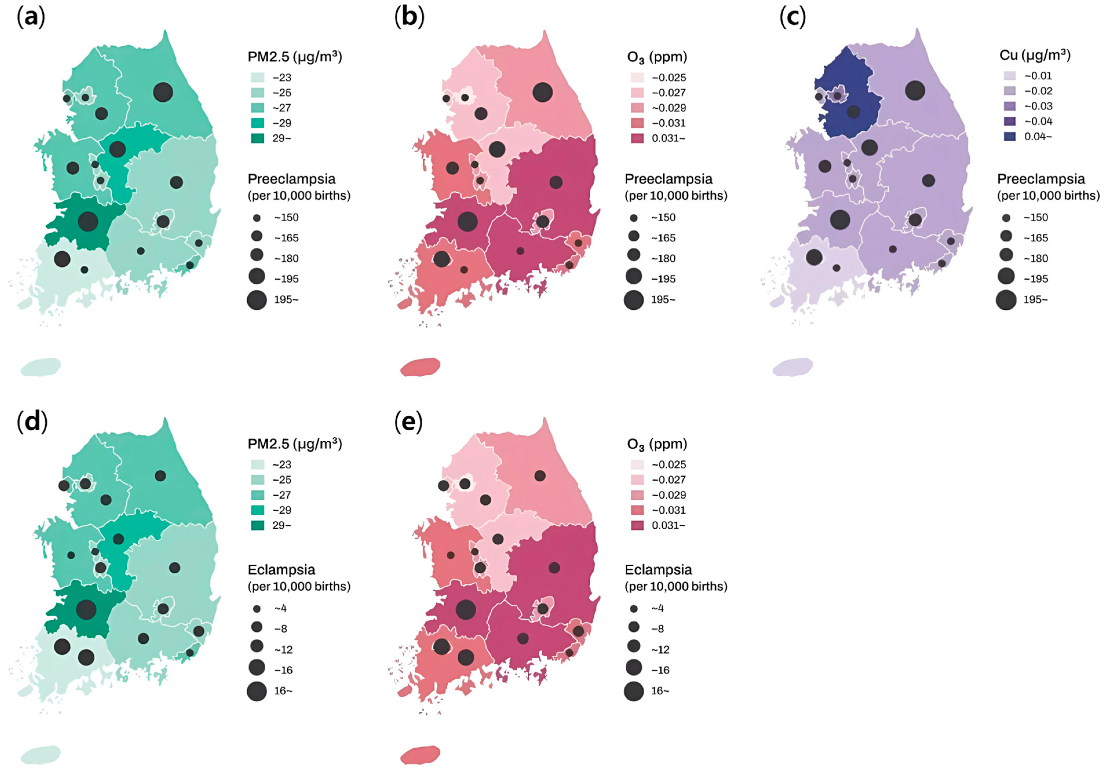
Table 2 summarizes the statistics on air pollutants and heavy metals for each case study. Figure 2 presents the spatial distribution of the mean PM2.5, O3, and Cu concentrations, as well as the number of preeclampsia and eclampsia cases across South Korea. Higher mean concentrations of PM2.5 and O3 were associated with an increased incidence of both preeclampsia and eclampsia, whereas elevated Cu concentrations were specifically linked to an increased incidence of preeclampsia.
Table 3 shows the association between exposure to specific air pollutants and heavy metals during the second trimester and the incidences of preeclampsia and eclampsia. Exposure to PM2.5, O3, and Cu was significantly associated with an increased risk of preeclampsia. O3 exposure was also significantly associated with an increased risk of eclampsia.
Table 4 presents the association between exposure to PM2.5, O3, and Cu and the risk of preeclampsia. In the univariate analysis, PM2.5 exposure was significantly associated with an increased risk of preeclampsia. In the multivariate model, Cu exposure remained significantly associated with an increased risk of preeclampsia (OR: 1.011, 95% CI: 1.001–1.023), suggesting that it may be an independent risk factor.
Table 5 illustrates the association between PM2.5 and O3 exposure and the risk of eclampsia. In the univariate analysis, O3 exposure was significantly associated with an increased risk of eclampsia, and this association remained significant in the multivariate model (OR: 1.113; 95% CI: 1.007–1.23), suggesting that O3 may be an independent risk factor for eclampsia. These findings suggest that elevated O3 exposure during pregnancy is associated with a higher risk of eclampsia.

4. Discussion

This study demonstrated that Cu exposure during the second trimester of pregnancy could be an independent risk factor for preeclampsia, while O3 exposure during the same period might independently contribute to the risk of eclampsia. Increased Cu exposure during the second trimester was associated with a higher incidence of preeclampsia, whereas increased O3 exposure during the same period was linked to a higher incidence of eclampsia.
The adverse effects of air pollution on human health have garnered growing attention worldwide. Recently, more research institutions have begun investigating the associations among preeclampsia, eclampsia, and air pollution. However, these studies have produced conflicting results and often oversimplify the analysis of air pollutants. Air pollutants consist of complex components, including nitrogen dioxide, sulfur dioxide, particulate matter, indirectly formed ozone, and heavy metals. Heavy metals often amalgamate with particulate matter and primarily originate from diesel and gasoline exhaust emissions, particularly in areas with high traffic and industrial activity. South Korea is an appropriate country to investigate the effects of heavy metals in air pollution on pregnant women, because its geographical characteristics and rapid urbanization and industrialization have resulted in higher concentrations of heavy metals in air pollution than the global average [24,25,26].
We analyzed data from 16,920 women who were diagnosed with preeclampsia out of 1,061,076 deliveries in Korea between January 2016 and December 2020. The incidence rate of preeclampsia was 1.59%, which is lower than that reported in other studies [27]. This discrepancy may be associated with the exclusion of patients with incomplete medical records or the fact that the study population was primarily Asian [28].
We examined the association between exposure to air pollution during pregnancy and the risk of preeclampsia, focusing on exposure during the second trimester. Although the etiology of preeclampsia has not been fully elucidated, one of the most commonly accepted theories is the two-stage model. In stage I, inadequate trophoblast invasion leads to shallow placentation and poor uteroplacental perfusion. In stage II, this results in widespread endothelial dysfunction and systemic clinical manifestations [29]. It is believed that stage I occurs before gestational age of 12 to 20 weeks, corresponding to the late first trimester and early second trimester, and stage II occurs thereafter [30]. Also, preeclampsia would require delivery or the termination of pregnancy under certain circumstances. Especially when the patient has not reached term pregnancy, there is an increased risk of preterm birth or pregnancy termination. As a result, third trimester data may be subject to selection survival bias. One study reported that the late first and second trimesters are likely the most influential periods for air pollution exposure and the development of preeclampsia, with the highest risk being associated with PM2.5 exposure identified in the fourth month of pregnancy [31]. Another study found that exposure to air pollutants in the second trimester was associated with an increased risk of gestational hypertension [32]. Additionally, Jia et al. showed that in the second trimester, multiple air pollutants were identified as risk factors for preeclampsia, with new pollutants that were not risk factors in the first trimester emerging as additional risks, indicating that the second trimester is more vulnerable to preeclampsia than the first [33]. Therefore, these authors concluded that exposure to airborne pollution particles has the greatest effect during the second trimester of pregnancy.
We confirmed that the risk factors for preeclampsia include adolescent pregnancy, working outside the home, lower economic status, infant sex, cold season, low birth weight, low gestational age, nulliparity, and comorbidities. This study also showed a significant association between exposure to PM2.5 and copper in air pollution during pregnancy and the risk of preeclampsia. Additionally, there was a significant association between exposure to O3 in air pollution during pregnancy and the risk of eclampsia.
This study demonstrated a significant association between exposure to PM2.5 during pregnancy and preeclampsia. Although the extent of placental exposure to PM2.5 is unknown, a recent study provided evidence that inhaled PM is translocated from the lungs to the placenta [34]. Generally, exposure to ambient PM is related to the inhibition of phagocytosis, enhancement of inflammatory responses, and elevated oxidative stress [35]. Additionally, an in vitro study found that exposure to PM2.5 increases the production of interleukin 6, a proinflammatory cytokine, and decreases the production of hCG in human first-trimester trophoblast cells [36]. It is speculated that PM2.5 may contribute to pathological changes in the placenta associated with preeclampsia. Similarly to our study, several meta-analyses have reported that exposure to PM2.5 during pregnancy is correlated with an increased risk of preeclampsia [37,38]. However, these meta-analyses have generally found that exposure to PM2.5 during pregnancy is positively associated with the risk of preeclampsia, whereas our study did not find a statistically significant association. This discrepancy may be attributed to the fact that our study was conducted in Korea, where PM2.5 concentrations are relatively uniform across regions because of the country’s small geographical area, unlike meta-analyses encompassing diverse countries and regions. Another meta-analysis reported that the risk of preeclampsia from an increase in PM2.5 during pregnancy was not statistically significant [39]. However, a subsequent meta-analysis that included more eligible studies reported a statistically significant association between these two factors [38].
Regarding exposure to O3 during pregnancy, this study found a modest association with the risk of preeclampsia. Evidence suggests that exposure to O3 during pregnancy may increase the risk of preeclampsia. Exposure to O3 during pregnancy was associated with an increased sFlt-1/PIGF ratio, a characteristic feature of preeclampsia [40,41]. In addition, a study using murine models found that maternal O3 exposure leads to alterations in the placental extracellular matrix, which is crucial for angiogenesis and vascular development [42]. Jia et al. reported a significant association between exposure to O3 during pregnancy and the risk of preeclampsia [33]. However, similarly to our results, Cheng et al. found no association between exposure to O3 during pregnancy and the risk of preeclampsia [43]. These discrepant results may be explained by the different levels of O3 pollution in various countries, regions, and populations. Jin et al. reported relatively higher levels of O3 pollution compared to those in our study and that of Cheng et al. Furthermore, Cheng et al. reported a significant association between O3 exposure and an increased risk of preeclampsia in older mothers, in contrast to the results observed in mothers of all ages.
Our results showed that the adjusted odds ratio and 95% CI for the association between preeclampsia and exposure to NO2 or SO2 during pregnancy were 0.826 (0.72–0.947) and 0.337 (0.158–0.718), respectively. We do not believe that the odds ratio indicates negative associations between NO2, SO2, and preeclampsia. Several studies have shown conflicting data regarding the association between NO2, SO2, and preeclampsia. Jia et al. reported that only the highest exposure to SO2 was a risk factor for preeclampsia in the second trimester [33]. Yan et al. found that ambient SO2 was not significantly associated with preeclampsia risk [44]. Other studies have reported that the risk of preeclampsia increases with an increase in NO2 concentration [33,45]. These results could indicate that the relationships between NO2, SO2, and preeclampsia are not typical linear relationships. Jiang et al. reported that NO2 and SO2 show a non-linear relationship with preeclampsia in a concentration-dependent manner [46]. This suggests the need for more precise exposure modeling in future studies.
In areas with air pollution, due to the ubiquity of Cu in the environment and its presence in airborne particulates, exposure to Cu through inhalation is commonplace, particularly in environments with elevated Cu concentrations [47,48]. The mechanism through which copper, a component of air pollutants, contributes to preeclampsia development remains unclear. However, due to their extremely small size, copper nanoparticles may cross biological barriers, enter the bloodstream, and eventually accumulate in other organs [49]. In vitro studies using human cell lines have shown that copper nanoparticles exhibit DNA-damaging effects, potentially mediated by reactive oxygen species and oxidative stress [50,51]. Nanoparticles can interact with organelles such as mitochondria and induce the production of reactive oxygen species, disrupting cellular functions and leading to cytotoxicity, genotoxicity, inflammation, or apoptosis [52]. In vivo study have demonstrated that Cu nanoparticles increased the production of reactive oxygen species associated with serious adverse effects such as genotoxicity, inflammation, and apoptosis [53]. Tadesse et al., using in vivo and in vitro models, demonstrated that oxidative stress-induced DNA damage is present in higher amounts in the placentas of women with preeclampsia and appears to be clustered in the genomic DNA of maternal decidual stromal cells [54]. It is reasonable to suggest that such DNA damage could affect the placentation, thereby contributing to the pathogenesis of preeclampsia. Despite these findings, the effect of Cu on preeclampsia remains epidemiologically inconclusive. Several studies have reported that Cu is associated with the development of preeclampsia [55,56], whereas other studies have found that Cu is not associated with the development of preeclampsia [57,58] or is associated with a low preeclampsia risk [59,60]. A recent meta-analysis reported that the discrepancies among these studies may be explained by a U-shaped effect, suggesting that abnormally low or high levels of serum Cu can lead to a high risk of preeclampsia [61]. In our study, we found a statistically significant association between air pollutant Cu exposure and preeclampsia, although the effect size was modest. This may be explained by another study conducted in the same region as ours, which showed that serum Cu levels in pregnancies with preeclampsia were neither abnormally high nor low [62].
This study shows a significant association between O3 exposure during pregnancy and the risk of eclampsia. However, as eclampsia represents a severe progression of preeclampsia, this result should be interpreted with caution. The low incidence and small number of patients with eclampsia may have contributed to this finding; therefore further research is warranted.
This study has several limitations. One is that we could not adjust for the potential impact of heavy metals originating from sources other than airborne exposure. Heavy metals are present not only in the air but also in soil and water and can enter the human body through the oral route. However, previous studies analyzing heavy metals in food and water in the same areas as in our study have reported that their levels were within acceptable ranges or at low concentrations [62,63,64]. Therefore, the influence of heavy metals in soil or water is likely negligible. Another is that our study did not adjust for the interaction between PM2.5 and heavy metals and did not include confounding factors such as pre-pregnancy BMI and occupational exposure. Although PM2.5 constitutes a small percentage of suspended particles, its large surface area and ability to adsorb various chemicals may result in varying magnitudes of effect depending on its composition. Jeon et al. showed that health risks may differ across regions due to variations in PM2.5 component concentrations [65]. According to Chen et al., different components of PM2.5 affect distinct metabolic pathways [66]. Due to these complexities, we did not conduct an interaction analysis. We included all high-risk factors for preeclampsia, as well as some moderate and other risk factors, based on the classifications provided by the National Institute for Health and Care Excellence guidelines. However, several moderate or other risk factors such as pre-pregnancy BMI and occupational exposure were not included due to the absence of corresponding data in the database. Finally, the identification of disease status based on ICD diagnostic codes may result in misclassification, potentially introducing bias into the findings.

5. Conclusions

This study demonstrated that exposure to PM2.5 and Cu during the second trimester of pregnancy is significantly associated with an increased risk of preeclampsia. These findings underscore the vulnerability of pregnant women to air pollutants, especially during the second trimester, a critical period for placental and vascular development. Despite several limitations, our findings highlight a direct link between heavy metals in air pollution and the risk of preeclampsia.

Author Contributions

Conceptualization, Y.J.C.; Methodology, S.H.K.; Validation, J.C.; Formal Analysis, H.W.S. and J.C.; Investigation, W.K.M.; Writing—Original Draft Preparation, K.S.L.; Writing—Review and Editing, K.S.L. and W.K.M.; Supervision, Y.J.C. All authors have read and agreed to the published version of the manuscript.

Funding

This research received no external funding.

Institutional Review Board Statement

This study was approved by the Institutional Review Board of Korea University Ansan Hospital (approval number 2022AS0033).

Informed Consent Statement

The requirement for informed consent was waived by the Institutional Review Board of Korea University Ansan Hospital because of the retrospective nature of the study.

Data Availability Statement

Since the database is managed by the government, analysis is restricted to designated premises, and data extraction is not permitted.

Conflicts of Interest

The authors declare no conflicts of interest.

Abbreviations

The following abbreviations are used in this manuscript:
PM2.5Particulate matter
NHINational Health Insurance
ORsOdds ratios
CIsConfidence intervals

References

  1. Garovic, V.D.; White, W.M.; Vaughan, L.; Saiki, M.; Parashuram, S.; Garcia-Valencia, O.; Weissgerber, T.L.; Milic, N.; Weaver, A.; Mielke, M.M. Incidence and Long-Term Outcomes of Hypertensive Disorders of Pregnancy. J. Am. Coll. Cardiol. 2020, 75, 2323–2334. [Google Scholar] [CrossRef] [PubMed]
  2. Torres-Torres, J.; Villafan-Bernal, J.R.; Martinez-Portilla, R.J.; Hidalgo-Carrera, J.A.; Estrada-Gutierrez, G.; Adalid-Martinez-Cisneros, R.; Rojas-Zepeda, L.; Acevedo-Gallegos, S.; Camarena-Cabrera, D.M.; Cruz-Martinez, M.Y.; et al. Performance of machine-learning approach for prediction of pre-eclampsia in a middle-income country. Ultrasound Obs. Gynecol. 2024, 63, 350–357. [Google Scholar] [CrossRef] [PubMed]
  3. Dimitriadis, E.; Rolnik, D.L.; Zhou, W.; Estrada-Gutierrez, G.; Koga, K.; Francisco, R.P.V.; Whitehead, C.; Hyett, J.; da Silva Costa, F.; Nicolaides, K.; et al. Pre-eclampsia. Nat. Rev. Dis. Primers 2023, 9, 8. [Google Scholar] [CrossRef]
  4. Shah, D.M. The role of RAS in the pathogenesis of preeclampsia. Curr. Hypertens. Rep. 2006, 8, 144–152. [Google Scholar] [CrossRef]
  5. Gestational Hypertension and Preeclampsia: ACOG Practice Bulletin, Number 222. Obs. Gynecol. 2020, 135, e237–e260. [CrossRef] [PubMed]
  6. Brown, M.A.; Magee, L.A.; Kenny, L.C.; Karumanchi, S.A.; McCarthy, F.P.; Saito, S.; Hall, D.R.; Warren, C.E.; Adoyi, G.; Ishaku, S. Hypertensive disorders of pregnancy: ISSHP classification, diagnosis, and management recommendations for international practice. Hypertension 2018, 72, 24–43. [Google Scholar] [CrossRef]
  7. Vuong, A.D.B.; Pham, X.T.T.; Nguyen, P.N. Posterior reversible encephalopathy syndrome (PRES) on the second postpartum day: Learning experience from a case report and literature review. Int. J. Emerg. Med. 2024, 17, 118. [Google Scholar] [CrossRef]
  8. North, R.A.; McCowan, L.M.; Dekker, G.A.; Poston, L.; Chan, E.H.; Stewart, A.W.; Black, M.A.; Taylor, R.S.; Walker, J.J.; Baker, P.N.; et al. Clinical risk prediction for pre-eclampsia in nulliparous women: Development of model in international prospective cohort. BMJ 2011, 342, d1875. [Google Scholar] [CrossRef] [PubMed]
  9. Ghosh, G.; Breborowicz, A.; Brazert, M.; Maczkiewicz, M.; Kobelski, M.; Dubiel, M.; Gudmundsson, S. Evaluation of third trimester uterine artery flow velocity indices in relationship to perinatal complications. J. Matern. Fetal Neonatal Med. 2006, 19, 551–555. [Google Scholar] [CrossRef]
  10. La Verde, M.; Torella, M.; Ronsini, C.; Riemma, G.; Cobellis, L.; Marrapodi, M.M.; Capristo, C.; Rapisarda, A.M.C.; Morlando, M.; De Franciscis, P. The association between fetal Doppler and uterine artery blood volume flow in term pregnancies: A pilot study. Ultraschall Med. 2024, 45, 184–189. [Google Scholar] [CrossRef]
  11. Ju, M.J.; Oh, J.; Choi, Y.-H. Changes in air pollution levels after COVID-19 outbreak in Korea. Sci. Total Environ. 2021, 750, 141521. [Google Scholar] [CrossRef]
  12. Ngarambe, J.; Joen, S.J.; Han, C.-H.; Yun, G.Y. Exploring the relationship between particulate matter, CO, SO2, NO2, O3 and urban heat island in Seoul, Korea. J. Hazard. Mater. 2021, 403, 123615. [Google Scholar] [CrossRef]
  13. Choi, E.; Yi, S.-M.; Lee, Y.S.; Jo, H.; Baek, S.-O.; Heo, J.-B. Sources of airborne particulate matter-bound metals and spatial-seasonal variability of health risk potentials in four large cities, South Korea. Environ. Sci. Pollut. Res. 2022, 29, 28359–28374. [Google Scholar] [CrossRef] [PubMed]
  14. Savitz, D.A.; Elston, B.; Bobb, J.F.; Clougherty, J.E.; Dominici, F.; Ito, K.; Johnson, S.; McAlexander, T.; Ross, Z.; Shmool, J.L.; et al. Ambient Fine Particulate Matter, Nitrogen Dioxide, and Hypertensive Disorders of Pregnancy in New York City. Epidemiology 2015, 26, 748–757. [Google Scholar] [CrossRef]
  15. Veras, M.M.; Damaceno-Rodrigues, N.R.; Caldini, E.G.; Maciel Ribeiro, A.A.; Mayhew, T.M.; Saldiva, P.H.; Dolhnikoff, M. Particulate urban air pollution affects the functional morphology of mouse placenta. Biol. Reprod. 2008, 79, 578–584. [Google Scholar] [CrossRef]
  16. Yoder, S.R.; Thornburg, L.L.; Bisognano, J.D. Hypertension in pregnancy and women of childbearing age. Am. J. Med. 2009, 122, 890–895. [Google Scholar] [CrossRef]
  17. Redman, C.W. Current topic: Pre-eclampsia and the placenta. Placenta 1991, 12, 301–308. [Google Scholar] [CrossRef] [PubMed]
  18. Roberts, J.M.; Redman, C.W. Pre-eclampsia: More than pregnancy-induced hypertension. Lancet 1993, 341, 1447–1451. [Google Scholar] [CrossRef] [PubMed]
  19. Cao, L.; Wang, L.; Wu, L.; Wang, T.; Cui, X.; Yu, L.; Diao, R.; Mao, H. Particulate matter and hypertensive disorders in pregnancy: Systematic review and meta-analysis. Public Health 2021, 200, 22–32. [Google Scholar] [CrossRef]
  20. Melody, S.M.; Wills, K.; Knibbs, L.D.; Ford, J.; Venn, A.; Johnston, F. Maternal Exposure to Ambient Air Pollution and Pregnancy Complications in Victoria, Australia. Int J Environ. Res Public Health 2020, 17, 2572. [Google Scholar] [CrossRef]
  21. Bearblock, E.; Aiken, C.E.; Burton, G.J. Air pollution and pre-eclampsia; associations and potential mechanisms. Placenta 2021, 104, 188–194. [Google Scholar] [CrossRef] [PubMed]
  22. Kang, K.; Kim, T.; Kim, H. Effect of indoor and outdoor sources on indoor particle concentrations in South Korean residential buildings. J. Hazard. Mater. 2021, 416, 125852. [Google Scholar] [CrossRef]
  23. Chen, H.; Fang, X.; Wong, T.H.; Chan, S.N.; Akinwunmi, B.; Ming, W.K.; Zhang, C.J.P.; Wang, Z. Physical Activity during Pregnancy: Comparisons between Objective Measures and Self-Reports in Relation to Blood Glucose Levels. Int. J. Environ. Res. Public Health 2022, 19, 8064. [Google Scholar] [CrossRef]
  24. Chon, H.T.; Kim, K.W.; Kim, J.Y. Metal contamination of soils and dusts in Seoul metropolitan city, Korea. Environ. Geochem. Health 1995, 17, 139–146. [Google Scholar] [CrossRef] [PubMed]
  25. Lim, J.-M.; Lee, J.-H.; Moon, J.-H.; Chung, Y.-S.; Kim, K.-H. Airborne PM10 and metals from multifarious sources in an industrial complex area. Atmos. Res. 2010, 96, 53–64. [Google Scholar] [CrossRef]
  26. Hieu, N.T.; Lee, B.-K. Characteristics of particulate matter and metals in the ambient air from a residential area in the largest industrial city in Korea. Atmos. Res. 2010, 98, 526–537. [Google Scholar] [CrossRef]
  27. Mol, B.W.J.; Roberts, C.T.; Thangaratinam, S.; Magee, L.A.; de Groot, C.J.M.; Hofmeyr, G.J. Pre-eclampsia. Lancet 2016, 387, 999–1011. [Google Scholar] [CrossRef]
  28. Fasanya, H.O.; Hsiao, C.J.; Armstrong-Sylvester, K.R.; Beal, S.G. A Critical Review on the Use of Race in Understanding Racial Disparities in Preeclampsia. J. Appl. Lab. Med. 2021, 6, 247–256. [Google Scholar] [CrossRef] [PubMed]
  29. Chang, K.J.; Seow, K.M.; Chen, K.H. Preeclampsia: Recent Advances in Predicting, Preventing, and Managing the Maternal and Fetal Life-Threatening Condition. Int. J. Environ. Res. Public Health 2023, 20, 2994. [Google Scholar] [CrossRef] [PubMed]
  30. Sato, Y.; Fujiwara, H.; Konishi, I. Mechanism of maternal vascular remodeling during human pregnancy. Reprod. Med. Biol. 2012, 11, 27–36. [Google Scholar] [CrossRef]
  31. Sun, Y.; Bhuyan, R.; Jiao, A.; Avila, C.C.; Chiu, V.Y.; Slezak, J.M.; Sacks, D.A.; Molitor, J.; Benmarhnia, T.; Chen, J.-C. Association between particulate air pollution and hypertensive disorders in pregnancy: A retrospective cohort study. PLoS Med. 2024, 21, e1004395. [Google Scholar] [CrossRef] [PubMed]
  32. Nobles, C.J.; Williams, A.; Ouidir, M.; Sherman, S.; Mendola, P. Differential Effect of Ambient Air Pollution Exposure on Risk of Gestational Hypertension and Preeclampsia. Hypertension 2019, 74, 384–390. [Google Scholar] [CrossRef] [PubMed]
  33. Jia, L.; Liu, Q.; Hou, H.; Guo, G.; Zhang, T.; Fan, S.; Wang, L. Association of Ambient air Pollution with risk of preeclampsia during pregnancy: A retrospective cohort study. BMC Public Health 2020, 20, 1663. [Google Scholar] [CrossRef]
  34. Liu, N.; Miyashita, L.; Mcphail, G.; Thangaratinam, S.; Grigg, J. Late Breaking Abstract-Do inhaled carbonaceous particles translocate from the lung to the placenta? In Proceedings of the ERS International Congress, Paris, France, 15–19 September 2018. [Google Scholar]
  35. Terzano, C.; Di Stefano, F.; Conti, V.; Graziani, E.; Petroianni, A. Air pollution ultrafine particles: Toxicity beyond the lung. Eur. Rev. Med. Pharmacol. Sci. 2010, 14, 809–821. [Google Scholar] [PubMed]
  36. Naav, A.; Erlandsson, L.; Isaxon, C.; Asander Frostner, E.; Ehinger, J.; Sporre, M.K.; Krais, A.M.; Strandberg, B.; Lundh, T.; Elmer, E.; et al. Urban PM2.5 Induces Cellular Toxicity, Hormone Dysregulation, Oxidative Damage, Inflammation, and Mitochondrial Interference in the HRT8 Trophoblast Cell Line. Front. Endocrinol. 2020, 11, 75. [Google Scholar] [CrossRef] [PubMed]
  37. Yu, H.; Yin, Y.; Zhang, J.; Zhou, R. The impact of particulate matter 2.5 on the risk of preeclampsia: An updated systematic review and meta-analysis. Environ. Sci. Pollut. Res. 2020, 27, 37527–37539. [Google Scholar] [CrossRef]
  38. Zhu, J.; Chen, J.; Wang, K.; Yan, H.; Liu, Q.; Lan, Y.; Ren, L.; Wu, S. Exposure to ambient black carbon and particulate matter during pregnancy in associations with risk of pre-eclampsia: A meta-analysis based on population-based studies. Environ. Pollut. 2024, 343, 123230. [Google Scholar] [CrossRef]
  39. Gogna, P.; Villeneuve, P.J.; Borghese, M.M.; King, W.D. An exposure-response meta-analysis of ambient PM(2.5) during pregnancy and preeclampsia. Environ. Res. 2022, 210, 112934. [Google Scholar] [CrossRef]
  40. Rasmussen, L.G.; Lykke, J.A.; Staff, A.C. Angiogenic biomarkers in pregnancy: Defining maternal and fetal health. Acta Obs. Gynecol. Scand. 2015, 94, 820–832. [Google Scholar] [CrossRef]
  41. Zheng, Y.; McElrath, T.; Cantonwine, D.; Hu, H. Longitudinal associations between ambient air pollution and angiogenic biomarkers among pregnant women in the LIFECODES Study, 2006–2008. Environ. Health Perspect. 2023, 131, 087005. [Google Scholar] [CrossRef]
  42. Hunter, R.; Baird, B.; Garcia, M.; Begay, J.; Goitom, S.; Lucas, S.; Herbert, G.; Scieszka, D.; Padilla, J.; Brayer, K.; et al. Gestational ozone inhalation elicits maternal cardiac dysfunction and transcriptional changes to placental pericytes and endothelial cells. Toxicol. Sci. 2023, 196, 238–249. [Google Scholar] [CrossRef]
  43. Cheng, Y.; Wang, P.; Zhang, L.; Shi, H.; Li, J.; Meng, X.; Xiao, X.; Dai, H.; Zhang, Y. Ozone Exposure During Pregnancy and Risk of Gestational Hypertension or Preeclampsia in China. JAMA Netw. Open 2023, 6, e236347. [Google Scholar] [CrossRef] [PubMed]
  44. Yan, M.; Liu, N.; Fan, Y.; Ma, L.; Guan, T. Associations of pregnancy complications with ambient air pollution in China. Ecotoxicol. Environ. Saf. 2022, 241, 113727. [Google Scholar] [CrossRef]
  45. Hu, H.; Ha, S.; Roth, J.; Kearney, G.; Talbott, E.O.; Xu, X. Ambient Air Pollution and Hypertensive Disorders of Pregnancy: A Systematic Review and Meta-analysis. Atmos. Environ. 2014, 97, 336–345. [Google Scholar] [CrossRef] [PubMed]
  46. Jiang, W.; Yu, G.; Wang, C.; Yin, S.; Huang, Y.; Chen, Q.; Sun, K.; Zhang, J. Exposure to multiple air pollutant mixtures and the subtypes of hypertensive disorders in pregnancy: A multicenter study. Int. J. Hyg. Environ. Health 2023, 253, 114238. [Google Scholar] [CrossRef]
  47. Freire, B.M.; Gonzaga, R.G.; Pedron, T.; Monteiro, L.R.; Lange, C.N.; Pedreira Filho, W.D.R.; Batista, B.L. Occupational exposure to potentially toxic elements in the foundry industry: An integrated environmental and biological monitoring. Environ. Sci. Pollut. Res. Int. 2021, 28, 34630–34641. [Google Scholar] [CrossRef] [PubMed]
  48. Galarneau, J.M.; Beach, J.; Cherry, N. Urinary Metals as a Marker of Exposure in Men and Women in the Welding and Electrical Trades: A Canadian Cohort Study. Ann. Work Expo. Health 2022, 66, 1111–1121. [Google Scholar] [CrossRef] [PubMed]
  49. Lee, I.-C.; Ko, J.-W.; Park, S.-H.; Lim, J.-O.; Shin, I.-S.; Moon, C.; Kim, S.-H.; Heo, J.-D.; Kim, J.-C. Comparative toxicity and biodistribution of copper nanoparticles and cupric ions in rats. Int. J. Nanomed. 2016, 11, 2883–2900. [Google Scholar]
  50. Alarifi, S.; Ali, D.; Verma, A.; Alakhtani, S.; Ali, B.A. Cytotoxicity and genotoxicity of copper oxide nanoparticles in human skin keratinocytes cells. Int. J. Toxicol. 2013, 32, 296–307. [Google Scholar] [CrossRef] [PubMed]
  51. Karlsson, H.L.; Cronholm, P.; Gustafsson, J.; Möller, L. Copper oxide nanoparticles are highly toxic: A comparison between metal oxide nanoparticles and carbon nanotubes. Chem. Res. Toxicol. 2008, 21, 1726–1732. [Google Scholar] [CrossRef] [PubMed]
  52. Yang, H.; Liu, C.; Yang, D.; Zhang, H.; Xi, Z. Comparative study of cytotoxicity, oxidative stress and genotoxicity induced by four typical nanomaterials: The role of particle size, shape and composition. J. Appl. Toxicol. 2009, 29, 69–78. [Google Scholar] [CrossRef] [PubMed]
  53. Ameh, T.; Sayes, C.M. The potential exposure and hazards of copper nanoparticles: A review. Environ. Toxicol. Pharmacol. 2019, 71, 103220. [Google Scholar] [CrossRef] [PubMed]
  54. Tadesse, S.; Kidane, D.; Guller, S.; Luo, T.; Norwitz, N.G.; Arcuri, F.; Toti, P.; Norwitz, E.R. In Vivo and In Vitro Evidence for Placental DNA Damage in Preeclampsia. PLoS ONE 2014, 9, e86791. [Google Scholar] [CrossRef] [PubMed]
  55. Sak, S.; Barut, M.; Celik, H.; Incebiyik, A.; Agacayak, E.; Uyanikoglu, H.; Kirmit, A.; Sak, M. Copper and ceruloplasmin levels are closely related to the severity of preeclampsia. J. Matern. Fetal Neonatal Med. 2020, 33, 96–102. [Google Scholar] [CrossRef]
  56. Gul, A.Z.; Atakul, N.; Selek, S.; Atamer, Y.; Sarikaya, U.; Yildiz, T.; Demirel, M. Maternal Serum Levels of Zinc, Copper, and Thiols in Preeclampsia Patients: A Case-Control Study. Biol. Trace Elem. Res. 2022, 200, 464–472. [Google Scholar] [CrossRef]
  57. Salima, S.; Daundy, K.H.; Mose, J.C.; Pramatirta, A.Y.; Suardi, D.; Pusianawati, D. Comparison Levels of Copper, Zinc, and Cu/Zn Ratio of in Pre-eclampsia and Normal Pregnancy. Open Access Maced. J. Med. Sci. 2022, 10, 2392–2398. [Google Scholar] [CrossRef]
  58. Farzin, L.; Sajadi, F. Comparison of serum trace element levels in patients with or without pre-eclampsia. J. Res. Med. Sci. Off. J. Isfahan Univ. Med. Sci. 2012, 17, 938. [Google Scholar]
  59. Chen, Y.; Ou, Q.X.; Chen, Y.; Zhu, Q.L.; Tan, M.H.; Zhang, M.M.; Wu, S.Z.; Xu, H.Y. Association between trace elements and preeclampsia: A retrospective cohort study. J. Trace Elem. Med. Biol. 2022, 72, 126971. [Google Scholar] [CrossRef]
  60. El Desouky, E.; Raslan, O.K.; El Magd, I.A.; Al Sheikh, W.; Eltonsy, M. Comparative study for serum zinc and copper levels in cases with normal pregnancy versus preeclampsia. Nat. Sci. 2020, 18, 180–184. [Google Scholar]
  61. Zhong, Z.; Yang, Q.; Sun, T.; Wang, Q. A global perspective of correlation between maternal copper levels and preeclampsia in the 21st century: A systematic review and Meta-analysis. Front. Public Health 2022, 10, 924103. [Google Scholar] [CrossRef] [PubMed]
  62. Choi, R.; Sun, J.; Yoo, H.; Kim, S.; Cho, Y.Y.; Kim, H.J.; Kim, S.W.; Chung, J.H.; Oh, S.Y.; Lee, S.Y. A Prospective Study of Serum Trace Elements in Healthy Korean Pregnant Women. Nutrients 2016, 8, 749. [Google Scholar] [CrossRef] [PubMed]
  63. Hee-Yun, K.; Jae-In, K.; Jin-Chul, K.; Ji-Eun, P.; Kyung-Jin, L.; Sung-Il, K.; Jae-Ho, O.; Young-Mi, J. Survey of Heavy Metal Contents of Circulating Agricultural Products in Korea. Korean J. Food Sci. Technol. 2009, 41, 238–244. [Google Scholar]
  64. Kim, M.; Kim, J.S.; Sho, Y.S.; Chung, S.-Y.; Lee, J.O. The Study on Heavy Metal Contents in Various Foods. Korean J. Food Sci. Technol. 2003, 35, 561–567. [Google Scholar]
  65. Jeon, J.-I.; Jung, J.-Y.; Park, S.-Y.; Lee, H.-W.; Lee, J.-I.; Lee, C.-M. A Comparison of Health Risks from PM2.5 and Heavy Metal Exposure in Industrial Complexes in Dangjin and Yeosu·Gwangyang. Toxics 2024, 12, 158. [Google Scholar] [CrossRef] [PubMed]
  66. Chen, J.; Zhou, Q.; Wang, Y.; Wu, Y.; Li, M.; Wang, H.; Zheng, G.; Hao, Y.; Cao, X.; Yang, W.; et al. Association of PM2.5-bound multiple metals co-exposure with early cardiovascular damage: A panel study in young adults combining metabolomics. Environ. Pollut. 2025, 371, 125964. [Google Scholar] [CrossRef]
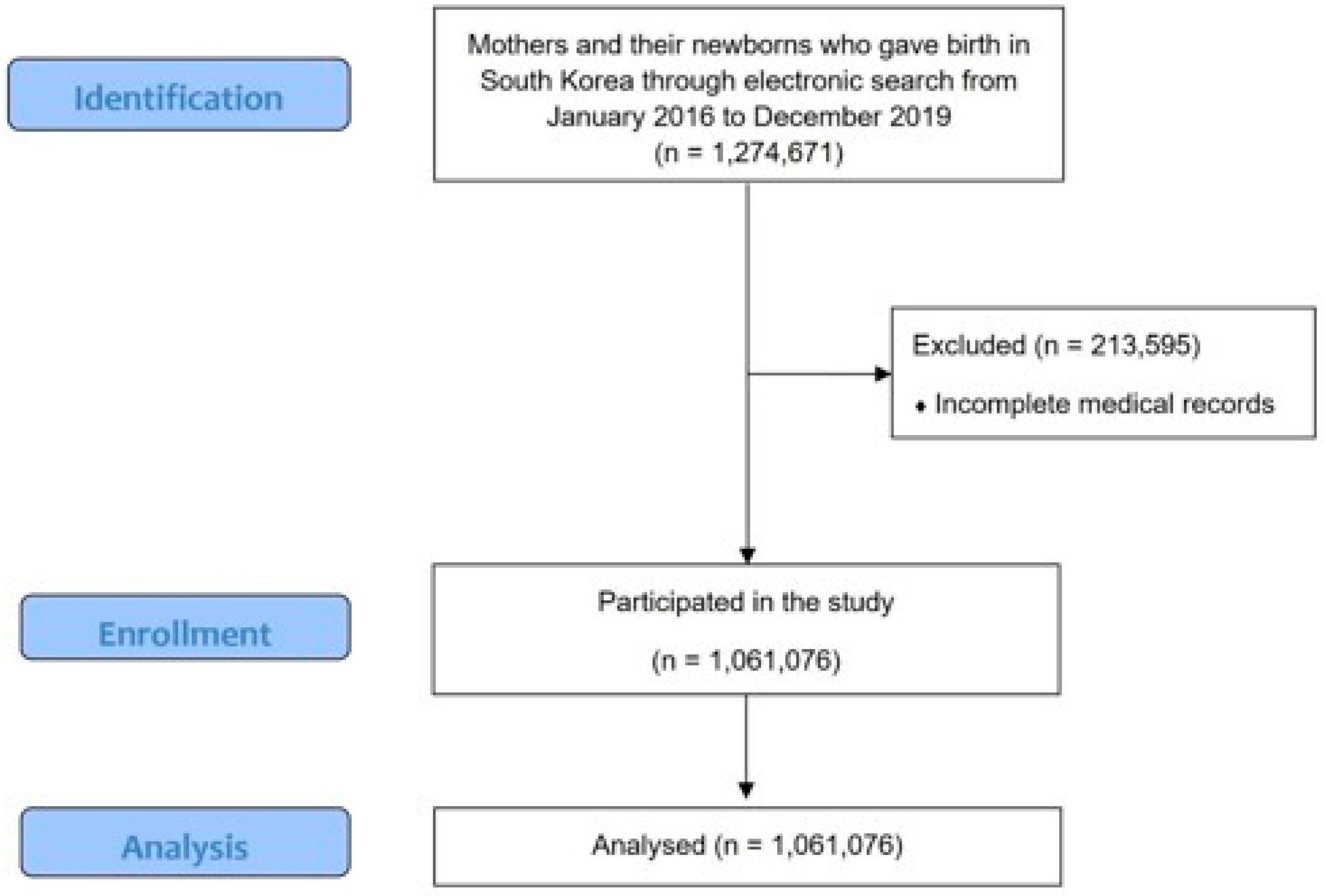
Figure 1. Consort flow diagram of this study.
Medicina 61 01146 g001
Figure 2. Spatial distribution of the mean PM2.5, O3, and Cu concentrations and the number of preeclampsia and eclampsia cases in South Korea. (a) PM2.5 and preeclampsia, (b) O3 and preeclampsia, (c) Cu and preeclampsia, (d) PM2.5 and eclampsia, (e) O3 and eclampsia. Data are presented as the mean concentration of each pollutant and the number of cases per 10,000 births.
Medicina 61 01146 g002
Table 1. Demographic and clinical characteristics of study participants.
TotalWith PreeclampsiaWith Eclampsia
Maternal age (years)
    Total1,061,07616,920592
    <20 *†203 (0.0)9 (0.1)1 (0.2)
    20–30107,623 (10.1)1578 (9.3)75 (12.7)
    30–40783,542 (73.8)11,780 (69.6)401 (67.7)
    40<169,708 (16.0)3553 (21.0)115 (19.4)
Occupational status *†
    Worked outside home429,667 (40.5)7191 (42.5)272 (45.9)
Household income
    Low (40%) *388,197 (36.6)6811 (40.3)234 (39.5)
    Middle (40–95%)649,430 (61.2)9783 (57.8)351 (59.3)
    High (95%<)23,449 (2.2)326 (1.9)7 (1.2)
Infant sex
    Female *†516,809 (48.7)8486 (50.2)316 (53.4)
Season
    Winter *†267,164 (25.2)4353 (25.7)152 (25.7)
    Spring279,390 (26.3)4273 (25.3)148 (25.0)
    Summer261,856 (24.7)4048 (23.9)137 (23.1)
    Fall252,666 (23.8)4246 (25.1)155 (26.2)
Body weight
    <1 kg *†2060 (0.2)442 (2.6)11 (1.9)
    1–2.5 kg35,697 (3.4)4038 (23.9)109 (18.4)
    2.5 kg<1,023,319 (96.4)12,440 (73.5)472 (79.7)
Premature
    <28 weeks *†1661 (0.2)192 (1.1)5 (0.8)
    28–36 weeks41,341 (3.9)4614 (27.3)120 (20.3)
    36 weeks<1,018,074 (95.9)12,114 (71.6)467 (78.9)
Multiple birth
    Singleton pregnancy *†1,039,880 (98.0)15,801 (93.4)557 (94.1)
    Twin pregnancy20,433 (1.9)1072 (6.3)32 (5.4)
    Multiple pregnancy763 (0.1)47 (0.3)3 (0.5)
Pre-existing hypertension *†37,236 (3.5)5759 (34.0)229 (38.7)
Gestational hypertension *†5508 (0.5)1003 (5.9)56 (9.5)
Superimposed preeclampsia *†1485 (0.1)600 (3.5)18 (3.0)
Pre-existing diabetes *†2877 (0.3)242 (1.4)10 (1.7)
Gestational diabetes *†229,918 (21.7)5026 (29.7)157 (26.5)
Kidney disease *†6438 (0.6)373 (2.2)15 (2.5)
Autoimmune disease *594 (0.1)16 (0.1)0 (0.0)
Thyroid disease *†374,703 (35.3)7683 (45.4)267 (45.1)
Dyslipidemia *†221,737 (20.9)6226 (36.8)234 (39.5)
Infertility *†157,150 (14.8)4210 (24.9)134 (22.6)
Data are presented as the number (%) of patients. * indicates p < 0.05 for comparison between groups with and without preeclampsia. † indicates p < 0.05 for comparison between groups with and without eclampsia.
Table 2. Summary statistics of air pollutants and heavy metals by case.
Air Pollutants and Heavy MetalsMeanSD
PM10 (μg/m3)46.29311.113
PM2.5 (μg/m3)25.5875.564
SO2 (ppm)0.0050.001
NO2 (ppm)0.0230.007
O3 (ppm)0.0270.008
CO (ppm)0.5030.110
Pb (μg/m3)0.0260.013
Cd (μg/m3)0.0010.001
Cr (μg/m3)0.0040.003
Cu (μg/m3)0.0250.014
Mn (μg/m3)0.0320.015
Fe (μg/m3)0.6640.267
Ni (μg/m3)0.0050.002
As (μg/m3)0.0040.002
Table 3. Adjusted odds ratios and 95% confidence intervals (CIs) for the association between preeclampsia or eclampsia and exposure to air pollutants and heavy metals during the second trimester of pregnancy, using a single-pollutant model.
With PreeclampsiaWith Eclampsia
OR (95% CI)OR (95% CI)
PM100.995 (0.993–0.997)1.003 (0.993–1.013)
PM2.51.004 (1–1.008)1.017 (0.997–1.037)
SO20.232 (0.201–0.268)0.337 (0.158–0.718)
NO20.916 (0.893–0.94)0.826 (0.72–0.947)
O31.05 (1.021–1.081)1.312 (1.132–1.521)
CO0.995 (0.993–0.997)0.998 (0.988–1.008)
Pb0.949 (0.936–0.962)0.894 (0.831–0.962)
Cd0.845 (0.693–1.031)0.17 (0.048–0.607)
Cr1.036 (0.98–1.095)0.541 (0.387–0.755)
Cu1.013 (1.002–1.024)0.968 (0.913–1.027)
Mn0.979 (0.968–0.989)0.891 (0.837–0.948)
Fe1 (0.999–1)0.996 (0.992–0.999)
Ni0.977 (0.917–1.042)0.49 (0.341–0.705)
As0.958 (0.897–1.024)0.693 (0.476–1.009)
The logistic regression model is adjusted for maternal age, infant sex, and season of conception.
Table 4. Logistic regression analysis of risk factors for preeclampsia.
UnivariateMultivariate
OR (95% CI)OR (95% CI)
Pre-existing hypertension15.187 (14.663–15.731)15.158 (14.643–15.692)
Twin and multiple pregnancy2.541 (2.372–2.723)2.537 (2.368–2.718)
Pre-existing diabetes and gestational diabetes1.305 (1.26–1.351)1.312 (1.267–1.358)
Age1.001 (0.997–1.004)
Kidney disease1.482 (1.32–1.665)1.483 (1.321–1.664)
Autoimmune disease1.221 (0.727–2.051)
Infertility1.499 (1.442–1.558)1.506 (1.45–1.564)
PM2.51.003 (1.001–1.006)
O31.005 (0.985–1.025)1.003 (0.984–1.023)
Cu1.006 (0.995–1.018)1.011 (1.001–1.023)
The logistic regression model is adjusted for maternal age, infant sex, and season of conception.
Table 5. Logistic regression analysis of risk factors for eclampsia.
UnivariateMultivariate
OR (95% CI)OR (95% CI)
Pre-existing hypertension16.549 (13.934–19.654)16.538 (13.984–19.558)
Twin and multiple pregnancy2.065 (1.447–2.947)2.056 (1.441–2.933)
Pre-existing diabetes and gestational diabetes1.102 (0.918–1.324)
Age0.984 (0.966–1.002)
Kidney disease1.433 (0.837–2.453)
Infertility1.318 (1.076–1.613)1.303 (1.067–1.593)
PM2.51.014 (0.998–1.029)1.013 (0.997–1.028)
O31.111 (1.005–1.228)1.113 (1.007–1.23)
The logistic regression model is adjusted for maternal age, infant sex, and season of conception.
Disclaimer/Publisher’s Note: The statements, opinions and data contained in all publications are solely those of the individual author(s) and contributor(s) and not of MDPI and/or the editor(s). MDPI and/or the editor(s) disclaim responsibility for any injury to people or property resulting from any ideas, methods, instructions or products referred to in the content.