Article

Comparison of Proximal Tibiofibular Joint Detachment with Tibial-Sided Osteotomy for Fibular Untethering in Lateral Closing-Wedge High Tibial Osteotomy: A Cadaveric Study

1
Department of Orthopaedic Surgery, Seoul St. Mary’s Hospital, College of Medicine, The Catholic University of Korea, Seoul 06591, Republic of Korea
2
Catholic Institute for Applied Anatomy, Department of Anatomy, College of Medicine, The Catholic University of Korea, Seoul 06591, Republic of Korea
*
Author to whom correspondence should be addressed.
Medicina 2025, 61(1), 161; https://doi.org/10.3390/medicina61010161

Submission received: 15 December 2024 / Revised: 4 January 2025 / Accepted: 17 January 2025 / Published: 19 January 2025
(This article belongs to the Special Issue Cutting-Edge Concepts in Knee Surgery)

Abstract

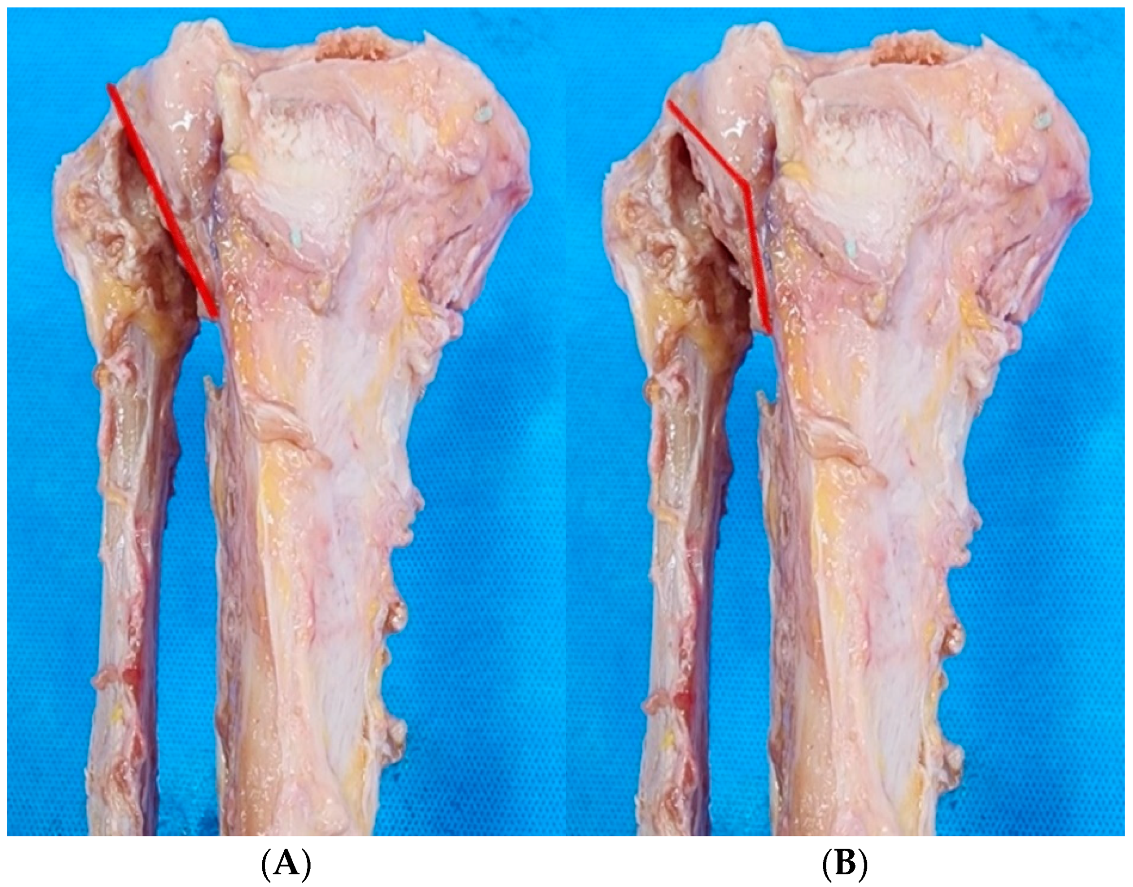
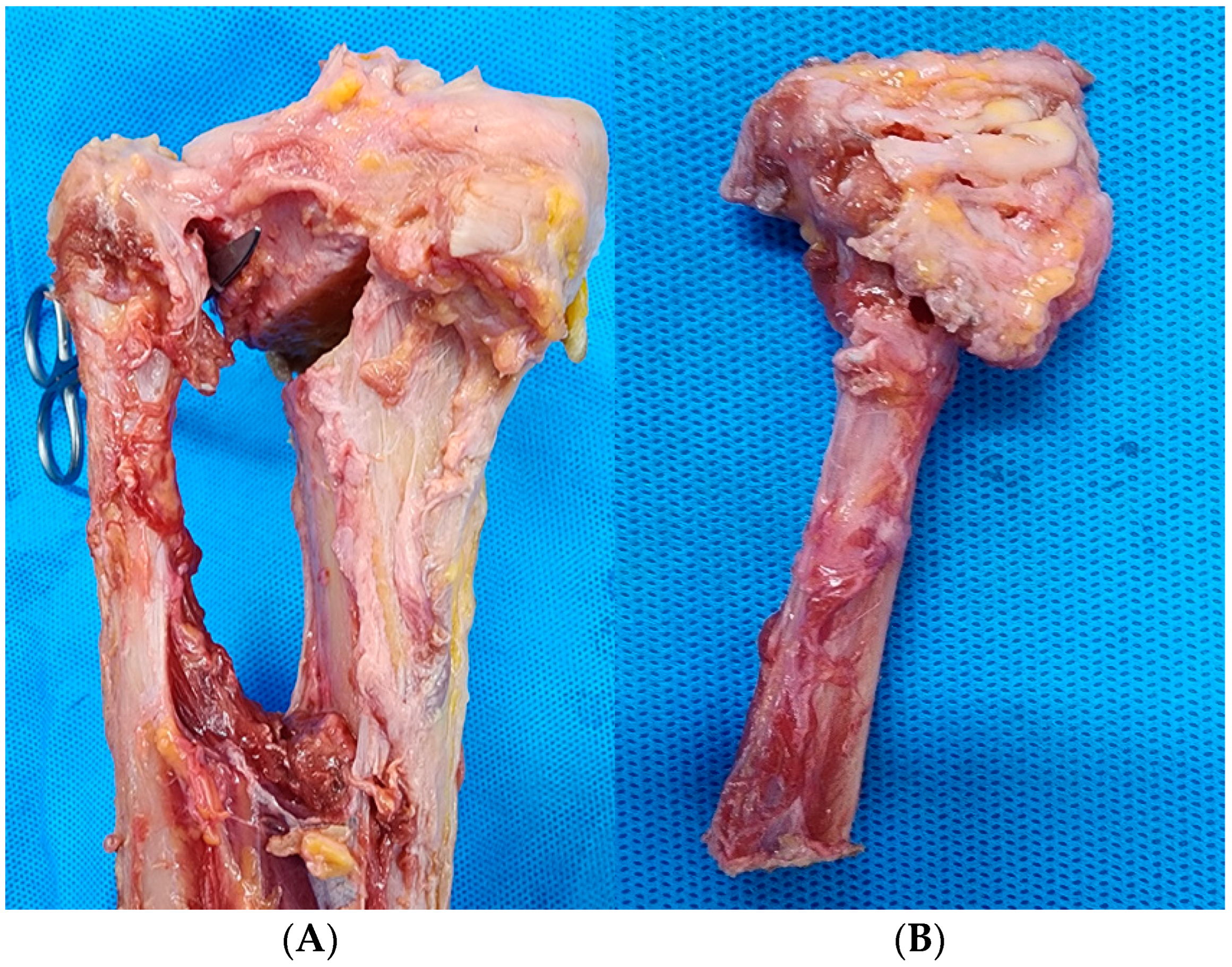
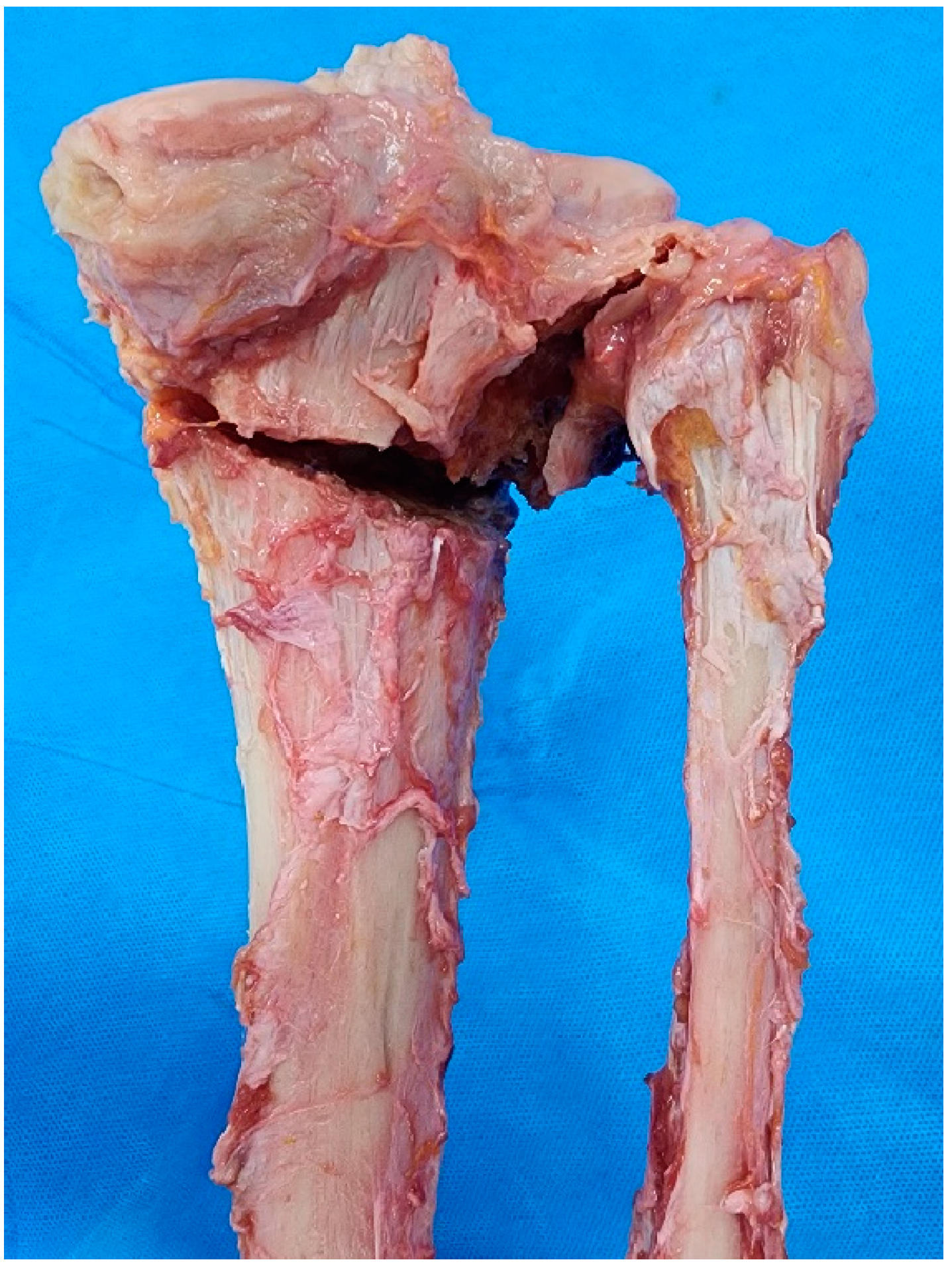
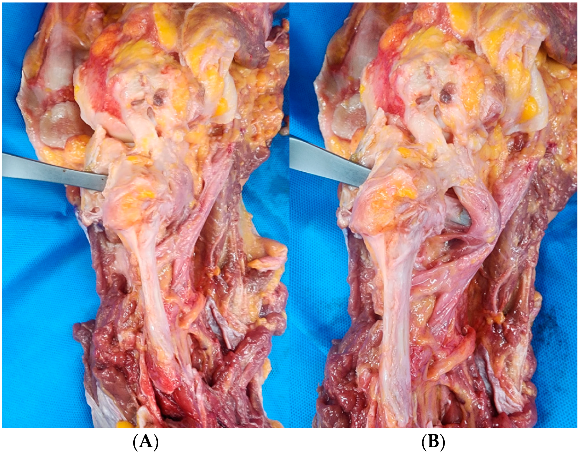
Background and Objectives: Proximal tibiofibular joint detachment (PTFJD) is a fibular untethering procedure during lateral closing-wedge high tibial osteotomy (LCWHTO) for varus knee osteoarthritis. However, the PTFJD procedure is technically demanding, and confirmation of clear joint separation is not straightforward. The aim of this study was to compare the degree of completion and safety of PTFJD versus tibial-sided osteotomy (TSO); this latter procedure is our novel technique for fibular untethering during LCWHTO. Materials and Methods: Sixteen fresh frozen cadaver knees from eight cadavers were included in the study. Among the eight pairs of knees, one knee was randomly assigned to undergo PTFJD and the other knee to undergo TSO, which separates the fibula by osteotomizing the lateral cortex of the proximal tibia at the medial side of the proximal tibiofibular joint for fibular untethering during LCWHTO. After each procedure with LCWHTO, the posterior compartment of each knee was dissected to compare the degree of procedural completion and the distance from the posterior detachment or osteotomy site to posterior neurovascular structures between PTFJD and TSO groups. The pass-through test crossing the separation site from anterior to posterior using an osteotome was also performed to evaluate the protective effect of the muscular structures of the posterior compartment. Results: In the PTFJD group, four of eight cases (50%) showed fibular head fractures rather than division of the proximal tibiofibular joint. In contrast, in all TSO cases, the lateral cortex of the proximal tibia was clearly osteotomized from the medial side of the posterior proximal tibiofibular joint. Distances from the posterior detachment or osteotomy site to the common peroneal nerve, popliteal artery, and anterior tibial artery in the PTFJD and TSO groups were 20.8 ± 3.3 mm and 22.9 ± 3.6 mm (p = 0.382), 11.0 ± 2.4 mm and 9.8 ± 2.8 mm (p = 0.382), and 14.8 ± 1.9 mm and 14.9 ± 2.5 mm (p = 0.721), respectively. In the pass-through test, an osteotome was able to pass anteriorly to posteriorly in all eight PTFJD group cases. However, the osteotome was blocked posteriorly by the popliteus muscle in the TSO group cases, indicating protection of posterior neurovascular structures during the TSO procedure. Conclusions: TSO, a novel fibular untethering procedure for LCWHTO, resulted in clear separation of the fibula from the lateral tibial cortex, and protection of posterior neurovascular structures by the popliteus muscle during the procedure. We anticipate that our novel surgical technique will provide more clear-cut and safer fibular untethering for LCWHTO.
Keywords: lateral closing-wedge high tibial osteotomy; proximal tibiofibular joint; fibular shaft osteotomy; proximal tibiofibular joint detachment; tibial-sided osteotomy lateral closing-wedge high tibial osteotomy; proximal tibiofibular joint; fibular shaft osteotomy; proximal tibiofibular joint detachment; tibial-sided osteotomy