
Understudied Hyperphosphatemia (Chronic Kidney Disease) Treatment Targets and New Biological Approaches


Abstract
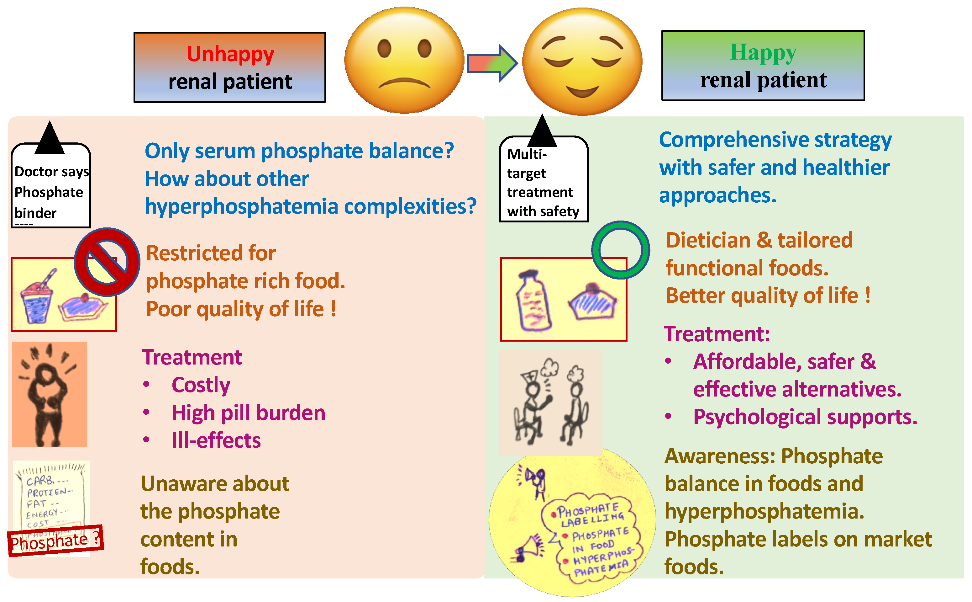
1. Introduction
2. Phosphate-Associated Toxicities
2.1. Calcification
2.2. Local pH
2.3. Phosphate Estimation
3. Hyperphosphatemia and Phosphate in Food
3.1. Socioeconomic Challenges
3.2. Public Education
4. Phosphate Binders in Hyperphosphatemia
Novel Phosphate Binders
5. Advanced CKD-Associated Understudied Diseases
5.1. Pulmonary and Cardiac Irregularities
5.2. Restless Leg Syndrome
5.3. Skin Disorders
6. Biological Approach for CKD and the Associated Hyperphosphatemia
6.1. Screening and Isolation of PAOs
6.2. Functional Food Formulation and In Vitro Studies
7. Conclusions
8. Future Directions
Supplementary Materials
Author Contributions
Funding
Institutional Review Board Statement
Informed Consent Statement
Data Availability Statement
Acknowledgments
Conflicts of Interest
References
- Global Facts About Kidney Disease. Available online: https://www.kidney.org/kidneydisease/global-facts-about-kidney-disease (accessed on 20 August 2021).
- Leaf, D.E.; Wolf, M. A Physiologic–Based Approach to the Evaluation of a Patient with Hyperphosphatemia. Am. J. Kidney Dis. 2013, 61, 330–336. [Google Scholar] [CrossRef] [PubMed]
- Fishbane, S.; Hazzan, A.D.; Halinski, C.; Mathew, A.T. Challenges and Opportunities in Late-Stage Chronic Kidney Disease. Clin. Kidney J. 2015, 8, 54–60. [Google Scholar] [CrossRef]
- Dhingra, R.; Sullivan, L.M.; Fox, C.S.; Wang, T.J.; D’Agostino, R.B.; Gaziano, J.M.; Vasan, R.S. Relations of Serum Phosphorus and Calcium Levels to the Incidence of Cardiovascular Disease in the Community. Arch. Intern. Med. 2007, 167, 879–885. [Google Scholar] [CrossRef]
- Eddington, H.; Hoefield, R.; Sinha, S.; Chrysochou, C.; Lane, B.; Foley, R.N.; Hegarty, J.; New, J.; O’Donoghue, D.J.; Middleton, R.J.; et al. Serum Phosphate and Mortality in Patients with Chronic Kidney Disease. Clin. J. Am. Soc. Nephrol. 2010, 5, 2251–2257. [Google Scholar] [CrossRef]
- Voormolen, N.; Noordzij, M.; Grootendorst, D.C.; Beetz, I.; Sijpkens, Y.W.; van Manen, J.G.; Boeschoten, E.W.; Huisman, R.M.; Krediet, R.T.; Dekker, F.W.; et al. High Plasma Phosphate as a Risk Factor for Decline in Renal Function and Mortality in Pre-dialysis Patients. Nephrol. Dial. Transplant. 2007, 22, 2909–2916. [Google Scholar] [CrossRef]
- Kestenbaum, B.; Sampson, J.N.; Rudser, K.D.; Patterson, D.J.; Seliger, S.L.; Young, B.; Sherrard, D.J.; Andress, D.L. Serum Phosphate Levels and Mortality Risk Among People with Chronic Kidney Disease. J. Am. Soc. Nephrol. 2005, 16, 520–528. [Google Scholar] [CrossRef] [PubMed]
- Katsumata, K.; Kusano, K.; Hirata, M.; Tsunemi, K.; Nagano, N.; Burke, S.K.; Fukushima, N. Sevelamer Hydrochloride Prevents Ectopic Calcification and Renal Osteodystrophy in Chronic Renal Failure Rats. Kidney Int. 2003, 64, 441–450. [Google Scholar] [CrossRef] [PubMed]
- Price, P.A.; Roublick, A.M.; Williamson, M.K. Artery Calcification in Uremic Rats Is Increased by a Low Protein Diet and Prevented by Treatment with Ibandronate. Kidney Int. 2006, 70, 1577–1583. [Google Scholar] [CrossRef]
- Moench, I.; Aravindhan, K.; Kuziw, J.; Schnackenberg, C.G.; Willette, R.N.; Toomey, J.R.; Gatto, G.J. High FGF23 Levels Failed to Predict Cardiac Hypertrophy in Animal Models of Hyperphosphatemia and Chronic Renal Failure. J. Endocr. Soc. 2021, 5, bvab066. [Google Scholar] [CrossRef] [PubMed]
- EFSA NDA Panel EFSA Panel on Dietetic Products N and A. Scientific Opinion on Dietary Reference Values for Phosphorus. EFSA J. 2015, 13, 4185–4239. [Google Scholar] [CrossRef]
- Shanahan, C.M.; Crouthamel, M.H.; Kapustin, A.; Giachelli, C.M. Arterial Calcification in Chronic Kidney Disease: Key Roles for Calcium and Phosphate. Circ. Res. 2011, 109, 697–711. [Google Scholar] [CrossRef]
- Vallet, M.; Metzger, M.; Haymann, J.P.; Flamant, M.; Gauci, C.; Thervet, E.; Boffa, J.J.; Vrtovsnik, F.; Froissart, M.; Stengel, B.; et al. Urinary Ammonia and Long-Term Outcomes in Chronic Kidney Disease. Kidney Int. 2015, 88, 137–145. [Google Scholar] [CrossRef]
- Kanda, E.; Ai, M.; Yoshida, M.; Kuriyama, R.; Shiigai, T. High Serum Bicarbonate Level Within the Normal Range Prevents the Progression of Chronic Kidney Disease in Elderly Chronic Kidney Disease Patients. BMC Nephrol. 2013, 14, 4. [Google Scholar] [CrossRef]
- Kopple, J.D.; Kalantar-Zadeh, K.; Mehrotra, R. Risks of Chronic Metabolic Acidosis in Patients with Chronic Kidney Disease. Kidney Int. Suppl. 2005, 95, S21–S27. [Google Scholar] [CrossRef]
- Kraut, J.A.; Kurtz, I. Metabolic Acidosis of CKD: Diagnosis, Clinical Characteristics, and Treatment. Am. J. Kidney Dis. 2005, 45, 978–993. [Google Scholar] [CrossRef] [PubMed]
- Goraya, N.; Simoni, J.; Jo, C.H.; Wesson, D.E. Treatment of Metabolic Acidosis in Patients with Stage 3 Chronic Kidney Disease with Fruits and Vegetables or Oral Bicarbonate Reduces Urine Angiotensinogen and Preserves Glomerular Filtration Rate. Kidney Int. 2014, 86, 1031–1038. [Google Scholar] [CrossRef] [PubMed]
- Dobre, M.; Yang, W.; Pan, Q.; Appel, L.; Bellovich, K.; Chen, J.; Feldman, H.; Fischer, M.J.; Ham, L.L.; Hostetter, T.; et al. Persistent High Serum Bicarbonate and the Risk of Heart Failure in Patients with Chronic Kidney Disease (CKD): A Report from the Chronic Renal Insufficiency Cohort (CRIC) Study. J. Am. Heart Assoc. 2015, 4, e001599. [Google Scholar] [CrossRef]
- Kovesdy, C.P.; Anderson, J.E.; Kalantar-Zadeh, K. Association of Serum Bicarbonate Levels with Mortality in Patients with Non-dialysis-Dependent CKD. Nephrol. Dial. Transplant. 2009, 24, 1232–1237. [Google Scholar] [CrossRef] [PubMed]
- Dhondup, T.; Qian, Q. Electrolyte and Acid-Base Disorders in Chronic Kidney Disease and End-Stage Kidney Failure. Blood Purif. 2017, 43, 179–188. [Google Scholar] [CrossRef] [PubMed]
- Wikipedia. Bohr Effect. Available online: https://en.wikipedia.org/wiki/Bohr_effect (accessed on 21 February 2022).
- Osuka, S.; Razzaque, M.S. Can Features of Phosphate Toxicity Appear in Normophosphatemia? J. Bone Miner. Metab. 2012, 30, 10–18. [Google Scholar] [CrossRef] [PubMed]
- Agar, B.U.; Akonur, A.; Lo, Y.C.; Cheung, A.K.; Leypoldt, J.K. Kinetic Model of Phosphorus Mobilization During and After Short and Conventional Hemodialysis. Clin. J. Am. Soc. Nephrol. 2011, 6, 2854–2860. [Google Scholar] [CrossRef] [PubMed]
- Hu, X.; Ma, X.; Luo, Y.; Xu, Y.; Xiong, Q.; Pan, X.; Bao, Y.; Jia, W. Contribution of Fibroblast Growth Factor 23 to Framingham Risk Score for Identifying Subclinical Atherosclerosis in Chinese Men. Nutr. Metab. Cardiovasc. Dis. 2017, 27, 147–153. [Google Scholar] [CrossRef] [PubMed]
- Benini, O.; D’Alessandro, C.; Gianfaldoni, D.; Cupisti, A. Extra-Phosphate Load from Food Additives in Commonly Eaten Foods: A Real and Insidious Danger for Renal Patients. J. Ren. Nutr. 2011, 21, 303–308. [Google Scholar] [CrossRef] [PubMed]
- Block, G.A.; Ix, J.H.; Ketteler, M.; Martin, K.J.; Thadhani, R.I.; Tonelli, M.; Wolf, M.; Jüppner, H.; Hruska, K.; Wheeler, D.C. Phosphate Homeostasis in CKD: Report of a Scientific Symposium Sponsored by the National Kidney Foundation. Am. J. Kidney Dis. 2013, 62, 457–473. [Google Scholar] [CrossRef]
- Kemi, V.E.; Rita, H.J.; Kärkkäinen, M.U.; Viljakainen, H.T.; Laaksonen, M.M.; Outila, T.A.; Lamberg-Allardt, C.J. Habitual High Phosphorus Intakes and Foods with Phosphate Additives Negatively Affect Serum Parathyroid Hormone Concentration: A Cross-Sectional Study on Healthy Premenopausal Women. Public Health Nutr. 2009, 12, 1885–1892. [Google Scholar] [CrossRef]
- Gutiérrez, O.M.; Luzuriaga-McPherson, A.; Lin, Y.; Gilbert, L.C.; Ha, S.W.; Beck, G.R. Impact of Phosphorus-Based Food Additives on Bone and Mineral Metabolism. J. Clin. Endocrinol. Metab. 2015, 100, 4264–4271. [Google Scholar] [CrossRef]
- Ketteler, M.; Block, G.A.; Evenepoel, P.; Fukagawa, M.; Herzog, C.A.; McCann, L.; Moe, S.M.; Shroff, R.; Tonelli, M.A.; Toussaint, N.D.; et al. Executive Summary of the 2017 KDIGO Chronic Kidney Disease-Mineral and Bone Disorder (CKD-MBD) Guideline Update: What’s Changed and Why It Matters. Kidney Int. 2017, 92, 26–36. [Google Scholar] [CrossRef]
- Noori, N.; Kalantar-Zadeh, K.; Kovesdy, C.P.; Bross, R.; Benner, D.; Kopple, J.D. Association of Dietary Phosphorus Intake and Phosphorus to Protein Ratio with Mortality in Hemodialysis Patients. Clin. J. Am. Soc. Nephrol. 2010, 5, 683–692. [Google Scholar] [CrossRef]
- Schmidt, D.R.; Holmstrom, S.R.; Fon Tacer, K.; Bookout, A.L.; Kliewer, S.A.; Mangelsdorf, D.J. Regulation of Bile Acid Synthesis by Fat-Soluble Vitamins A and D. J. Biol. Chem. 2010, 285, 14486–14494. [Google Scholar] [CrossRef]
- Moe, S.M.; Zidehsarai, M.P.; Chambers, M.A.; Jackman, L.A.; Radcliffe, J.S.; Trevino, L.L.; Donahue, S.E.; Asplin, J.R. Vegetarian Compared with Meat Dietary Protein Source and Phosphorus Homeostasis in Chronic Kidney Disease. Clin. J. Am. Soc. Nephrol. 2011, 6, 257–264. [Google Scholar] [CrossRef] [PubMed]
- Nelson, S.M.; Sarabia, S.R.; Christilaw, E.; Ward, E.C.; Lynch, S.K.; Adams, M.A.; Holden, R.M. Phosphate-Containing Prescription Medications Contribute to the Daily Phosphate Intake in a Third of Hemodialysis Patients. J. Ren. Nutr. 2017, 27, 91–96. [Google Scholar] [CrossRef] [PubMed]
- León, J.B.; Sullivan, C.M.; Sehgal, A.R. The Prevalence of Phosphorus-Containing Food Additives in Top-Selling Foods in Grocery Stores. J. Ren. Nutr. 2013, 23, 265–270.e2. [Google Scholar] [CrossRef]
- Calvo, M.S.; Uribarri, J. Public Health Impact of Dietary Phosphorus Excess on Bone and Cardiovascular Health in the General Population. Am. J. Clin. Nutr. 2013, 98, 6–15. [Google Scholar] [CrossRef] [PubMed]
- Karp, H.; Ekholm, P.; Kemi, V.; Itkonen, S.; Hirvonen, T.; Närkki, S.; Lamberg-Allardt, C. Differences Among Total and In Vitro Digestible Phosphorus Content of Plant Foods and Beverages. J. Ren. Nutr. 2012, 22, 416–422. [Google Scholar] [CrossRef]
- Kimura, M.; Itokawa, Y. Cooking Losses of Minerals in Foods and Its Nutritional Significance. J. Nutr. Sci. Vitaminol. 1990, 36 (Suppl. 1), S25–S32; discussion S33. [Google Scholar] [CrossRef]
- Jezo, I.; Luzak, I. Chemical Papers. Chem Zvesti. 1963. Available online: https://www.chempap.org/?id=7&paper=6273 (accessed on 1 March 2018).
- Jezo, I.; Luzak, I. Chemical Papers. Chem Zvesti. 1966. Available online: https://www.chempap.org/?id=7&paper=6192 (accessed on 1 March 2018).
- Jezo, I.; Luzak, I. Chemical Papers. Chem Zvesti. 1966. Available online: https://www.chempap.org/?id=7&paper=6351 (accessed on 1 March 2018).
- Agyei-Aye, K.; Chian, M.X.; Lauterbach, J.H.; Moldoveanu, S.C. The Role of the Anion in the Reaction of Reducing Sugars with Ammonium Salts. Carbohydr. Res. 2002, 337, 2273–2277. [Google Scholar] [CrossRef] [PubMed]
- Sadek, T.; Mazouz, H.; Bahloul, H.; Oprisiu, R.; El Esper, N.; El Esper, I.; Boitte, F.; Brazier, M.; Moriniere, P.; Fournier, A. Sevelamer Hydrochloride with or Without Alphacalcidol or Higher Dialysate Calcium vs Calcium Carbonate in Dialysis Patients: An Open-Label, Randomized Study. Nephrol. Dial. Transplant. 2003, 18, 582–588. [Google Scholar] [CrossRef]
- Goldfarb, D.S.; Modersitzki, F.; Asplin, J.R. A Randomized, Controlled Trial of Lactic Acid Bacteria for Idiopathic Hyperoxaluria. Clin. J. Am. Soc. Nephrol. 2007, 2, 745–749. [Google Scholar] [CrossRef]
- Sullivan, C.; Sayre, S.S.; Leon, J.B.; Machekano, R.; Love, T.E.; Porter, D.; Marbury, M.; Sehgal, A.R. Effect of Food Additives on Hyperphosphatemia Among Patients with End-Stage Renal Disease: A Randomized Controlled Trial. JAMA 2009, 301, 629–635. [Google Scholar] [CrossRef]
- Karavetian, M.; de Vries, N.; Rizk, R.; Elzein, H. Dietary Educational Interventions for Management of Hyperphosphatemia in Hemodialysis Patients: A Systematic Review and Meta-analysis. Nutr. Rev. 2014, 72, 471–482. [Google Scholar] [CrossRef]
- Wilhelm, M.; Gaillard, S.; Rakov, V.; Funk, F. The Iron-Based Phosphate Binder PA21 Has Potent Phosphate Binding Capacity and Minimal Iron Release Across a Physiological pH Range In Vitro. Clin. Nephrol. 2014, 81, 251–258. [Google Scholar] [CrossRef] [PubMed]
- Saupe, J.; Belmega, G.; Krause, R.; Bennhold, I. Management of Hyperphosphatemia with Calcium Citrate in Hemodialysis Patients. Nephron 1989, 52, 93–94. [Google Scholar] [CrossRef]
- Chan, S.; Au, K.; Francis, R.S.; Mudge, D.W.; Johnson, D.W.; Pillans, P.I. Phosphate Binders in Patients with Chronic Kidney Disease. Aust. Prescr. 2017, 40, 10–14. [Google Scholar] [CrossRef] [PubMed]
- Malberti, F. Hyperphosphataemia: Treatment Options. Drugs 2013, 73, 673–688. [Google Scholar] [CrossRef]
- Sekar, A.; Kaur, T.; Nally, J.V.; Rincon-Choles, H.; Jolly, S.; Nakhoul, G.N. Phosphorus Binders: The New and the Old, and How to Choose. Clevel. Clin. J. Med. 2018, 85, 629–638. [Google Scholar] [CrossRef]
- Floege, J.; Covic, A.C.; Ketteler, M.; Rastogi, A.; Chong, E.M.F.; Gaillard, S.; Lisk, L.J.; Sprague, S.M.; PA21 Study Group. A Phase III Study of the Efficacy and Safety of a Novel Iron-Based Phosphate Binder in Dialysis Patients. Kidney Int. 2014, 86, 638–647. [Google Scholar] [CrossRef]
- Peter, W.L.S.; Wazny, L.D.; Weinhandl, E.; Cardone, K.E.; Hudson, J.Q. A Review of Phosphate Binders in Chronic Kidney Disease: Incremental Progress or Just Higher Costs? Drugs 2017, 77, 1155–1186. [Google Scholar] [CrossRef]
- Ginsberg, C.L. Nicotinamide and Phosphate Homeostasis in Chronic Kidney Disease. Curr. Opin. Nephrol. Hypertens. 2016, 25, 285–291. [Google Scholar] [CrossRef] [PubMed]
- Pergola, P.E.; Rosenbaum, D.P.; Yang, Y.; Chertow, G.M. A Randomized Trial of Tenapanor and Phosphate Binders as a Dual-Mechanism Treatment for Hyperphosphatemia in Patients on Maintenance Dialysis (AMPLIFY). J. Am. Soc. Nephrol. 2021, 32, 1465–1473. [Google Scholar] [CrossRef]
- Niacinamide Uses, Side Effects and More. Available online: https://www.webmd.com/drugs/2/drug-6926/niacinamide-oral/details#:~:text=Niacinamide%20(nicotinamide)%20is%20a%20form,swelling%2C%20and%20peeling%20red%20skin (accessed on 15 April 2023).
- Semantic Scholar. Treatment of Hyperphosphatemia in Hemodialysis Patients: The Calcium Acetate Renagel Evaluation (CARE Study). Available online: https://www.semanticscholar.org/paper/Treatment-of-hyperphosphatemia-in-hemodialysis-pat-Qunibi-Hootkins/8d9a510f174f9b0aec70d4bda08d8a62e2faac5e (accessed on 1 March 2018).
- Kempson, S.A.; Colon-Otero, G.; Ou, S.Y.; Turner, S.T.; Dousa, T.P. Possible Role of Nicotinamide Adenine Dinucleotide as an Intracellular Regulator of Renal Transport of Phosphate in the Rat. J. Clin. Investig. 1981, 67, 1347–1360. [Google Scholar] [CrossRef] [PubMed]
- Takahashi, Y.; Tanaka, A.; Nakamura, T.; Fukuwatari, T.; Shibata, K.; Shimada, N.; Ebihara, I.; Koide, H. Nicotinamide Suppresses Hyperphosphatemia in Hemodialysis Patients. Kidney Int. 2004, 65, 1099–1104. [Google Scholar] [CrossRef]
- Rottembourg, J.B.; Launay-Vacher, V.; Massard, J. Thrombocytopenia Induced by Nicotinamide in Hemodialysis Patients. Kidney Int. 2005, 68, 2911–2912. [Google Scholar] [CrossRef]
- Yusuf, A.A.; Howell, B.L.; Powers, C.A.; St Peter, W.L. Utilization and Costs of Medications Associated with CKD Mineral and Bone Disorder in Dialysis Patients Enrolled in Medicare Part D. Am. J. Kidney Dis. 2014, 64, 770–780. [Google Scholar] [CrossRef]
- Nagla, M.; Hafez, A.; Gamal, M.; Abdel-Hafez, G.; Agmy, G.; Mohamed, M. Pulmonary Dysfunctions in End Stage Chronic Renal Failure Children on Regular Hemodialysis; Possible Risk Factors. Paediatr. Respir. Physiol. Health Dis. 2021, 5, 8–9. [Google Scholar]
- Akmal, M.; Barndt, R.R.; Ansari, A.N.; Mohler, J.G.; Massry, S.G. Excess PTH in CRF Induces Pulmonary Calcification, Pulmonary Hypertension and Right Ventricular Hypertrophy. Kidney Int. 1995, 47, 158–163. [Google Scholar] [CrossRef] [PubMed]
- Conger, J.D.; Hammond, W.S.; Alfrey, A.C.; Contiguglia, S.R.; Stanford, R.E.; Huffer, W.E. Pulmonary Calcification in Chronic Dialysis Patients. Clinical and Pathologic Studies. Ann. Intern. Med. 1975, 83, 330–336. [Google Scholar] [CrossRef]
- Faubert, P.F.; Shapiro, W.B.; Porush, J.G.; Chou, S.Y.; Gross, J.M.; Bondi, E.; Gomez-Leon, G. Pulmonary Calcification in Hemodialyzed Patients Detected by Technetium−99m Diphosphonate Scanning. Kidney Int. 1980, 18, 95–102. [Google Scholar] [CrossRef] [PubMed]
- Berndt, T.J.; Liang, M.; Tyce, G.M.; Knox, F.G. Intrarenal Serotonin, Dopamine, and Phosphate Handling in Remnant Kidneys. Kidney Int. 2001, 59, 625–630. [Google Scholar] [CrossRef] [PubMed]
- National Kidney Federation. Skin Care in Renal Transplant Patients. Available online: https://www.kidney.org.uk/skin-care-in-renal-transplant-patients (accessed on 8 July 2021).
- Calciphylaxis. Pictures, Definition, Symptoms, Treatment, and Outlook. Available online: https://www.healthline.com/health/calciphylaxis (accessed on 8 September 2001).
- Simenhoff, M.L.; Dunn, S.R.; Zollner, G.P.; Fitzpatrick, M.E.; Emery, S.M.; Sandine, W.E.; Ayres, J.W. Biomodulation of the Toxic and Nutritional Effects of Small Bowel Bacterial Overgrowth in End-Stage Kidney Disease Using Freeze-Dried Lactobacillus acidophilus. Miner. Electrolyte Metab. 1996, 22, 92–96. [Google Scholar]
- Anand, A.; Sato, M.; Aoyagi, H. Screening of Phosphate-Accumulating Probiotics for Potential Use in Chronic Kidney Disorder. Food Sci. Technol. Res. 2019, 25, 89–96. [Google Scholar] [CrossRef]
- Anand, A.; Aoyagi, H. Estimation of Microbial Phosphate-Accumulation Abilities. Sci. Rep. 2019, 9, 4879. [Google Scholar] [CrossRef]
- Anand, A.; Yoshida, S.; Aoyagi, H. Tailored Synbiotic Powder (Functional Food) to Prevent Hyperphosphataemia (Kidney Disorder). Sci. Rep. 2021, 11, 16485. [Google Scholar] [CrossRef]
- Perico, L.; Benigni, A.; Remuzzi, G. Should COVID-19 concern nephrologists? Why and to what extent? The emerging impasse of angiotensin blockade. Nephron 2020, 144, 213–221. [Google Scholar] [CrossRef]
- Larsen, C.P.; Bourne, T.D.; Wilson, J.D.; Saqqa, O.S.M. Collapsing glomerulopathy in a patient with coronavirus disease 2019 (COVID-19). Kidney Int. Rep. 2020, 5, 935–939. [Google Scholar] [CrossRef] [PubMed]
- Cheng, Y.; Luo, R.; Wang, K.; Zhang, M.; Wang, Z.; Dong, L.; Li, J.; Yao, Y.; Ge, S.; Xu, G. Kidney impairment is associated with in- hospital death of COVID-19 patients. Kidney Int. 2020, 97, 829–838. [Google Scholar] [CrossRef] [PubMed]
- Banerjee, D.; Popoola, J.; Shah, S.; Ster, I.C.; Quan, V.; Phanish, M. COVID-19 infection in kidney transplant recipients. Kidney Int. 2020, 97, 1076–1082. [Google Scholar] [CrossRef] [PubMed]
- Huang, C.; Wang, Y.; Li, X.; Ren, L.; Zhao, J.; Hu, Y.; Zhang, L.; Fan, G.; Xu, J.; Gu, X.; et al. Clinical features of patients infected with 2019 novel coronavirus in Wuhan, China. Lancet 2020, 395, 497–506. [Google Scholar] [CrossRef]
- Oyadela, T.; Alqahtani, J.C.G. Prognosis of COVID-19 in patients with Liver and Kidney diseases: An early systemative review and meta-analysis. Trop. Med. Infect. Dis. 2020, 5, 80. [Google Scholar]
- Chen, N.; Dong, X.; Qu, J.; Gong, F.; Han, Y.; Qiu, Y.; Qiu, Y.; Wang, J.; Liu, Y.; Wei, Y.; et al. Epidemiological and clinical characteristics of 99 cases of 2019 novel coronavirus pneumonia in Wuhan, China: A descriptive study. Lancet 2020, 395, 507–513. [Google Scholar] [CrossRef]
- Dawei, W.; Bo, H.; Chang, H.; Fangfang, Z.; Xing, L.; Jing, Z.; Wang, B.; Xiang, H.; Cheng, Z.; Xiong, Y.; et al. Clinical characteristics of 138 hospitalized patients with 2019 novel coronavirus- infected pneumonia in Wuhan, China. JAMA 2020, 7323, 1061–1069. [Google Scholar]
- Diao, B.; Wang, C.; Wang, R.; Feng, Z.; Tan, Y.; Wang, H.; Tan, Y.; Wang, H.; Wang, C.; Liu, L.; et al. Human kidney is a target for novel severe acute respiratory syndrome coronavirus 2 (SARS-CoV-2) infection. medRxiv 2020. [Google Scholar] [CrossRef]
- Hong, X.; Chi, Z.; Liu, G.; Huang, H.; Guo, S.; Fan, J.-R.; Lin, X.-W.; Qu, L.-Z.; Chen, R.-L.; Wu, L.-J.; et al. Analysis of early renal injury in COVID-19 and diagnostic value of multi-index combined detection. medRxiv 2020. [Google Scholar] [CrossRef]
- Su, H.; Yang, M.; Wan, C.; Yi, L.-X.; Tang, F.; Zhu, H.-Y.; Lin, X.-W.; Qu, L.-Z.; Chen, R.-L.; Wu, L.-J.; et al. Renal histopathological analysis of 26 postmortem findings of patients with COVID-19 in China. Kidney Int. 2020, 98, 219–227. [Google Scholar] [CrossRef] [PubMed]
- Pan, X.; Xu, D.; Zhang, H.; Zhou, W.; Wang, L.; Cui, X.G. Identification of a potential mechanism of acute kidney injury during the COVID-19 outbreak: A study based on single-cell transcriptome analysis. Intensive Care Med. 2020, 46, 1114–1116. [Google Scholar] [CrossRef]
- Khoshdelrad, N.; Zahmatkesh, E.; Shpichka, A.; Timashev, P.V.M. Outbreak of chronic renal failure: Will this be a delayed heritage of COVID-19? J. Nephrol. 2020, 2, 3–5. [Google Scholar]
- Ma, Y.; Diao, B.; Lv, X.; Zhu, J.; Liang, W.; Liu, L.; Zhang, S.; Shen, B.; Wang, H. COVID-19 in hemodialysis (HD) patients: Report from one HD center in Wuhan, China. medRxiv 2020. [Google Scholar] [CrossRef]
- Zhu, L.; Xu, X.; Ma, K.; Yang, J.; Guan, H.; Chen, S.; Chen, Z.; Chen, G. Successful recovery of COVID-19 pneumonia in a renal transplant recipient with long-term immunosuppression. Am. J. Transplant. 2020, 20, 1859–1863. [Google Scholar] [CrossRef]


| Phosphate Binders | Dosage (per Day) | Advantages | Disadvantages |
|---|---|---|---|
| Calcium carbonate | 500–600 mg, 3 times | Relatively inexpensive and first-line treatment | Can potentially lead to hypercalcemia, cardiovascular disorder (CVD), gastrointestinal risks, and vascular and bone calcification |
| Calcium citrate | 4.5 g | Cost-effective | Can potentially lead to hypercalcemia and enhanced intestinal aluminium absorption |
| Calcium acetate | 667–6000 mg, 9 times | Less calcium than calcium carbonate | Needs prescription |
| Aluminium hydroxide | 600–1200 mg, 3 times | No calcium and effective across a wide range of pH levels | Can potentially lead to aluminium deposition into bones and requires strict monitoring of aluminium levels, and haematological and neurological toxicity |
| Lanthanum carbonate | 0.5–1 g, 3 times | No calcium and effective across a wide range of pH levels | Expensive, there is deposition into bones, and can lead to toxicity, muscular-ache, and gastrointestinal risks |
| Magnesium carbonate | - | Effective and relatively inexpensive | Gastrointestinal risks and requires strict monitoring of magnesium levels |
| Sevelamer oxyhydroxide | 0.5–3 g, 3 times | Effective across a wide range of pH levels, no calcium and lanthanum, reduces low-density cholesterol, and minimal assimilation | Expensive, has gastrointestinal risks, can lead to metabolic acidosis and hinder the assimilation of fat-soluble vitamins |
| Sucroferric oxyhydroxide | 500 mg, 3 times | Effective with low pill burden | Gastrointestinal risks and expensive |
| Ferric citrate | 210 mg, 9 times | Effective for iron and phosphate parameters | Expensive |
| Nicotinamide | 1.5 g | Effective with low pill burden and treats pellagra | Exacerbates hyperuricemia and can lead to nausea and hyperglycaemia |
| Tenapanor | 30 mg, 2 times | Effective with low pill burden and treats constipation | Diarrhoea and nausea |
Disclaimer/Publisher’s Note: The statements, opinions and data contained in all publications are solely those of the individual author(s) and contributor(s) and not of MDPI and/or the editor(s). MDPI and/or the editor(s) disclaim responsibility for any injury to people or property resulting from any ideas, methods, instructions or products referred to in the content. |
© 2023 by the authors. Licensee MDPI, Basel, Switzerland. This article is an open access article distributed under the terms and conditions of the Creative Commons Attribution (CC BY) license (https://creativecommons.org/licenses/by/4.0/).
Share and Cite
Anand, A.; Aoyagi, H. Understudied Hyperphosphatemia (Chronic Kidney Disease) Treatment Targets and New Biological Approaches. Medicina 2023, 59, 959. https://doi.org/10.3390/medicina59050959
Anand A, Aoyagi H. Understudied Hyperphosphatemia (Chronic Kidney Disease) Treatment Targets and New Biological Approaches. Medicina. 2023; 59(5):959. https://doi.org/10.3390/medicina59050959
Chicago/Turabian StyleAnand, Ajeeta, and Hideki Aoyagi. 2023. "Understudied Hyperphosphatemia (Chronic Kidney Disease) Treatment Targets and New Biological Approaches" Medicina 59, no. 5: 959. https://doi.org/10.3390/medicina59050959
APA StyleAnand, A., & Aoyagi, H. (2023). Understudied Hyperphosphatemia (Chronic Kidney Disease) Treatment Targets and New Biological Approaches. Medicina, 59(5), 959. https://doi.org/10.3390/medicina59050959