Article

Immediate Effects of Sprinter-Pattern Exercise on the Lordotic Curve and Abdominal Muscle Activity in Individuals with Hyperlordosis

1
Department of Physical Therapy, Graduate School of Sahmyook University, 815, Hwarang-ro, Nowon-gu, Seoul 01795, Republic of Korea
2
Neuromusculoskeletal Science Laboratory, 15, Gangnam-daero 84-gil, Seoul 06232, Republic of Korea
3
Institute of SMART Rehabilitation, Sahmyook University, 815, Hwarang-ro, Nowon-gu, Seoul 01795, Republic of Korea
4
Department of Physical Therapy, Sahmyook University, 815, Hwarang-ro, Nowon-gu, Seoul 01795, Republic of Korea
*
Author to whom correspondence should be addressed.
Medicina 2023, 59(12), 2177; https://doi.org/10.3390/medicina59122177
Submission received: 9 November 2023 / Revised: 27 November 2023 / Accepted: 11 December 2023 / Published: 15 December 2023
(This article belongs to the Section Sports Medicine and Sports Traumatology)

Abstract

:
Background and Objectives: Abdominal muscle exercises with limb movements are more effective for trunk stabilization than traditional exercises involving trunk flexion alone. This study examined the effects of abdominal exercises incorporating sprinter pattern and crunch exercises on changes in the lordotic curve and abdominal muscle activation in individuals with low back pain caused by hyperlordosis resulting from weak abdominal muscles. Materials and Methods: In this single-blind, randomized controlled trial, a total of 40 participants with hyperlordosis were recruited and randomly assigned to perform either sprinter-pattern abdominal exercises or crunch exercises. The participants assigned to each group performed three sets of ten abdominal exercises. The lumbar lordotic angle (LLA) and sacrohorizontal angle (SHA) were assessed prior to and following the intervention, whereas abdominal muscle activity was gauged throughout the intervention period. Changes in the LLA and SHA were measured by radiography. Abdominal muscle activity was measured using electromyography. Results: The LLA and SHA decreased significantly in both groups (p < 0.001), while the sprinter-pattern exercise group showed a statistically significant decrease compared to the crunch exercise group (p < 0.001). In the activity of the abdominal muscles, there was no significant difference in the rectus abdominis muscle between the two groups (p > 0.005). However, a significant difference between the external and internal oblique muscles was observed, and the activities of both muscles were significantly higher in the sprinter-pattern exercise group than in the crunch exercise group (p < 0.005). Conclusions: Abdominal exercise using a sprinter pattern may be effective in reducing lumbar lordosis by strengthening the abdominal muscles in patients with hyperlordosis.

1. Introduction

Chronic back pain has become a pressing health issue due to factors such as an increased sedentary lifestyles, lack of physical activity, and unawareness of body imbalances [1]. One of the causes of chronic back pain is the shortening of the erector spinae muscles and the weakening of the abdominal muscles, which can result in changes to the curvature of the spine in the lumbar spine [2]. This combination can lead to hyperlordosis, which puts substantial stress on the back, resulting in back pain [2,3,4].
A deficiency in the abdominal muscles causes hyperlordosis, which leads to a deviation from the normal alignment of the spine, pelvis, and sacrum, resulting in instability in the lumbar segment [5,6,7]. Hyperlordosis can induce pain by reducing the space in the intervertebral foramen [4] and misshaping the spine’s natural curve [2].
Abdominal muscle weakness is commonly found in patients with back pain [3]. Strengthening the abdominal muscles is known to be essential for the spine to maintain neutrality and to restore segmental stability [3]. Therefore, strong abdominal muscles are crucial not only for individuals with back instability, but also for those who are susceptible to developing back pain, as these muscles play an important role in preventing back pain by increasing the space in the intervertebral foramen, thus relieving stress on the facet joints resulting from hyperlordosis and maintaining proper postural control [8,9,10].
Although traditional exercises such as sit-ups and curl-ups are widely used to strengthen the abdomen, they can also increase the risk of lower back injuries and potentially lead to degenerative spinal damage owing to increased pressure on the spine [11,12]. To mitigate these risks, it is recommended to engage in stability exercises that integrate the functional movements of the arms and legs to enhance abdominal activity [13]. Building on this knowledge, Dietz [14] designed integrated pattern training, combining proprioceptor neuromuscular facilitation patterns with walking movement patterns, termed sprinter and skater motions.
In particular, the sprinter pattern enhances body stabilization by strengthening the abdominal muscles, and employs coordinated arm and leg movements to stabilize the trunk [15]. This movement activates the rectus abdominis (RA), external oblique (EO), and internal oblique (IO) [16]. This activation is consistent with research emphasizing the significance of exercises that strengthen posture-related muscles for trunk stabilization and activate crucial muscles for spinal alignment [15,16,17].
While extensive research has been conducted on abdominal exercises to examine their effects on hyperlordosis and the activation of abdominal muscles, the current literature lacks conclusive results regarding the specific exercises that effectively reduce lumbar lordosis and increase activation of abdominal muscles. Therefore, this study aimed to assess and compare the effects of abdominal exercises involving sprinter-pattern motions with traditional crunches, focusing on lumbar anterior flexion and abdominal muscle activity in patients with hypertrophic lumbar lordosis with angles exceeding the norm.

2. Materials and Methods

2.1. Study Design

This single-blind, randomized controlled trial was conducted from 29 October to 24 November 2019, and adhered to the ethical principles outlined in the Declaration of Helsinki. This study followed the reporting guidelines of the Consolidated Standards of Reporting Trials (CONSORT).

2.2. Participants and Ethics

A total of 40 participants (20 males, 20 females) between the ages of 20 and 50 who visited the Shinbaro Orthopedic Clinic in Seoul, Republic of Korea for low back pain were enrolled in the study. Study participant recruitment began in October 2019, and all the data were obtained between October 2019 and November 2019. Participants were voluntarily recruited through a clinic’s bulletin board. Participants were assessed for eligibility using inclusion [18,19,20] and exclusion criteria [21,22,23,24]. Subsequently, participants who met the eligibility criteria were provided with detailed written and verbal explanations of the study procedures and objectives. They voluntarily expressed their willingness to participate and signed the informed consent form before the commencement of the study.
The study procedures were registered with the Clinical Research Information Service, affiliated with the World Health Organization’s International Clinical Trials Registry Platform in the Republic of Korea (No. KCT0005232). Prior to initiating the experiment, we obtained ethical approval from the Sahmyook University Ethics Committee (No. 2-7001793-AB-N012019086HR, 29 October 2019).

2.2.1. Inclusion Criteria

  • Adults aged 20–50 years;
  • Chronic back pain for more than 3 months;
  • Lumbar lordotic angle (LLA) > 50°;
  • Sacrohorizontal angle (SHA) > 40°.

2.2.2. Exclusion Criteria

  • Orthopedic malformations or neurosurgical diseases;
  • Disc degeneration or other conditions that may affect the interpretation of results (severe fibromyalgia and rheumatoid arthritis, in combination with other treatments);
  • Surgery or pregnancy within 30 d;
  • Breastfeeding;
  • Wearing a hyperlordosis treatment device;
  • Participation in similar studies.

2.3. Sample Size

In previous studies, a reduction in the LLA resulted in an effect size of d = 0.92 [19]. To achieve this effect size, calculations were made using G*power 3.1 software (developed by Franz Faul at University of Kiel, Kiel, Germany), setting α at 0.05 and β at 0.20. These calculations indicated that 20 participants were required in each group. To accommodate the dropout rate, an additional 20% of participants were included. Therefore, 48 participants were included.

2.4. Randomization and Blinding
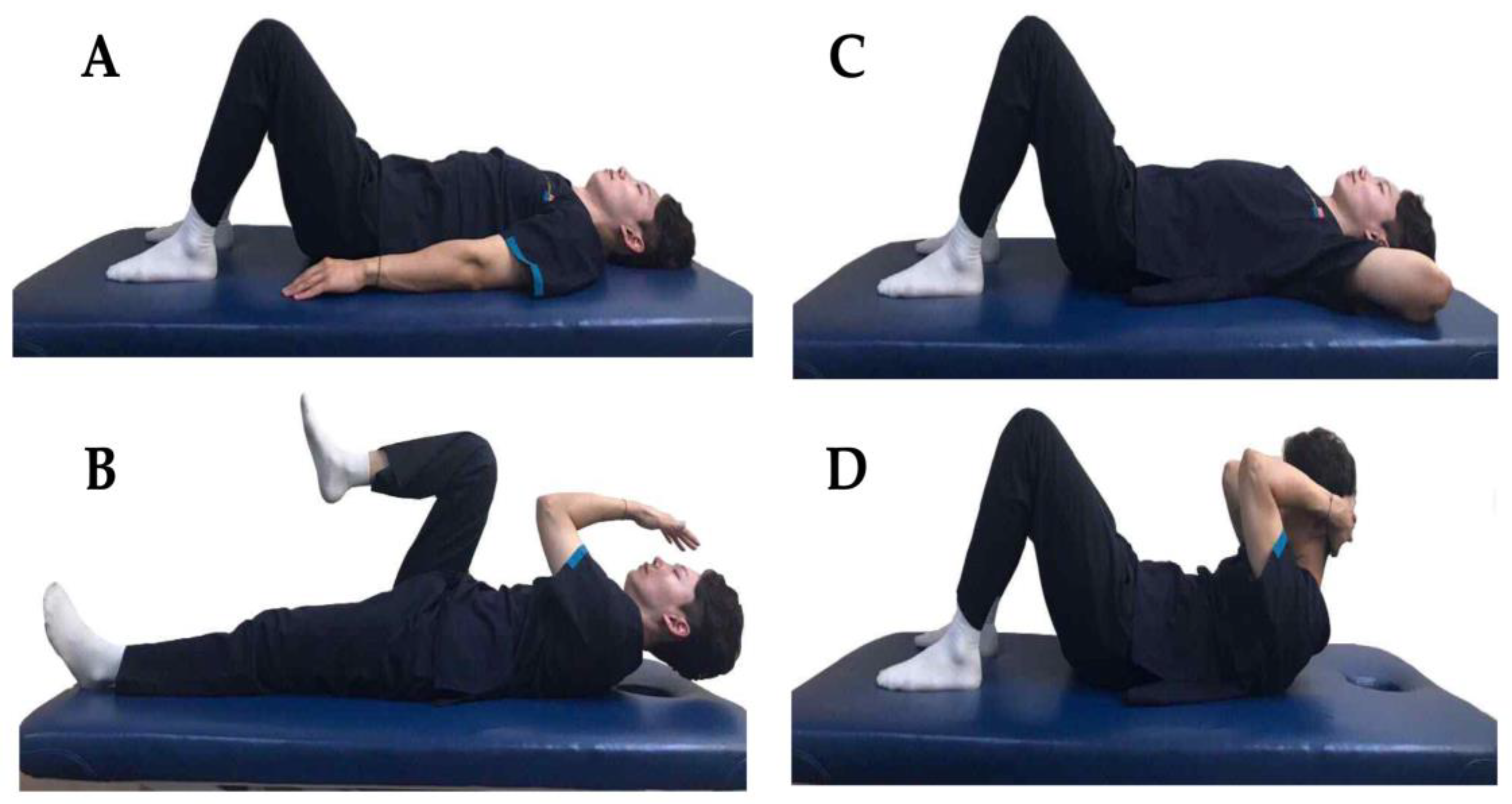
The participants were randomly allocated to two groups using Random Allocation Software for Windows ver. 2.0, developed by Isfahan University, Iran. One group was assigned to perform abdominal exercises using the sprinter pattern (n = 20), whereas the other was assigned to perform the crunch exercises (n = 20). This study was conducted as a single-blind intervention; until the commencement of the exercise, participants remained unaware of the assigned exercises for both their own group and the other group. In addition, the radiographer was kept unaware of the assignment of exercises to specific patients. Both researchers and assessors were informed about the interventions during the evaluation phases, including the pre- and post-tests.

2.5. Intervention

Both groups initially underwent a pre-test in which the root of the mean square reference values was assessed using electromyography (EMG), and radiographic imaging was employed to measure the lordotic curve. Subsequently, the participants performed three sets of exercises according to their group: sprinter-pattern exercises (SPEs) or crunch exercises (CEs). Each set consisted of ten repetitions, with each movement held for 10 s. A 15 s rest interval was provided between sets to prevent muscle fatigue. The participants performed the intervention thrice. Abdominal muscle activity was measured using EMG during the exercise, and both groups underwent immediate post-exercise radiographic imaging for the post-test. To ensure accuracy during the study, the participants were instructed in advance by a trained physical therapist on the measurement methods and general procedure of the intervention program, and the same physical therapist performed the interventions throughout. Interventions were individually performed to ensure adequate attention and precision in the exercises. Furthermore, participants were closely monitored throughout the study for any adverse events.

2.5.1. Sprinter-Pattern Exercise

For the SPE, the participants lay on their backs with their knees bent at a 45° angle and their arms resting beside them on the floor. Upon receiving a given signal, they raised their heads off the mat and simultaneously performed a series of coordinated arm and leg movements: flexing, adducting, and externally rotating one arm, while extending and internally rotating the corresponding leg. Conversely, they extended, abducted, and internally rotated the other arm while flexing, adducting, and externally rotating the opposing leg [14,25] (Figure 1A,B).

2.5.2. Crunch Exercise

For the CE, the participants lay on their backs with their knees bent at a 45° angle and placed their hands behind their heads. When given the start signal, they lifted their heads and shoulders, elevating the inferior angle of the scapula from the mat [25] (Figure 1C,D).

2.6. Outcomes

The LLA and SHA were measured before and after the intervention, while abdominal muscle activity was measured during the intervention period.

2.6.1. Lordotic Curve

The LLA and SHA were measured using an Accuray-525R diagnostic X-ray machine (Don Kang Medical System, Seoul, Republic of Korea). Radiographs were captured at distances of 2 m and 50 cm, with the following settings: exposure time, 0.8 s; tube voltage, 90 kVp; and tube current, 200 mA. Images were taken from the sagittal plane covering the pelvis to the lower spine while the subjects maintained an upright and functionally appropriate posture [26]. Throughout the study, a consistent and experienced radiologist from the hospital captured the radiographs. Although the clinical method for measuring the LLA has a decent reliability of 0.70–0.85 [27], radiographical methods have a higher reliability (0.87) [28]. Radiographic images were captured both before and after the intervention and subsequently underwent digitization before being transferred to a computer connected to the X-ray machine. The angles were then analyzed using built-in Viex Rex ver1.3.3.3.0 software (TechHeim, Seoul, Republic of Korea) on a vertical monitor with a resolution of 1080 × 1920.
  • Lumbar Lordotic Angle
The LLA measures the degree of lumbar curvature, which typically falls within a normal range of 40–50° [29]. This measurement is obtained by drawing lines along the upper plane of the L1 and S1 vertebrae, adding perpendicular lines to the converging side of the two, and subsequently measuring the angle of intersection [28]. This measurement technique is the prevailing standard in contemporary assessments of lumbar lordosis [30] (Figure 2).
2.
Sacrohorizontal Angle
The SHA indicates the degree of sacral tilt with a normal angle of 40° [5]. There is a strong correlation between the SHA and lumbar lordosis (r = 0.84). An increase in the SHA typically results in an increase in lumbar lordosis [31,32]. This angle is determined by the alignment of a line parallel to the upper surface of the first sacral vertebra with the horizontal plane [33] (Figure 2).

2.6.2. Abdominal Muscle Activity

Muscle activity was measured using surface EMG (Mini-wave Infinity Waterproof, Cometa Systems, Italy). The EMG signals were converted from analog to digital, and an EMG analysis software (Myoresearch XP Master ver. 1.07, Noraxon, Scottsdale, AZ, USA) was utilized, with the sampling rate set to 2000 Hz. The raw EMG data underwent full-wave rectification and were then quantified to an RMS of 150 ms. The data were filtered within a range of 20–450 Hz using a digital band-pass filter (Lancosh FIR). Ag/AgCl electrodes were used for the measurements. Before attaching the electrodes, to minimize skin resistance, any hair was removed, and the skin was exfoliated with sandpaper and cleaned with an alcohol swab. The specific attachment sites for each muscle are listed in Table 1 [34].
EMG was performed on the RA, EO, and IO muscles. To determine the baseline values for these muscles, the participants were asked to lie down with their knees bent and to lift both feet approximately 8 cm off the ground for 3 s. The average effective value was then calculated [35]. This action was used as a standard and normalized for comparison (% reference voluntary contraction (%RVC)).
Abdominal muscle activity was measured using EMG during the intervention. The average EMG signals were derived from the muscle activity measured during the assigned exercises, excluding the first and last second of each set. The effective values of each muscle recorded during the exercise motion were compared with the contraction values of the standard posture for analysis.

2.7. Data Analysis

Statistical analyses of the collected data were performed using SPSS Statistics 22 (IBM, New York, NY, USA). All participants underwent the Shapiro–Wilk normality test, and all variables were confirmed to follow a normal distribution. Descriptive statistics were used to assess participants’ general characteristics, while homogeneity tests were conducted using the chi-square test and the independent samples t-test. To compare the LLA, SHA, and muscle activity between the groups, an independent samples t-test was used. Moreover, to compare pre- and post-intervention within the groups, a paired sample t-test was used. The statistical significance level (α) for all data was set at 0.05. The interpretation based on the effect size utilized Cohen’s d, where 0.2, 0.5 and 0.8 indicates a small, medium, and large effect, respectively [36].

3. Results

3.1. General Characteristics

Based on the study recruitment documents, a total of 40 individuals who voluntarily expressed interest and met the inclusion and exclusion criteria participated in the study (Figure 3). No significant differences were observed in the general characteristics of the enrolled participants between the groups (p > 0.05) (Table 2).

3.2. Changes in the Lumbar Lordotic Angle by Exercise Method

The changes in the LLA between the groups according to the exercise method are shown in Table 3 and Figure 3. Both the SPE and CE groups showed significant immediate post-intervention changes (p < 0.001). The difference in change from pre- and post-intervention showed a more positive improvement in the SPE group, which decreased by 4.62° compared to the CE group, which decreased by 2.24° (p < 0.001). Significant differences were found between the groups post-intervention, with the SPE group showing a statistically significant decrease compared to the CE group (p < 0.001).

3.3. Changes in Sacrohorizontal Angle by Exercise Method

The changes in the SHA between the groups according to the exercise method are shown in Table 3 and Figure 3. Both the SPE and CE groups showed significant immediate post-intervention changes (p < 0.001). The difference in change from pre- and post-intervention showed a more positive improvement in the SPE group, which decreased by 3.30°, compared to the CE group, which decreased by 1.81° (p < 0.001). Significant differences were observed between the groups post-intervention, and the SPE group showed a statistically significant decrease compared to the CE group (p < 0.01).

3.4. Changes in Muscle Activity by Exercise Method

The differences in the muscle activity between the groups are based on the exercise methods and are shown in Table 3 and Figure 4. No significant differences were observed in the muscle activity of the RA (p > 0.05). However, significant differences were observed in the EO and IO, with a significant increase in muscle activity observed in both muscles within the SPE group (p < 0.05).

4. Discussion

In this study, we examined the immediate effects of the SPE and CE on patients with hyperlordosis. Based on the research results, both LLA and SHA significantly decreased in both groups after the completion of the interventions (p < 0.001). These findings align with previous research by Olmedo-Buenrostro et al. [37], who reported that interventions, including Williams’ trunk-bending exercises, led to a reduction in both LLA and SHA. Similarly, Balasubramaniyam et al. [38] observed a decrease in LLA in participants with excessive lumbar lordosis after performing abdominal strengthening exercises. Therefore, increasing abdominal muscle strength is critical to treating hyperlordosis, which occurs when the pelvis tilts forward due to weakened abdominal muscles [7].
The present study emphasized the significance of abdominal exercises in reducing lumbar lordosis [5,29]. The RA, EO, and IO muscles, attached to the pelvis and ribs, contribute to trunk flexion through co-contraction, while trunk rotation is achieved through the co-contraction of the EO and IO muscles [5,39,40]. The abdominal muscles can influence lumbar lordosis by altering the position of the pelvis, and strong abdominal muscles can induce a posterior tilt of the pelvis, potentially reducing lumbar lordosis [41,42,43,44].
In this study, it was suggested that both the SPE and the CE may contribute to the reduction of LLA and SHA by increasing abdominal strength. According to the results, both groups closely reached the normal ranges of 40–50° for LLA and 40° for SHA [5,29]. Specifically, the SPE group showed a greater decrease, with LLA and SHA decreasing by 4.62° and 3.30°, respectively, compared to the CE group, where LLA and SHA decreased by 2.24° and 1.51°, respectively. This is thought to be because the SPE increases the activity of the EO and IO through the additional rotational resistance of trunk flexion and simultaneous crossed arm and leg movements, in coordination with the RA, whereas the CE primarily activates the RA over the EO and IO [45,46]. This indicates a significantly stronger effect of the SPE, suggesting the potential superiority of this specific exercise program in improving hyperlordosis. The critical findings of this study underscore the importance of the SPE in reducing hyperlordosis.
While this study provides evidence that both the SPE and the CE increase the activation of the abdominal muscles, the SPE group showed significantly higher activity in the EO and IO muscles, resulting in a more significant contribution to the reduction of LLA and SHA compared to the CE group (p < 0.05). However, the activity of the RA muscle was higher in the CE group compared to the SPE group, although there was no statistically significant difference (p > 0.05). The finding of higher activation shown in the EO and IO muscles in the SPE group, in comparison to the CE group, aligns with previous research indicating that abdominal exercises involving the arms and legs result in increased muscle activity in the EO and IO muscles [47,48,49]. This is likely due to the increased activity in the EO and IO muscles that occurs with a greater magnitude when movements from additional arm and leg exercises are applied to the trunk [50,51,52]. Therefore, abdominal exercises showing high muscle activity in these two muscles are suggested to be suitable exercises for individuals with low back pain [53]. Also, this multi-faceted muscle engagement is instrumental in fostering spinal movement and trunk stabilization, contributing to enhanced spinal and pelvic stability [47,54].
Carolyn Richardson et al. [55] suggested that abdominal exercises resisting trunk rotation to maintain a neutral spinal position are suitable for core stabilization exercises. These exercises minimize the contraction of the RA while inducing cooperative contractions of the IO and EO, making them suitable for core stabilization exercises [55]. In contrast, conventional abdominal exercises that involve excessive flexion and extension of the spine, such as sit-ups, which activate the abdominal muscles, increase the risk of low back pain by generating significant shear force and compressive force on the intervertebral discs and lumbar vertebrae [56,57]. Our research findings indicate the positive effect of the SPE in reducing lumbar lordosis by strengthening the abdominal muscles. Therefore, the SPE could be considered as a beneficial and targeted intervention for individuals dealing with lumbar lordosis.
While this study provides valuable insight into the immediate effects of the SPE and CE on lumbar lordosis and abdominal muscle activity, it is important to recognize certain limitations. The immediate post-intervention measurements captured in this study, while informative, do not necessarily reflect the potential for long-term effects or sustained relief of back pain. As suggested by McPherson and Watson [58], short-term interventions may not guarantee long-term benefits. Therefore, future research efforts should prioritize the investigation of the sustained effects of these exercises, including assessments of back pain severity and comparisons with other commonly used abdominal exercises. In addition, it is crucial to recognize the inherent individual variability in response to these exercises. Thus, future research should take a more personalized approach and tailor exercise programs to individual needs. There is also a need to assess the adaptability and responsiveness of different demographic groups to these interventions. Therefore, it is imperative to include a diverse pool of participants and to account for individual baseline characteristics. Moreover, exploring the underlying mechanisms by which these exercises contribute to muscle activity and spinal alignment holds promise for informing therapeutic interventions. Understanding these mechanisms may contribute to more tailored exercise recommendations for individuals with hyperlordosis. This comprehensive approach may lead to a more nuanced understanding of how these exercises work in different populations. It will allow researchers to formulate personalized therapeutic strategies that address the unique needs of individuals, contributing to a more effective and targeted approach to the treatment of hyperlordosis. This expanded knowledge base would not only improve the effectiveness of interventions but also contribute to a broader understanding of lumbar spine health.

5. Conclusions

In conclusion, this study provides valuable insight into the immediate effects of the SPE and CE on lumbar lordosis and abdominal muscle activity in individuals with hyperlordosis. The SPE and CE have the potential to strengthen the abdominal muscles and consequently reduce lumbar lordosis in patients with hyperlordosis. In particular, the SPE shows superior efficacy in stimulating abdominal muscles and reducing lumbar lordosis compared to the CE. Therefore, the SPE might be an ideal option for the intervention in patients with hyperlordosis.

Author Contributions

Conceptualization, S.L. (Sangbong Lee), J.J. and S.L. (Seungwon Lee); methodology, S.L. (Sangbong Lee) and S.L. (Seungwon Lee); software, S.L. (Sangbong Lee) and H.K.; validation, S.L. (Sangbong Lee), J.J. and S.L. (Seungwon Lee); formal analysis, S.L. (Sangbong Lee) and S.L. (Seungwon Lee); investigation, S.L. (Sangbong Lee) and S.L. (Seungwon Lee); resources, S.L. (Sangbong Lee); data curation, S.L. (Sangbong Lee), H.K. and S.L. (Seungwon Lee); writing—original draft preparation, S.L. (Sangbong Lee); writing—review and editing, S.L. (Sangbong Lee), J.J., H.K. and S.L. (Seungwon Lee); supervision, S.L. (Seungwon Lee); project administration, S.L. (Sangbong Lee). All authors have read and agreed to the published version of the manuscript.

Funding

This research received no external funding.

Institutional Review Board Statement

This study was conducted in accordance with the guidelines of the Declaration of Helsinki and approved by the Institutional Review Board (IRB) of Sahmyook University (No. 2-7001793-AB-N012019086HR, 29 October 2019).

Informed Consent Statement

Informed consent was obtained from all the participants involved in this study.

Data Availability Statement

The data presented in this study are available on request from the corresponding author.

Conflicts of Interest

The authors declare no conflict of interest.

References

  1. Lee, J.; Moon, S.; So, Y. Effects of the Lumbar Stabilization Exercise on Scoliosis, Local Muscle Area, VAS and Muscle Function in Female University Students. Exerc. Sci. 2011, 20, 61–70. [Google Scholar]
  2. Fatemi, R.; Javid, M.; Najafabadi, E.M. Effects of William Training on Lumbosacral Muscles Function, Lumbar Curve and Pain. J. Back. Musculoskelet. Rehabil. 2015, 28, 591–597. [Google Scholar] [CrossRef]
  3. Kendall, F.P.; McCreary, E.K.; Provance, P.G.; Rodgers, M.M.; Romani, W.A. Muscles: Testing and Function with Posture and Pain; Lippincott Williams & Wilkins: Baltimore, MD, USA, 2005; Volume 5. [Google Scholar]
  4. Muscolino, J.E. Kinesiology-E-Book: The Skeletal System and Muscle Function; Elsevier Health Sciences: Philadelphia, PA, USA, 2014. [Google Scholar]
  5. Neumann, D.A. Kinesiology of the Musculoskeletal System-e-Book: Foundations for Rehabilitation; Elsevier Health Sciences: Philadelphia, PA, USA, 2016. [Google Scholar]
  6. Rone-Adams, S.A.; Shamus, E.; Hileman, M. Physical Therapists’ Evaluation of the Trunk Flexors in Patients with Low Back Pain. Internet J. Allied Health Sci. Pr. 2004, 2, 3. [Google Scholar] [CrossRef]
  7. Workman, J.C.; Docherty, D.; Parfrey, K.C.; Behm, D.G. Influence of Pelvis Position on the Activation of Abdominal and Hip Flexor Muscles. J. Strength. Cond. Res. 2008, 22, 1563–1569. [Google Scholar] [CrossRef] [PubMed]
  8. Rodacki, C.L.N.; Rodacki, A.L.F.; Ugrinowitsch, C.; Zielinski, D.; da Costa, R.B. Spinal Unloading after Abdominal Exercises. Clin. Biomech. 2008, 23, 8–14. [Google Scholar] [CrossRef]
  9. Shin, M.G. The Effect of Abdominal Muscle Contraction on Center of Gravity Moving and Lumbar Lordosis: In Low Back Pain Patients with Pelvic Anterior Shift; Kyonggi University: Suwon, Republic of Korea, 2010. [Google Scholar]
  10. Rahmani, N.; Mohseni-Bandpei, M.A.; Salavati, M.; Vameghi, R.; Abdollahi, I. Comparative Study of Abdominal Muscle Thickness on Ultrasonography in Healthy Adolescents and Patients with Low Back Pain. J. Ultrasound Med. 2018, 37, 905–912. [Google Scholar] [CrossRef]
  11. Childs, J.D.; Teyhen, D.S.; Casey, P.R.; McCoy-Singh, K.A.; Feldtmann, A.W.; Wright, A.C.; Dugan, J.L.; Wu, S.S.; George, S.Z. Effects of Traditional Sit-Up Training versus Core Stabilization Exercises on Short-Term Musculoskeletal Injuries in US Army Soldiers: A Cluster Randomized trial. Phys. Ther. 2010, 90, 1404–1412. [Google Scholar] [CrossRef]
  12. McGill, S. Core Training: Evidence Translating to Better Performance and Injury Prevention. Strength. Cond. J. 2010, 32, 33–46. [Google Scholar] [CrossRef]
  13. Kim, S.-Y.; Kang, M.-H.; Kim, E.-R.; Jung, I.-G.; Seo, E.-Y.; Oh, J.-S. Comparison of EMG Activity on Abdominal Muscles during Plank Exercise with Unilateral and Bilateral Additional Isometric Hip Adduction. J. Electromyogr. Kinesiol. 2016, 30, 9–14. [Google Scholar] [CrossRef]
  14. Dietz, B. Let’s Sprint, Let’s Skate; Springer Science & Business Media: Berlin, Germany, 2009. [Google Scholar]
  15. Lee, H.J.; Kim, S.Y. Comparison of the Effects of Abdominal Draw-in and Expansion Maneuvers on Trunk Stabilization in Patients with Low Back Pain and Lumbar Spine Instability. Phys. Ther. Korea 2015, 22, 37–48. [Google Scholar] [CrossRef]
  16. França, F.R.; Burke, T.N.; Caffaro, R.R.; Ramos, L.A.; Marques, A.P. Effects of Muscular Stretching and Segmental Stabilization on Functional Disability and Pain in Patients with Chronic Low Back Pain: A Randomized, Controlled Trial. J. Manip. Physiol. Ther. 2012, 35, 279–285. [Google Scholar] [CrossRef]
  17. Grenier, S.G.; McGill, S.M. Quantification of Lumbar Stability by Using 2 Different Abdominal Activation Strategies. Arch. Phys. Med. Rehabil. 2007, 88, 54–62. [Google Scholar] [CrossRef] [PubMed]
  18. Shortz, S.K.; Haas, M. Relationship Between Radiographic Lumbosacral Spine Mensuration and Chronic Low Back Pain Intensity: A Cross-Sectional Study. J. Chiropr. Med. 2018, 17, 1–6. [Google Scholar] [CrossRef] [PubMed]
  19. Javid, M.; Najafabadi, E.; Motlagh, K.; Fatemi, R. The Effects of 8 Weeks Corrective Training on Lumbar Angle and Flexibility of Lumbosacral Muscles in Females with Hyperlordosis; William’s Intervention. Int. J. Res. Stud. Biosci. 2014, 2, 116–125. [Google Scholar]
  20. Lee, W.-J.; Park, S.; Park, J.-W. Inf Luence of Trunk Stabilization Exercise upon the Lumbar Stabilization and Foot Pressure in Patients with Back Pain. J. Korean Phys. Ther. 2014, 26, 21–26. [Google Scholar]
  21. Shirazi, S.A.; Haghighi, F.M.; Alavi, S.M.; Nezhad, F.F.; Emami, F. Is Application of Kinesio Tape to Treat Hyperlordosis More Effective on Abdominal Muscles or Hamstrings? J. Res. Med. Sci. 2018, 23, 9. [Google Scholar] [CrossRef]
  22. Carvalho, C.; Caetano, J.M.; Cunha, L.; Rebouta, P.; Kaptchuk, T.J.; Kirsch, I. Open-Label Placebo Treatment in Chronic Low Back Pain: A Randomized Controlled Trial. Pain 2016, 157, 2766. [Google Scholar] [CrossRef]
  23. van Vugt, V.A.; van der Wouden, J.C.; Essery, R.; Yardley, L.; Twisk, J.W.R.; van der Horst, H.E.; Maarsingh, O.R. Internet Based Vestibular Rehabilitation with and without Physiotherapy Support for Adults Aged 50 and Older with a Chronic Vestibular Syndrome in General Practice: Three Armed Randomised Controlled Trial. BMJ 2019, 367, l5922. [Google Scholar] [CrossRef]
  24. Zhou, J.; Ning, X.; Fathallah, F. Differences in Lumbopelvic Rhythm between Trunk Flexion and Extension. Clin. Biomech. 2016, 32, 274–279. [Google Scholar] [CrossRef]
  25. Nelson, G.A.; Bent-Forsythe, D.A.; Roopchand-Martin, S.C. Electromyographic Activity of the Rectus Abdominis during a Traditional Crunch and the Basic Jackknife Exercise with the Ab Lounge™. J. Strength. Cond. Res. 2012, 26, 1584–1588. [Google Scholar] [CrossRef]
  26. Marks, M.; Stanford, C.; Newton, P. Which Lateral Radiographic Positioning Technique Provides the Most Reliable and Functional Representation of a Patient’s Sagittal Balance? Spine 2009, 34, 949–954. [Google Scholar] [CrossRef]
  27. Penha, P.J.; Casarotto, R.A.; Sacco, I.C.N.; Marques, A.P.; João, S.M.A. Qualitative Postural Analysis among Boys and Girls of Seven to Ten Years of Age. Braz. J. Phys. Ther. 2008, 12, 386–391. [Google Scholar] [CrossRef]
  28. Vrtovec, T.; Pernus, F.; Likar, B. A Review of Methods for Quantitative Evaluation of Spinal Curvature. Eur. Spine J. 2009, 18, 593–607. [Google Scholar] [CrossRef]
  29. Flint, J.; Linneman, T.; Pederson, R.; Storstad, M. EMG Analysis of Latissimus Dorsi, Erector Spinae and Middle Trapezius Muscle Activity during Spinal Rotation: A Pilot Study. Phys. Ther. Sch. Proj. 2015, 571. Available online: https://commons.und.edu/pt-grad/571 (accessed on 8 November 2023).
  30. Schuller, S.; Charles, Y.P.; Steib, J.P. Sagittal Spinopelvic Alignment and Body Mass Index in Patients with Degenerative Spondylolisthesis. Eur. Spine J. 2011, 20, 713–719. [Google Scholar] [CrossRef]
  31. Morvan, G.; Mathieu, P.; Vuillemin, V.; Guerini, H.; Bossard, P.; Zeitoun, F.; Wybier, M. Standardized Way for Imaging of the Sagittal Spinal Balance. Eur. Spine J. 2011, 20 (Suppl. 5), 602–608. [Google Scholar] [CrossRef]
  32. Rajnics, P.; Templier, A.; Skalli, W.; Lavaste, F.; Illés, T. The Association of Sagittal Spinal and Pelvic Parameters in Asymptomatic Persons and Patients with Isthmic Spondylolisthesis. J. Spinal Disord. Tech. 2002, 15, 24–30. [Google Scholar] [CrossRef]
  33. Evcik, D.; Yücel, A. Lumbar Lordosis in Acute and Chronic Low Back Pain Patients. Rheumatol. Int. 2003, 23, 163–165. [Google Scholar] [CrossRef]
  34. Criswell, E. Cram’s Introduction to Surface Electromyography; Jones & Bartlett Publishers: Sudbury, MA, USA, 2010. [Google Scholar]
  35. Dankaerts, W.; O’Sullivan, P.B.; Burnett, A.F.; Straker, L.M.; Danneels, L.A. Reliability of EMG Measurements for Trunk Muscles During Maximal and Sub-Maximal Voluntary Isometric Contractions in Healthy Controls and CLBP Patients. J. Electromyogr. Kinesiol. 2004, 14, 333–342. [Google Scholar] [CrossRef]
  36. Cohen, J. Statistical Power Analysis for the Behavioral Sciences; Academic Press: New York, NY, USA, 2013. [Google Scholar]
  37. Olmedo-Buenrostro, B.A.; Trujillo-Hernández, B.; Pérez-Vargas, F.D.; Díaz-Giner, V.R.; Delgado-Enciso, I.; Muñiz-Murguía, J.; Huerta, M.; Trujillo, X.; Mora-Brambila, A.B.; Vásquez, C. Comparison of Three Therapeutic Exercises Protocols to Lumbar Hyperlordosis Improvement in Asyntomatic Youths. Rev. Investig. Clin. 2010, 62, 568–576. [Google Scholar]
  38. Arun, B.; Suganya, M.; Ashok, A. Myofascial Release Therapy in Addition to the Posterior Pelvic Tilting in Hyperlordosis Individuals. Indones. J. Health Sci. 2019, 3, 71–77. [Google Scholar]
  39. Jensen, K.K.; Kjaer, M.; Jorgensen, L.N. Abdominal Muscle Function and Incisional Hernia: A Systematic Review. Hernia 2014, 18, 481–486. [Google Scholar] [CrossRef]
  40. Nikita, A.; Richer, P. Quick Reference Evidence Based Muscle Manual; Professional Health System: Hong Kong, China, 2008. [Google Scholar]
  41. Been, E.; Kalichman, L. Lumbar lordosis. Spine J. 2014, 14, 87–97. [Google Scholar] [CrossRef]
  42. Drysdale, C.L.; Earl, J.E.; Hertel, J. Surface Electromyographic Activity of the Abdominal Muscles during Pelvic-Tilt and Abdominal-Hollowing Exercises. J. Athl. Train. 2004, 39, 32. [Google Scholar]
  43. Shields, R.K.; Heiss, D.G. An Electromyographic Comparison of Abdominal Muscle Synergies during Curl and Double Straight Leg Lowering Exercises with Control of the Pelvic Position. Spine 1997, 22, 1873–1879. [Google Scholar] [CrossRef]
  44. Urquhart, D.M.; Hodges, P.W.; Allen, T.J.; Story, I.H. Abdominal Muscle Recruitment during a Range of Voluntary Exercises. Man. Ther. 2005, 10, 144–153. [Google Scholar] [CrossRef]
  45. Contreras, B.; Schoenfeld, B. To Crunch or Not to Crunch: An Evidence-Based Examination of Spinal Flexion Exercises, Their Potential Risks, and Their Applicability to Program Design. Strength. Cond. J. 2011, 33, 8–18. [Google Scholar] [CrossRef]
  46. Souza, G.M.; Baker, L.L.; Powers, C.M. Electromyographic Activity of Selected Trunk Muscles during Dynamic Spine Stabilization Exercises. Arch. Phys. Med. Rehabil. 2001, 82, 1551–1557. [Google Scholar] [CrossRef]
  47. Escamilla, R.F.; Lewis, C.; Bell, D.; Bramblet, G.; Daffron, J.; Lambert, S.; Pecson, A.; Imamura, R.; Paulos, L.; Andrews, J.R. Core Muscle Activation During Swiss Ball and Traditional Abdominal Exercises. J. Orthop. Sports Phys. Ther. 2010, 40, 265–276. [Google Scholar] [CrossRef]
  48. Escamilla, R.F.; Babb, E.; DeWitt, R.; Jew, P.; Kelleher, P.; Burnham, T.; Busch, J.; D’Anna, K.; Mowbray, R.; Imamura, R.T. Electromyographic analysis of Traditional and Nontraditional Abdominal Exercises: Implications for Rehabilitation and Training. Phys. Ther. 2006, 86, 656–671. [Google Scholar] [CrossRef]
  49. Escamilla, R.F.; Lewis, C.; Pecson, A.; Imamura, R.; Andrews, J.R. Muscle Activation among Supine, Prone, and Side Position Exercises with and without a Swiss Ball. Sports Health 2016, 8, 372–379. [Google Scholar] [CrossRef]
  50. Aruin, A.S.; Latash, M.L. Directional Specificity of Postural Muscles in Feed-Forward Postural Reactions during Fast Voluntary Arm Movements. Exp. Brain Res. 1995, 103, 323–332. [Google Scholar] [CrossRef]
  51. Hodges, P.; Cresswell, A.; Thorstensson, A. Preparatory Trunk Motion Accompanies Rapid upper Limb Movement. Exp. Brain Res. 1999, 124, 69–79. [Google Scholar] [CrossRef]
  52. Lee, D.; Kang, M.; Kim, J.; Kim, Y.; Park, J.; Oh, J. Effects of Non-Paretic Arm Exercises Using a Tubing Band on Abdominal Muscle Activity in Stroke Patients. NeuroRehabilitation 2013, 33, 605–610. [Google Scholar] [CrossRef]
  53. Pereira, I.L.R.; Queiroz, B.; Loss, J.; Amorim, C.; Sacco, I.C.N. Trunk Muscle Emg During Intermediate Pilates Mat Exercises in Beginner Healthy and Chronic Low Back Pain Individuals. J. Manip. Physiol. Ther. 2017, 40, 350–357. [Google Scholar] [CrossRef]
  54. Meakin, J.R.; Fulford, J.; Seymour, R.; Welsman, J.R.; Knapp, K.M. The Relationship between Sagittal Curvature and Extensor Muscle Volume in the Lumbar Spine. J. Anat. 2013, 222, 608–614. [Google Scholar] [CrossRef]
  55. Richardson, C.; Toppenberg, R.; Jull, G. An Initial Evaluation of Eight Abdominal Exercises for Their Ability to Provide Stabilisation for the Lumbar Spine. Aust. J. Physiother. 1990, 36, 6–11. [Google Scholar] [CrossRef]
  56. Norris, C.M. Abdominal Muscle Training in Sport. Br. J. Sports Med. 1993, 27, 19–27. [Google Scholar] [CrossRef]
  57. Richardson, C.A.; Jull, G.A. Muscle Control–Pain Control. What Exercises Would You Prescribe? Manu. Ther. 1995, 1, 2–10. [Google Scholar] [CrossRef]
  58. McPherson, S.L.; Watson, T. Training of Transversus Abdominis Activation in the Supine Position with Ultrasound Biofeedback Translated to Increased Transversus Abdominis Activation during Upright Loaded Functional Tasks. PMR 2014, 6, 612–623. [Google Scholar] [CrossRef]
Figure 1. Interventions. (A) Position before the sprinter-pattern exercise, (B) sprinter-pattern exercise, (C) position before the crunch exercise, (D) crunch exercise.
Medicina 59 02177 g001
Figure 2. The lumbar lordotic angle (LLA) and sacrohorizontal angle (SHA).
Medicina 59 02177 g002
Figure 3. CONSORT (Consolidated Standards for Reporting of Trials) study flow.
Medicina 59 02177 g003
Figure 4. Effect size by exercise method. CE: crunch exercise, EO: external oblique, IO: internal oblique, LLA: lumbar lordotic angle, RA: rectus abdominis, SHA: sacrohorizontal angle, SPE: sprinter-pattern exercise.
Medicina 59 02177 g004
Table 1. Electromyography attachment sites.
Abdominal MusclesAttachment Site
Rectus abdominisA point approximately 2 cm lateral to and above the umbilicus, parallel to the muscle fibers.
External obliqueA point above the anterior superior iliac spine on the lateral surface of the rectus abdominis.
Internal oblique The midpoint between the border of the inguinal ligament and outer corner of the rectus abdominis and the triangle formed by the anterior superior iliac spine and the umbilicus.
Table 2. General characteristics of participants.
SPE (n = 20)CE (n = 20)p
Sex (male/female)9/1111/90.539
Age (years)34.40 ± 7.1834.25 ± 6.960.413
Height (cm)168.80 ± 7.19169.40 ± 7.370.625
Weight (kg)61.35 ± 10.0061.97 ± 9.110.839
Body mass index (kg/m2)21.34 ± 1.7521.32 ± 1.420.968
Values are presented as mean ± standard deviation. CE: crunch exercise, SPE: sprinter-pattern exercise.
Table 3. Changes in each variable depending on the exercise method.
VariablesSPE (n = 20)CE (n = 20)Between-Group Differences
Lumbar lordotic angle (°)
Baseline (A)56.24 ± 2.5755.49 ± 3.11
Immediate (B)51.63 ± 2.2153.25 ± 2.79−1.62 [−3.02, −0.22]
Change from A to B4.62 ± 1.312.24 ± 1.18
Change from A to B 1.876 [4.05, 5.19] ***0.739 [1.72, 2.76] ***1.851 [1.58, 3.18] ***
Sacrohorizontal angle (°)
Baseline (A)42.78 ± 1.5041.98 ± 1.18
Immediate (B)39.48 ± 1.9740.17 ± 1.42−0.69 [−1.68, 0.30]
Change from A to B3.30 ± 1.601.81 ± 1.17
Change from A to B 1.831 [2.60, 4.01] ***1.352 [1.30, 2.32] ***1.031 [0.59, 2.40] **
Muscle activation (%)
Rectus abdominis155.90 ± 38.17179.03 ± 49.71−23.12 [−48.47, 2.23]
External oblique75.96 ± 21.0062.90 ± 17.2013.06 [1.44, 24.68] †,*
Internal oblique 165.76 ± 46.96132.08 ± 39.8733.68 [4.34, 63.02] †,*
Values are presented as mean ± standard deviation. CE, crunch exercise; SPE, sprinter-pattern exercise. Mean change (95% confidence interval). Cohen’s d (95% confidence interval). * p < 0.05, ** p < 0.01, *** p < 0.001.
Disclaimer/Publisher’s Note: The statements, opinions and data contained in all publications are solely those of the individual author(s) and contributor(s) and not of MDPI and/or the editor(s). MDPI and/or the editor(s) disclaim responsibility for any injury to people or property resulting from any ideas, methods, instructions or products referred to in the content.

