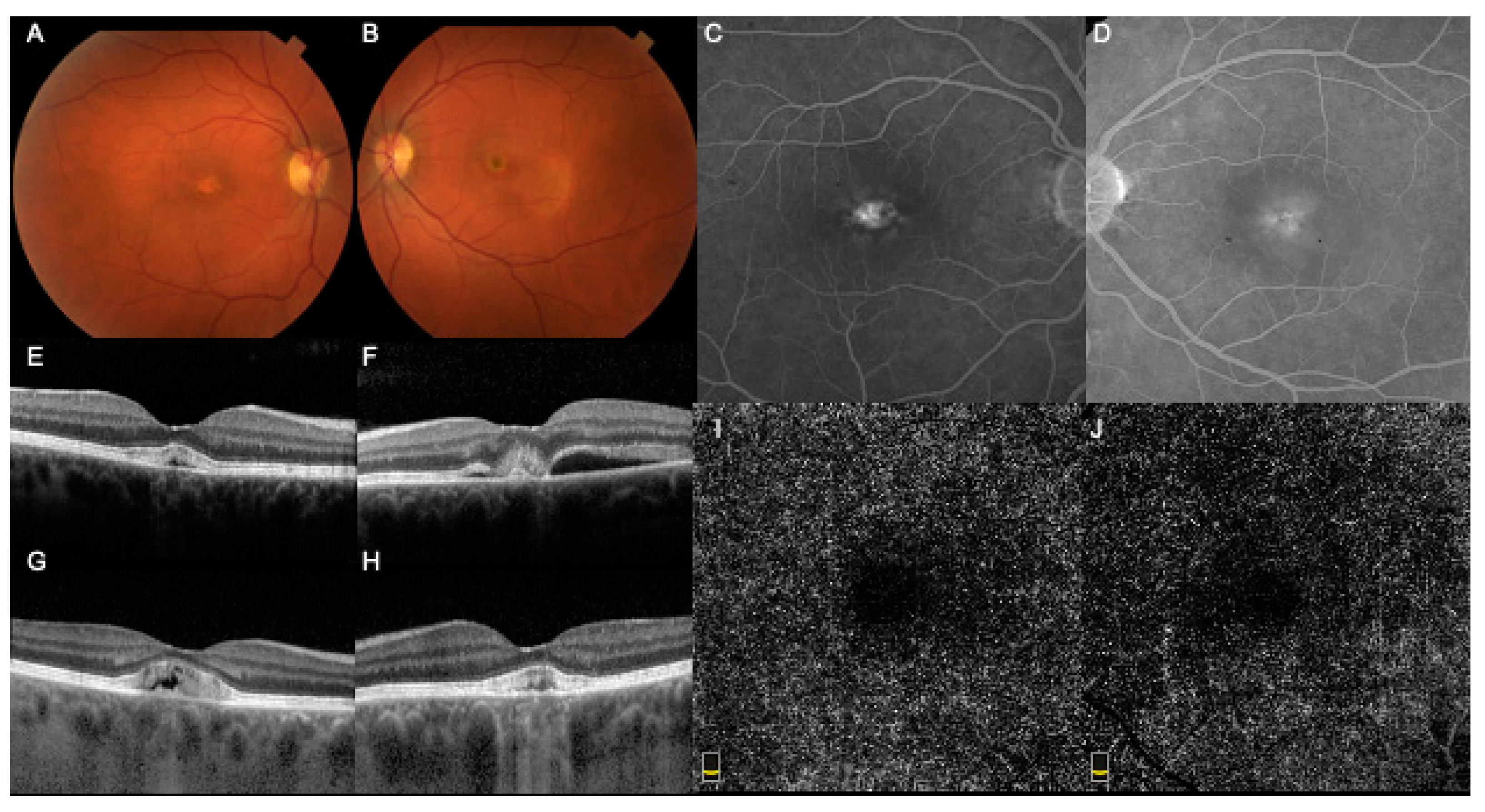
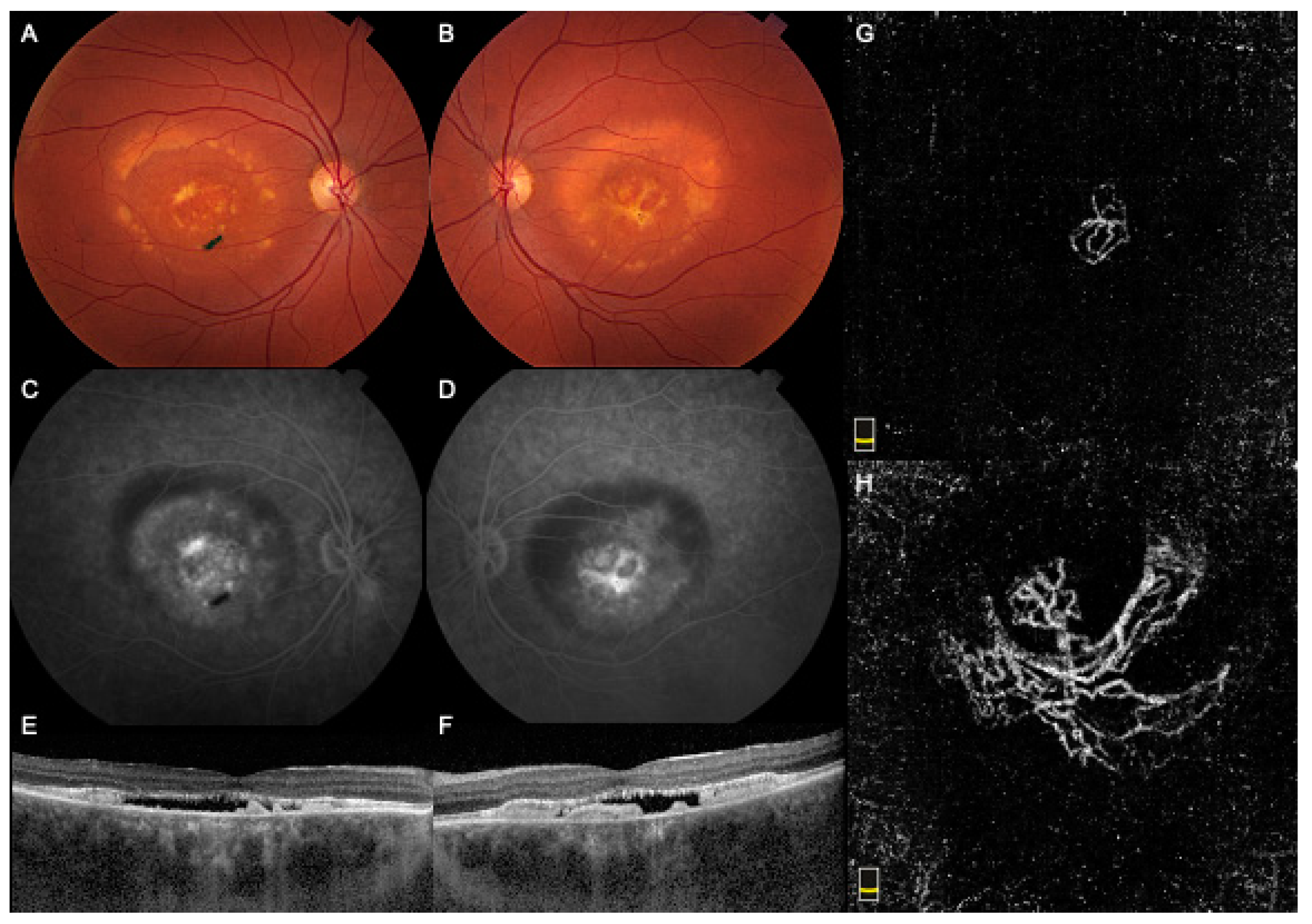
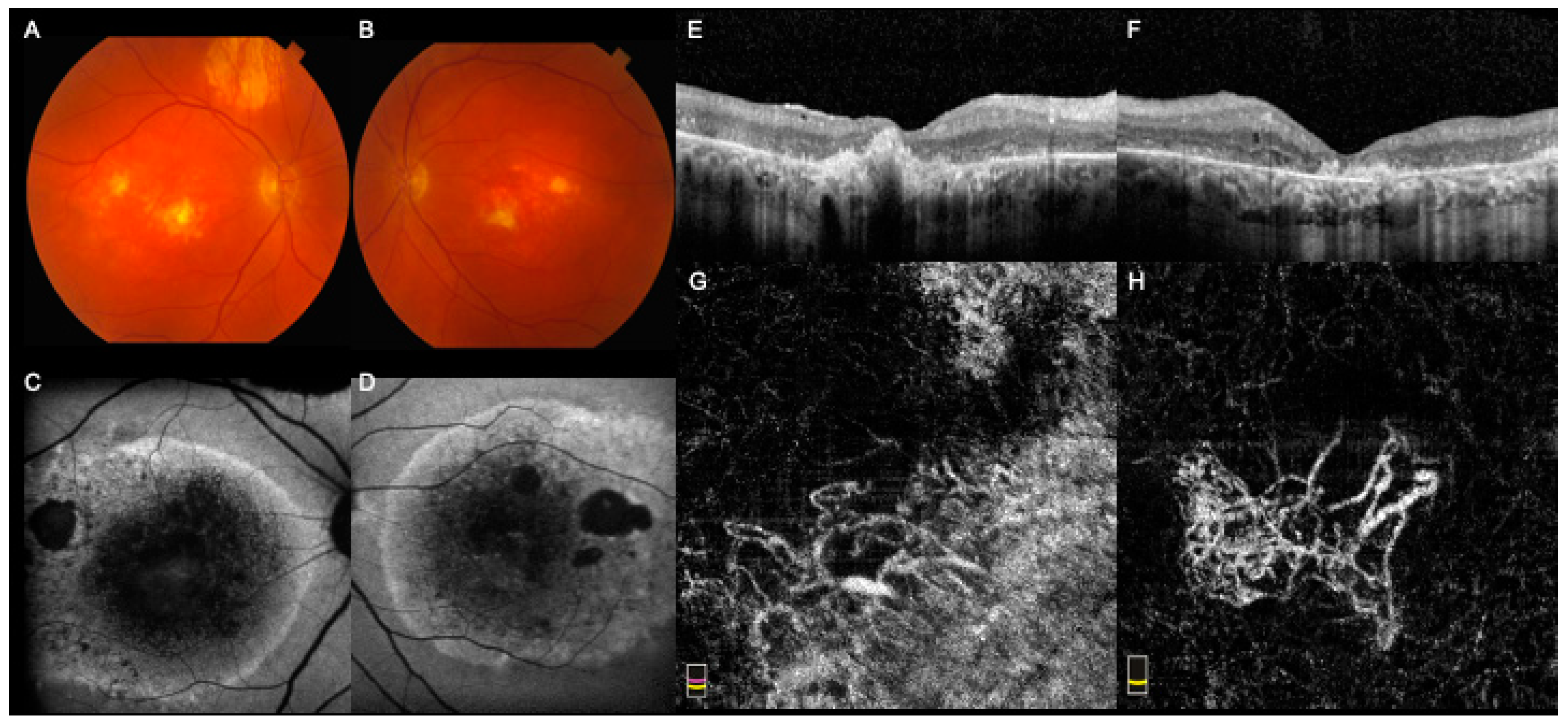
The Role of Optical Coherence Tomography Angiography (OCTA) in Detecting Choroidal Neovascularization in Different Stages of Best Macular Dystrophy: A Case Series

Abstract
1. Introduction
2. Case Reports
3. Discussion
4. Conclusions
Author Contributions
Funding
Institutional Review Board Statement
Informed Consent Statement
Data Availability Statement
Conflicts of Interest
References
- Casalino, G.; Khan, K.N.; Armengol, M.; Wright, G.; Pontikos, N.; Georgiou, M.; Webster, A.R.; Robson, A.G.; Grewal, P.S.; Michaelides, M. Autosomal recessive bestrophinopathy: clinical features, natural history and genetic findings in preparation for clinical trials. Ophthalmology 2020. [Google Scholar] [CrossRef] [Scilit] [PubMed]
- MacDonald, I.M.; Lee, T. Best Vitelliform Macular Dystrophy. In GeneReviews((R)); Adam, M.P., Ardinger, H.H., Pagon, R.A., Wallace, S.E., Bean, L.J.H., Stephens, K., Amemiya, A., Eds.; University of Washington: Seattle, WA, USA, 1993. [Google Scholar]
- Battaglia Parodi, M.; Romano, F.; Arrigo, A.; Di Nunzio, C.; Buzzotta, A.; Alto, G.; Bandello, F. Natural course of the vitelliform stage in best vitelliform macular dystrophy: A five-year follow-up study. Graefes Arch. Clin. Exp. Ophthalmol. 2020, 258, 297–301. [Google Scholar] [CrossRef] [Scilit] [PubMed]
- Chhablani, J.; Jalali, S. Intravitreal bevacizumab for choroidal neovascularization secondary to Best vitelliform macular dystrophy in a 6-year-old child. Eur. J. Ophthalmol. 2012, 22, 677–679. [Google Scholar] [CrossRef] [Scilit] [PubMed]
- Mandal, S.; Sinha, S.; Venkatesh, P.; Vashisht, N. Intravitreal bevacizumab in choroidal neovascularization associated with Best’s vitelliform dystrophy. Indian J. Ophthalmol. 2011, 59, 262–263. [Google Scholar] [CrossRef] [Scilit] [PubMed]
- Battaglia Parodi, M.; Romano, F.; Cicinelli, M.V.; Rabiolo, A.; Arrigo, A.; Pierro, L.; Iacono, P.; Bandello, F. Retinal Vascular Impairment in Best Vitelliform Macular Dystrophy Assessed by Means of Optical Coherence Tomography Angiography. Am. J. Ophthalmol. 2018, 187, 61–70. [Google Scholar] [CrossRef] [Scilit] [PubMed]
- Wang, X.N.; You, Q.S.; Li, Q.; Li, Y.; Mao, Y.; Hu, F.; Zhao, H.Y.; Tsai, F.F.; Peng, X.Y. Findings of Optical Coherence Tomography Angiography in Best Vitelliform Macular Dystrophy. Ophthalmic Res. 2018, 60, 214–220. [Google Scholar] [CrossRef] [Scilit] [PubMed]
- Elbany, S.; Kodjikian, L.; Boucher, S.; Loria, O.; Burillon, C.; Mathis, T. Neovascularized Best Disease in Child: Contribution of Optical Coherence Tomography Angiography. Ophthalmic Surg. Lasers Imaging Retina 2019, 50, 597–601. [Google Scholar] [CrossRef] [Scilit]
- Parodi, M.B.; Arrigo, A.; Bandello, F. Optical Coherence Tomography Angiography Quantitative Assessment of Macular Neovascularization in Best Vitelliform Macular Dystrophy. Investig. Ophthalmol. Vis. Sci. 2020, 61, 61. [Google Scholar] [CrossRef] [Scilit] [PubMed]
- Querques, G.; Regenbogen, M.; Quijano, C.; Delphin, N.; Soubrane, G.; Souied, E.H. High-definition optical coherence tomography features in vitelliform macular dystrophy. Am. J. Ophthalmol. 2008, 146, 501–507. [Google Scholar] [CrossRef] [Scilit]
- Fishman, G.A.; Baca, W.; Alexander, K.R.; Derlacki, D.J.; Glenn, A.M.; Viana, M. Visual acuity in patients with best vitelliform macular dystrophy. Ophthalmology 1993, 100, 1665–1670. [Google Scholar] [CrossRef] [Scilit]
- Guduru, A.; Gupta, A.; Tyagi, M.; Jalali, S.; Chhablani, J. Optical coherence tomography angiography characterisation of Best disease and associated choroidal neovascularisation. Br. J. Ophthalmol. 2018, 102, 444–447. [Google Scholar] [CrossRef] [Scilit] [PubMed]
- Zatreanu, L.; Freund, K.B.; Leong, B.C.S.; Yu, H.G.; Teke, M.Y.; Yzer, S.; Sadda, S.R.; Sarraf, D. Serous Macular Detachment in Best Disease: A Masquerade Syndrome. Retina 2019. [Google Scholar] [CrossRef] [Scilit] [PubMed]
- Murro, V.; Mucciolo, D.P.; Giorgio, D.; Sodi, A.; Passerini, I.; Cipollini, F.; Virgili, G.; Giansanti, F. Optical coherence tomography angiography cyclic remodeling of CNV in patients affected by Best macular dystrophy. Ophthalmic Genet. 2020, 41, 440–447. [Google Scholar] [CrossRef] [Scilit] [PubMed]
- Spaide, R.F.; Fujimoto, J.G.; Waheed, N.K. Image Artifacts in Optical Coherence Tomography Angiography. Retina 2015, 35, 2163–2180. [Google Scholar] [CrossRef] [Scilit] [PubMed]

Publisher’s Note: MDPI stays neutral with regard to jurisdictional claims in published maps and institutional affiliations. |
© 2021 by the authors. Licensee MDPI, Basel, Switzerland. This article is an open access article distributed under the terms and conditions of the Creative Commons Attribution (CC BY) license (http://creativecommons.org/licenses/by/4.0/).
Share and Cite
Qaseem, Y.; German, O.; Cicinelli, M.V.; Mirza, R.G. The Role of Optical Coherence Tomography Angiography (OCTA) in Detecting Choroidal Neovascularization in Different Stages of Best Macular Dystrophy: A Case Series. Medicina 2021, 57, 213. https://doi.org/10.3390/medicina57030213
Qaseem Y, German O, Cicinelli MV, Mirza RG. The Role of Optical Coherence Tomography Angiography (OCTA) in Detecting Choroidal Neovascularization in Different Stages of Best Macular Dystrophy: A Case Series. Medicina. 2021; 57(3):213. https://doi.org/10.3390/medicina57030213
Chicago/Turabian StyleQaseem, Yaqoob, Olga German, Maria Vittoria Cicinelli, and Rukhsana G. Mirza. 2021. "The Role of Optical Coherence Tomography Angiography (OCTA) in Detecting Choroidal Neovascularization in Different Stages of Best Macular Dystrophy: A Case Series" Medicina 57, no. 3: 213. https://doi.org/10.3390/medicina57030213
APA StyleQaseem, Y., German, O., Cicinelli, M. V., & Mirza, R. G. (2021). The Role of Optical Coherence Tomography Angiography (OCTA) in Detecting Choroidal Neovascularization in Different Stages of Best Macular Dystrophy: A Case Series. Medicina, 57(3), 213. https://doi.org/10.3390/medicina57030213