Dermoscopic Features of Lichen Amyloidosis in Caucasians—A Case Series and Literature Review


Abstract
1. Introduction
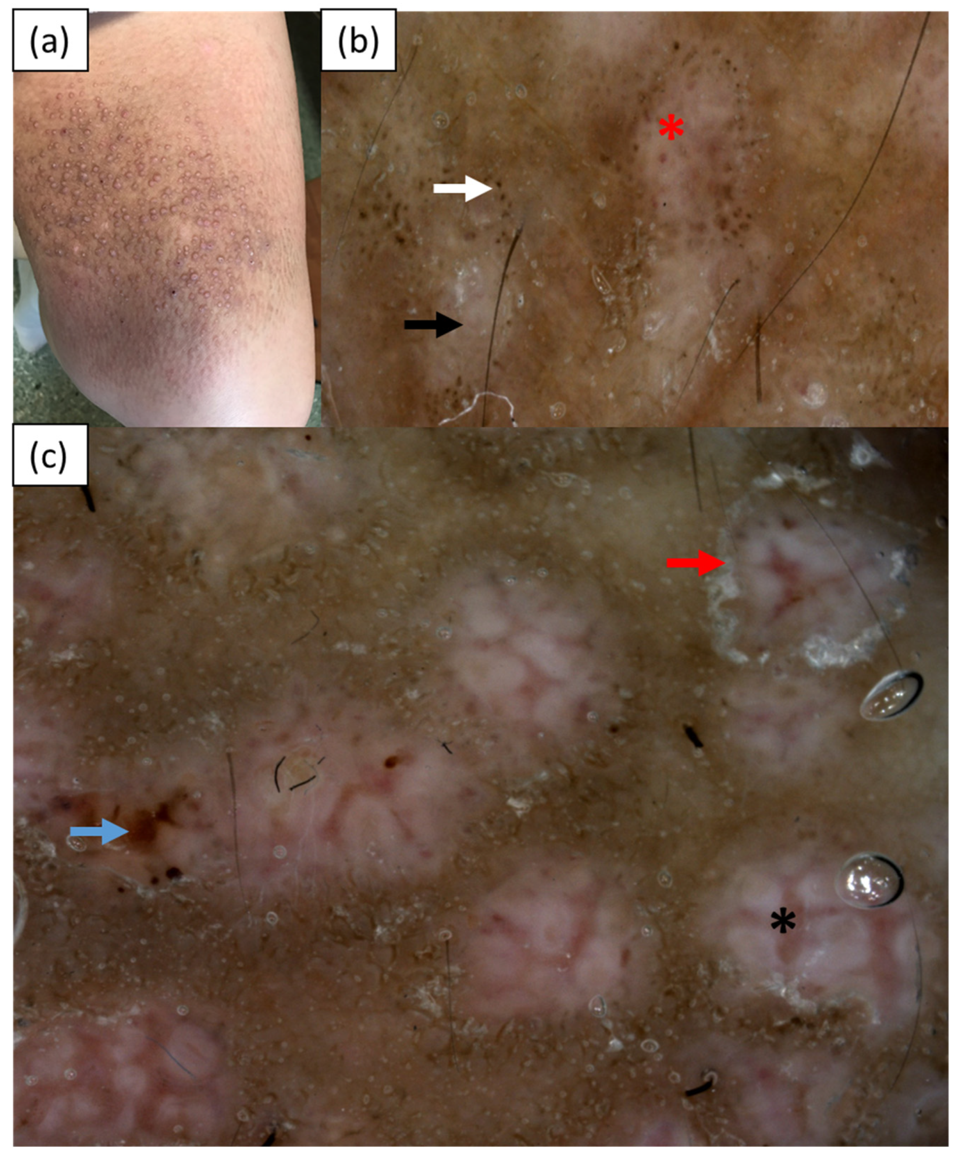
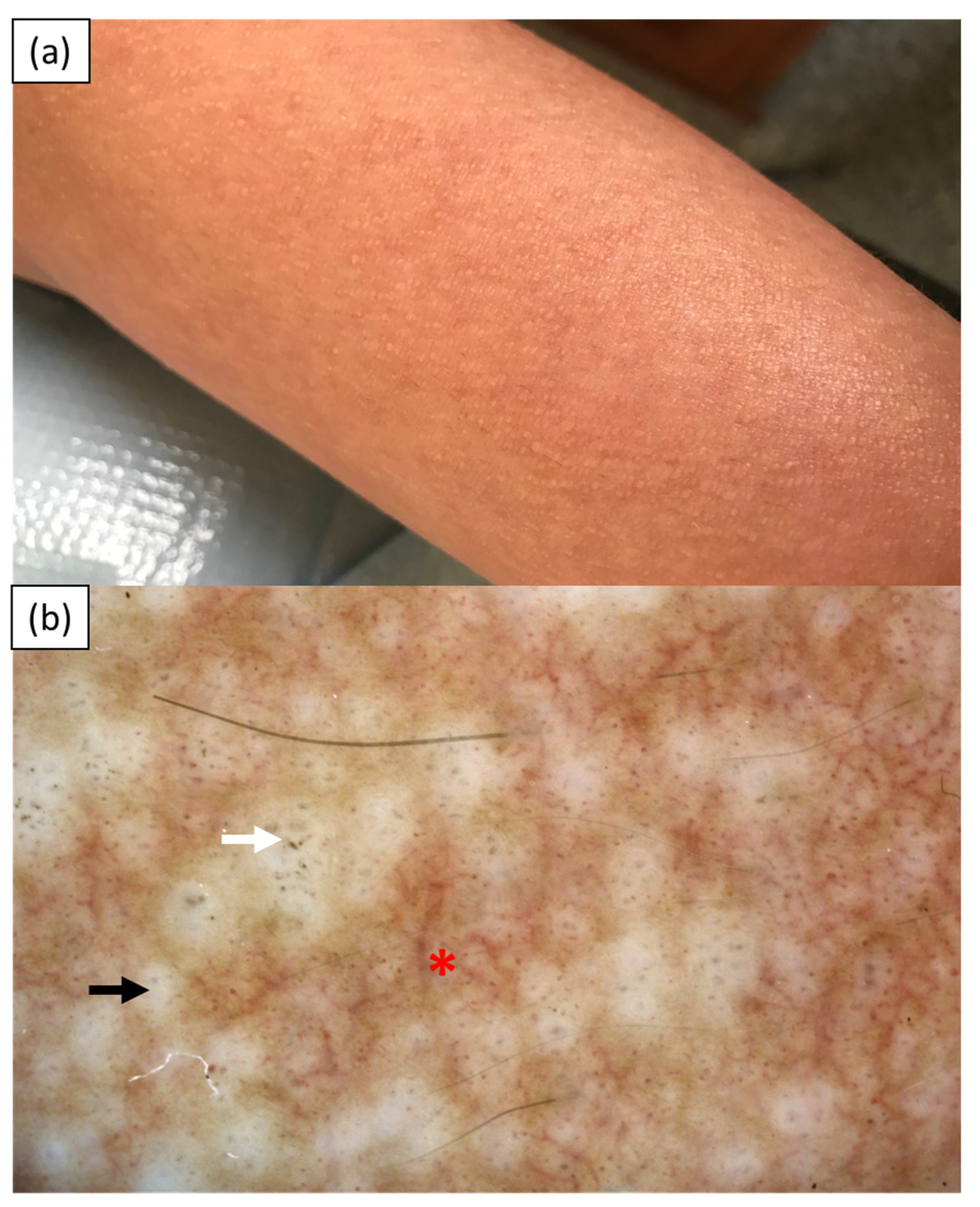
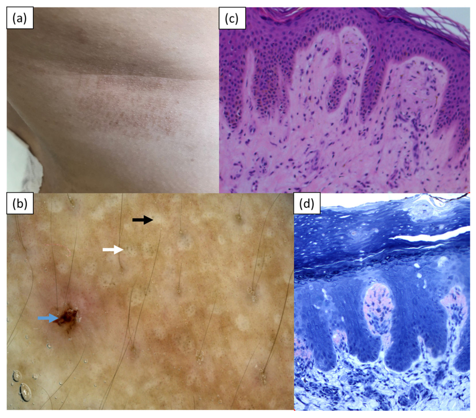
2. Case Reports
3. Discussion
4. Conclusions
Author Contributions
Funding
Institutional Review Board Statement
Informed Consent Statement
Data Availability Statement
Conflicts of Interest
References
- Arnold, S.J.; Bowling, J.C.R. ‘Shiny white streaks’ in lichen amyloidosis: A clue to diagnosis. Australas J. Dermatol. 2012, 53, 272–273. [Google Scholar] [CrossRef] [Scilit] [PubMed]
- Chuang, Y.Y.; Lee, D.D.; Lin, C.S.; Chang, Y.J.; Tanaka, M.; Chang, Y.T.; Liu, H.N. Characteristic dermoscopic features of primary cutaneous amyloidosis: A study of 35 cases. Br. J. Dermatol. 2012, 167, 548–554. [Google Scholar] [CrossRef] [Scilit] [PubMed]
- Wang, X.; Wang, H.; Zhong, Z.; Zheng, L.; Wang, Y.; Guo, Z.; Li, H.; Gao, M. Case report: Diagnosis of primary cutaneous amyloidosis using dermoscopy and reflectance confocal microscopy. Front. Med 2021, 7, 619907. [Google Scholar] [CrossRef] [Scilit] [PubMed]
- Rongioletti, F.; Atzori, L.; Ferreli, C.; Pinna, A.; Aste, N.; Pau, M. A unique dermoscopy pattern of primary cutaneous nodular amyloidosis mimicking a granulomatous disease. J. Am. Acad. Dermatol. 2016, 74, e9–e10. [Google Scholar] [CrossRef] [Scilit] [PubMed]
- Di Meo, N.; Noal, C.; Fadel, M.; Trevisan, G. Yellow teardrop-like structures in primary nodular skin amyloidosis. G. Ital. Derm. Venereol. 2018, 153, 118–119. [Google Scholar]
- Wang, L.; Jiang, X.; Zhang, N.; Liu, L.; Zhou, H.; Liu, H.J. Case of amyloidosis cutis dyschromica with dermoscopy. J. Dermatol. 2019, 46, e77–e79. [Google Scholar] [CrossRef] [Scilit] [PubMed]
- Moscarella, E.; Ronchi, A.; Agozzino, M.; Franco, R.; Argenziano, G. Image gallery: Dermoscopy of lichen amyloidosis. Br. J. Dermatol. 2018, 179, e231. [Google Scholar] [CrossRef] [Scilit] [PubMed]
- Kaliyadan, F.; Alkhateeb, A.; Kuruvilla, J.; Swaroop, K.; Alabdulsalam, A.A. Dermoscopy of primary cutaneous amyloidosis in skin of color. Dermatol. Pr. Concept 2019, 9, 232–234. [Google Scholar] [CrossRef] [Scilit] [PubMed]
- Ferreira, I.L.O.; Fernandes, E.L.; Lapins, J.; Benini, T.; Silva, L.C.; Lanzoni, M.A.; Steiner, D. Primary localized cutaneous nodular amyloidosis on a toe: Clinical presentation, histopathology, and dermoscopy findings. Dermatol. Pr. Concept 2019, 9, 235–236. [Google Scholar] [CrossRef] [Scilit] [PubMed]
- Sonthalia, S.; Agrawal, M.; Sehgal, V.N. Dermoscopy of macular amyloidosis. Indian Dermatol. Online J. 2020, 12, 203–205. [Google Scholar] [PubMed]
- Madarkar, M.S.; Koti, V.R. FotoFinder dermoscopy analysis and histopathological correlation in primary localized cutaneous amyloidosis. Dermatol. Pr. Concept 2021, 11, e2021057. [Google Scholar] [CrossRef] [Scilit] [PubMed]
- Behera, B.; Kumari, R.; Mohan Thappa, D.; Gochhait, D.; Hanuman Srinivas, B.; Ayyanar, P. Dermoscopic features of primary cutaneous amyloidosis in skin of colour: A retrospective analysis of 48 patients from South India. Australas J. Dermatol. 2021, 6. [Google Scholar] [CrossRef] [Scilit]



| Lichen Amyloidosis | ||||||
|---|---|---|---|---|---|---|
| Arnold et al., 2012 (India/NA) n = 2 | Chuang et al., 2012 (Taiwan) n = 17 | Moscarella et al., 2018 (Italy) n = 1 | Madarkar et al., 2021 (India) n = 18 | Behera et al., 2021 (India) n = 30 | Current cases (Poland) n = 3 | |
| Type of examination | polarized | Polarized and nonpolarized | N/A | Contact polarized | Contact nonpolarized | Contact nonpolarized |
| Two-zone pattern | - | - | - | - | 25 | - |
| Jigsaw puzzle pattern | - | - | - | - | 1 | - |
| Ridge and groove pattern | - | - | - | - | 4 | - |
| Hub and spoke pattern | - | - | - | - | 5 | - |
| Central hub | - | - | - | - | - | 3 |
| -white | - | 7 | 1 | 5 | - | 3 |
| -brown | - | - | - | 2 | - | - |
| -white and brown | - | - | - | 1 | - | - |
| White scar-like center | - | 10 | - | 10 | - | - |
| White structureless areas | - | 7 | - | 10 | - | - |
| Pigment dots/globules/ peppering | - | 3 | 1 | - | 30 | 3 |
| -clustered | - | - | - | - | 18 | 2 |
| -discrete | - | - | - | - | 23 | 1 |
| -starbust | - | - | - | - | 12 | - |
| -perieccrine | - | - | - | - | 3 | - |
| Blue-grey ovoid nest-like area | - | - | - | - | 5 | - |
| Concentric structures | - | - | - | - | 5 | - |
| White keratotic areas | - | - | - | - | 5 | - |
| Rim of white collarette | - | 3 | - | present | - | 1 |
| Diffuse white scaling | - | - | - | present | - | - |
| Comedo-like opening | - | - | - | - | 1 | - |
| Milia-like cyst | - | - | - | - | 1 | - |
| Red/purple dots and globules | - | - | - | - | 7 | 2 |
| Thick branching vessels | - | - | - | - | - | 2 |
| Shiny white streaks | 2 | - | - | - | - | - |
| Rosettes | - | - | - | 1 | - | - |
| Grayish-brown background | - | - | - | present | - | 1 |
| ‘Daily lily flower’ structures | - | - | - | present | - | - |
| Erosions | - | - | - | - | - | 2 |
| Macular Amyloidosis | ||||||
| Chuang et al., 2012 (Taiwan) n = 18 | Madarkar et al., 2021 (India) n = 12 | Wang et al., 2021 (China) n = 1 | Behera et al., 2021 (India) n = 18 | Sonthalia et al., 2020 (India) n = 2 | ||
| Type of examination | Polarized and nonpolarized | Contact polarized | NA | Contact nonpolarized | polarized | |
| Jigsaw puzzle pattern | - | - | - | 17 | - | |
| Ridge and groove pattern | - | - | - | 8 | - | |
| Hub and spoke pattern | - | - | - | 4 | 1 | |
| Central hub | 18 | - | 1 | - | 1 | |
| -white | 11 | 3 | - | - | 1 | |
| -brown | 4 | 8 | - | - | 1 | |
| -white and brown | 3 | 1 | - | - | 1 | |
| Pigment dots/globules/ peppering | - | 5 | 1 | 18 | - | |
| -clustered | - | - | - | 17 | - | |
| -discrete | - | - | - | 10 | - | |
| -starbust | - | - | - | 8 | - | |
| -perieccrine | - | - | - | 7 | - | |
| Peripheral pigmentation (fine streaks/leaf-like extensions/bulbous projections) | 18 | - | - | - | 2 | |
| Semicircular hyperpigmented structures | - | 10 | - | - | - | |
| Blue-grey ovoid nest-like area | - | - | - | 1 | - | |
| ‘daily lily flower’ structures | - | 2 | - | - | - | |
| Erythematous background | - | 3 | - | - | - | |
| Perifollicular depigmented halo | - | - | - | - | 1 | |
| Fine white scales | - | - | - | - | 1 | |
| White-to-off white colored intersecting lines | - | - | - | - | 1 | |
| white-to-light brown colored structureless areas | - | - | - | - | 1 | |
| Nodular Amyloidosis | ||||||
| Rongioletti et al., 2016 (Italy) n = 1 | Ferreira et al., 2019 (Brazil) n = 1 | DI Meo et al., 2018 (Italy) n = 1 | ||||
| Type of examination | polarized | polarized | NA | |||
| Shiny white streaks | - | 1 | - | |||
| Orange-yellowish homogenous background | 1 | - | - | |||
| Yellow teardrop-like structures | - | - | 1 | |||
| Orange-pink background | - | 1 | - | |||
| Elongated teleangiectasias | 1 | - | - | |||
| Serpentine teleangiectasias | 1 | - | - | |||
| Biphasic amyloidosis | ||||||
| Kaliyadan et al., 2019 (Saudi Arabia) n = 1 | Wang et al., 2021 (China) n = 1 | |||||
| Type of examination | polarized | NA | ||||
| Brown stripes | - | 1 | ||||
| Irregular brown center | - | 1 | ||||
| White central hub | 1 | 1 | ||||
| Dotted/globular pigmentation | 1 | 1 | ||||
| Amyloidosis Cutis Dyschromica | ||||||
| Wang et al., 2019 (China) n = 1 | ||||||
| Type of examination | NA | |||||
| Elliptical/round white spots | 1 | |||||
| Dotted/globular pigmentation | 1 | |||||
| Reticular pigment spots | 1 | |||||
| Striped hypopigmented spots | 1 | |||||
| Monotonous hypopigmented spots | 1 | |||||
Publisher’s Note: MDPI stays neutral with regard to jurisdictional claims in published maps and institutional affiliations. |
© 2021 by the authors. Licensee MDPI, Basel, Switzerland. This article is an open access article distributed under the terms and conditions of the Creative Commons Attribution (CC BY) license (https://creativecommons.org/licenses/by/4.0/).
Share and Cite
Żychowska, M.; Pięta, K.; Rudy, I.; Skubisz, A.; Reich, A. Dermoscopic Features of Lichen Amyloidosis in Caucasians—A Case Series and Literature Review. Medicina 2021, 57, 1027. https://doi.org/10.3390/medicina57101027
Żychowska M, Pięta K, Rudy I, Skubisz A, Reich A. Dermoscopic Features of Lichen Amyloidosis in Caucasians—A Case Series and Literature Review. Medicina. 2021; 57(10):1027. https://doi.org/10.3390/medicina57101027
Chicago/Turabian StyleŻychowska, Magdalena, Karolina Pięta, Izabela Rudy, Aleksandra Skubisz, and Adam Reich. 2021. "Dermoscopic Features of Lichen Amyloidosis in Caucasians—A Case Series and Literature Review" Medicina 57, no. 10: 1027. https://doi.org/10.3390/medicina57101027
APA StyleŻychowska, M., Pięta, K., Rudy, I., Skubisz, A., & Reich, A. (2021). Dermoscopic Features of Lichen Amyloidosis in Caucasians—A Case Series and Literature Review. Medicina, 57(10), 1027. https://doi.org/10.3390/medicina57101027