Eosinophilic Esophagitis as a Side Effect of Food Oral Immunotherapy

Abstract
1. Introduction
2. Food Allergy Oral Immunotherapy (OIT)
3. Eosinophilic Esophagitis
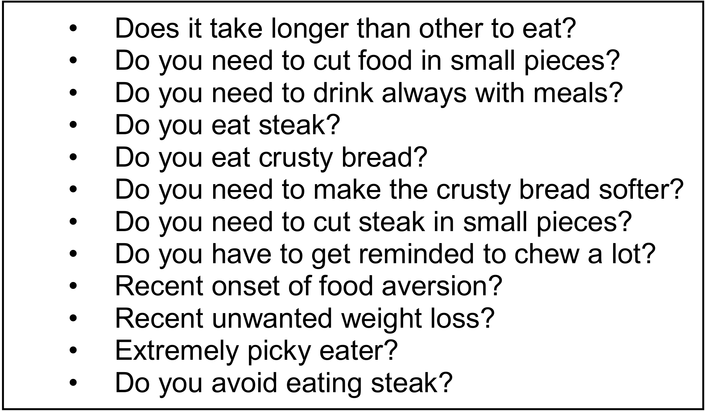
4. OIT Risk of Developing EoE
5. Conclusions
Funding
Conflicts of Interest
References
- Jones, S.M.; Burks, A.W.; Dupont, C. State of the art on food allergen immunotherapy: Oral, sublingual, and epicutaneous. J. Allergy Clin. Immunol. 2014, 133, 318–323. [Google Scholar] [CrossRef] [PubMed]
- Boyce, J.A.; Assa’a, A.; Burks, A.W.; Jones, S.M.; Sampson, H.A.; Wood, R.A.; Plaut, M.; Cooper, S.F.; Fenton, M.J.; Arshad, S.H.; et al. Guidelines for the diagnosis and management of food allergy in the United States: Summary of the NIAID-Sponsored Expert Panel Report. Nutrition 2011, 27, 253–267. [Google Scholar] [CrossRef] [PubMed]
- Joo Chan , C.; Richardo, T.; Lim, R.L.H. Current Trend in Immunotherapy for Peanut Allergy. Int. Rev. Immunol. 2018, 37, 279–290. [Google Scholar] [CrossRef]
- Hill, D.A.; Grundmeier, R.W.; Ram, G.; Spergel, J.M. The epidemiologic characteristics of healthcare provider-diagnosed eczema, asthma, allergic rhinitis, and food allergy in children: A retrospective cohort study. BMC Pediatr. 2016, 16, 133. [Google Scholar] [CrossRef] [PubMed]
- Cianferoni, A.; Spergel, J.M. Food allergy: Review, classification and diagnosis. Allergol. Int. 2009, 58, 457–466. [Google Scholar] [CrossRef] [PubMed]
- Cianferoni, A.; Ruffner, M.A.; Guzek, R.; Guan, S.; Brown-Whitehorn, T.; Muir, A.; Spergel, J.M. Elevated expression of activated TH2 cells and milk-specific TH2 cells in milk-induced eosinophilic esophagitis. Ann. Allergy Asthma Immunol. 2018, 120, 177–183. [Google Scholar] [CrossRef]
- Sampson, H.A. Food allergy. Part 2: Diagnosis and management. J. Allergy Clin. Immunol. 1999, 103, 981–989. [Google Scholar] [CrossRef]
- Sampson, H.A. Food allergy. Part 1: Immunopathogenesis and clinical disorders. J. Allergy Clin. Immunol. 1999, 103, 717–728. [Google Scholar] [CrossRef]
- Nowak-Wegrzyn, A.; Sampson, H.A. Adverse reactions to foods. Med. Clin. N. Am. 2006, 90, 97–127. [Google Scholar] [CrossRef] [PubMed]
- Lee, L.A.; Burks, A.W. Food allergies: Prevalence, molecular characterization, and treatment/prevention strategies. Annu. Rev. Nutr. 2006, 26, 539–565. [Google Scholar] [CrossRef] [PubMed]
- Cianferoni, A.; Muraro, A. Food-induced anaphylaxis. Immunol. Allergy Clin. No. Am. 2012, 32, 165–195. [Google Scholar] [CrossRef] [PubMed]
- Dellon, E.S.; Gonsalves, N.; Hirano, I.; Furuta, G.T.; Liacouras, C.A.; Katzka, D.A. ACG clinical guideline: Evidenced based approach to the diagnosis and management of esophageal eosinophilia and eosinophilic esophagitis (EoE). Am. J. Gastroenterol. 2013, 108, 679–692. [Google Scholar] [CrossRef] [PubMed]
- Cianferoni, A.; Spergel, J. Eosinophilic Esophagitis: A Comprehensive Review. Clin. Rev. Allergy Immunol. 2016, 50, 159–174. [Google Scholar] [CrossRef] [PubMed]
- Goswami, R.; Blazquez, A.B.; Kosoy, R.; Rahman, A.; Nowak-Wegrzyn, A.; Berin, M.C. Systemic innate immune activation in food protein-induced enterocolitis syndrome. J. Allergy Clin. Immunol. 2017, 139, 1885–1896. [Google Scholar] [CrossRef] [PubMed]
- Turner, P.J.; Jerschow, E.; Umasunthar, T.; Lin, R.; Campbell, D.E.; Boyle, R.J. Fatal Anaphylaxis: Mortality Rate and Risk Factors. J. Allergy Clin. Immunol. Pract. 2017, 5, 1169–1178. [Google Scholar] [CrossRef]
- Cianferoni, A.; Novembre, E.; Mugnaini, L.; Lombardi, E.; Bernardini, R.; Pucci, N.; Vierucci, A. Clinical features of severe acute anaphylaxis in patients admitted to an university hospital: An 11 year retrospective review (1985–1996). J. Allergy Clin. Immunol. 2001, 107, S57. [Google Scholar]
- Cianferoni, A.; Novembre, E.; Mugnaini, L.; Lombardi, E.; Bernardini, R.; Pucci, N.; Vierucci, A. Clinical features of acute anaphylaxis in patients admitted to a university hospital: An 11-year retrospective review (1985–1996). Ann. Allergy Asthma Immunol. 2001, 87, 27–32. [Google Scholar] [CrossRef]
- Cianferoni, A.; Novembre, E.; Pucci, N.; Lombardi, E.; Bernardini, R.; Vierucci, A. Anaphylaxis: A 7-year follow-up survey of 46 children. Ann. Allergy Asthma Immunol. 2004, 92, 464–468. [Google Scholar] [CrossRef]
- Lee, J.; Rodio, B.; Lavelle, J.; Lewis, M.O.; English, R.; Hadley, S.; Molnar, J.; Jacobstein, C.; Cianferoni, A.; Spergel, J.; et al. Improving Anaphylaxis Care: The Impact of a Clinical Pathway. Pediatrics 2018, 141, e20171616. [Google Scholar] [CrossRef]
- Novembre, E.; Cianferoni, A.; Bernardini, R.; Mugnaini, L.; Caffarelli, C.; Cavagni, G.; Giovane, A.; Vierucci, A. Anaphylaxis in children: Clinical and allergologic features. Pediatrics 1998, 101, e8. [Google Scholar] [CrossRef]
- Spergel, J.M.; Beausoleil, J.L.; Fiedler, J.M.; Ginsberg, J.; Wagner, K.; Pawlowski, N.A. Correlation of initial food reactions to observed reactions on challenges. Ann. Allergy Asthma Immunol. 2004, 92, 217–224. [Google Scholar] [CrossRef]
- Mori, F.; Cianferoni, A.; Brambilla, A.; Barni, S.; Sarti, L.; Pucci, N.; de Martino, M.; Novembre, E. Side effects and their impact on the success of milk oral immunotherapy (OIT) in children. Int. J. Immunopathol. Pharmacol. 2017, 30, 182–187. [Google Scholar] [CrossRef] [PubMed]
- Saltzman, R.W.; Kelleher, M.; Brown-Whitehorn, T.; Fiedler, J.; Corry, J.; Gober, L.; Dudek, E.; Cianferoni, A.; Spergel, J. Milk Oral Immunotherapy: A Single-Center Pilot Study of Safety and Efficacy. J. Allergy Clin. Immunol. 2012, 129, AB25. [Google Scholar] [CrossRef]
- Pajno, G.B.; Fernandez-Rivas, M.; Arasi, S.; Roberts, G.; Akdis, C.A.; Alvaro-Lozano, M.; Beyer, K.; Bindslev-Jensen, C.; Burks, W.; Ebisawa , M.; et al. EAACI Guidelines on allergen immunotherapy: IgE-mediated food allergy. Allergy 2018, 73, 799–815. [Google Scholar] [CrossRef]
- PALISADE Group of Clinical Investigators. AR101 Oral Immunotherapy for Peanut Allergy. N. Engl. J. Med. 2018, 379, 1991–2001. [Google Scholar] [CrossRef]
- Bird, J.A.; Spergel, J.M.; Jones, S.M.; Rachid, R.; Assa’ad, A.H.; Wang, J.; Leonard, S.A.; Laubach, S.S.; Kim, E.H.; Vickery, B.P.; et al. Efficacy and Safety of AR101 in Oral Immunotherapy for Peanut Allergy: Results of ARC001, a Randomized, Double-Blind, Placebo-Controlled Phase 2 Clinical Trial. J. Allergy Clin. Immunol. Pract. 2017, 6, 476–485. [Google Scholar] [CrossRef]
- Buchanan, A.D.; Green, T.D.; Jones, S.M.; Scurlock, A.M.; Christie, L.; Althage, K.A.; Steele, P.H.; Pons, L.; Helm, R.M.; Lee, L.A.; et al. Egg oral immunotherapy in nonanaphylactic children with egg allergy. J. Allergy Clin. Immunol. 2007, 119, 199–205. [Google Scholar] [CrossRef]
- Burks, A.W.; Jones, S.M. Egg oral immunotherapy in non-anaphylactic children with egg allergy: Follow-up. J. Allergy Clin. Immunol. 2008, 121, 270–271. [Google Scholar] [CrossRef]
- Burks, A.W.; Jones, S.M.; Wood, R.A.; Fleischer, D.M.; Sicherer, S.H.; Lindblad, R.W.; Stablein, D.; Henning, A.K.; Vickery, B.P.; Liu, A.H.; et al. Oral immunotherapy for treatment of egg allergy in children. N. Engl. J. Med. 2012, 367, 233–243. [Google Scholar] [CrossRef]
- Jones, S.M.; Burks, A.W.; Keet, C.; Vickery, B.P.; Scurlock, A.M.; Wood, R.A.; Liu, A.H.; Sicherer, S.H.; Henning, A.K.; Lindblad, R.W.; et al. Long-term treatment with egg oral immunotherapy enhances sustained unresponsiveness that persists after cessation of therapy. J. Allergy Clin. Immunol. 2016, 137, 1117–1127. [Google Scholar] [CrossRef]
- Narisety, S.D.; Frischmeyer-Guerrerio, P.A.; Keet, C.A.; Gorelik, M.; Schroeder, J.; Hamilton, R.G.; Wood, R.A. A randomized, double-blind, placebo-controlled pilot study of sublingual versus oral immunotherapy for the treatment of peanut allergy. J. Allergy Clin. Immunol. 2016, 137, 1117–1127. [Google Scholar]
- Narisety, S.D.; Skripak, J.M.; Steele, P.; Hamilton, R.G.; Matsui, E.C.; Burks, A.W.; Wood, R.A. Open-label maintenance after milk oral immunotherapy for IgE-mediated cow’s milk allergy. J. Allergy Clin. Immunol. 2009, 124, 610–612. [Google Scholar] [CrossRef]
- Pajno, G.B.; Caminiti, L.; Ruggeri, P.; De Luca, R.; Vita, D.; La Rosa, M.; Passalacqua, G. Oral immunotherapy for cow’s milk allergy with a weekly up-dosing regimen: A randomized single-blind controlled study. Ann. Allergy Asthma Immunol. 2010, 105, 376–381. [Google Scholar] [CrossRef] [PubMed]
- Skripak, J.M.; Nash, S.D.; Rowley, H.; Brereton, N.H.; Oh, S.; Hamilton, R.G.; Matsui, E.C.; Burks, A.W.; Wood, R.A. A randomized, double-blind, placebo-controlled study of milk oral immunotherapy for cow’s milk allergy. J. Allergy Clin. Immunol. 2008, 122, 1154–1160. [Google Scholar] [CrossRef] [PubMed]
- Still, L.M.; Jones, S.M. Sustained unresponsiveness to peanut in subjects who have completed peanut oral immunotherapy. Pediatrics 2014, 134 (Suppl. S3), S155. [Google Scholar] [CrossRef]
- Varshney, P.; Jones, S.M.; Scurlock, A.M.; Perry, T.T.; Kemper, A.; Steele, P.; Hiegel, A.; Kamilaris, J.; Carlisle, S.; Yue, X.; et al. A randomized controlled study of peanut oral immunotherapy: Clinical desensitization and modulation of the allergic response. J. Allergy Clin. Immunol. 2011, 127, 654–660. [Google Scholar] [CrossRef]
- Vickery, B.P.; Scurlock, A.M.; Kulis, M.; Steele, P.H.; Kamilaris, J.; Berglund, J.P.; Burk, C.; Hiegel, A.; Carlisle, S.; Christie, L.; et al. Sustained unresponsiveness to peanut in subjects who have completed peanut oral immunotherapy. J. Allergy Clin. Immunol. 2014, 133, 468–475. [Google Scholar] [CrossRef]
- Cianferoni, A.; Saltzman, R.; Saretta, F.; Barni, S.; Dudek, E.; Kelleher, M.; Spergel, J.M. Invariant natural killer cells change after an oral allergy desensitization protocol for cow’s milk. Clin. Exp. Allergy 2017, 47, 1390–1397. [Google Scholar] [CrossRef]
- Longo, G.; Barbi, E.; Berti, I.; Meneghetti, R.; Pittalis, A.; Ronfani, L.; Ventura, A. Specific oral tolerance induction in children with very severe cow’s milk-induced reactions. J. Allergy Clin. Immunol. 2008, 121, 343–347. [Google Scholar] [CrossRef]
- Salmivesi, S.; Korppi, M.; Makela, M.J.; Paassilta, M. Milk oral immunotherapy is effective in school-aged children. Acta Paediatr. 2013, 102, 172–176. [Google Scholar] [CrossRef]
- Martorell, A.; De la Hoz, B.; Ibanez, M.D.; Bone, J.; Terrados, M.S.; Michavila, A.; Plaza, A.M.; Alonso, E.; Garde, J.; Nevot, S.; et al. Oral desensitization as a useful treatment in 2-year-old children with cow’s milk allergy. Clin. Exp. Allergy 2011, 41, 1297–1304. [Google Scholar] [CrossRef] [PubMed]
- Varshney, P.; Steele, P.H.; Vickery, B.P.; Bird, J.A.; Thyagarajan, A.; Scurlock, A.M.; Perry, T.T.; Jones, S.M.; Burks, A.W. Adverse reactions during peanut oral immunotherapy home dosing. J. Allergy Clin. Immunol. 2009, 124, 1351–1352. [Google Scholar] [CrossRef] [PubMed]
- Dellon, E.S.; Jensen, E.T.; Martin, C.F.; Shaheen, N.J.; Kappelman, M.D. Prevalence of Eosinophilic Esophagitis in the United States. Clin. Gastroenterol. Hepatol. 2014, 12, 589–596. [Google Scholar] [CrossRef]
- Liacouras, C.A.; Furuta, G.T.; Hirano, I.; Atkins, D.; Attwood, S.E.; Bonis, P.A.; Burks, A.W.; Chehade, M.; Collins, M.H.; Dellon, E.S.; et al. Eosinophilic esophagitis: Updated consensus recommendations for children and adults. J. Allergy Clin. Immunol. 2011, 128, 3–20. [Google Scholar] [CrossRef] [PubMed]
- Spergel, J.M.; Andrews, T.; Brown-Whitehorn, T.F.; Beausoleil, J.L.; Liacouras, C.A. Treatment of eosinophilic esophagitis with specific food elimination diet directed by a combination of skin prick and patch tests. Ann. Allergy Asthma Immunol. 2005, 95, 336–343. [Google Scholar] [CrossRef]
- Spergel, J.M.; Brown-Whitehorn, T.F.; Beausoleil, J.L.; Franciosi, J.; Shuker, M.; Verma, R.; Liacouras, C.A. 14 years of eosinophilic esophagitis: Clinical features and prognosis. J. Pediatr. Gastroenterol. Nutr. 2009, 48, 30–36. [Google Scholar] [CrossRef] [PubMed]
- Noel, R.J.; Putnam, P.E.; Rothenberg, M.E. Eosinophilic esophagitis. N. Engl. J. Med. 2004, 351, 940–941. [Google Scholar] [CrossRef]
- Gonsalves, N.; Policarpio-Nicolas, M.; Zhang, Q.; Rao, M.S.; Hirano, I. Histopathologic variability and endoscopic correlates in adults with eosinophilic esophagitis. Gastrointest. Endosc. 2006, 64, 313–319. [Google Scholar] [CrossRef]
- Schaefer, E.T.; Fitzgerald, J.F.; Molleston, J.P.; Croffie, J.M.; Pfefferkorn, M.D.; Corkins, M.R.; Lim, J.D.; Steiner, S.J.; Gupta, S.K. Comparison of oral prednisone and topical fluticasone in the treatment of eosinophilic esophagitis: A randomized trial in children. Clin. Gastroenterol. Hepatol. 2008, 6, 165–173. [Google Scholar] [CrossRef]
- Lucendo, A.J.; Molina-Infante, J.; Arias, Á.; von Arnim, U.; Bredenoord, A.J.; Bussmann, C.; Dias, J.A.; Bove, M.; González-Cervera, J.; Larsson, H.; et al. Guidelines on eosinophilic esophagitis: evidence-based statements and recommendations for diagnosis and management in children and adults. United Eur. Gastroenterol. J. 2017, 5, 335–358. [Google Scholar] [CrossRef]
- Furuta, G.T.; Liacouras, C.A.; Collins, M.H.; Gupta, S.K.; Justinich, C.; Putnam, P.E.; Bonis, P.; Hassall, E.; Straumann, A.; Rothenberg, M.E.; et al. Eosinophilic esophagitis in children and adults: A systematic review and consensus recommendations for diagnosis and treatment. Gastroenterology 2007, 133, 1342–1363. [Google Scholar]
- Markowitz, J.E.; Spergel, J.M.; Ruchelli, E.; Liacouras, C.A. Elemental diet is an effective treatment for eosinophilic esophagitis in children and adolescents. Am. J. Gastroenterol. 2003, 98, 777–782. [Google Scholar]
- Peterson, K.A.; Byrne, K.R.; Vinson, L.A.; Ying, J.; Boynton, K.K.; Fang, J.C.; Gleich, G.J.; Adler, D.G.; Clayton, F. Elemental diet induces histologic response in adult eosinophilic esophagitis. Am. J. Gastroenterol. 2013, 108, 759–766. [Google Scholar]
- Warners, M.J.; Vlieg-Boerstra, B.J.; Verheij, J.; Van Hamersveld, P.H.; Van Rhijn, B.D.; Van Ampting, M.T.; Harthoorn, L.F.; de Jonge, W.J.; Smout, A.J.P.M.; Bredenoord, A.J. Esophageal and Small Intestinal Mucosal Integrity in Eosinophilic Esophagitis and Response to an Elemental Diet. Am. J. Gastroenterol. 2017, 112, 1061–1071. [Google Scholar] [PubMed]
- Warners, M.J.; Vlieg-Boerstra, B.J.; Verheij, J.; van Rhijn, B.D.; Van Ampting, M.T.J.; Harthoorn, L.F.; de Jonge, W.J.; Smout, A.J.P.M.; Bredenoord, A.J. Elemental diet decreases inflammation and improves symptoms in adult eosinophilic oesophagitis patients. Aliment. Pharmacol. Ther. 2017, 45, 777–787. [Google Scholar]
- Cianferoni, A.; Spergel, J. The importance of TSLP in allergic disease and its role as a potential therapeutic target. Expert Rev. Clin. Immunol. 2014, 10, 1463–1474. [Google Scholar]
- Godwin, B.; Liacouras, C.; Mehta , V.; Eisenberg, J.; Agawu, A.; Brown-Whitehorn, T.; Ruffner, M.A.; Verma, R.; Cianferoni, A.; Spergel, J.M.; et al. A Review of Tertiary Referrals for Management of Pediatric Esophageal Eosinophilia. Front. Pediatr. 2018, 20, 6–173. [Google Scholar] [CrossRef]
- Lucendo, A.J.; Arias, A.; Molina-Infante, J. Efficacy of Proton Pump Inhibitor Drugs for Inducing Clinical and Histologic Remission in Patients with Symptomatic Esophageal Eosinophilia: A Systematic Review and Meta-Analysis. Clin. Gastroenterol. Hepatol. 2016, 14, 13–22. [Google Scholar] [PubMed]
- Dellon, E.S.; Liacouras, C.A.; Molina-Infante, J.; Furuta, G.T.; Spergel, J.M.; Zevit, N.; Spechler, S.J.; Attwood, S.E.; Straumann, A.; Aceves, S.S.; et al. Updated international consensus diagnostic criteria for eosinophilic esophagitis: Proceedings of the AGREE conference. Gastroenterology 2018, 155, 1022–1033. [Google Scholar] [PubMed]
- Spergel, J.M.; Dellon, E.S.; Liacouras, C.A.; Hirano, I.; Molina-Infante, J.; Bredenoord, A.J.; Furuta, G.T. Summary of the updated international consensus diagnostic criteria for eosinophilic esophagitis: AGREE conference. Ann. Allergy Asthma Immunol. 2018, 121, 281–284. [Google Scholar]
- Liacouras, C.A.; Spergel, J.M.; Ruchelli, E.; Verma, R.; Mascarenhas, M.; Semeao, E.; Flick, J.; Kelly, J.; Brown-Whitehorn, T.; Mamula, P.; et al. Eosinophilic esophagitis: A 10-year experience in 381 children. Clin. Gastroenterol. Hepatol. 2005, 3, 1198–1206. [Google Scholar] [CrossRef]
- Caldwell, J.M.; Blanchard, C.; Collins, M.H.; Putnam, P.E.; Kaul, A.; Aceves, S.S.; Bouska, C.A.; Rothenberg, M.E. Glucocorticoid-regulated genes in eosinophilic esophagitis: A role for FKBP51. J. Allergy Clin. Immunol. 2010, 125, 879–888. [Google Scholar] [CrossRef] [PubMed]
- Aceves, S.S.; Bastian, J.F.; Newbury, R.O.; Dohil, R. Oral viscous budesonide: A potential new therapy for eosinophilic esophagitis in children. Am. J. Gastroenterol. 2007, 102, 2271–2279. [Google Scholar] [PubMed]
- Dohil, R.; Newbury, R.; Fox, L.; Bastian, J.; Aceves, S. Oral viscous budesonide is effective in children with eosinophilic esophagitis in a randomized, placebo-controlled trial. Gastroenterology 2010, 139, 418–429. [Google Scholar] [CrossRef] [PubMed]
- Simon, D.; Cianferoni, A.; Spergel, J.M.; Aceves, S.; Holbreich, M.; Venter, C.; Rothenberg, M.E.; Terreehorst, I.; Muraro, A.; Lucendo, A.J.; et al. Eosinophilic esophagitis is characterized by a non-IgE-mediated food hypersensitivity. Allergy 2016, 71, 611–620. [Google Scholar] [CrossRef]
- Spergel, J.M.; Brown-Whitehorn, T.F.; Cianferoni, A.; Shuker, M.; Wang, M.L.; Verma, R.; Liacouras, C.A. Identification of causative foods in children with eosinophilic esophagitis treated with an elimination diet. J. Allergy Clin. Immunol. 2012, 130, 461–467. [Google Scholar]
- Gonsalves, N.; Yang, G.Y.; Doerfler, B.; Ritz, S.; Ditto, A.M.; Hirano, I. Elimination diet effectively treats eosinophilic esophagitis in adults; food reintroduction identifies causative factors. Gastroenterology 2012, 142, 1451–1459. [Google Scholar] [CrossRef]
- Kagalwalla, A.F.; Sentongo, T.A.; Ritz, S.; Hess, T.; Nelson, S.P.; Emerick, K.M.; Melin-Aldana, H.; Li, B.U.K. Effect of six-food elimination diet on clinical and histologic outcomes in eosinophilic esophagitis. Clin. Gastroenterol. Hepatol. 2006, 4, 1097–1102. [Google Scholar] [CrossRef]
- Lucendo, A.J. Meta-Analysis-Based Guidance for Dietary Management in Eosinophilic Esophagitis. Curr. Gastroenterol. Rep. 2015, 17, 464. [Google Scholar] [CrossRef]
- Lucendo, A.J.; Arias, A.; Gonzalez-Cervera, J.; Yagüe-Compadre, J.L.; Guagnozzi, D.; Angueira, T.; Jiménez-Contreras, S.; González-Castillo, S.; Rodríguez-Domíngez, B.; De Rezende, L.C.; et al. Empiric 6-food elimination diet induced and maintained prolonged remission in patients with adult eosinophilic esophagitis: A prospective study on the food cause of the disease. J. Allergy Clin. Immunol. 2013, 131, 797–804. [Google Scholar]
- Molina-Infante, J.; Arias, A.; Alcedo, J.; Garcia-Romero, R.; Casabona-Frances, S.; Prieto-Garcia, A.; Modolell, I.; Gonzalez-Cordero, P.L.; Perez-Martinez, I.; Martin-Lorente, J.L.; et al. Step-up empiric elimination diet for pediatric and adult eosinophilic esophagitis: The 2-4-6 study. J. Allergy Clin. Immunol. 2018, 141, 1365–1372. [Google Scholar] [CrossRef] [PubMed]
- Molina-Infante, J.; Arias, A.; Barrio, J.; Rodriguez-Sanchez, J.; Sanchez-Cazalilla, M.; Lucendo, A.J. Four-food group elimination diet for adult eosinophilic esophagitis: A prospective multicenter study. J. Allergy Clin. Immunol. 2014, 134, 1093–1099. [Google Scholar] [CrossRef]
- Kelly, K.J.; Lazenby, A.J.; Rowe, P.C.; Yardley, J.H.; Perman, J.A.; Sampson, H.A. Eosinophilic esophagitis attributed to gastroesophageal reflux: Improvement with an amino acid-based formula. Gastroenterology 1995, 109, 1503–1512. [Google Scholar] [CrossRef]
- Cianferoni, A.; Shuker, M.; Brown-Whitehorn, T.; Hunter, H.; Venter, C.; Spergel, J.M. Food avoidance strategies in eosinophilic oesophagitis. Clin. Exp. Allergy 2019, 49, 269–284. [Google Scholar] [CrossRef] [PubMed]
- Spergel, J.; Aceves, S.S. Allergic components of eosinophilic esophagitis. J. Allergy Clin. Immunol. 2018, 142, 1–8. [Google Scholar] [CrossRef] [PubMed]
- Assa’ad, A.H.; Putnam, P.E.; Collins, M.H.; Akers, R.M.; Jameson, S.C.; Kirby, C.L.; Buckmeier, B.K.; Bullock, J.Z.; Collier, A.R.; Konikoff, M.R.; et al. Pediatric patients with eosinophilic esophagitis: An 8-year follow-up. J. Allergy Clin. Immunol. 2007, 119, 731–738. [Google Scholar] [CrossRef] [PubMed]
- Chehade, M.; Jones, S.M.; Pesek, R.D.; Burks, A.W.; Vickery, B.P.; Wood, R.A.; Leung, D.Y.M.; Furuta, G.T.; Fleischer, D.M.; Henning, A.K.; et al. Phenotypic Characterization of Eosinophilic Esophagitis in a Large Multi-Center Patient Population from the Consortium for Food Allergy Research. J. Allergy Clin. Immunol. Pract. 2018, 6, 1534–1544. [Google Scholar] [CrossRef]
- Blanchard, C.; Wang, N.; Rothenberg, M.E. Eosinophilic esophagitis: Pathogenesis, genetics, and therapy. J. Allergy Clin. Immunol. 2006, 118, 1054–1059. [Google Scholar] [CrossRef]
- Capucilli, P.; Cianferoni, A.; Grundmeier, R.W.; Spergel, J.M. Comparison of comorbid diagnoses in children with and without eosinophilic esophagitis in a large population. Ann. Allergy Asthma Immunol. 2018, 121, 711–716. [Google Scholar] [CrossRef]
- Wallace, D.V.; Dykewicz, M.S.; Bernstein, D.I.; Blessing-Moore, J.; Cox, L.; Khan, D.A.; Lang, D.M.; Nicklas, R.A.; Oppenheimer, J.; Portnoy, J.M.; et al. The diagnosis and management of rhinitis: An updated practice parameter. J. Allergy Clin. Immunol. 2008, 122, S1–S84. [Google Scholar] [CrossRef]
- Sicherer, S.H.; Sampson, H.A. Food allergy. J. Allergy Clin. Immunol. 2006, 117, S470–S475. [Google Scholar] [CrossRef] [PubMed]
- Adams, P.F.; Barnes, P.M.; Vickerie, J.L. Summary health statistics for the U.S. population: National Health Interview Survey, 2007. Vital Health Stat. 2008, 10, 1–104. [Google Scholar]
- Hill, D.A.; Dudley, J.W.; Spergel, J.M. The Prevalence of Eosinophilic Esophagitis in Pediatric Patients with IgE-Mediated Food Allergy. J. Allergy Clin. Immunol. Pract. 2017, 5, 369–375. [Google Scholar] [CrossRef] [PubMed]
- Maggadottir, S.M.; Hill, D.A.; Ruymann, K.; Brown-Whitehorn, T.F.; Cianferoni, A.; Shuker, M.; Wang, M.-L.; Chikwava, K.; Verma, R.; Liacouras, C.A.; et al. Resolution of acute IgE-mediated allergy with development of eosinophilic esophagitis triggered by the same food. J. Allergy Clin. Immunol. 2014, 133, 1487–1489. [Google Scholar] [CrossRef] [PubMed]
- Bene, J.; Ley, D.; Roboubi, R.; Gottrand, F.; Gautier, S. Eosinophilic esophagitis after desensitization to dust mites with sublingual immunotherapy. Ann. Allergy Asthma Immunol. 2016, 116, 583–584. [Google Scholar] [CrossRef]
- Miehlke, S.; Alpan, O.; Schroder, S.; Straumann, A. Induction of eosinophilic esophagitis by sublingual pollen immunotherapy. Case Rep. Gastroenterol. 2013, 7, 363–368. [Google Scholar] [CrossRef] [PubMed]
- Rokosz, M.; Bauer, C.; Schroeder, S. Eosinophilic esophagitis induced by aeroallergen sublingual immunotherapy in an enteral feeding tube-dependent pediatric patient. Ann. Allergy Asthma Immunol. 2017, 119, 88–89. [Google Scholar] [CrossRef]
- Lucendo, A.J.; Arias, A.; Tenias, J.M. Relation between eosinophilic esophagitis and oral immunotherapy for food allergy: A systematic review with meta-analysis. Ann. Allergy Asthma Immunol. 2014, 113, 624–629. [Google Scholar] [CrossRef]
- Petroni, D.; Spergel, J.M. Eosinophilic esophagitis and symptoms possibly related to eosinophilic esophagitis in oral immunotherapy. Ann. Allergy Asthma Immunol. 2018, 120, 237–240. [Google Scholar] [CrossRef]
- Echeverria-Zudaire, L.A.; Fernandez-Fernandez, S.; Rayo-Fernandez, A.; Munoz-Archidona, C.; Checa-Rodriguez, R. Primary eosinophilic gastrointestinal disorders in children who have received food oral immunotherapy. Allergol. Immunopathol. 2016, 44, 531–536. [Google Scholar] [CrossRef]
- Goldberg, M.R.; Elizur, A.; Nachshon, L.; Appel, M.Y.; Levy, M.B.; Golobov, K.; Goldberg, R.; Stein, M.; Rothenberg, M.E.; Katz, Y. Oral immunotherapy-induced gastrointestinal symptoms and peripheral blood eosinophil responses. J. Allergy Clin. Immunol. 2017, 139, 1388–1390. [Google Scholar] [CrossRef] [PubMed]
- Konikoff, M.R.; Blanchard, C.; Kirby, C.; Buckmeier, B.K.; Cohen, M.B.; Heubi, J.E.; Putnam, P.E.; Rothenberg, M.E. Potential of blood eosinophils, eosinophil-derived neurotoxin, and eotaxin-3 as biomarkers of eosinophilic esophagitis. Clin. Gastroenterol. Hepatol. 2006, 4, 1328–1336. [Google Scholar] [CrossRef] [PubMed]
- Burk, C.M.; Dellon, E.S.; Steele, P.H.; Virkud, Y.V.; Kulis, M.; Burks, A.W.; Vickery, B.P. Eosinophilic esophagitis during peanut oral immunotherapy with omalizumab. J. Allergy Clin. Immunol. Pract. 2017, 5, 498–501. [Google Scholar] [CrossRef] [PubMed]
- Wright, B.L.; Fernandez-Becker, N.Q.; Kambham, N.; Purington, N.; Tupa, D.; Zhang, W.; Rank, M.A.; Kita, H.; Shim, K.P.; Bunning, B.J.; et al. Baseline Gastrointestinal Eosinophilia Is Common in Oral Immunotherapy Subjects With IgE-Mediated Peanut Allergy. Front. Immunol. 2018, 9, 2624. [Google Scholar] [CrossRef] [PubMed]
- Wright, B.L.; Fernandez-Becker, N.Q.; Kambham, N.; Purington, N.; Cao, S.; Tupa, D.; Zhang, W.; Sindher, S.B.; Rank, M.A.; Kita, H.; et al. Gastrointestinal Eosinophil Responses in a Longitudinal, Randomized Trial of Peanut Oral Immunotherapy. Clin. Gastroenterol. Hepatol. 2020. [Google Scholar] [CrossRef]


| Author | Year | Food | Dose | Total pts | EoE | % |
|---|---|---|---|---|---|---|
| Babaie D | 2017 | Milk | 120 cc | 18 | 1 | 5.6 |
| Goldberg, | 2017 | Various | Various | 794 | 3 | 0.4 |
| Echeverría-Zudaire | 2015 | Milk and or egg | 8 g | 128 | 8 | 6.25 |
| MacGinnitie | 2017 | Peanut + Xolair | 4 g | 37 | 1 | 2.702703 |
| Palisade | 2018 | Peanut | 300 mg | 372 | 1 | 0.268817 |
| Burk CM | 2017 | Peanut + Xolair | 4 g | 13 | 2 | 15.4 |
| MacGinnitie | 2017 | Peanut + Xolair | 4 g | 37 | 1 | 2.702703 |
| Andorf, S | 2018 | Multifood- + Xolair | 1 g | 45 | 0 | 0 |
Publisher’s Note: MDPI stays neutral with regard to jurisdictional claims in published maps and institutional affiliations. |
© 2020 by the author. Licensee MDPI, Basel, Switzerland. This article is an open access article distributed under the terms and conditions of the Creative Commons Attribution (CC BY) license (http://creativecommons.org/licenses/by/4.0/).
Share and Cite
Cianferoni, A. Eosinophilic Esophagitis as a Side Effect of Food Oral Immunotherapy. Medicina 2020, 56, 618. https://doi.org/10.3390/medicina56110618
Cianferoni A. Eosinophilic Esophagitis as a Side Effect of Food Oral Immunotherapy. Medicina. 2020; 56(11):618. https://doi.org/10.3390/medicina56110618
Chicago/Turabian StyleCianferoni, Antonella. 2020. "Eosinophilic Esophagitis as a Side Effect of Food Oral Immunotherapy" Medicina 56, no. 11: 618. https://doi.org/10.3390/medicina56110618
APA StyleCianferoni, A. (2020). Eosinophilic Esophagitis as a Side Effect of Food Oral Immunotherapy. Medicina, 56(11), 618. https://doi.org/10.3390/medicina56110618