Abstract
There are various biomaterials, but none fulfills all requirements. Cellulose biopolymers have advanced biomedicine to satisfy high market demand and circumvent many ecological concerns. This review aims to present an overview of cellulose knowledge and technical biomedical applications such as antibacterial agents, antifouling, wound healing, drug delivery, tissue engineering, and bone regeneration. It includes an extensive bibliography of recent research findings from fundamental and applied investigations. Cellulose-based materials are tailorable to obtain suitable chemical, mechanical, and physical properties required for biomedical applications. The chemical structure of cellulose allows modifications and simple conjugation with several materials, including nanoparticles, without tedious efforts. They render the applications cheap, biocompatible, biodegradable, and easy to shape and process.
1. Introduction
The word ‘’biomedicine’’ refers to ‘’clinical medicine based on physiology and biochemistry’’ [1]. Biomedicine aims to provide new strategies for biosensing, bioimaging, biomimetic structures, and processes for the regeneration of tissues or organs [2,3,4,5]. It is a multidisciplinary research area involving biologists, chemists, biomaterials scientists, and physicists. An ideal biomaterial for biomedicine should meet specific requirements, including high biocompatibility (no toxicity), high hemocompatibility (no blood coagulation), good biodegradability and bio-absorbability, the promotion of cellular and tissue interactions, the facilitation of an interconnected porous structure, and good mechanical properties such as high wear resistance. Biomaterials should be able to sustain loads and generate tiny wear debris.
Cellulose is a natural linear structural biopolymer of anhydroglucose monomers linked via β-(1–4) bonds ((C6H10O5)n; n is the degree of polymerization; n equals 10,000–5000, depending on the source used for cellulose extraction) [6,7,8,9,10,11,12]. Plants, seaweeds, sugarcane bagasse, tunicate, marine algae, and bacteria are predominant sources of cellulose [13,14,15,16]. The annual production of cellulose is more than several hundred billion tons. The market demand has continuously increased over the years. Cellulose exhibits good mechanical, physical, and chemical properties, such as high stability under acidic conditions, chirality, high tensile strength, good elastic modulus (130–150 GPa), low density or lightweight (density of 1.6 g/cm3), high biodegradability, and abundant hydroxyl functional groups on their surfaces, enabling chemical modification with high wettability. Thus, cellulose has advanced energy, environmental, and health-based applications [17,18,19,20,21,22,23,24,25,26,27].
Cellulose-based materials include composites with advanced organic and inorganic biomedical applications [28,29,30,31,32,33,34,35,36,37,38,39,40,41,42,43,44]. They have been reported as antibacterial agents [45,46,47,48,49,50] and materials for wound dressing [51,52,53,54,55,56,57], drug delivery [24,58,59,60,61], tissue engineering [33,43,62], artificial blood vessels [63,64], and protection from ultraviolet (UV) radiation [65,66]. Cellulose can be processed into different forms, such as hydrogels [67,68], aerogels [69], membranes [70], and three-dimensional (3D) scaffolds [71,72]. They possess good binding properties [73]. The surface chemistry of cellulose can be modified with several functional groups and compounds [74,75,76,77]. Cellulose-based materials exhibit great potential in biomedicine [78].
This review summarizes the applications of cellulose-based materials in biomedicine. It covers the potential of cellulose and its composites with other materials for drug delivery, tissue engineering, wound healing, antifouling, and antimicrobial agents (Figure 1). Cellulose-based materials exhibit several advantages: high biocompatibility, transparency, production from renewable sources, low cost, high mechanical and physical strength, ease of shaping and processing, and simple procedures for conjugation with other materials.
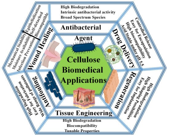
Figure 1.
Overview of biomedical applications covered in this review.
2. Cellulose Nanoparticles
Cellulose has been marketed in several forms, including microfibrillated cellulose (MFC), microcrystalline cellulose (MCC), nanofibrillated cellulose (NFC), cellulose nanocrystals (CNCs), microfibrils, and bacterial cellulose (BC). It can be obtained in micro- and nanoscale regimes (Figure 2). MCC is composed of micro-sized cellulose particles with a length of up to 1 µm and a width of more than 1 µm. MFCs are commonly produced via the mechanical or chemical treatment of wood. Microfibrils are individual fibers with a length of >10 µm and a width of 2–20 µm. At the nanoscale, cellulose nanomaterial or nanocellulose (Figure 2). CNCs, 2,2,6,6-tetramethyl-piperidine-1-oxyl (TEMPO)-mediated oxidized cellulose nanofibers (TOCNFs), and BC are common cellulose nanoparticles. The production method of TOCNFs depends on the oxidation of C6 (i.e., -CH2OH) of glucose using TEMPO as an oxidant in NaClO2 at pH 10. The steric hindrance in TEMPO due to the methyl groups allows only the oxidation of C6. CNCs are common products of the acid hydrolysis of cellulose-based materials [79]. The size distribution of CNCs is in the range of 50–500 nm and 2–20 nm in length and width, respectively. BCs are produced by bacterial cells. They exhibit high crystallinity with good elastic and mechanical properties. The length of BC tends to be 200–3000 nm, and the width can be 10–75 nm (Figure 2). Cellulose extraction at the nanoscale removes defects associated with the hierarchical structure and leads to new properties, such as a large surface area.
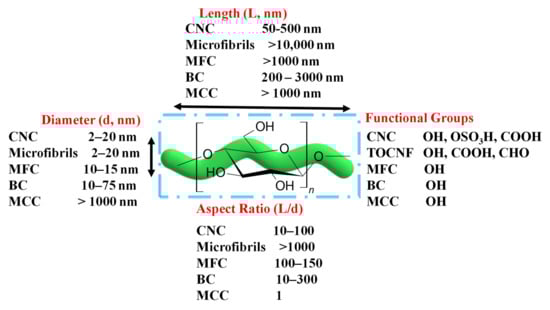
Figure 2.
Summary of cellulose nanoparticles with sizes and functional groups.
Cellulose nanomaterials exhibit a degree of crystallinity of 50–90%. The joint functional group of cellulose is the hydroxyl group. NFC can be further oxidized to form carboxylic functional groups via TEMPO-mediated oxidation, resulting in TOCNFs. The cellulose surface can be modified using several methods, including adsorption or chemical modification via covalent bond formation [5]. The surface charge of cellulose is an essential parameter for the material’s characterization. The cellulose colloids’ high charge ensures high stability and prevents nanoparticle aggregation [80].
3. Applications of Cellulose Nanoparticles as Antibacterial Agents
Cellulose exhibits no intrinsic biocidal activity, in contrast to other natural biopolymers such as cationic chitosan [81]. However, it can be used as an antibacterial agent via several methods, including surface modification and conjugation with antibacterial agents, including organic and inorganic materials (Figure 3) [47,82]. The following section summarizes the applications of cellulose-based materials as antibacterial agents.
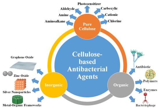
Figure 3.
Cellulose-based materials as antibacterial agents.
3.1. Pure Cellulose for Antibacterial Agents
The antibacterial properties of pure cellulose can be improved via surface modification with several functional groups, such as carboxylic groups, aldehyde, amine, alkylamine, and quaternary ammonium groups (Figure 3). The functional groups of cellulose can be modified with organic molecules that exhibit photosensitization properties. A summary of some of the antibacterial agents prepared using cellulose-based materials is tabulated in Table 1.
The antibacterial activity of 2,3-dialdehyde nanofibrillated cellulose (DANFC) was investigated against Staphylococcus aureus (S. aureus) and methicillin-resistant S. aureus (MRSA) [83]. Dialdehyde formation was performed via the oxidative cleavage of C2 and C3 bonds in the D-glucose monomer of cellulose using a chemical reagent, such as sodium periodate (NaIO4). The antimicrobial activity of DANFC is enhanced by increasing the time of oxidation. The antibacterial effect of DACNF is due to its aldehyde groups, which cause a drop in the pH value (5.7–6.2) [83]. Dialdehyde microcrystalline cellulose (DAMC) antibacterial activity was also reported [84]. DAMC with aldehyde contents of 5.14 mmol/g showed the most potent antibacterial activity against S. aureus, Bacillus subtilis (B. subtilis), E. coli, and Salmonella typhimurium (S. Typhimurium). It exhibited minimum inhibitory concentration (MIC) values of 15, 15, 15, and 30 mg/mL for S. aureus, B. subtilis, E. coli, and S. Typhimurium, respectively [84]. Cellulose was extracted from ginger residues, denoted as GNFs (ginger nanofibers), via acid hydrolysis and high-pressure homogenization [85]. GNFs were tested for antibacterial activity [85]. The MIC values of GNFs were 14 ± 2, 13 ± 1, 18 ± 0, and 31 ± 0 µg/mL for B. cereus, E. coli, S. aureus, and S. Typhimurium, respectively [85].
Cellulose with carboxylic groups shows high antibacterial activity (Table 1). A gel of TOCNFs (0.2–0.8 wt.% in water) inhibited the growth of a wound infected with the pathogen P. aeruginosa [86]. The physical, chemical, and antibacterial activities of carboxylated CNFs can be modified via treatments such as autoclaving [87]. Autoclave treatment at 121 °C for 20 min reduced the gels’ viscosity, increased the ultraviolet-visible absorbance maxima to 250 nm, and increased the aldehyde content. Autoclaved carboxylated CNFs showed high antibacterial activity with minimal toxicity toward L929 mouse fibroblasts and reconstructed human epidermis (RhE) [87]. The antibacterial activity of carboxylated CNFs with different oxidation levels was evaluated against P. aeruginosa and S. aureus [55,87]. Oxygenated CNFs exhibited higher antibacterial activity than non-oxygenated CNF dispersion [55,87].
The antibacterial activity of pure cellulose nanoparticles could be due to several mechanisms, including a decrease in the mobility of bacterial cells [86], the surrounding and entrapping of bacteria via the formation of a network [55], and a reduced pH value due to the increase in aldehyde groups in CNFs [83,87]. However, further investigations should be carried out to understand the key parameters affecting the antibacterial activity of pure cellulose nanomaterials.
Pure cellulose with suitable functional groups possesses good antibacterial activity. However, it is crucial to consider the presence of foreign species such as endotoxins or lipopolysaccharides that can cause inflammation leading to antibacterial activity [88]. A modified TEMPO-mediated oxidation method using sodium hydroxide as a pre-treatment produced CNFs showing an endotoxin level of 45 endotoxin units (EU) per gram of cellulose [89]. This value may be non-toxic at low concentrations. However, it can be critical at high concentrations [90]. The presence of endotoxin should be considered during the evaluation of the antibacterial activity of cellulose-based materials.
3.2. Photoactive Cellulose for Antibacterial Agents
Photo-based light radiation treatments are promising for antibiotic-resistant bacteria (Table 1) [91,92,93,94]. They require the presence of a photosensitizer molecule that converts the light radiation to thermal energy (photothermal therapy) or generates reactive species (i.e., photodynamic treatment) such as reactive oxygen species (ROS). Pure cellulose lacks photosensitizer properties. Thus, it is usually modified with small molecules via covalent or non-covalent interactions to absorb light. Most of these photosensitizers are bacteria-inactivating materials. However, they are effective for bacterial inactivation using cheap light sources such as light-emitting diodes (LEDs) [95].
Photodynamical inactivation (PDI) of bacteria using CNC [96] and hairy aminated nanocrystalline cellulose (ANCC) was reported [97]. ROS can be generated under light via the modification of cellulose with molecules such as anthraquinone vat dyes [98], 3,3′,4,4′-benzophenone tetracarboxylic acid [99], ketoprofen [100], hypocrellin [101], xanthene [77], BODIPY (Dipyrromethene boron difluoride) [91,102], chlorin-e6 [103], phthalocyanines [104,105], protoporphyrin-IX [106,107,108], and porphyrin [96,97,109,110,111,112,113,114]. CNC was chemically modified with cationic porphyrin, denoted as CNC-Por, via Cu(I)-catalyzed Huisgen–Meldal–Sharpless 1,3-dipolar cycloaddition (Figure 4a). The reaction occurs between the azide and alkyne groups on the cellulosic and porphyrinic molecules, respectively (Figure 4a). PDI of Mycobacterium smegmatis, S. aureus, and E. coli by CNC-Por was investigated under white-light radiation (400–700 nm, 60 mW/cm2) [96]. CNC-Por exhibited high PDI against M. smegmatis and S. aureus and insignificant activity against E. coli after illumination for 15 min. However, after 60 min, the PDI activity of the material against all bacteria was higher than 99% (99.9999% for S. aureus) [96]. ANCC was modified with the natural photosensitizer Rose Bengal (RB) via covalent bonds (Figure 4b) [97]. RB-ANCC showed PDI of over 80% against the pathogens Listeria monocytogenes and S. Typhimurium under illumination using normal light irradiation. Interestedly, ANCC improved the PDI of free RB against S. Typhimurium [97].
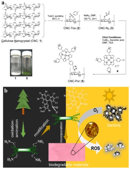
Figure 4.
(a) Synthesis of CNC-Porphyrin; (1) CNC preparation via acid hydrolysis, (2) surface tosylation of CNC, CNC-Tos, (3) synthesis of azide-bearing CNC-N3, (4) click reaction of CNC-N3 with Porphyrin. Reprinted with permission from Ref. [96]. 2011, ACS (2011). (b) Chemical modification of ANCC with Rose Bengal as photosensitizer. Reprinted with permission from Ref. [97]. 2021, American Chemical Society (ACS).
Cationic porphyrin (Por(+))-conjugated cellulose was formed as paper for scalable antimicrobial treatment using PDI [81]. Por(+)- modified cellulosic paper was illuminated using visible light with a wavelength and power of 400–700 nm and 65 ±5 mW/cm2, respectively, for 30 min. The antibacterial and antiviral efficacies were investigated against bacteria (S. aureus, vancomycin-resistant Enterococcus faecium (VER), Acinetobacter baumannii, P. aeruginosa, and Klebsiella pneumoniae) and viruses such as dengue-1 virus, influenza A, and human adenovirus-5 with efficiencies of 99.995%, 99.5%, and 99%, respectively [91]. The inactivation efficiencies for all investigated species, i.e., bacteria and viruses, were higher than 99.9% [91].
PDI using cellulose-based materials exhibits several advantages, such as high antibacterial efficiency (≈100%, Table 1). The method can be applied to the treatment of antibiotic-resistant bacteria. Cellulose chemistry allows the fabrication of materials such as paper [91], fibers [110], or textiles [115] with scalable and straightforward uses for antibacterial treatment. It allows immediate covalent modification using advanced methods such as photo-strain-triggered click ligation [116]. It may open a new avenue for photoactive textiles [117].
3.3. Cationic Cellulose for Antibacterial Agents
Similar to chitosan (CTS), cationic cellulose exhibits intrinsic antibacterial activity. The mechanism of the antibacterial action of cationic biopolymers depends on the high binding affinity between the positive charge of these polymers and the negative charge of bacterial cells. The surface of bacterial cells, i.e., Gram-positive and Gram-negative, is negative due to hostile phosphate groups in peptidoglycan and phospholipids. Following this principle, creating a positive charge on cellulose enables high antibacterial activity (Table 1). Cationic CNCs can also be used as immune modulators [118].
Cationic cellulose can be achieved via modification with quaternary ammonium compounds such as poly(isopropanol dimethyl ammonium) chloride (PIDMAC) [119], quaternized poly (2-(dimethyl aminoethyl) methacrylate) (PDMAEMA) [120], cetyltrimethyl ammonium bromide (CTAB) [121], 3-chloro-2-hydroxypropyl-tri-methyl ammonium chloride 3-chloro-2-hydroxypropyl-trimethyl ammonium chloride (CHPTAC) [122], pyridinium/N-chloramine [123], benzalkonium chloride [124], and quinolinium silane salt [125]. Cellulose grafted with DMAEMA can be prepared via polymerization using a reversible addition-fragmentation chain transfer (RAFT) reaction [120]. Cationic cellulose can also be prepared via direct covalent bonding of quaternary ammonium moieties without a linker [126]. The silane group of 3-(trimethoxy silyl)-propyl-dimethyl octadecyl ammonium chloride reacted with the hydroxyl functional groups in cellulose, forming Si–O–Si bonds [126]. Cellulose-QA showed complete inactivation of E. coli and P. aeruginosa after one hour and 10-fold inactivation of B. cereus [126].
Cellulose can be modified with quaternary ammonium and porphyrin moieties via esterification [108]. The material’s treatment requires relatively low porphyrin concentrations and can be applied under a low dosage of white-light irradiation (2.4 J/cm) [103]. The protoporphyrin IX (PpIX) moiety provides white-light radiation photosensitization (Figure 5). Quaternary ammonium moieties offer dual functionality: antibacterial activity and the prevention of the aggregation of porphyrins that cause the quenching of ROS generation. Photodynamic treatment of bacteria using quaternary ammonium-porphyrin-modified cellulose showed effective antibacterial activity against antibiotic-resistant E. coli and S. aureus strains (Figure 5). The antibacterial activity of the materials is due to the intrinsic bioactivity of quaternary ammonium moieties and the generation of ROS [108].
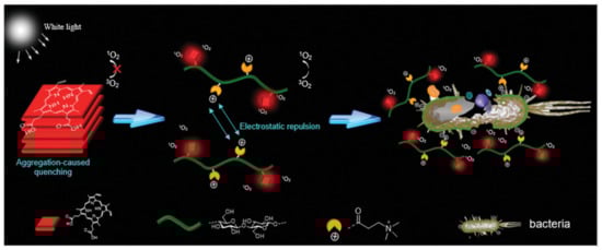
Figure 5.
The antibacterial mechanism for porphyrin and quaternary ammonium-modified cellulose under light radiation. Reprinted with permission from Ref. [108]. 2019, John Wiley & Sons.
The antibacterial activity of cationic cellulose can be due to several mechanisms, such as destabilization of the bacterial intercellular membranes due to Ca2+ or Mg2+ ion exchange, membrane disruption because of the release of potassium (K+) ions, the formation of ROS, an increase in the number amine groups [127], or an increase in lipophilicity using amino-alkyl [128].
3.4. Organic-Modified Cellulose as Antibacterial Agents
The antibacterial activity of cellulose can be enhanced via chemical modification with organic bioactive molecules, including antibiotics, antimicrobial peptides, N-halamines, aminoalkyl groups, bacteriophages, and polymers (Figure 3).
3.4.1. Antibiotic-Modified Cellulose
Antibiotics are widely used for bacterial treatments. Cellulose has been grafted with antibiotics, including β-lactam antibiotic benzylpenicillin [129], ciprofloxacin [130], tetracycline hydrochloride [131], silver sulfadiazine (Ag SD) [132], 3-pentadactylphenol [133], allicin [134], and amoxicillin [135]. Antibiotics such as penicillin can be covalently modified with cellulose via ester bond formation (Figure 6) [129]. The covalent modification of cellulose ensures high durability with good antibacterial activity. Using thermal treatment, the materials can be processed into the film without destroying the formed bonds (Figure 6).
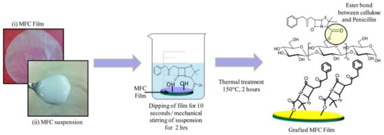
Figure 6.
Schematic representation of suspension and film of MFC and chemical modification with Benzyl Penicillin via esterification. Reprinted with permission from Ref. [129]. 2015, ACS.
The cationic cellulose filter paper was reported for water treatment with antibacterial activity [119]. The cellulose filter paper was coated with PIDMAC as a cationic polyelectrolyte binder (CPE). It was loaded with amphiphilic block copolymer micelles containing triclosan (antibacterial and antifungal agent). The micelles interacted with CPE via polystyrene-block-polyacrylic acid (PS-b-PAA) as the block copolymer. The materials included two antibacterial agents i.e., triclosan (hydrophobic) and an ammonium compound [119]. Thus, they showed high antibacterial activity [119].
Cellulose is good support for antibacterial agents, including antibiotics [136]. The use of conventional antibacterial agents such as antibiotics ensures high antibacterial activity for cellulose-based materials. However, the release of antibiotics into water causes environmental concerns and raises the risk of highly antibiotic-resistant bacteria.
3.4.2. Aminoalkyl-Modified Cellulose
Cellulose modified with aminoalkyl functional groups exhibits antibacterial activity [128,137,138,139,140]. The modification takes place via a reaction with silanol groups as coupling agents. The process involves the formation of covalent bonds between silanol groups (Si-OH) and hydroxyl groups (OH) of cellulose, i.e., the formation of Si–O–C bonds. BC membranes modified with aminoalkyl groups using APMS ((3-aminopropyl)trimethoxysilane) exhibit antibacterial activity against S. aureus and E. coli [128]. The antibacterial activity of APMS-modified cellulose is due to the polycationic nature of the membrane and the alkyl chains in APMS. The long chains of the alkyl groups increase lipophilicity, which ensures strong interactions with the cytoplasmic membrane of the investigated bacterial cells [128]. Alkyl groups with up to 10 carbons exhibited a broad spectrum of antibacterial and antifungal activities [141]. A cellulose membrane with aminoalkyl groups showed non-toxic properties in human adipose-derived mesenchymal stem cells (hAMSC) [128] and human embryonic kidney 293 cells (HEK-293) [141].
Besides the chain length, the increase in amine groups ensures high antibacterial activity [127]. Cellulose was modified with aminoalkyl containing different groups of 1, 2, and 3 amines using APMS, 2-aminoethyl 3-aminopropyl trimethoxysilane (DAMS), and 3-2-(2-aminoethyl amino) ethyl aminopropyl trimethoxysilane (TAMS), respectively. The increase in the number of amine groups results in high antibacterial activity. Cellulose–TAMS showed the most increased antibacterial activity against Gram-positive bacteria [127].
3.4.3. N-halamine@Cellulose
N-halamines, halogen atoms linked to nitrogen-containing compounds, modified cellulose [142,143,144]. The functional groups of N-halamine, such as epoxy groups or organosiloxane, enable grafting onto cellulose via a reaction with hydroxyl groups [145]. The materials exhibit durable antibacterial activity [145]. The N-halamine of the s-triazine-based quaternized molecule was used to modify cellulose. The material showed a 6-log reduction in S. aureus and E. coli after treatment for 1–5 min [144]. The oxidant chlorine in N-halamine molecules showed 50% retention of its activity even after 50 cycles of washing and 30 days of storage. The remaining 50% of the material’s activity could be regenerated after exposure to a bleach solution [144]. N-halamine-modified cellulose can be restored via simple methods, such as treatment with a diluted bleach solution [146].
3.4.4. Antimicrobial Peptide-Modified Cellulose
Antimicrobial peptides are attractive compared to small organic molecules used as antibiotics [147,148]. Cellulose has been modified with several peptides, such as gentamicin [149]. Bacterial cellulose (BC) was chemically modified with RGDC peptide (R, G, D, and C refer to arginine, glycine, aspartic acid, and cysteine, respectively) and gentamicin via covalent bonds [149]. The process involves the cross-coupling of RGDC peptide to BC using a coupling agent such as 3-aminopropyltriethoxysilane (APTES). Gentamicin was then attached to the surface of the RGDC-BC membrane [149]. The gentamicin–RGDC-grafted BC membranes exhibited high antibacterial activity against Streptococcus mutans without significant toxicity toward human dermal fibroblasts [149].
3.4.5. Polymer-Modified Cellulose
Cellulose has been modified with polymers to enhance antibacterial activity. Polymers can be directly blended (anchored or grafted) with cellulose using a simple procedure (post-synthetic procedure) or via in situ polymerizations of the monomers in the presence of cellulose (in situ procedure). Cellulose was modified with several polymers, including: (1) biopolymers such as chitosan [150] and (2) synthetic polymers such as polypyrrole [151], polypropylene [133,152], polyethyleneimine [153], polyhexamethylene guanidine hydrochloride (PHMG-Cl) [154], polyvinyl [155], 2-aminoethyl methacrylate [156], and poly(3-hydroxy-acetylthioalkanoate-co-3-hydroxy alkanoates) [157].
Fibers of dialdehyde cellulose (DAC)/CTS composite were prepared via stirring and filtration [150]. The antimicrobial activity of DAC/CTS against E. coli and S. aureus was investigated. Data analysis showed antibacterial activity of 90.2% and 95.1% against E. coli and S. aureus, respectively [150]. The antibacterial activity is due to the aldehyde groups of cellulose and the intrinsic antibacterial activity of CTS [150].
Polyrhodanine (PR) was prepared in the presence of CNC [158]. The synthesis procedure involves the polymerization of rhodanine on the surface of CNC using ferric chloride (FeCl3) as the initiator and oxidant. The negatively charged surface of CNC assisted the polymerization and led to the formation of core-sheath nanoparticles of CNC@PR. Using the plate colony-counting method, the antibacterial activities were evaluated against E. coli and B. subtilis. CNC@PR showed good antibacterial activity with good MICs [158].
Cellulose has been modified with polymers such as guanidine polymer [159]. In situ polymerization of 2-aminoethyl methacrylate (AEM) into a BC network was reported with and without a cross-linker such as N,N-methylenbis(acrylamide) (MBA) [156]. The polymerization took place on the BC network via a radical-based reaction, resulting in BC/poly-AEM/MBA. The synthesized polymer filled the pores of the BC network. BC/poly-AEM and BC/poly-AEM/MBA materials were evaluated for antimicrobial activity against E. coli. BC/poly-AEM exhibited higher antibacterial activity than cross-linked BC/poly-AEM/MBA material. The authors explained that cross-linking reduced the diffusion of the bacteria into the BC network, leading to low contact between E. coli and the ammonium groups [156]. Thus, cross-linking of the network reduced the antibacterial activity [156].
Post-synthetic modification of BC with octenidine dihydrochloride was reported [160]. The material showed significant antimicrobial activity against S. aureus even after six months of storage. It exhibited minimal cytotoxic effects against human keratinocytes [160].
3.4.6. Bacteriophage-Modified Cellulose
Cellulose can be modified with a virus form called a bacteriophage (or phage for a simple description) to induce DNA or RNA inside cells [161,162,163,164] or bacteriophage endolysins (enzymes that cause hydrolysis) [165,166]. The phage can be immobilized on cellulose via non-specific interactions (adsorption) [167] or covalent bonds [168].
Several enzymes, such as lysozyme (muramidase), can be used as antibacterial agents. The mechanism of most enzymes is the cleavage of bonds in the cell membrane, such as the peptidoglycan layer of bacteria, leading to cell lysis [169]. Enzymes such as lysozyme cause hydrolysis of the 1,4-β-linkage between N-acetylmuramic acid and N-acetylglucosamine [169]. CNC was used to immobilize hen egg-white lysozyme (HEWL) and T4 lysozyme (T4L, Figure 7) [168]. The immobilization of lysozyme on CNC does not cause a decrease in the enzyme’s enzymatic activity for lysis and hydrolysis. The process involves covalent coupling via carbodiimide-activated CNC via carboxylate groups to glutaraldehyde-activated aminated CNC (Am-CNC, Figure 7). Am-CNC-HEWL and Am-CNC-T4L showed lytic activity of 86.3% and 78.3%, respectively. After immobilization, the enzyme exhibited high bactericidal activity compared to the free enzyme. They also showed high stability during storage at 4 °C and 22 °C [168]. A thin film of CNC–lysozyme composite was prepared using the evaporation-induced self-assembly method [170]. The technique produces a film with a HEWL enzyme loading of 10 wt.%. However, it showed insignificant antibacterial activity against Gram-positive bacteria. The optimization of enzyme-modified cellulose is critical to achieving high antibacterial activity.
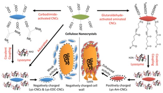
Figure 7.
Schematic illustration of the immobilization of lysozymes on CNCs for antibacterial activity. Reprinted with permission from Ref. [168]. 2017, ACS.
Enzyme-based antibacterial agents offer several advantages, such as selectivity. For example, lysozyme exhibits higher activity against Gram-positive bacteria than Gram-negative bacteria. This selectivity is because the peptidoglycan layer exists only in Gram-positive bacterial cells [20]. Antibacterial agents should be active against a broad spectrum of bacteria.
Bacteriophages exhibit high antibacterial activity. However, they are usually negatively charged at their heads. Thus, they have difficulty interacting directly with the negative charge of cellulose. They interact easily with the positively charged surface via electrostatic interactions. The presence of functional groups such as amine and carboxylic acid on the phage surface enables functionalization via covalent bonds. The latter method allows highly durable and robust phage attachment to cellulose materials.
Cellulose–organic composites with antibacterial activity can be synthesized via several methods. The simple attachment of both components via grafting or non-covalent interactions requires no tedious efforts or expensive equipment. However, they lack high stability or long-term activity due to leaching. Thus, strong bonds such as covalent bonds are required to ensure high strength and long-term activity [171].
3.5. Cellulose–Inorganic Nanoparticles for Antibacterial Agents
Cellulose has been modified with inorganic nanoparticles such as carbon nanomaterials, metal oxides, metallic nanoparticles, and metal-organic frameworks (MOFs) (Figure 3) [172,173,174,175,176,177]. Most of these materials exhibit intrinsic antibacterial activity, leading to high performance against Gram-positive and Gram-negative strains.
Carbon nanomaterials exhibit high antibacterial activity. They were conjugated with cellulose. A composite of BC and graphene oxide (GO) was reported as an antimicrobial agent against E. coli and S. aureus [178]. Electrostatic modification improved the antimicrobial activity of GO/BC nanocomposites [179]. The antibacterial activity of carbon nanosheets such as GO is mainly due to the sharp edges of the sheets, which act as a knife to cut the cell membrane, causing the rupture of the outer envelope [180,181,182,183].
Nanoparticles with metal oxides such as zinc oxide (ZnO) exhibit high antibacterial activity. The antibacterial activity of ZnO-based nanomaterials depends on the ZnO contents in composites [175]. A composite of cellulose acetate (CA) and hydrophobic polysulfone (PSf) polymer was used to modify the material with 0.1 wt.% ZnO NPs [173]. Membranes containing ZnO NPs showed good antibacterial activity against E. coli [173]. ZnO nanoparticles were prepared via an in situ procedure [174]. Zn2+ ions were adsorbed onto cellulose before precipitation using ammonium hydroxide [174]. The surface functional groups of cellulose, such as hydroxyl groups, enable the adsorption of Zn2+ ions via electrostatic interactions and control the morphology and particle size.
Metal oxide nanoparticles such as CuO [184] and TiO2 nanoparticles [185,186], faujasite [187], and montmorillonite (MMT) [188] were modified with cellulose for antibacterial activity. Cellulose acetate/TiO2 nanoparticles exhibited high antibacterial activity [186]. A faujasite–cellulose composite membrane was used to purify water contaminated with bacteria (E. coli, Enterococci, and Clostridium). It showed high removal efficiency, resulting in <100 colonies/100 mL [187]. The presence of MMT in the cellulose membrane enabled modification with several metal ions, such as Na, Ca, and Cu [188]. BC/Cu-MMT composites showed the highest antibacterial activity against the investigated bacteria [188].
Silver nanoparticles (Ag NPs) are very active antibacterial agents [189]. They have been widely modified with cellulose nanoparticles for antibacterial activity [190,191,192,193,194,195,196,197,198]. Ag NPs were synthesized directly into electrospinning fiber cellulose acetate (CA) [199]. The procedure involved the adsorption of silver ions reduced via photons onto Ag NPs. The prepared materials exhibited high antibacterial activity against S. aureus, E. coli, K. pneumoniae, and P. aeruginosa [199]. A nanofiltration (NF) membrane consisting of CNC/silver (CNC/Ag) and polyamide was fabricated using the interfacial polymerization (IP) method [200]. The membrane with 0.01 wt.% CNC/Ag showed antibacterial activity of 99.4% against E. coli viability [200]. The high antibacterial activity of silver-based nanomaterials can be explained by several mechanisms, such as ionization and the release of silver ions [198]. Silver nanoparticles offer several advantages. They can be synthesized into cellulose textiles via an in situ procedure [201]. They can be conjugated with other materials such as magnetic nanoparticles (MNPs) [202] and gold nanoparticles [203]. Furthermore, cellulose serves as a biodegradable support for Ag NPs [204].
The conjugation of cellulose with inorganic nanomaterials ensures high antibacterial activity. Most of the antibacterial activity is due to the inorganic materials present in the cellulose composite [205]. However, they can be toxic to mammalian cells or environmentally unfriendly due to toxic heavy metal ions released into drinking water.
4. Cellulose-Based Materials for Antifouling
Pure cellulose nanoparticles with residual lignin [206,207] or carboxylic functional groups [208] possess antifouling properties. Thus, they are widely used for membranes for antifouling applications [208]. Cellulose nanoparticle-based membranes exhibit high antifouling performance with high reflux compared to commercial membranes such as Millipore GS9035 [209]. A membrane of TOCNF, poly(vinyl alcohol) (PVA), and polyethersulfone (PES) was fabricated for antifouling application. The TOCNF/PVA@PES membrane exhibited high antifouling capability [208]. A micro/nanocellulose membrane grafted with zwitterionic poly(cysteine methacrylate) (PCysMA) showed excellent antibacterial and antifouling properties. It showed a reduction efficiency of 85% in the biofilm formation of S. aureus [207].
Photocatalytic disinfection using photoactive substances is based on generating free radicals under a light. A CNC/silver/polyamide membrane containing CNC/Ag (0.01 wt.%) showed high antifouling activity of 92.6% using humic acid with the antibacterial activity of 99.4% against E. coli viability [200]. A membrane of CMC/GO/magnesium oxide (MgO) nanoparticles (CMC/GO/MgO) was prepared for photocatalytic antifouling [210]. It can be used as an antifouling membrane due to the generation of electrons and ROS. Thus, it can be used to oxidize organic pollutants [210].
Cellulose–inorganic hybrids exhibited high antibacterial performance. CNC/silver/polyamide membrane demonstrated effective antifouling properties using humic acid with a flux recovery of 92.6% [200]. Composite nanofibers of CA and polysulfone (PSf) with 0.1 wt.% ZnO exhibited high antibacterial activity against E. coli [211].

Table 1.
Antibacterial applications for cellulose-based materials.
Table 1.
Antibacterial applications for cellulose-based materials.
| Materials | Preparation Methods | Form | Microorganism | Methods | Time | Efficiency | Mechanism | Ref. |
|---|---|---|---|---|---|---|---|---|
| DANFC | 1. Mechanical grinding 2. Enzyme treatment 3. NaIO4 oxidation 4. Dialysis 5. Freeze-drying | Mat | S. aureus MERSA | Zone inhibition Plate counting method | 24 h | 100% | Drop-in pH value | [83] |
| TOCNF | 1. TEMPO oxidation 2. Oxygenation | Suspension | P. aeruginosa S. aureus | Plate counting method | 24 h | 71% | The formation of a network surrounding the bacteria | [55] |
| 1. TEMPO oxidation 2. Autoclaving (121 °C, 20 min) | Gels | 24 h | 71% | Increase in aldehydes, drop in pH value | [87] | |||
| CNC-Porphyrin | 1. HBr acid hydrolysis of Whatman filter paper 2. Cu(I)-catalyzed Huisgen–Meldal–Sharpless 1,3-dipolar cycloaddition | Suspension | Mycobacterium smegmatis, S. aureus, E. coli | 60 min | >99% | Generation of ROS, photodynamic | [96] | |
| Porphyrin-cellulose paper | Cu(I)-catalyzed Huisgen–Meldal–Sharpless cycloaddition | Paper | S. aureus, VER, Enterococcus faecium, Acinetobacter baumannii, P. aeruginosa, Klebsiella pneumoniae | 30 min | >99.9% | [91] | ||
| NFC-Porphyrin | Cyanuric chloride coupling | Paper | MRSA, VER, E. faecium, A. baumannii K. pneumoniae | 30 min | 99.999% | [114] | ||
| CHPTAC-Cellulose triacetate | 1. Immersion precipitation technique 2. Alkaline hydrolysis 3. Esterification | Membrane | S. aureus E. coli | 24 h | 78.7–89.0% 64.7–76.6% | Cationic charge | [122] | |
| BC-aminoalkyl | Stirring for 5 h at 25 °C | Dynamic shake flask method | 24 h | >99.9% | Increase in lipophilicity | [128] | ||
| DAC/CTS | 1. NaIO4 oxidation 2. Stirring 3. Filtration | Fibers | Plate counting method | 24 h | 95.1% 90.2% | Drop in pH, cationic CTS | [150] | |
| T7 phage-Cellulose acetate | Electrospinning of cellulose acetate | Membrane | E. coli | Plaque-forming units (PFU) | 24 h | 6 log(PFU/mL) | Release of phage and hydrolysis | [167] |
| Am-CNC-HEWL Am-CNC-T4L | 1. Ammonium persulfate oxidation 2. Modification 3. Coupling | Suspension | M. lysodeikticus, Corynebacterium sp., E. coli, P. smendocina | Time–kill study with Alamar Blue assay | 24 h | 100% | Lytic activity | [168] |
| CNC-Lysozyme | 1. Sulfuric acid hydrolysis 2. Evaporation-induced self-assembly | Thin film | E. coli S. aureus | Diffusion assays | 24 h | 0% | [170] | |
| ZnO-BC | In situ synthesis | Sheets | Inhibition zone | 24 h | 5.7 ± 0.29 mm 2.9 ± 0.75 mm | Formation of ROS | [212] | |
| Ex situ synthesis | Film | E. coli | Inhibition zone | 24 h | 34–41 mm | [213] | ||
| TiO2-BC | Ex situ synthesis | Pieces | Optical density (OD600) | 12 h | 60% | [185] | ||
| Faujasite-cellulose | Hydrothermal | Membrane | E. coli, Enterococci Clostridium | Standard FS ISO9308 and ISO17025 | 80% | [187] | ||
| SSD/BC | Impregnation and ultrasonication | Commercial membrane | P. aeruginosa, E. coli S. aureus | Zone inhibition | 24 h | 6.5 mm | [214] |
Inorganic nanoparticles improved the antibacterial activity of cellulose-based materials. Direct contact with metallic ions causes damage to the cell membrane due to the generation of ROS that can permeate the cell wall. The formed ROS damage the phospholipid layer of the cell membrane and can also cause cell disruption via the oxidation of thiol groups of amino acids present in the bacterial cells.
5. Cellulose Nanoparticles for Wound Dressing
During burn treatment, special care should be taken to avoid microbial infection [55]. Wound healing requires fast re-epithelialization within 10–14 days to prevent complications. Chronic wounds may undergo critical physiological changes or initiate tumor growth, leading to the significant destruction of tissues or organs. This situation is vital for diabetic patients who suffer from chronic ulcers. Thus, the dressing becomes an essential active component during the healing process and is no longer considered a supplement. The sauce should offer a warm and moist environment to provide a fast and effective healing process. It should also be biocompatible and easy to detach, act as a thermal insulator, and provide antimicrobial activity to prevent infections.
Natural polymers such as cellulose have been widely investigated for wound dressing because of their high biocompatibility and biodegradability (Table 2). Bacterial cellulose (BC) has been extensively used for wound healing [57,215]. Several methods have been reported to modify BC to improve the material’s properties, such as biocompatibility and the uptake/release of water or drug molecules, and ensure high antimicrobial activity. Cellulose can be fabricated into different forms, such as films, foams, hydrocolloids, hydrogels, and nonwovens [216]. It can also supply wounds with drugs, growth factors, and antimicrobial agents. The traditional gauze of cotton is usually modified with sodium periodate via oxidation, i.e., the formation of dialdehyde [217]. The oxidation process decreases elastase and can support the healing of a chronic wound. It can also improve the antibacterial activity and offers high protection due to the drop in pH value caused by the aldehyde functional group in cellulose [83]. BC-based wound dressings reduce the closure time (i.e., recovery rate) of the wound [218,219] without significant inflammation (Table 2) [220].
A wound dressing of pure cellulose such as NFC was reported [53]. The antimicrobial properties of dressing materials were investigated against bacterial pathogens such as S. aureus and P. aeruginosa [53]. NFC-based wound dressing showed no antibacterial activity against the tested strains. The BC-based film exhibits good wound healing (Figure 8) [220]. The performance is lower than that of traditional wound dressing (gauze). However, it may be improved. The sides of BC film (i.e., bottom and top) exhibit different performances (Figure 8) [220]. The antibacterial activity of cellulose can be improved via carboxylated CNFs [55]. Autoclaved CNFs showed a strong antibacterial effect against bacteria that infect wounds [87].
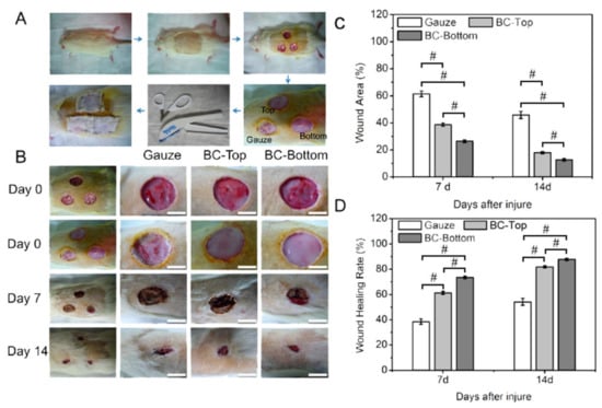
Figure 8.
Wound healing treatment using BC-based dressing: (A) description of the operation on the skin injury model and the dynamic healing of a rat, (B) the progress (0–14 days) of healing for the skin injury model on Wistar rat using gauze and BC-based dressing of two sides, top and bottom (all scale bars equal 10 mm), (C) wound area progression after the injury and (D) wound healing rate. Error bars represent means ± standard deviation (SD) for n = 5 (# p < 0.01).Reprinted with permission from Ref. [220]. 2015, American Chemical Society (ACS, 2015).
Cellulose nanofibrils were prepared via the defibrillation of wood via a mechanical method [51]. The fibrils were developed into a membrane via filtration and drying under mild pressure. The prepared cellulose membrane was used as a wound dressing. It offered strong adhesion to the wound and promoted epithelialization after four days without the observation of allergic reactions or inflammation during the treatment [51]. A cellulose hydrogel was prepared from the alkali treatment and TEMPO-mediated oxidation of ginger fibers (T-GNFs) [221]. It was designed via a simple vacuum-assisted filtration using ginger nanofibers without cross-linking treatments. The preparation method required low energy and few components for the production procedure. The material’s antibacterial and wound healing properties were investigated [221]. The material enabled the closure of the wound without significant antibacterial activity against E. coli and S. aureus [221]. The 3D printing of NC hydrogel scaffolds was reported for wound healing [222]. The process included a double cross-linking procedure via two steps: (i) in situ Ca2+ cross-linking during 3D printing and (ii) chemical cross-linking with 1,4-butanediol diglycidyl ether (BDDE) after printing. The scaffolds were used to support the proliferation of fibroblast cells with an attachment percentage of 84–86.5% [222]. Cellulose exhibits high performance in wound healing and can be processed via several procedures.
Modulating the wound’s pH value enhanced the wound treatment [223]. Intact skin exhibits a slightly acidic pH value of 4–6 due to the secretion of organic acids by keratinocytes to regulate bacterial flora and prevent infection, while the infected wound exhibits a pH value of 7–7.5. The neutral pH value of the injury is an ideal condition for the growth of bacterial cells. The drop in the pH of the wound using aldehyde-modified cellulose enabled high protection against bacterial infection [83]. Dialdehyde of microcrystalline cellulose (DAMC) [84] and nanofibrillated cellulose (DANFC) [83] with aldehyde contents of 6.5 mmol/g and 1.5 mmol/g, respectively, showed high antibacterial activity.
The antibacterial activity of a cellulose-based wound dressing can be improved by loading it with antibiotics. Chloramphenicol (CAP)-loaded dialdehyde (DABC) and non-oxidized BC membranes were investigated in terms of their antimicrobial efficiency against E. coli, S. aureus, and Streptococcus pneumoniae (S. pneumoniae) [224]. The CAP drug-loading capacity using DABC was low (0.1 mg/cm2) than the BC-based membrane, which showed a loading capacity of 5 mg/cm2. However, both membranes, i.e., oxidized and non-oxidized BC, showed high antimicrobial activity against the tested bacteria. CAP/DABC showed high adhesion and proliferation of the fibroblast cell line L929 compared to non-oxidized BC. This study highlighted the potential for using newly developed CAP/DABC dressing materials in wound treatment [224].
Cellulose-modified organic nanocomposite exhibits high performance as a wound dressing. A three-dimensional (3D) network of collagen I (Col-I), hydroxypropyl trimethyl ammonium chloride chitosan (HACC), and BC was prepared via a membrane–liquid interface (MLI) method [225]. HACC exerted antibacterial activity during wound healing [225]. Inerpan (a polymer of L-leucine and methyl L-glutamate) and Procel-Super (SOD) accelerated the healing of burn wounds with BC-based wound dressing by 17.0 and 5.5%, respectively [226].
Cellulose nanocomposites have been widely reported for wound dressing. A membrane of BC and chitosan (BC–CTS) was fabricated via the immersion of BC in a solution of CTS followed by freeze-drying [227]. The procedure can be applied for large-scale production. The presence of antibacterial agents such as CTS significantly inhibited the growth of bacterial cells, e.g., E. coli and S. aureus [227]. The BC-CTS membrane exhibited higher antibacterial activity compared to the pure BC membrane. BC and BC-CTS membranes demonstrated antibacterial inhibition of 49.2% and 99.9% against E. coli and 30.4% and 99.9% against S. aureus, respectively. The BC-CTS membrane showed high epithelialization and regeneration during wound healing compared to wounds treated with BC only or a commercial dressing such as TegadermTM [227].
A sponge of carboxylated brown algae cellulose nanofibers (BACNFs) was prepared to contain organic rectorite (OREC) via freeze-drying [228]. OREC was organized via intercalation of chitosan (positively charged) into negative silicate layers via ion exchange. The sponge of BACNFs/OREC exhibited high antibacterial activity against E. coli and S. aureus without significant toxicity toward mouse fibroblasts (L929). It also showed effective wound closure (100%) after 12 days compared to commercial gauze. It also exhibited effective prevention of bacterial infections without a significant inflammatory response [228]. It is essential to mention that BACNFs showed no antibacterial activity [228].
Cellulose has been conjugated with several inorganic-based antimicrobial agents, such as silver nanoparticles [229], graphene oxide (GO) [230], and ZnO [231]. Silver-based drugs such as silver sulfadiazine (SSD) are widely used as antibacterial agents. BC/SSD was prepared via the impregnation of a BC membrane with SSD via ultrasonication [214]. The BC/SSD membrane showed significant antibacterial activities against several bacteria, such as P. aeruginosa, E. coli, and S. aureus [214]. The membrane exhibited high biocompatibility [214]. Methyl cellulose improved the dispersion of GO [230]. A cytocompatibility assay using [3-(4,5-dimethylthiazol)-2-yl]-2,5-diphenyltetrazolium bromide (MTT) and live/dead assays of EA.hy926 human endothelial cells (ECs) exhibited high biocompatibility. Cell migration under the effect of GO–cellulose using an induced wound scratch model of EA.hy926 ECs was reported (Figure 9A). GO–cellulose accelerated cell migration. The in vivo wound healing of full-thickness wounds in the dorsum of rats (diameter of 8 mm) was evaluated using different measurements, including wound closure measurements (Figure 9B,C). GO–cellulose exhibited a significant wound closure rate compared to control experiments (Figure 9B,C). Data analysis revealed high wound re-epithelization and neovascularization with the significant formation of extensive collagen deposition [230].
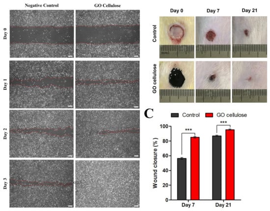
Figure 9.
(A) Cell migration with and without GO–cellulose nanocomposite; red-dotted lines represent the wound edges, scale bar = 200 μm; (B) in vivo evaluation of the skin wounds of rats with and without GO–cellulose nanocomposite for post-wound induction on days 0, 7, and 21; and (C) the percentage of wound closure: significant differences were evaluated using one-way ANOVA, where *** p < 0.0001. Reprinted with permission from Ref. [230]. 2021, Elsevier.
Cellulose offers several advantages for wound dressing (Table 2). The required cost is low. It can be fabricated into applicable forms such as membranes using cheap sources such as wood [51]. A cellulose-based membrane exhibited higher performance as a wound dressing than a commercial porous regenerating membrane [51]. The epithelialization of wood-based dressings such as NFC showed faster healing compared to Suprathel® (commercial lactocapromer-based wound dressing) [53]. BC dressings are cheaper than conventional synthetic fiber dressings [232].
The surface properties of cellulose-based wound materials can be modified via several methods, such as cross-linking with silane-based reagents [233]. The material was investigated for femoral artery and liver injury models. Chemical modification with organosilane enabled the formation of a hydrophobic layer that stopped blood penetration (blood loss < 50%) and accelerated the process of blood clotting. It required a short time for hemostasis in both models [233]. The high surface charge of cellulose nanomaterials enhanced protein adsorption and could promote cell adhesion [162].
A dressing prepared from cellulose is usually transparent, allowing the evaluation of the wound treatment without removing or exchanging the dressing [51]. A cellulose-based membrane offers good adhesion to the moist wound surface due to the abundant hydroxyl groups present in the cellulose structure without significant allergic or inflammatory responses [51]. Cellulose-based dressings enable faster self-detachment than commercially available wound healing dressings [53]. They can be used for infected wounds [234]. Thymol-enriched BC hydrogel can be used to treat and heal third-degree burn injuries [235].
Among several cellulose types, BC-based membranes are widely used for wound healing. However, the extraction process produces a low yield and is expensive. Nanocellulose extracted from wood pulp fibers can be an alternative to BC. Cellulose-based dressings suffer from swelling behavior due to their high affinity toward water molecules. The surface properties should be improved to meet requirements for wound healing. BC shows no antimicrobial activity. Thus, antimicrobial agents are usually required.
Table 2.
Cellulose-based materials for wound dressing.
Table 2.
Cellulose-based materials for wound dressing.
| Materials | Fabrication | Cellulose Source | Form | Study | Closure (%) | Time (days) | Bacteria | Efficiency | Ref. |
|---|---|---|---|---|---|---|---|---|---|
| BC–CTS | Immersing BC in chitosan followed by freeze-drying | Acetobacter xylinum | Membrane | In vivo | 85 | 8 | E. coli S. aureus | 99.9% | [227] |
| Cellulose nanofibrils | Filtration technique | Birch pulp fibers | Membrane | In vitro In vivo Clinical studies | 8–9 | P. aeruginosa, S. aureus | None | [53] | |
| Carboxylated CNF | 1. Autoclaved using NaOH 2. TEMPO-mediated oxidation | Pinus radiata bleached kraft pulp fibers | Gels | In vitro In vivo | 24 h | 60% | [87] | ||
| SSD/BC | Impregnation of BC with SSD via ultrasonication | Commercial membrane | Membrane | In vitro | P. aeruginosa, E. coli, S. aureus | 6.5 mm | [214] | ||
| BC | Cultured bacteria in Hestrin and Schramm (HS) | Acetobacter xylinum | Film | In vitro In vivo | 90 | 24 h | [220] | ||
| T-GNF | 1. Alkali treatment 2. TEMPO-mediated oxidation | Ginger fibers | Hydrogels | In vitro | 67 | 72 h | E. coli, S. aureus | 0 | [221] |
| BACNF/QCR | 1. Cation exchange 2. Freeze-drying | Brown algae | Sponge | In vitro In vivo | 100 | 12 d | 6 mm | [228] |
6. Drug and Gene Delivery Using Cellulose-Based Materials
Cellulose-based materials have advanced drug delivery [61,236,237,238,239,240]. For multifunctional applications, they can be conjugated with nanomaterials such as magnetic nanoparticles (MNPs) [241]. Cellulose can encapsulate drugs [242]. The functional groups of carboxymethyl cellulose (CMC) enabled its modification with folate for the selective release of the anticancer agent 2,4-dihydroxy-5-fluorpyrimidin (5-FU) [243]. Cellulose’s surface modification with folic acid ensures selective cell uptake and binding via a folate-receptor-mediated cellular mechanism [244,245]. Cellulose serves as an effective carrier for delivering hydrophobic drugs such as docetaxel, paclitaxel (PTX), and etoposide [246].
Hydroxypropyl methylcellulose improved the cellular uptake of curcumin (CUR) to treat prostate cancer cells [247]. CUR-conjugated cellulose exhibited significant changes in apoptosis compared to CUR alone. Cellulose also showed the highest cellular uptake compared to other carriers, such as β-cyclodextrin (CD), poly(lactic-co-glycolic acid) (PLGA), MNPs, and dendrimers [247]. TOCNFs and MOFs such as zeolitic imidazolate frameworks (ZIF-8) and Material Institute of Lavoisier (MIL-101 (Fe)) were used for the drug delivery of CUR (Figure 10) [248]. TOCNF/ZIF-8 underwent material processing into a 3D network via 3D printing [248]. Cellulose enables the printing of MOF powder. It serves as a binder and template for MOFs during in situ synthesis. The materials can release the CUR drug at physiological pH (5.5) [248].
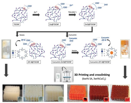
Figure 10.
The synthesis procedure of cellulose-ZIF8 bioink and their processing into the 3D network via 3D printing. Reprinted with permission from Ref. [248]. 2019, John Wiley & Sons.
Cellulose-based materials were also used to deliver oligonucleotides such as siRNAs [249,250]. They can be considered non-viral vectors [251,252]. CNCs were modified with poly(2-dimethylamino)ethyl methacrylate) (PDMAEMA) via atom transfer radical polymerization (ATRP) for gene delivery of pDNA (Figure 11). Polymerization occurred via the formation of disulfide (SS) bonds, and the product was denoted as CNC-SS-PDs (Figure 11) [253]. The CNC-SS-PDs exhibited good transfection efficacy with low cytotoxicity (Figure 11) [253]. Cellulose-based materials are promising as non-viral vectors for gene delivery [251,254,255,256,257,258,259].
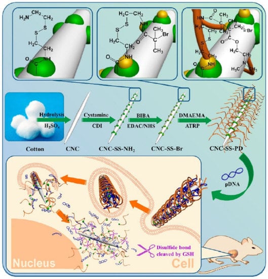
Figure 11.
Schematic representation of the preparation of CNC-SS-PD and their use for gene delivery. Reprinted with permission from Ref. [253]. 2015, ACS.
Cellulose-based materials offer several advantages for drug delivery. They can be used to release water-soluble and insoluble drugs and ionizable and hydrophobic drugs [246,260]. Thus, they enable the co-delivery of two drugs [261]. CNC hydrogels promoted locally targeted drug release [262] with sustainable properties [263]. Cellulose can be fabricated as capsules without gelatin [264]. It can be used for oral drug delivery [265]. Drug delivery using cellulose-based hydrogels can be simulated for pH- and temperature-responsive release.
7. Scaffolds for Skin, Bone, and Tissue Engineering
Nanocellulose-based materials offer several advantages as scaffolds for tissue engineering (Table 3) [266]. They show high biocompatibility, good water absorption, high water retention, high optical transparency, and good mechanical properties. They can be fabricated using comprehensive methods, including solvent casting, electrospinning, freeze-drying, and 3D printing [266]. Cellulose materials offer easy custom-made processes for tissue engineering of damaged tissues or organs. They can be optimized to ensure the required hierarchical structure, pore size, surface functional groups, and mechanical properties. Thus, cellulose-based materials are promising for engineering blood vessels, skin, and organs [266]. Scaffolds should meet several essential requirements, including high biocompatibility to mimic the natural extracellular matrix (ECM) of native tissue. They should also support cell growth, proliferation, and differentiation (Table 3).
Bacterial cellulose (BC) was used in tissue-engineered blood vessels (TEBV) [267]. BC-based TEBV is promising for caliber vascular grafts to reconstruct tissues associated with vascular diseases [267]. BC was used to fabricate artificial blood vessels that can be used for microsurgery [63]. The vessels were abbreviated as BASYC®, referring to BActerial SYnthesized Cellulose. The prepared vessels exhibited high mechanical strength in their wet form with high water retention. They also showed low roughness of the inner surface and provided complete “vitalization” in the rat. BASYC® was proposed as an artificial blood vessel in microsurgery [63].
BC was reported as a temporary skin substitute [254] and a scaffold for cartilage tissue engineering [268]. It was used for the in vitro seeding of cells, such as L929 cells/human osteoblasts [269]; human adipose-derived stem cells (hASCs) [270]; human urine-derived stem cells (hUSCs) [271]; human keratinocytes (HaCats) [272]; human vein endothelial cells (HVECs) [273,274]; human umbilical vein endothelial cells (HUVECs) [275]; equine-derived bone marrow mesenchymal stem cells (EqMSCs) [276]; human embryonic kidney 293 cells (HEK) [277]; bovine smooth muscle cells (SMCs) [275]; endothelial cells (ECs) [278]; chondrocytes [268]; epidermal cells [214]; and mouse leukemic monocyte-macrophage cells (RAW 264.7, Table 3) [220]. It showed high biocompatibility and promoted the proliferation of cells such as SMCs, leading to ingrowth of size > 40 μm after two weeks of culture on BC pellicles [267]. The proliferation of hASCs on BC film showed a plateau phase after nine days, indicating a single layer on the film [270]. Thus, BC is promising for tissue engineering.
BC pellicles showed the formation of an exemplary network similar to a collagen network [267]. It formed porous scaffolds with different pore sizes using sterile paraffin particles of varying size ranges (90–150 μm, 150–300 μm, and 300–500 μm) [271]. Biomolecules such as alginate were used to prepare sponges for oral tissue regeneration [272]. It can also be shaped into tubes using tubular template materials such as PDMS (polydimethylsiloxane) [275].
Cellulose offers several advantages. The cellulose composite exhibits high cell compatibility, water uptake, and mechanical strength. Cellulose-based materials exhibit good water and phosphate buffer saline (PBS) buffer stability. BC enabled the growth of multipotent mesenchymal stem cells (MSCs) [276]. BC scaffolds improved the adhesion, proliferation, and differentiation of MSCs [268]. Thus, BC/MSC hydrogel can be used to construct musculoskeletal tissue. Composites of polyvinyl alcohol (PVA) and BC can be promising biomaterials for soft tissue replacement [279].
The proliferation rates of several cells such as HaCats and gingival fibroblasts (GFs) using different supports were ordered in the following order: tissue culture plastic (TCP) > BC > BC-alginate (BCA) > alginate [272]. This character depends on the cell type. BC showed significantly higher chondrocyte growth levels than TCP and calcium alginate [268]. Unmodified BC supported the proliferation of chondrocytes at 50% of the collagen type II substrate [268]. However, BC-based materials exhibited good mechanical properties [268].
The 3D printing of bioink consisting of NFC and alginate was reported [280]. NFC ensures high shear-thinning properties, while alginate enables fast cross-linking ability. The 3D printing of bioink can be used for cartilage structures, including a human ear and sheep meniscus (Figure 12). The method can be used to print human chondrocyte nanocellulose-based bioink. Cellulose bioink showed 73% and 86% high cell viability after 1 and 7 days, respectively [280].
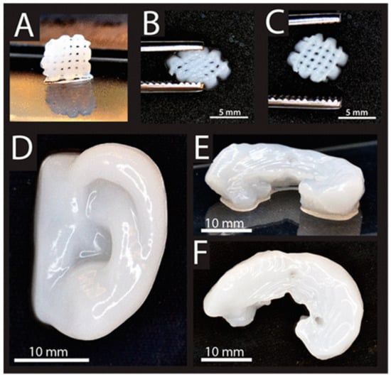
Figure 12.
The 3D printing of NFC–alginate into (A) small grids (7.2 × 7.2 mm2), (B) after squeezing, and (C) restored after squeezing; (D–F) 3D-printed human ear in different views. Reprinted with permission from Ref. [280]. 2015, ACS.
Table 3.
Applications of cellulose-based materials for tissue engineering.
Table 3.
Applications of cellulose-based materials for tissue engineering.
| Cellulose | Source | Form | Fabrication Method | Study Type | Cells | Time (d) | Evaluation Method | Comments | Ref. |
|---|---|---|---|---|---|---|---|---|---|
| BC | Acetobacter xylinum | Films | Shaken in a culture flask | In vitro In vivo | hASCs | 9 | Optical density (OD) | [270] | |
| Scaffold | Fermentation into sterile paraffin particles | hUSC | 7 | Histology | No effect of pore size | [271] | |||
| BC/alginate (BCA) | Sponge | Freeze-drying | In vitro | HaCat | 2 | MTT assay | 30% alginate | [272] | |
| BC | Scaffold | Culture on TCP | In vitro | EqMSCs | 14 | OD | The seeded cells were metabolically active | [276] | |
| BC | Tubes | Culture on PDMS tubes | In vitro In vivo | SMCs | 7 | No signs of inflammation | [275] | ||
| BC-CMC | Gluconacetobacter saccharivorans | Gel | Agitation overnight at room temperature | In vitro | HEK | 1 | Optical microscope | [281] | |
| BC | Acetobacter xylinum | Tubes | Fermentation in glass tubes using a silicone support | In vitro | ECs | 28 | F Fluorescence microscope | [278] | |
| Scaffold | Freeze-drying | In-vitro In-vivo | Chondrocyte | 8 | [268] |
Notes: MTT (3-(4,5-dimethyl thiazolyl-2)-2,5-diphenyltetrazolium bromide) assay; Acetobacter xylinum and Gluconacetobacter xylinus are used for the same strains.
Cellulose has been used for bone regeneration. BC assisted the synthesis of hydroxyapatite (HAP) with calcium-deficient and crystallite particles of nanoscale size [282,283]. BC was oxidized before the formation of HAP to produce highly degradable materials. HAP/BC was used for bone regeneration using osseous tissue [282]. The material can be degraded under physiological conditions, i.e., pH and temperature of 7 and 37 °C, respectively. Thus, it was proposed to stimulate bone colonization [282]. A composite of HAP/BC was reported to treat non-critical bone defects in rats [284]. The bone defects were repaired entirely after 16 days [284].
A nanocomposite material of BC networks and HAP was prepared using a wet chemical precipitation method using aqueous calcium nitrate and di-ammonium phosphate salts [277]. The dispersion can be improved by adding CMC (1% w/v). CMC increased the pore size of BC by 47.8%. The prepared composite, i.e., BC/HAP/CMC, supported the growth of HEK cells [277].
BC scaffolds were used for the growth of EqMSCs to apply in bone and cartilage tissue engineering [276]. They can be fabricated via freeze-drying. They can be prepared with fiber diameters and pore sizes of 32.08 ± 10.85 nm and 254.16 ± 76.65 nm, respectively. They exhibited high cytocompatibility and supported the adhesion and proliferation of the cells. They maintained the differentiation of EqMSCs. EqMSC/BC scaffolds are promising for bone and cartilage regeneration tissue engineering, similar to those reported using tissue-culture-treated plastic (TCP) [276].
Cellulose materials have also been reported for other biomedical implants, such as artificial cornea [285], urinary conduits [271], neuronal regeneration [286], and endodontic treatment [287]. Cellulose and its composites are promising and can be implemented in other applications.
Cellulose-based materials offer several advantages for tissue engineering. They provided high biocompatibility [288] and self-healing properties [289]. Cellulose materials can be conjugated with polymers to improve their properties. A rigid composite of tannic acid (TA)/CNC (TA@CNC) was incorporated into poly(vinyl alcohol) (PVA)-borax networks [290]. TA@CNC/PVA-borax hydrogels offered high toughness with self-healing properties [290]. The structure of cellulose materials such as BC hydrogel enables the proliferation of HVECs horizontally and their migration vertically [273,274]. Cell penetration into BC hydrogels depends on oxygen availability inside the hydrogels.
8. Advantages and Disadvantages of Cellulose-Based Nanomaterials
Cellulose exhibits high biocompatibility and hemocompatibility with good biodegradability. Mesenchymal stem cells can grow on a cellulose membrane with viability higher than 95% [291]. However, cellulose materials show slow or no degradation in vivo and in vitro. This property makes them suitable for applications as a scaffold. The degradability of cellulose can be improved via chemical oxidation of the amorphous regions of BC using TEMPO-mediated oxidation [285,292], periodate [285], exposure to γ-radiation [293], or incorporation with an enzyme [294]. The chemical oxidation form 2,3-dialdehyde cellulose can easily degrade in vivo and in vitro. BC membranes can also be degraded via γ-radiation within 2–4 weeks [293].
The mechanical strength of nanomaterials can be improved using cellulose nanomaterials. Cellulose exhibits good mechanical properties [295]. Both air-dried and hot-pressed BC exhibit Young’s moduli higher than 18 GPa. Air-dried and hot-pressed BC show tensile strengths of >260 MPa and 216 MPa, respectively. Atomic force microscopy (AFM) was used to measure the mechanical properties of single BC nanofibers with diameters of 35–90 nm [296]. Data analysis revealed a high Young’s modulus of 78 ± 17 GPa [296]. BC-based hydrogel exhibited an elastic modulus of 1.7 MPa [297]. It possessed a very high water-holding capacity, even under pressures as high as 3.7 MPa. It recovered its original shape, even after repeated compression [297].
Cellulose retains moisture. The hydroxyl groups of cellulose material maintain the material’s hydrophilicity. The hydroxyl groups have high water capacity with good mechanical and thermal properties. These functional groups play an essential role in interfacial interactions during a composite [285].
BC offers protection from UV radiation and heavy metal stress. Cellulose exhibits resistance to aerobic environments.
The homogeneous distribution of cells on cellulose is vital for tissue engineering. The native BC membrane suffers from cell aggregation [298]. However, the modification of cellulose with RGD protein improved the distribution of cells [298]. The growth rate of muscle-derived cells can be increased by increasing the starch content [299]. The increase in the cell growth rate may be due to the porous nature of the composite [299].
9. Outlook
CNCs have been commercialized by several companies, such as Bio Vision (Canada), CelluForce (Canada; the trade name is NCC™), and US Forest Service Forest Products Laboratory (USA), while CNFs have been marketed by several European companies, such as Centre Technique du Papier (France)), Borregaard ChemCell (Norway), Innventia AB (Sweden), Stora Enso (Finland), and UPM fibril cellulose (Finland). Plant-based cellulose has been commercialized in several products, such as cellophane (transparent films), Rayon or Tencel™ (synthetic textile fibers), Bionext® (commercial BC extracted from Acetobacter xylinum), Surgicel™, and Interceed®. BC-based materials have been commercialized in several products, such as Bioprocess®, BASYC®, Biofill®, XCell®, and Gengiflex®. Microbial-based cellulose is free of lignin and hemicelluloses, unlike cellulose extracted from plant sources. For example, bacterial cellulose (BC) exhibits higher purity and better crystallinity than plant-derived cellulose. Thus, BC is suitable for biomedical applications. However, microbial-based cellulose requires high-security precautions to avoid the presence of microbial species inside the extracted cellulose. Cellulose can be produced from cheap sources and waste materials [300,301,302]. However, most of the current technologies for cellulose fabrication at the nanoscale are still expensive, require tedious efforts, and lack large-scale production for industrial and biomedical applications. The output of nanocellulose with high purity and the absence of endotoxins is highly needed for biomedical applications.
Cellulose-based antimicrobial agents are promising for the fabrication of membranes [50], fabrics [152], textiles [303,304,305], and food packaging [306,307]. They possess durable antibacterial activity with tunable properties such as hydrophobicity [308]. They can be modified with different antibacterial agents with permanent antibacterial properties [309]. Nanoparticles exhibit undesirable toxicity. Thus, some precautions or post-synthetic treatments should be performed. For instance, sulfidation was proposed to transform Ag NPs into highly insoluble forms for minimal cytotoxicity [310].
Cellulose-based materials can serve as green cross-linkers [311] and flexible platforms [312] for tissue engineering. They can be modified via in situ and ex-situ procedures [313]. They can be processed into several forms using various methods. Most of the available techniques lack large-scale production and require other materials such as binders or modifiers. Further investigations are needed to develop cellulose materials that meet customized requirements at a minimal cost.
Author Contributions
Conceptualization, H.N.A.; methodology, H.N.A.; software, H.N.A.; validation, H.N.A.; formal analysis, H.N.A.; investigation, H.N.A.; resources, H.N.A.; data curation, H.N.A.; writing—original draft preparation, H.N.A.; writing—review and editing, H.N.A. and A.P.M.; visualization, H.N.A.; supervision, H.N.A.; project administration, H.N.A.; funding acquisition, H.N.A. All authors have read and agreed to the published version of the manuscript.
Funding
This research was funded by The Swedish Foundation for Strategic Environmental Research (Mistra) under MISTRA TerraClean (project No. 2015/31).
Informed Consent Statement
Not applicable.
Data Availability Statement
The data presented in this study are available on request from the corresponding author.
Acknowledgments
Stockholm University Library sponsors open-access publications. This project is funded by The Swedish Foundation for Strategic Environmental Research (Mistra) under MISTRA TerraClean (project No. 2015/31). The preprint of this review was published in Ref. [314].
Conflicts of Interest
The authors declare no conflict of interest.
References
- Quirke, V.; Gaudillière, J.-P. The Era of Biomedicine: Science, Medicine, and Public Health in Britain and France after the Second World War. Med. Hist. 2008, 52, 441–452. [Google Scholar] [CrossRef] [PubMed]
- Wu, D.; Zhou, J.; Creyer, M.N.; Yim, W.; Chen, Z.; Messersmith, P.B.; Jokerst, J.V. Phenolic-enabled nanotechnology: Versatile particle engineering for biomedicine. Chem. Soc. Rev. 2021, 50, 4432–4483. [Google Scholar] [CrossRef] [PubMed]
- Nikzamir, M.; Akbarzadeh, A.; Panahi, Y. An overview on nanoparticles used in biomedicine and their cytotoxicity. J. Drug Deliv. Sci. Technol. 2021, 61, 102316. [Google Scholar] [CrossRef]
- Liu, X.; Liang, T.; Zhang, R.; Ding, Q.; Wu, S.; Li, C.; Lin, Y.; Ye, Y.; Zhong, Z.; Zhou, M. Iron-Based Metal–Organic Frameworks in Drug Delivery and Biomedicine. ACS Appl. Mater. Interfaces 2021, 13, 9643–9655. [Google Scholar] [CrossRef]
- Swingler, S.; Gupta, A.; Gibson, H.; Kowalczuk, M.; Heaselgrave, W.; Radecka, I. Recent Advances and Applications of Bacterial Cellulose in Biomedicine. Polymers 2021, 13, 412. [Google Scholar] [CrossRef]
- Klemm, D.; Heublein, B.; Fink, H.-P.; Bohn, A. Cellulose: Fascinating Biopolymer and Sustainable Raw Material. Angew. Chem. Int. Ed. 2005, 44, 3358–3393. [Google Scholar] [CrossRef]
- Klemm, D.; Kramer, F.; Moritz, S.; Lindström, T.; Ankerfors, M.; Gray, D.; Dorris, A. Nanocelluloses: A New Family of Nature-Based Materials. Angew. Chem. Int. Ed. 2011, 50, 5438–5466. [Google Scholar] [CrossRef]
- Czaja, W.K.; Young, D.J.; Kawecki, M.; Brown, R.M. The Future Prospects of Microbial Cellulose in Biomedical Applications. Biomacromolecules 2007, 8, 1–12. [Google Scholar] [CrossRef]
- Kaushik, M.; Moores, A. Review: Nanocelluloses as versatile supports for metal nanoparticles and their applications in catalysis. Green Chem. 2016, 18, 622–637. [Google Scholar] [CrossRef] [Green Version]
- Moon, R.J.; Martini, A.; Nairn, J.; Simonsen, J.; Youngblood, J. Cellulose nanomaterials review: Structure, properties and nanocomposites. Chem. Soc. Rev. 2011, 40, 3941. [Google Scholar] [CrossRef]
- Lam, E.; Hemraz, U.D. Preparation and Surface Functionalization of Carboxylated Cellulose Nanocrystals. Nanomaterials 2021, 11, 1641. [Google Scholar] [CrossRef] [PubMed]
- Calvino, C.; Macke, N.; Kato, R.; Rowan, S.J. Development, processing and applications of bio-sourced cellulose nanocrystal composites. Prog. Polym. Sci. 2020, 103, 101221. [Google Scholar] [CrossRef]
- Zaki, M.; Khali, H.P.S.A.; Sabaruddin, F.A.; Bairwan, R.D.; Oyekanmi, A.A.; Alfatah, T.; Danish, M.; Mistar, E.M.; Abdullah, C.K. Microbial treatment for nanocellulose extraction from marine algae and its applications as sustainable functional material. Bioresour. Technol. Rep. 2021, 16, 100811. [Google Scholar] [CrossRef]
- Hafid, H.S.; Omar, F.N.; Zhu, J.; Wakisaka, M. Enhanced crystallinity and thermal properties of cellulose from rice husk using acid hydrolysis treatment. Carbohydr. Polym. 2021, 260, 117789. [Google Scholar] [CrossRef] [PubMed]
- Baghel, R.S.; Reddy, C.R.K.; Singh, R.P. Seaweed-based cellulose: Applications, and future perspectives. Carbohydr. Polym. 2021, 267, 118241. [Google Scholar] [CrossRef] [PubMed]
- Abdel-Halim, E.S. Chemical modification of cellulose extracted from sugarcane bagasse: Preparation of hydroxyethyl cellulose. Arab. J. Chem. 2014, 7, 362–371. [Google Scholar] [CrossRef] [Green Version]
- Dutta, S.; Kim, J.; Ide, Y.; Ho Kim, J.; Hossain, M.S.A.; Bando, Y.; Yamauchi, Y.; Wu, K.C.-W. 3D network of cellulose-based energy storage devices and related emerging applications. Mater. Horiz. 2017, 4, 522–545. [Google Scholar] [CrossRef] [Green Version]
- Grishkewich, N.; Mohammed, N.; Tang, J.; Tam, K.C. Recent advances in the application of cellulose nanocrystals. Curr. Opin. Colloid Interface Sci. 2017, 29, 32–45. [Google Scholar] [CrossRef]
- Georgouvelas, D.; Abdelhamid, H.N.; Li, J.; Edlund, U.; Mathew, A.P. All-cellulose functional membranes for water treatment: Adsorption of metal ions and catalytic decolorization of dyes. Carbohydr. Polym. 2021, 264, 118044. [Google Scholar] [CrossRef]
- Cheng, H.; Lijie, L.; Wang, B.; Feng, X.; Mao, Z.; Vancso, G.J.; Sui, X. Multifaceted applications of cellulosic porous materials in environment, energy, and health. Prog. Polym. Sci. 2020, 106, 101253. [Google Scholar] [CrossRef]
- Pelegrini, B.L.; Ré, F.; de Oliveira, M.M.; Fernandes, T.; Oliveira, J.H.; Junior, A.G.O.; Girotto, E.M.; Nakamura, C.V.; Sampaio, A.R.; Valim, A.; et al. Cellulose Nanocrystals as a Sustainable Raw Material: Cytotoxicity and Applications on Healthcare Technology. Macromol. Mater. Eng. 2019, 304, 1900092. [Google Scholar] [CrossRef]
- Lv, P.; Lu, X.; Wang, L.; Feng, W. Nanocellulose-Based Functional Materials: From Chiral Photonics to Soft Actuator and Energy Storage. Adv. Funct. Mater. 2021, 31, 2104991. [Google Scholar] [CrossRef]
- Chen, C.; Ding, W.; Zhang, H.; Zhang, L.; Huang, Y.; Fan, M.; Yang, J.; Sun, D. Bacterial cellulose-based biomaterials: From fabrication to application. Carbohydr. Polym. 2022, 278, 118995. [Google Scholar] [CrossRef] [PubMed]
- Karimian, A.; Parsian, H.; Majidinia, M.; Rahimi, M.; Mir, S.M.; Kafil, H.S.; Shafiei-Irannejad, V.; Kheyrollah, M.; Ostadi, H.; Yousefi, B. Nanocrystalline cellulose: Preparation, physicochemical properties, and applications in drug delivery systems. Int. J. Biol. Macromol. 2019, 133, 850–859. [Google Scholar] [CrossRef] [PubMed]
- Abdelhamid, H.N.; Mathew, A.P. Cellulose-zeolitic imidazolate frameworks (CelloZIFs) for multifunctional environmental remediation: Adsorption and catalytic degradation. Chem. Eng. J. 2021, 426, 131733. [Google Scholar] [CrossRef]
- Abdelhamid, H.N.; Mathew, A.P. Cellulose-Based Materials for Water Remediation: Adsorption, Catalysis, and Antifouling. Front. Chem. Eng. 2021, 3, 790314. [Google Scholar] [CrossRef]
- Abdelhamid, H.N.; Mathew, A.P. In-situ growth of zeolitic imidazolate frameworks into a cellulosic filter paper for the reduction of 4-nitrophenol. Carbohydr. Polym. 2021, 274, 118657. [Google Scholar] [CrossRef]
- Yang, J.; Li, J. Self-assembled cellulose materials for biomedicine: A review. Carbohydr. Polym. 2018, 181, 264–274. [Google Scholar] [CrossRef]
- Agarwal, C.; Csóka, L. Surface-modified cellulose in biomedical engineering. In Materials for Biomedical Engineering; Elsevier: Amsterdam, The Netherlands, 2019; pp. 215–261. [Google Scholar] [CrossRef]
- Ul-Islam, M.; Khan, S.; Khattak, W.A.; Ullah, M.W.; Park, J.K. Synthesis, Chemistry, and Medical Application of Bacterial Cellulose Nanocomposites. In Eco-Friendly Polymer Nanocomposites; Advanced Structured Materials; Thakur, V., Thakur, M., Eds.; Springer: New Delhi, India, 2015; Volume 74, pp. 399–437. [Google Scholar] [CrossRef]
- Nehra, P.; Chauhan, R.P. Eco-friendly nanocellulose and its biomedical applications: Current status and future prospect. J. Biomater. Sci. Polym. Ed. 2021, 32, 112–149. [Google Scholar] [CrossRef]
- Parvulescu, O.C.; Isopencu, G.; Busuioc, C.; Raducanu, C.; Mocanu, A.; Deleanu, I.; Stoica-Guzun, A. Antimicrobial bacterial cellulose composites as textile materials. In Antimicrobial Textiles from Natural Resources; Woodhead Publishing, Elsevier: Amsterdam, The Netherlands, 2021; pp. 513–556. [Google Scholar] [CrossRef]
- Ullah, S.; Chen, X. Fabrication, applications and challenges of natural biomaterials in tissue engineering. Appl. Mater. Today 2020, 20, 100656. [Google Scholar] [CrossRef]
- Jorfi, M.; Foster, E.J. Recent advances in nanocellulose for biomedical applications. J. Appl. Polym. Sci. 2015, 132, 41719. [Google Scholar] [CrossRef]
- Long, W.; Ouyang, H.; Hu, X.; Liu, M.; Zhang, X.; Feng, Y.; Wei, Y. State-of-art review on preparation, surface functionalization and biomedical applications of cellulose nanocrystals-based materials. Int. J. Biol. Macromol. 2021, 186, 591–615. [Google Scholar] [CrossRef] [PubMed]
- Rahimi, S.K.; Aeinehvand, R.; Kim, K.; Otaigbe, J.U. Structure and Biocompatibility of Bioabsorbable Nanocomposites of Aliphatic-Aromatic Copolyester and Cellulose Nanocrystals. Biomacromolecules 2017, 18, 2179–2194. [Google Scholar] [CrossRef] [PubMed]
- Sunasee, R.; Hemraz, U.D.; Ckless, K. Cellulose nanocrystals: A versatile nanoplatform for emerging biomedical applications. Expert Opin. Drug Deliv. 2016, 13, 1243–1256. [Google Scholar] [CrossRef]
- Lin, N.; Dufresne, A. Nanocellulose: Biomedical Nanomaterial Applications. In Encyclopedia of Biomedical Polymers and Polymeric Biomaterials; Taylor & Francis: New York, NY, USA, 2015; pp. 5077–5100. [Google Scholar] [CrossRef]
- Oz, Y.E.; Keskin-Erdogan, Z.; Safa, N.; Tuna, E.E.H. A review of functionalised bacterial cellulose for targeted biomedical fields. J. Biomater. Appl. 2021, 36, 648–681. [Google Scholar] [CrossRef]
- Blanco, F.G.; Hernández, N.; Rivero-Buceta, V.; Maestro, B.; Sanz, J.M.; Mato, A.; Hernández-Arriaga, A.M.; Prieto, M.A. From Residues to Added-Value Bacterial Biopolymers as Nanomaterials for Biomedical Applications. Nanomaterials 2021, 11, 1492. [Google Scholar] [CrossRef]
- Thomas, P.; Duolikun, T.; Rumjit, N.P.; Moosavi, S.; Lai, C.W.; Bin Johan, M.R.; Fen, L.B. Comprehensive review on nanocellulose: Recent developments, challenges and future prospects. J. Mech. Behav. Biomed. Mater. 2020, 110, 103884. [Google Scholar] [CrossRef]
- Ullah, H.; Wahid, F.; Santos, H.A.; Khan, T. Advances in biomedical and pharmaceutical applications of functional bacterial cellulose-based nanocomposites. Carbohydr. Polym. 2016, 150, 330–352. [Google Scholar] [CrossRef]
- Abitbol, T.; Rivkin, A.; Cao, Y.; Nevo, Y.; Abraham, E.; Ben-Shalom, T.; Lapidot, S.; Shoseyov, O. Nanocellulose, a tiny fiber with huge applications. Curr. Opin. Biotechnol. 2016, 39, 76–88. [Google Scholar] [CrossRef]
- Rajwade, J.M.; Paknikar, K.M.; Kumbhar, J.V. Applications of bacterial cellulose and its composites in biomedicine. Appl. Microbiol. Biotechnol. 2015, 99, 2491–2511. [Google Scholar] [CrossRef]
- Khattak, S.; Wahid, F.; Liu, L.-P.; Jia, S.-R.; Chu, L.-Q.; Xie, Y.-Y.; Li, Z.-X.; Zhong, C. Applications of cellulose and chitin/chitosan derivatives and composites as antibacterial materials: Current state and perspectives. Appl. Microbiol. Biotechnol. 2019, 103, 1989–2006. [Google Scholar] [CrossRef] [PubMed]
- Sun, L.; Ding, C.; Qian, X.; An, X. Effective and simple one-step strategy for preparation of “clickable” cellulose modules: Support to build antibacterial materials. Cellulose 2019, 26, 1961–1976. [Google Scholar] [CrossRef]
- Li, J.; Cha, R.; Mou, K.; Zhao, X.; Long, K.; Luo, H.; Zhou, F.; Jiang, X. Nanocellulose-Based Antibacterial Materials. Adv. Healthc. Mater. 2018, 7, 1800334. [Google Scholar] [CrossRef] [PubMed]
- Vázquez, E.; Duarte, L.; López-Saucedo, F.; Flores-Rojas, G.G.; Bucio, E. Cellulose-Based Antimicrobial Materials. In Advanced Antimicrobial Materials and Applications; Springer Nature: Singapore, 2021; pp. 61–85. [Google Scholar] [CrossRef]
- Alavi, M. Modifications of microcrystalline cellulose (MCC), nanofibrillated cellulose (NFC), and nanocrystalline cellulose (NCC) for antimicrobial and wound healing applications. e-Polymers 2019, 19, 103–119. [Google Scholar] [CrossRef]
- Coma, V.; Freire, C.S.R.; Silvestre, A.J.D. Recent Advances on the Development of AntibacterialAntibacterial activity Polysaccharide-Based MaterialsBiomedical products and devices. In Polysaccharides; Springer International Publishing: Cham, Switzerland, 2015; pp. 1751–1803. [Google Scholar] [CrossRef]
- Claro, F.C.; Jordão, C.; de Viveiros, B.M.; Isaka, L.J.E.; Junior, J.A.V.; Magalhães, W.L.E. Low cost membrane of wood nanocellulose obtained by mechanical defibrillation for potential applications as wound dressing. Cellulose 2020, 27, 10765–10779. [Google Scholar] [CrossRef]
- Resch, A.; Staud, C.; Radtke, C. Nanocellulose-based wound dressing for conservative wound management in children with second-degree burns. Int. Wound J. 2021, 18, 478–486. [Google Scholar] [CrossRef]
- Hakkarainen, T.; Koivuniemi, R.; Kosonen, M.; Escobedo-Lucea, C.; Sanz-Garcia, A.; Vuola, J.; Valtonen, J.; Tammela, P.; Mäkitie, A.; Luukko, K.; et al. Nanofibrillar cellulose wound dressing in skin graft donor site treatment. J. Control. Release 2016, 244, 292–301. [Google Scholar] [CrossRef]
- Cattelaens, J.; Turco, L.; Berclaz, L.M.; Huelsse, B.; Hitzl, W.; Vollkommer, T.; Bodenschatz, K.J. The Impact of a Nanocellulose-Based Wound Dressing in the Management of Thermal Injuries in Children: Results of a Retrospective Evaluation. Life 2020, 10, 212. [Google Scholar] [CrossRef]
- Knutsen, M.F.; Agrenius, K.; Ugland, H.; Petronis, S.; Haglerod, C.; Håkansson, J.; Chinga-Carrasco, G. Oxygenated Nanocellulose—A Material Platform for Antibacterial Wound Dressing Devices. ACS Appl. Bio Mater. 2021, 4, 7554–7562. [Google Scholar] [CrossRef]
- He, W.; Wu, J.; Xu, J.; Mosselhy, D.A.; Zheng, Y.; Yang, S. Bacterial Cellulose: Functional Modification and Wound Healing Applications. Adv. Wound Care 2021, 10, 623–640. [Google Scholar] [CrossRef]
- Sulaeva, I.; Henniges, U.; Rosenau, T.; Potthast, A. Bacterial cellulose as a material for wound treatment: Properties and modifications. A review. Biotechnol. Adv. 2015, 33, 1547–1571. [Google Scholar] [CrossRef] [PubMed]
- Venkatesan, J.; Anil, S.; Kim, S.-K.; Shim, M. Seaweed Polysaccharide-Based Nanoparticles: Preparation and Applications for Drug Delivery. Polymers 2016, 8, 30. [Google Scholar] [CrossRef] [Green Version]
- Islam, S.U.; Ul-Islam, M.; Ahsan, H.; Ahmed, M.B.; Shehzad, A.; Fatima, A.; Sonn, J.K.; Lee, Y.S. Potential applications of bacterial cellulose and its composites for cancer treatment. Int. J. Biol. Macromol. 2021, 168, 301–309. [Google Scholar] [CrossRef] [PubMed]
- Seabra, A.B.; Bernardes, J.S.; Fávaro, W.J.; Paula, A.J.; Durán, N. Cellulose nanocrystals as carriers in medicine and their toxicities: A review. Carbohydr. Polym. 2018, 181, 514–527. [Google Scholar] [CrossRef] [PubMed]
- Kupnik, K.; Primožič, M.; Kokol, V.; Leitgeb, M. Nanocellulose in Drug Delivery and Antimicrobially Active Materials. Polymers 2020, 12, 2825. [Google Scholar] [CrossRef] [PubMed]
- Khalil, H.P.S.A.; Jummaat, F.; Yahya, E.B.; Olaiya, N.G.; Adnan, A.S.; Abdat, M.; Nasir, N.A.M.; Halim, A.S.; Kumar, U.S.U.; Bairwan, R.; et al. A Review on Micro- to Nanocellulose Biopolymer Scaffold Forming for Tissue Engineering Applications. Polymers 2020, 12, 2043. [Google Scholar] [CrossRef]
- Klemm, D.; Schumann, D.; Udhardt, U.; Marsch, S. Bacterial synthesized cellulose—artificial blood vessels for microsurgery. Prog. Polym. Sci. 2001, 26, 1561–1603. [Google Scholar] [CrossRef]
- Tang, J.; Bao, L.; Li, X.; Chen, L.; Hong, F.F. Potential of PVA-doped bacterial nano-cellulose tubular composites for artificial blood vessels. J. Mater. Chem. B 2015, 3, 8537–8547. [Google Scholar] [CrossRef]
- Sadeghifar, H.; Venditti, R.; Jur, J.; Gorga, R.E.; Pawlak, J.J. Cellulose-Lignin Biodegradable and Flexible UV Protection Film. ACS Sustain. Chem. Eng. 2017, 5, 625–631. [Google Scholar] [CrossRef]
- Mendoza, D.J.; Mouterde, L.M.M.; Browne, C.; Raghuwanshi, V.S.; Simon, G.P.; Garnier, G.; Allais, F. Grafting Nature-Inspired and Bio-Based Phenolic Esters onto Cellulose Nanocrystals Gives Biomaterials with Photostable Anti-UV Properties. ChemSusChem 2020, 13, 6552–6561. [Google Scholar] [CrossRef]
- Du, H.; Liu, W.; Zhang, M.; Si, C.; Zhang, X.; Li, B. Cellulose nanocrystals and cellulose nanofibrils based hydrogels for biomedical applications. Carbohydr. Polym. 2019, 209, 130–144. [Google Scholar] [CrossRef] [PubMed]
- Rahmanian, V.; Pirzada, T.; Wang, S.; Khan, S.A. Cellulose-Based Hybrid Aerogels: Strategies toward Design and Functionality. Adv. Mater. 2021, 33, 2102892. [Google Scholar] [CrossRef] [PubMed]
- De France, K.J.; Hoare, T.; Cranston, E.D. Review of Hydrogels and Aerogels Containing Nanocellulose. Chem. Mater. 2017, 29, 4609–4631. [Google Scholar] [CrossRef]
- Douglass, E.F.; Avci, H.; Boy, R.; Rojas, O.J.; Kotek, R. A Review of Cellulose and Cellulose Blends for Preparation of Bio-derived and Conventional Membranes, Nanostructured Thin Films, and Composites. Polym. Rev. 2018, 58, 102–163. [Google Scholar] [CrossRef]
- Sultan, S.; Siqueira, G.; Zimmermann, T.; Mathew, A.P. 3D printing of nano-cellulosic biomaterials for medical applications. Curr. Opin. Biomed. Eng. 2017, 2, 29–34. [Google Scholar] [CrossRef]
- Shavandi, A.; Hosseini, S.; Okoro, O.V.; Nie, L.; Babadi, F.E.; Melchels, F. 3D Bioprinting of Lignocellulosic Biomaterials. Adv. Healthc. Mater. 2020, 9, 2001472. [Google Scholar] [CrossRef]
- Tayeb, A.; Amini, E.; Ghasemi, S.; Tajvidi, M. Cellulose Nanomaterials—Binding Properties and Applications: A Review. Molecules 2018, 23, 2684. [Google Scholar] [CrossRef] [Green Version]
- Yang, Y.; Lu, Y.; Zeng, K.; Heinze, T.; Groth, T.; Zhang, K. Recent Progress on Cellulose-Based Ionic Compounds for Biomaterials. Adv. Mater. 2021, 33, 2000717. [Google Scholar] [CrossRef] [Green Version]
- Tortorella, S.; Buratti, V.V.; Maturi, M.; Sambri, L.; Franchini, M.C.; Locatelli, E. Surface-Modified Nanocellulose for Application in Biomedical Engineering and Nanomedicine: A Review. Int. J. Nanomed. 2020, 15, 9909–9937. [Google Scholar] [CrossRef]
- Habibi, Y. Key advances in the chemical modification of nanocelluloses. Chem. Soc. Rev. 2014, 43, 1519–1542. [Google Scholar] [CrossRef]
- Eyley, S.; Thielemans, W. Surface modification of cellulose nanocrystals. Nanoscale 2014, 6, 7764–7779. [Google Scholar] [CrossRef] [Green Version]
- Abdelhamid, H.N.; Badr, G. Nanobiotechnology as a platform for the diagnosis of COVID-19: A review. Nanotechnol. Environ. Eng. 2021, 6, 19. [Google Scholar] [CrossRef]
- Mali, P.; Sherje, A.P. Cellulose nanocrystals: Fundamentals and biomedical applications. Carbohydr. Polym. 2022, 275, 118668. [Google Scholar] [CrossRef] [PubMed]
- Yang, H.; Alam, M.N.; van de Ven, T.G.M. Highly charged nanocrystalline cellulose and dicarboxylated cellulose from periodate and chlorite oxidized cellulose fibers. Cellulose 2013, 20, 1865–1875. [Google Scholar] [CrossRef]
- Dash, M.; Chiellini, F.; Ottenbrite, R.M.; Chiellini, E. Chitosan—A versatile semi-synthetic polymer in biomedical applications. Prog. Polym. Sci. 2011, 36, 981–1014. [Google Scholar] [CrossRef]
- Tavakolian, M.; Jafari, S.M.; van de Ven, T.G.M. A Review on Surface-Functionalized Cellulosic Nanostructures as Biocompatible Antibacterial Materials. Nano-Micro Lett. 2020, 12, 73. [Google Scholar] [CrossRef] [Green Version]
- Mou, K.; Li, J.; Wang, Y.; Cha, R.; Jiang, X. 2,3-Dialdehyde nanofibrillated cellulose as a potential material for the treatment of MRSA infection. J. Mater. Chem. B 2017, 5, 7876–7884. [Google Scholar] [CrossRef]
- Zhang, L.; Ge, H.; Xu, M.; Cao, J.; Dai, Y. Physicochemical properties, antioxidant and antibacterial activities of dialdehyde microcrystalline cellulose. Cellulose 2017, 24, 2287–2298. [Google Scholar] [CrossRef]
- Jacob, J.; Haponiuk, J.; Thomas, S.; Peter, G.; Gopi, S. Use of Ginger Nanofibers for the Preparation of Cellulose Nanocomposites and Their Antimicrobial Activities. Fibers 2018, 6, 79. [Google Scholar] [CrossRef] [Green Version]
- Jack, A.A.; Nordli, H.R.; Powell, L.C.; Powell, K.A.; Kishnani, H.; Johnsen, P.O.; Pukstad, B.; Thomas, D.W.; Chinga-Carrasco, G.; Hill, K.E. The interaction of wood nanocellulose dressings and the wound pathogen P. aeruginosa. Carbohydr. Polym. 2017, 157, 1955–1962. [Google Scholar] [CrossRef]
- Chinga-Carrasco, G.; Johansson, J.; Heggset, E.B.; Leirset, I.; Björn, C.; Agrenius, K.; Stevanic, J.S.; Håkansson, J. Characterization and Antibacterial Properties of Autoclaved Carboxylated Wood Nanocellulose. Biomacromolecules 2021, 22, 2779–2789. [Google Scholar] [CrossRef] [PubMed]
- Gorbet, M.B.; Sefton, M.V. Endotoxin: The uninvited guest. Biomaterials 2005, 26, 6811–6817. [Google Scholar] [CrossRef] [PubMed]
- Nordli, H.R.; Chinga-Carrasco, G.; Rokstad, A.M.; Pukstad, B. Producing ultrapure wood cellulose nanofibrils and evaluating the cytotoxicity using human skin cells. Carbohydr. Polym. 2016, 150, 65–73. [Google Scholar] [CrossRef] [Green Version]
- Čolić, M.; Mihajlović, D.; Mathew, A.; Naseri, N.; Kokol, V. Cytocompatibility and immunomodulatory properties of wood based nanofibrillated cellulose. Cellulose 2015, 22, 763–778. [Google Scholar] [CrossRef]
- Pham, T.C.; Nguyen, V.-N.; Choi, Y.; Lee, S.; Yoon, J. Recent Strategies to Develop Innovative Photosensitizers for Enhanced Photodynamic Therapy. Chem. Rev. 2021, 121, 13454–13619. [Google Scholar] [CrossRef]
- Peddinti, B.S.T.; Morales-Gagnon, N.; Pourdeyhimi, B.; Scholle, F.; Spontak, R.J.; Ghiladi, R.A. Photodynamic Coatings on Polymer Microfibers for Pathogen Inactivation: Effects of Application Method and Composition. ACS Appl. Mater. Interfaces 2021, 13, 155–163. [Google Scholar] [CrossRef]
- Maldonado-Carmona, N.; Ouk, T.-S.; Calvete, M.J.F.; Pereira, M.M.; Villandier, N.; Leroy-Lhez, S. Conjugating biomaterials with photosensitizers: Advances and perspectives for photodynamic antimicrobial chemotherapy. Photochem. Photobiol. Sci. 2020, 19, 445–461. [Google Scholar] [CrossRef]
- Mesquita, M.Q.; Dias, C.J.; Neves, M.G.P.M.S.; Almeida, A.; Faustino, M.A.F. Revisiting Current Photoactive Materials for Antimicrobial Photodynamic Therapy. Molecules 2018, 23, 2424. [Google Scholar] [CrossRef] [Green Version]
- George, L.; Hiltunen, A.; Santala, V.; Efimov, A. Photo-antimicrobial efficacy of zinc complexes of porphyrin and phthalocyanine activated by inexpensive consumer LED lamp. J. Inorg. Biochem. 2018, 183, 94–100. [Google Scholar] [CrossRef]
- Feese, E.; Sadeghifar, H.; Gracz, H.S.; Argyropoulos, D.S.; Ghiladi, R.A. Photobactericidal Porphyrin-Cellulose Nanocrystals: Synthesis, Characterization, and Antimicrobial Properties. Biomacromolecules 2011, 12, 3528–3539. [Google Scholar] [CrossRef]
- Koshani, R.; Zhang, J.; van de Ven, T.G.M.; Lu, X.; Wang, Y. Modified Hairy Nanocrystalline Cellulose as Photobactericidal Nanofillers for Food Packaging Application. ACS Sustain. Chem. Eng. 2021, 9, 10513–10523. [Google Scholar] [CrossRef]
- Zhuo, J.; Sun, G. Antimicrobial Functions on Cellulose Materials Introduced by Anthraquinone Vat Dyes. ACS Appl. Mater. Interfaces 2013, 5, 10830–10835. [Google Scholar] [CrossRef] [PubMed]
- Hou, A.; Feng, G.; Zhuo, J.; Sun, G. UV Light-Induced Generation of Reactive Oxygen Species and Antimicrobial Properties of Cellulose Fabric Modified by 3,3′,4,4′-Benzophenone Tetracarboxylic Acid. ACS Appl. Mater. Interfaces 2015, 7, 27918–27924. [Google Scholar] [CrossRef] [PubMed]
- Sun, B.; Zhang, Y.; Li, W.; Xu, X.; Zhang, H.; Zhao, Y.; Lin, J.; Sun, D. Facile synthesis and light-induced antibacterial activity of ketoprofen functionalized bacterial cellulose membranes. Colloids Surf. A Physicochem. Eng. Asp. 2019, 568, 231–238. [Google Scholar] [CrossRef]
- Wang, T.; Xu, L.; Shen, H.; Cao, X.; Wei, Q.; Ghiladi, R.A.; Wang, Q. Photoinactivation of bacteria by hypocrellin-grafted bacterial cellulose. Cellulose 2020, 27, 991–1007. [Google Scholar] [CrossRef]
- Chauhan, P.; Yan, N. Novel bodipy—cellulose nanohybrids for the production of singlet oxygen. RSC Adv. 2016, 6, 32070–32073. [Google Scholar] [CrossRef]
- Le Guern, F.; Ouk, T.-S.; Grenier, K.; Joly, N.; Lequart, V.; Sol, V. Enhancement of photobactericidal activity of chlorin-e6-cellulose nanocrystals by covalent attachment of polymyxin B. J. Mater. Chem. B 2017, 5, 6953–6962. [Google Scholar] [CrossRef]
- Anaya-Plaza, E.; van de Winckel, E.; Mikkilä, J.; Malho, J.-M.; Ikkala, O.; Gulías, O.; Bresolí-Obach, R.; Agut, M.; Nonell, S.; Torres, T.; et al. Photoantimicrobial Biohybrids by Supramolecular Immobilization of Cationic Phthalocyanines onto Cellulose Nanocrystals. Chem. A Eur. J. 2017, 23, 4320–4326. [Google Scholar] [CrossRef]
- Hu, L.; Zhang, H.; Gao, A.; Hou, A. Functional modification of cellulose fabrics with phthalocyanine derivatives and the UV light-induced antibacterial performance. Carbohydr. Polym. 2018, 201, 382–386. [Google Scholar] [CrossRef]
- Dong, J.; Ghiladi, R.A.; Wang, Q.; Cai, Y.; Wei, Q. Protoporphyrin-IX conjugated cellulose nanofibers that exhibit high antibacterial photodynamic inactivation efficacy. Nanotechnology 2018, 29, 265601. [Google Scholar] [CrossRef]
- Dong, J.; Ghiladi, R.A.; Wang, Q.; Cai, Y.; Wei, Q. Protoporphyrin IX conjugated bacterial cellulose via diamide spacer arms with specific antibacterial photodynamic inactivation against Escherichia coli. Cellulose 2018, 25, 1673–1686. [Google Scholar] [CrossRef]
- Jia, R.; Tian, W.; Bai, H.; Zhang, J.; Wang, S.; Zhang, J. Sunlight-Driven Wearable and Robust Antibacterial Coatings with Water-Soluble Cellulose-Based Photosensitizers. Adv. Healthc. Mater. 2019, 8, 1801591. [Google Scholar] [CrossRef] [PubMed]
- Hasanin, M.S.; Abdelraof, M.; Fikry, M.; Shaker, Y.M.; Sweed, A.M.K.; Senge, M.O. Development of Antimicrobial Laser-Induced Photodynamic Therapy Based on Ethylcellulose/Chitosan Nanocomposite with 5,10,15,20-Tetrakis(m-Hydroxyphenyl)porphyrin. Molecules 2021, 26, 3551. [Google Scholar] [CrossRef] [PubMed]
- Wang, T.; Ke, H.; Chen, S.; Wang, J.; Yang, W.; Cao, X.; Liu, J.; Wei, Q.; Ghiladi, R.A.; Wang, Q. Porous protoporphyrin IX-embedded cellulose diacetate electrospun microfibers in antimicrobial photodynamic inactivation. Mater. Sci. Eng. C 2021, 118, 111502. [Google Scholar] [CrossRef] [PubMed]
- Fadavi, F.; Abdulkhani, A.; Hamzeh, Y.; Bacher, M.; Gorfer, M.; Bandian, D.; Rosenau, T.; Hettegger, H. Photodynamic Antimicrobial Cellulosic Material Through Covalent Linkage of Protoporphyrin IX onto Lyocell Fibers. J. Wood Chem. Technol. 2019, 39, 57–74. [Google Scholar] [CrossRef]
- Chauhan, P.; Hadad, C.; Sartorelli, A.; Zarattini, M.; Herreros-López, A.; Mba, M.; Maggini, M.; Prato, M.; Carofiglio, T. Nanocrystalline cellulose–porphyrin hybrids: Synthesis, supramolecular properties, and singlet-oxygen production. Chem. Commun. 2013, 49, 8525. [Google Scholar] [CrossRef]
- Carpenter, B.L.; Feese, E.; Sadeghifar, H.; Argyropoulos, D.S.; Ghiladi, R.A. Porphyrin-Cellulose Nanocrystals: A Photobactericidal Material that Exhibits Broad Spectrum Antimicrobial Activity†. Photochem. Photobiol. 2012, 88, 527–536. [Google Scholar] [CrossRef]
- Alvarado, D.R.; Argyropoulos, D.S.; Scholle, F.; Peddinti, B.S.T.; Ghiladi, R.A. A facile strategy for photoactive nanocellulose-based antimicrobial materials. Green Chem. 2019, 21, 3424–3435. [Google Scholar] [CrossRef]
- Song, L.; Sun, L.; Zhao, J.; Wang, X.; Yin, J.; Luan, S.; Ming, W. Synergistic Superhydrophobic and Photodynamic Cotton Textiles with Remarkable Antibacterial Activities. ACS Appl. Bio Mater. 2019, 2, 2756–2765. [Google Scholar] [CrossRef]
- Denis, M.; Gindre, D.; Felpin, F.-X. Ultra-fast covalent molecular printing on cellulose paper by photo-strain-triggered click ligation: UV LED versus laser irradiations. J. Mater. Sci. 2021, 56, 5006–5014. [Google Scholar] [CrossRef]
- Zhuo, J. Photoactive chemicals for antimicrobial textiles. In Antimicrobial Textiles; Elsevier: New York, NY, USA, 2016; pp. 197–223. [Google Scholar] [CrossRef]
- Imtiaz, Y.; Tuga, B.; Smith, C.W.; Rabideau, A.; Nguyen, L.; Liu, Y.; Hrapovic, S.; Ckless, K.; Sunasee, R. Synthesis and Cytotoxicity Studies of Wood-Based Cationic Cellulose Nanocrystals as Potential Immunomodulators. Nanomaterials 2020, 10, 1603. [Google Scholar] [CrossRef] [PubMed]
- Vyhnalkova, R.; Mansur-Azzam, N.; Eisenberg, A.; van de Ven, T.G.M. Ten Million Fold Reduction of Live Bacteria by Bactericidal Filter Paper. Adv. Funct. Mater. 2012, 22, 4096–4100. [Google Scholar] [CrossRef]
- Roy, D.; Knapp, J.S.; Guthrie, J.T.; Perrier, S. Antibacterial Cellulose Fiber via RAFT Surface Graft Polymerization. Biomacromolecules 2008, 9, 91–99. [Google Scholar] [CrossRef] [PubMed]
- Abitbol, T.; Marway, H.; Cranston, E.D. Surface modification of cellulose nanocrystals with cetyltrimethylammonium bromide. Nord. Pulp Pap. Res. J. 2014, 29, 46–57. [Google Scholar] [CrossRef]
- Fei, P.; Liao, L.; Meng, J.; Cheng, B.; Hu, X.; Song, J. Non-leaching antibacterial cellulose triacetate reverse osmosis membrane via covalent immobilization of quaternary ammonium cations. Carbohydr. Polym. 2018, 181, 1102–1111. [Google Scholar] [CrossRef]
- Chen, Y.; Ma, Y.; He, Q.; Han, Q.; Zhang, Q.; Chen, Q. Construction of pyridinium/N-chloramine polysiloxane on cellulose for synergistic biocidal application. Cellulose 2019, 26, 5033–5049. [Google Scholar] [CrossRef]
- Wei, B.; Yang, G.; Hong, F. Preparation and evaluation of a kind of bacterial cellulose dry films with antibacterial properties. Carbohydr. Polym. 2011, 84, 533–538. [Google Scholar] [CrossRef]
- Hassanpour, A.; Asghari, S.; Lakouraj, M.M. Synthesis, characterization and antibacterial evaluation of nanofibrillated cellulose grafted by a novel quinolinium silane salt. RSC Adv. 2017, 7, 23907–23916. [Google Scholar] [CrossRef] [Green Version]
- Poverenov, E.; Shemesh, M.; Gulino, A.; Cristaldi, D.A.; Zakin, V.; Yefremov, T.; Granit, R. Durable contact active antimicrobial materials formed by a one-step covalent modification of polyvinyl alcohol, cellulose and glass surfaces. Colloids Surf. B Biointerfaces 2013, 112, 356–361. [Google Scholar] [CrossRef]
- Saini, S.; Belgacem, M.N.; Bras, J. Effect of variable aminoalkyl chains on chemical grafting of cellulose nanofiber and their antimicrobial activity. Mater. Sci. Eng. C 2017, 75, 760–768. [Google Scholar] [CrossRef]
- Fernandes, S.C.M.; Sadocco, P.; Alonso-Varona, A.; Palomares, T.; Eceiza, A.; Silvestre, A.J.D.; Mondragon, I.; Freire, C.S.R. Bioinspired Antimicrobial and Biocompatible Bacterial Cellulose Membranes Obtained by Surface Functionalization with Aminoalkyl Groups. ACS Appl. Mater. Interfaces 2013, 5, 3290–3297. [Google Scholar] [CrossRef] [PubMed]
- Saini, S.; Belgacem, N.; Mendes, J.; Elegir, G.; Bras, J. Contact Antimicrobial Surface Obtained by Chemical Grafting of Microfibrillated Cellulose in Aqueous Solution Limiting Antibiotic Release. ACS Appl. Mater. Interfaces 2015, 7, 18076–18085. [Google Scholar] [CrossRef] [PubMed]
- Sun, L.; Yang, S.; Qian, X.; An, X. Cyclodextrin and cellulose combination product developed by click chemistry: Fascinating design for inclusion of ciprofloxacin. Cellulose 2020, 27, 5955–5970. [Google Scholar] [CrossRef]
- Shao, W.; Wang, S.; Liu, X.; Liu, H.; Wu, J.; Zhang, R.; Min, H.; Huang, M. Tetracycline hydrochloride loaded regenerated cellulose composite membranes with controlled release and efficient antibacterial performance. RSC Adv. 2016, 6, 3068–3073. [Google Scholar] [CrossRef]
- Dellera, E.; Bonferoni, M.C.; Sandri, G.; Rossi, S.; Ferrari, F.; Del Fante, C.; Perotti, C.; Grisoli, P.; Caramella, C. Development of chitosan oleate ionic micelles loaded with silver sulfadiazine to be associated with platelet lysate for application in wound healing. Eur. J. Pharm. Biopharm. 2014, 88, 643–650. [Google Scholar] [CrossRef]
- Shin, H.; Kim, S.; Kim, J.; Kong, S.; Lee, Y.; Lee, J. Preparation of 3-pentadecylphenol-modified cellulose nanocrystal and its application as a filler to polypropylene nanocomposites having improved antibacterial and mechanical properties. J. Appl. Polym. Sci. 2022, 139, 51848. [Google Scholar] [CrossRef]
- Jebali, A.; Hekmatimoghaddam, S.; Behzadi, A.; Rezapor, I.; Mohammadi, B.H.; Jasemizad, T.; Yasini, S.A.; Javadzadeh, M.; Amiri, A.; Soltani, M.; et al. Antimicrobial activity of nanocellulose conjugated with allicin and lysozyme. Cellulose 2013, 20, 2897–2907. [Google Scholar] [CrossRef]
- Anirudhan, T.S.; Rejeena, S.R. Poly(acrylic acid-co-acrylamide-co-2-acrylamido-2-methyl-1-propanesulfonic acid)-grafted nanocellulose/poly(vinyl alcohol) composite for the in vitro gastrointestinal release of amoxicillin. J. Appl. Polym. Sci. 2014, 131. [Google Scholar] [CrossRef]
- Ahmad, H. Celluloses as support materials for antibacterial agents: A review. Cellulose 2021, 28, 2715–2761. [Google Scholar] [CrossRef]
- He, W.; Zhang, Z.; Zheng, Y.; Qiao, S.; Xie, Y.; Sun, Y.; Qiao, K.; Feng, Z.; Wang, X.; Wang, J. Preparation of aminoalkyl-grafted bacterial cellulose membranes with improved antimicrobial properties for biomedical applications. J. Biomed. Mater. Res. Part A 2020, 108, 1086–1098. [Google Scholar] [CrossRef]
- Darpentigny, C.; Sillard, C.; Menneteau, M.; Martinez, E.; Marcoux, P.R.; Bras, J.; Jean, B.; Nonglaton, G. Antibacterial Cellulose Nanopapers via Aminosilane Grafting in Supercritical Carbon Dioxide. ACS Appl. Bio Mater. 2020, 3, 8402–8413. [Google Scholar] [CrossRef] [PubMed]
- Meng, J.; Zhang, X.; Ni, L.; Tang, Z.; Zhang, Y.; Zhang, Y.; Zhang, W. Antibacterial cellulose membrane via one-step covalent immobilization of ammonium/amine groups. Desalination 2015, 359, 156–166. [Google Scholar] [CrossRef]
- Saini, S.; Belgacem, M.N.; Salon, M.-C.B.; Bras, J. Non leaching biomimetic antimicrobial surfaces via surface functionalisation of cellulose nanofibers with aminosilane. Cellulose 2016, 23, 795–810. [Google Scholar] [CrossRef]
- Shao, W.; Wu, J.; Liu, H.; Ye, S.; Jiang, L.; Liu, X. Novel bioactive surface functionalization of bacterial cellulose membrane. Carbohydr. Polym. 2017, 178, 270–276. [Google Scholar] [CrossRef]
- Chen, Y.; Chen, Q.; Wang, Y.; Zhang, Q.; Han, Q. Fluorocarbon-assisted surface orientation of N-halamine groups on cellulose in supercritical CO2: An effective and eco-friendly approach for developing higher biocidability. Appl. Surf. Sci. 2021, 535, 147702. [Google Scholar] [CrossRef]
- Chen, Y.; Wang, Y.; Feng, C.; He, Q.; Chen, Q.; Wang, Z.; Han, Q. Novel quat/di-N-halamines silane unit with enhanced synergism polymerized on cellulose for development of superior biocidability. Int. J. Biol. Macromol. 2020, 154, 173–181. [Google Scholar] [CrossRef]
- Jiang, Z.; Qiao, M.; Ren, X.; Zhu, P.; Huang, T.-S. Preparation of antibacterial cellulose with s-triazine-based quaternarized N -halamine. J. Appl. Polym. Sci. 2017, 134. [Google Scholar] [CrossRef]
- Liu, S.; Sun, G. Durable and Regenerable Biocidal Polymers: Acyclic N -Halamine Cotton Cellulose. Ind. Eng. Chem. Res. 2006, 45, 6477–6482. [Google Scholar] [CrossRef]
- Liu, Y.; Li, J.; Cheng, X.; Ren, X.; Huang, T.S. Self-assembled antibacterial coating by N-halamine polyelectrolytes on a cellulose substrate. J. Mater. Chem. B 2015, 3, 1446–1454. [Google Scholar] [CrossRef]
- Xu, A.Y.; McGillivray, D.J.; Dingley, A.J. Active antibacterial coating of cotton fabrics with antimicrobial proteins. Cellulose 2021, 28, 8077–8094. [Google Scholar] [CrossRef]
- Basmaji, P.; de Olyveira, G.M.; dos Santos, M.L.; Guastaldi, A.C. Novel Antimicrobial Peptides Bacterial Cellulose Obtained by Symbioses Culture Between Polyhexanide Biguanide (PHMB) and Green Tea. J. Biomater. Tissue Eng. 2014, 4, 59–64. [Google Scholar] [CrossRef]
- Rouabhia, M.; Asselin, J.; Tazi, N.; Messaddeq, Y.; Levinson, D.; Zhang, Z. Production of Biocompatible and Antimicrobial Bacterial Cellulose Polymers Functionalized by RGDC Grafting Groups and Gentamicin. ACS Appl. Mater. Interfaces 2014, 6, 1439–1446. [Google Scholar] [CrossRef] [PubMed]
- Hou, Q.; Liu, W.; Liu, Z.; Duan, B.; Bai, L. Characteristics of antimicrobial fibers prepared with wood periodate oxycellulose. Carbohydr. Polym. 2008, 74, 235–240. [Google Scholar] [CrossRef]
- Hanif, Z.; Shin, D.; Choi, D.; Park, S.J. Development of a vapor phase polymerization method using a wet-on-wet process to coat polypyrrole on never-dried nanocellulose crystals for fabrication of compression strain sensor. Chem. Eng. J. 2020, 381, 122700. [Google Scholar] [CrossRef]
- Rong, L.; Yang, D.; Wang, B.; Xiao, D.; Lu, M.; Mao, Z.; Xu, H.; Gu, Y.; Feng, X.; Sui, X. Durable and Effective Antibacterial Cotton Fabric Collaborated with Polypropylene Tissue Mesh for Abdominal Wall Defect Repair. ACS Biomater. Sci. Eng. 2020, 6, 3868–3877. [Google Scholar] [CrossRef] [PubMed]
- Wahid, F.; Bai, H.; Wang, F.-P.; Xie, Y.-Y.; Zhang, Y.-W.; Chu, L.-Q.; Jia, S.-R.; Zhong, C. Facile synthesis of bacterial cellulose and polyethyleneimine based hybrid hydrogels for antibacterial applications. Cellulose 2020, 27, 369–383. [Google Scholar] [CrossRef]
- Kukharenko, O.; Bardeau, J.-F.; Zaets, I.; Ovcharenko, L.; Tarasyuk, O.; Porhyn, S.; Mischenko, I.; Vovk, A.; Rogalsky, S.; Kozyrovska, N. Promising low cost antimicrobial composite material based on bacterial cellulose and polyhexamethylene guanidine hydrochloride. Eur. Polym. J. 2014, 60, 247–254. [Google Scholar] [CrossRef]
- Liu, H.; Hu, Y.; Zhu, Y.; Wu, X.; Zhou, X.; Pan, H.; Chen, S.; Tian, P. A simultaneous grafting/vinyl polymerization process generates a polycationic surface for enhanced antibacterial activity of bacterial cellulose. Int. J. Biol. Macromol. 2020, 143, 224–234. [Google Scholar] [CrossRef]
- Figueiredo, A.R.P.; Figueiredo, A.G.P.R.; Silva, N.H.C.S.; Barros-Timmons, A.; Almeida, A.; Silvestre, A.J.D.; Freire, C.S.R. Antimicrobial bacterial cellulose nanocomposites prepared by in situ polymerization of 2-aminoethyl methacrylate. Carbohydr. Polym. 2015, 123, 443–453. [Google Scholar] [CrossRef]
- Rivero-Buceta, V.; Aguilar, M.R.; Hernández-Arriaga, A.M.; Blanco, F.G.; Rojas, A.; Tortajada, M.; Ramírez-Jiménez, R.A.; Vázquez-Lasa, B.; Prieto, A. Anti-staphylococcal hydrogels based on bacterial cellulose and the antimicrobial biopolyester poly(3-hydroxy-acetylthioalkanoate-co-3-hydroxyalkanoate). Int. J. Biol. Macromol. 2020, 162, 1869–1879. [Google Scholar] [CrossRef]
- Tang, J.; Song, Y.; Tanvir, S.; Anderson, W.A.; Berry, R.M.; Tam, K.C. Polyrhodanine Coated Cellulose Nanocrystals: A Sustainable Antimicrobial Agent. ACS Sustain. Chem. Eng. 2015, 3, 1801–1809. [Google Scholar] [CrossRef]
- Sun, L.; Yang, S.; Qian, X.; An, X. High-efficacy and long term antibacterial cellulose material: Anchored guanidine polymer via double “click chemistry”. Cellulose 2020, 27, 8799–8812. [Google Scholar] [CrossRef]
- Moritz, S.; Wiegand, C.; Wesarg, F.; Hessler, N.; Müller, F.A.; Kralisch, D.; Hipler, U.-C.; Fischer, D. Active wound dressings based on bacterial nanocellulose as drug delivery system for octenidine. Int. J. Pharm. 2014, 471, 45–55. [Google Scholar] [CrossRef] [PubMed]
- Abouhmad, A.; Mamo, G.; Dishisha, T.; Amin, M.A.; Hatti-Kaul, R. T4 lysozyme fused with cellulose-binding module for antimicrobial cellulosic wound dressing materials. J. Appl. Microbiol. 2016, 121, 115–125. [Google Scholar] [CrossRef] [Green Version]
- Anany, H.; Chen, W.; Pelton, R.; Griffiths, M.W. Biocontrol of Listeria monocytogenes and Escherichia coli O157:H7 in Meat by Using Phages Immobilized on Modified Cellulose Membranes. Appl. Environ. Microbiol. 2011, 77, 6379–6387. [Google Scholar] [CrossRef] [Green Version]
- Lone, A.; Anany, H.; Hakeem, M.; Aguis, L.; Avdjian, A.-C.; Bouget, M.; Atashi, A.; Brovko, L.; Rochefort, D.; Griffiths, M.W. Development of prototypes of bioactive packaging materials based on immobilized bacteriophages for control of growth of bacterial pathogens in foods. Int. J. Food Microbiol. 2016, 217, 49–58. [Google Scholar] [CrossRef]
- Meyer, A.; Greene, M.; Kimmelshue, C.; Cademartiri, R. Stabilization of T4 bacteriophage at acidic and basic pH by adsorption on paper. Colloids Surf. B Biointerfaces 2017, 160, 169–176. [Google Scholar] [CrossRef]
- Jackson, E.; Correa, S.; Betancor, L. Cellulose-Based Nanosupports for Enzyme Immobilization. In Cellulose-Based Superabsorbent Hydrogels; Polymers and Polymeric Composites: A Reference Series; Mondal, M., Ed.; Springer: Cham, Switzerland, 2019; pp. 1235–1253. [Google Scholar] [CrossRef]
- Koshani, R.; van de Ven, T.G.M. Carboxylated Cellulose Nanocrystals Developed by Cu-Assisted H2O2 Oxidation as Green Nanocarriers for Efficient Lysozyme Immobilization. J. Agric. Food Chem. 2020, 68, 5938–5950. [Google Scholar] [CrossRef]
- Abdelhamid, H.N.; Georgouvelas, D.; Edlund, U.; Mathew, A.P. CelloZIFPaper: Cellulose-ZIF Hybrid Paper for Heavy Metal Removal and Electrochemical Sensing. Chem. Eng. J. 2022, 136614. [Google Scholar] [CrossRef]
- Abouhmad, A.; Dishisha, T.; Amin, M.A.; Hatti-Kaul, R. Immobilization to Positively Charged Cellulose Nanocrystals Enhances the Antibacterial Activity and Stability of Hen Egg White and T4 Lysozyme. Biomacromolecules 2017, 18, 1600–1608. [Google Scholar] [CrossRef]
- Chipman, D.M.; Sharon, N. Mechanism of Lysozyme Action. Science 1969, 165, 454–465. [Google Scholar] [CrossRef] [PubMed]
- De France, K.J.; Kummer, N.; Ren, Q.; Campioni, S.; Nyström, G. Assembly of Cellulose Nanocrystal–Lysozyme Composite Films with Varied Lysozyme Morphology. Biomacromolecules 2020, 21, 5139–5147. [Google Scholar] [CrossRef] [PubMed]
- Yatvin, J.; Gao, J.; Locklin, J. Durable defense: Robust and varied attachment of non-leaching poly“-onium” bactericidal coatings to reactive and inert surfaces. Chem. Commun. 2014, 50, 9433–9442. [Google Scholar] [CrossRef] [PubMed]
- Abdelhamid, H.N.; Mathew, A. Cellulose-Metal Organic Frameworks (CelloMOFs) Hybrid Materials and their Multifaceted Applications: A Review. Coord. Chem. Rev. 2022, 451, 214263. [Google Scholar] [CrossRef]
- Yang, Y.; Guo, Z.; Huang, W.; Zhang, S.; Huang, J.; Yang, H.; Zhou, Y.; Xu, W.; Gu, S. Fabrication of multifunctional textiles with durable antibacterial property and efficient oil-water separation via in situ growth of zeolitic imidazolate framework-8 (ZIF-8) on cotton fabric. Appl. Surf. Sci. 2020, 503, 144079. [Google Scholar] [CrossRef]
- Emam, H.E.; Darwesh, O.M.; Abdelhameed, R.M. Protective Cotton Textiles via Amalgamation of Cross-Linked Zeolitic Imidazole Frameworks. Ind. Eng. Chem. Res. 2020, 59, 10931–10944. [Google Scholar] [CrossRef]
- Qian, L.; Lei, D.; Duan, X.; Zhang, S.; Song, W.; Hou, C.; Tang, R. Design and preparation of metal-organic framework papers with enhanced mechanical properties and good antibacterial capacity. Carbohydr. Polym. 2018, 192, 44–51. [Google Scholar] [CrossRef]
- Duan, C.; Meng, X.; Liu, C.; Lu, W.; Liu, J.; Dai, L.; Wang, W.; Zhao, W.; Xiong, C.; Ni, Y. Carbohydrates-rich corncobs supported metal-organic frameworks as versatile biosorbents for dye removal and microbial inactivation. Carbohydr. Polym. 2019, 222, 115042. [Google Scholar] [CrossRef]
- Abdelhamid, H.N. Advanced Functional Porous Materials; Engineering Materials; Uthaman, A., Thomas, S., Li, T., Maria, H., Eds.; Springer International Publishing: Cham, Switzerland, 2022. [Google Scholar] [CrossRef]
- Shao, W.; Liu, H.; Liu, X.; Wang, S.; Zhang, R. Anti-bacterial performances and biocompatibility of bacterial cellulose/graphene oxide composites. RSC Adv. 2015, 5, 4795–4803. [Google Scholar] [CrossRef]
- Yang, X.-N.; Xue, D.-D.; Li, J.-Y.; Liu, M.; Jia, S.-R.; Chu, L.-Q.; Wahid, F.; Zhang, Y.-M.; Zhong, C. Improvement of antimicrobial activity of graphene oxide/bacterial cellulose nanocomposites through the electrostatic modification. Carbohydr. Polym. 2016, 136, 1152–1160. [Google Scholar] [CrossRef]
- Abdelhamid, H.N.; Wu, H.-F. Multifunctional graphene magnetic nanosheet decorated with chitosan for highly sensitive detection of pathogenic bacteria. J. Mater. Chem. B 2013, 1, 3950–3961. [Google Scholar] [CrossRef] [PubMed]
- Wu, B.-S.; Abdelhamid, H.N.; Wu, H.-F. Synthesis and antibacterial activities of graphene decorated with stannous dioxide. RSC Adv. 2014, 4, 3722. [Google Scholar] [CrossRef]
- Hua, P.-Y.; Manikandan, M.; Abdelhamid, H.N.; Wu, H.-F. Graphene nanoflakes as an efficient ionizing matrix for MALDI-MS based lipidomics of cancer cells and cancer stem cells. J. Mater. Chem. B 2014, 2, 7334–7343. [Google Scholar] [CrossRef] [PubMed]
- Khan, M.S.; Abdelhamid, H.N.; Wu, H.-F. Near infrared (NIR) laser mediated surface activation of graphene oxide nanoflakes for efficient antibacterial, antifungal and wound healing treatment. Colloids Surf. B. Biointerfaces 2015, 127C, 281–291. [Google Scholar] [CrossRef]
- Saravanakumar, K.; Sathiyaseelan, A.; Mariadoss, A.V.A.; Xiaowen, H.; Wang, M.-H. Physical and bioactivities of biopolymeric films incorporated with cellulose, sodium alginate and copper oxide nanoparticles for food packaging application. Int. J. Biol. Macromol. 2020, 153, 207–214. [Google Scholar] [CrossRef]
- Khan, S.; Ul-Islam, M.; Khattak, W.A.; Ullah, M.W.; Park, J.K. Bacterial cellulose-titanium dioxide nanocomposites: Nanostructural characteristics, antibacterial mechanism, and biocompatibility. Cellulose 2015, 22, 565–579. [Google Scholar] [CrossRef]
- Gao, Y.; Wang, X.; Li, X.; Dai, H. An antibacterial composite film based on cellulose acetate/TiO2 nanoparticles. New J. Chem. 2020, 44, 20751–20758. [Google Scholar] [CrossRef]
- Baghdad, K.; Hasnaoui, A.M. Zeolite–cellulose composite membranes: Synthesis and applications in metals and bacteria removal. J. Environ. Chem. Eng. 2020, 8, 104047. [Google Scholar] [CrossRef]
- Hong, S.-I.; Rhim, J.-W. Antimicrobial Activity of Organically Modified Nano-Clays. J. Nanosci. Nanotechnol. 2008, 8, 5818–5824. [Google Scholar] [CrossRef]
- Karade, V.C.; Patil, R.B.; Parit, S.B.; Kim, J.H.; Chougale, A.D.; Dawkar, V.V. Insights into Shape-Based Silver Nanoparticles: A Weapon to Cope with Pathogenic Attacks. ACS Sustain. Chem. Eng. 2021, 9, 12476–12507. [Google Scholar] [CrossRef]
- Liu, C.; Yang, D.; Wang, Y.; Shi, J.; Jiang, Z. Fabrication of antimicrobial bacterial cellulose–Ag/AgCl nanocomposite using bacteria as versatile biofactory. J. Nanoparticle Res. 2012, 14, 1084. [Google Scholar] [CrossRef]
- Wu, J.; Zheng, Y.; Wen, X.; Lin, Q.; Chen, X.; Wu, Z. Silver nanoparticle/bacterial cellulose gel membranes for antibacterial wound dressing: Investigation in vitro and in vivo. Biomed. Mater. 2014, 9, 035005. [Google Scholar] [CrossRef] [PubMed]
- Shao, W.; Liu, H.; Liu, X.; Sun, H.; Wang, S.; Zhang, R. pH-responsive release behavior and anti-bacterial activity of bacterial cellulose-silver nanocomposites. Int. J. Biol. Macromol. 2015, 76, 209–217. [Google Scholar] [CrossRef] [PubMed]
- Barud, H.S.; Regiani, T.; Marques, R.F.C.; Lustri, W.R.; Messaddeq, Y.; Ribeiro, S.J.L. Antimicrobial Bacterial Cellulose-Silver Nanoparticles Composite Membranes. J. Nanomater. 2011, 2011, 1–8. [Google Scholar] [CrossRef] [Green Version]
- Pinto, R.J.B.; Marques, P.A.A.P.; Neto, C.P.; Trindade, T.; Daina, S.; Sadocco, P. Antibacterial activity of nanocomposites of silver and bacterial or vegetable cellulosic fibers. Acta Biomater. 2009, 5, 2279–2289. [Google Scholar] [CrossRef]
- Berndt, S.; Wesarg, F.; Wiegand, C.; Kralisch, D.; Müller, F.A. Antimicrobial porous hybrids consisting of bacterial nanocellulose and silver nanoparticles. Cellulose 2013, 20, 771–783. [Google Scholar] [CrossRef]
- Islam, M.S.; Akter, N.; Rahman, M.M.; Shi, C.; Islam, M.T.; Zeng, H.; Azam, M.S. Mussel-Inspired Immobilization of Silver Nanoparticles toward Antimicrobial Cellulose Paper. ACS Sustain. Chem. Eng. 2018, 6, 9178–9188. [Google Scholar] [CrossRef]
- Liu, G.; Xiang, J.; Xia, Q.; Li, K.; Yan, H.; Yu, L. Fabrication of Durably Antibacterial Cotton Fabrics by Robust and Uniform Immobilization of Silver Nanoparticles via Mussel-Inspired Polydopamine/Polyethyleneimine Coating. Ind. Eng. Chem. Res. 2020, 59, 9666–9678. [Google Scholar] [CrossRef]
- Lazić, V.; Vivod, V.; Peršin, Z.; Stoiljković, M.; Ratnayake, I.S.; Ahrenkiel, P.S.; Nedeljković, J.M.; Kokol, V. Dextran-coated silver nanoparticles for improved barrier and controlled antimicrobial properties of nanocellulose films used in food packaging. Food Packag. Shelf Life 2020, 26, 100575. [Google Scholar] [CrossRef]
- Son, W.K.; Youk, J.H.; Lee, T.S.; Park, W.H. Preparation of Antimicrobial Ultrafine Cellulose Acetate Fibers with Silver Nanoparticles. Macromol. Rapid Commun. 2004, 25, 1632–1637. [Google Scholar] [CrossRef]
- Xu, C.; Chen, W.; Gao, H.; Xie, X.; Chen, Y. Cellulose nanocrystal/silver (CNC/Ag) thin-film nanocomposite nanofiltration membranes with multifunctional properties. Environ. Sci. Nano 2020, 7, 803–816. [Google Scholar] [CrossRef]
- Shahid-ul-Islam; Butola, B.S.; Kumar, A. Green chemistry based in-situ synthesis of silver nanoparticles for multifunctional finishing of chitosan polysaccharide modified cellulosic textile substrate. Int. J. Biol. Macromol. 2020, 152, 1135–1145. [Google Scholar] [CrossRef] [PubMed]
- Sureshkumar, M.; Siswanto, D.Y.; Lee, C.-K. Magnetic antimicrobial nanocomposite based on bacterial cellulose and silver nanoparticles. J. Mater. Chem. 2010, 20, 6948. [Google Scholar] [CrossRef]
- Tsai, T.-T.; Huang, T.-H.; Chang, C.-J.; Ho, N.Y.-J.; Tseng, Y.-T.; Chen, C.-F. Antibacterial cellulose paper made with silver-coated gold nanoparticles. Sci. Rep. 2017, 7, 3155. [Google Scholar] [CrossRef] [PubMed]
- Elayaraja, S.; Liu, G.; Zagorsek, K.; Mabrok, M.; Ji, M.; Ye, Z.; Zhu, S.; Rodkhum, C. TEMPO-oxidized biodegradable bacterial cellulose (BBC) membrane coated with biologically-synthesized silver nanoparticles (AgNPs) as a potential antimicrobial agent in aquaculture (In vitro). Aquaculture 2021, 530, 735746. [Google Scholar] [CrossRef]
- Abdelhamid, H.N. Zeolitic Imidazolate Frameworks (ZIF-8) for Biomedical Applications: A Review. Curr. Med. Chem. 2021, 28, 7023–7075. [Google Scholar] [CrossRef]
- Georgouvelas, D.; Jalvo, B.; Valencia, L.; Papawassiliou, W.; Pell, A.J.; Edlund, U.; Mathew, A.P. Residual Lignin and Zwitterionic Polymer Grafts on Cellulose Nanocrystals for Antifouling and Antibacterial Applications. ACS Appl. Polym. Mater. 2020, 2, 3060–3071. [Google Scholar] [CrossRef]
- Valencia, L.; Kumar, S.; Jalvo, B.; Mautner, A.; Salazar-Alvarez, G.; Mathew, A.P. Fully bio-based zwitterionic membranes with superior antifouling and antibacterial properties prepared via surface-initiated free-radical polymerization of poly(cysteine methacrylate). J. Mater. Chem. A 2018, 6, 16361–16370. [Google Scholar] [CrossRef] [Green Version]
- Aguilar-Sanchez, A.; Jalvo, B.; Mautner, A.; Rissanen, V.; Kontturi, K.S.; Abdelhamid, H.N.; Tammelin, T.; Mathew, A.P. Charged ultrafiltration membranes based on TEMPO-oxidized cellulose nanofibrils/poly(vinyl alcohol) antifouling coating. RSC Adv. 2021, 11, 6859–6868. [Google Scholar] [CrossRef]
- Ma, H.; Burger, C.; Hsiao, B.S.; Chu, B. Ultra-fine cellulose nanofibers: New nano-scale materials for water purification. J. Mater. Chem. 2011, 21, 7507. [Google Scholar] [CrossRef]
- El-Shafai, N.M.; Beltagi, A.M.; Ibrahim, M.M.; Ramadan, M.S.; El-Mehasseb, I. Enhancement of the photocurrent and electrochemical properties of the modified nanohybrid composite membrane of cellulose/graphene oxide with magnesium oxide nanoparticle (GO@CMC.MgO) for photocatalytic antifouling and supercapacitors applications. Electrochim. Acta 2021, 392, 138989. [Google Scholar] [CrossRef]
- Mousa, H.M.; Alfadhel, H.; Nasr, E.A. Engineering and Characterization of Antibacterial Coaxial Nanofiber Membranes for Oil/Water Separation. Polymers 2020, 12, 2597. [Google Scholar] [CrossRef] [PubMed]
- Katepetch, C.; Rujiravanit, R.; Tamura, H. Formation of nanocrystalline ZnO particles into bacterial cellulose pellicle by ultrasonic-assisted in situ synthesis. Cellulose 2013, 20, 1275–1292. [Google Scholar] [CrossRef]
- Ul-Islam, M.; Khattak, W.A.; Ullah, M.W.; Khan, S.; Park, J.K. Synthesis of regenerated bacterial cellulose-zinc oxide nanocomposite films for biomedical applications. Cellulose 2014, 21, 433–447. [Google Scholar] [CrossRef]
- Luan, J.; Wu, J.; Zheng, Y.; Song, W.; Wang, G.; Guo, J.; Ding, X. Impregnation of silver sulfadiazine into bacterial cellulose for antimicrobial and biocompatible wound dressing. Biomed. Mater. 2012, 7, 065006. [Google Scholar] [CrossRef]
- Kwak, M.H.; Kim, J.E.; Go, J.; Koh, E.K.; Song, S.H.; Son, H.J.; Kim, H.S.; Yun, Y.H.; Jung, Y.J.; Hwang, D.Y. Bacterial cellulose membrane produced by Acetobacter sp. A10 for burn wound dressing applications. Carbohydr. Polym. 2015, 122, 387–398. [Google Scholar] [CrossRef]
- Liu, S.; Li, J.; Zhang, S.; Zhang, X.; Ma, J.; Wang, N.; Wang, S.; Wang, B.; Chen, S. Template-Assisted Magnetron Sputtering of Cotton Nonwovens for Wound Healing Application. ACS Appl. Bio Mater. 2020, 3, 848–858. [Google Scholar] [CrossRef]
- Edwards, J.V.; Yager, D.R.; Cohen, I.K.; Diegelmann, R.F.; Montante, S.; Bertoniere, N.; Bopp, A.F. Modified cotton gauze dressings that selectively absorb neutrophil elastase activity in solution. Wound Repair Regen. 2001, 9, 50–58. [Google Scholar] [CrossRef]
- Portal, O.; Clark, W.A.; Levinson, D.J. Microbial cellulose wound dressing in the treatment of nonhealing lower extremity ulcers. Wounds Compend. Clin. Res. Pract. 2009, 21, 1–3. [Google Scholar]
- Solway, D.R.; Clark, W.A.; Levinson, D.J. A parallel open-label trial to evaluate microbial cellulose wound dressing in the treatment of diabetic foot ulcers. Int. Wound J. 2011, 8, 69–73. [Google Scholar] [CrossRef]
- Li, Y.; Wang, S.; Huang, R.; Huang, Z.; Hu, B.; Zheng, W.; Yang, G.; Jiang, X. Evaluation of the Effect of the Structure of Bacterial Cellulose on Full Thickness Skin Wound Repair on a Microfluidic Chip. Biomacromolecules 2015, 16, 780–789. [Google Scholar] [CrossRef] [PubMed]
- Squinca, P.; Berglund, L.; Hanna, K.; Rakar, J.; Junker, J.; Khalaf, H.; Farinas, C.S.; Oksman, K. Multifunctional Ginger Nanofiber Hydrogels with Tunable Absorption: The Potential for Advanced Wound Dressing Applications. Biomacromolecules 2021, 22, 3202–3215. [Google Scholar] [CrossRef] [PubMed]
- Xu, C.; Zhang Molino, B.; Wang, X.; Cheng, F.; Xu, W.; Molino, P.; Bacher, M.; Su, D.; Rosenau, T.; Willför, S.; et al. 3D printing of nanocellulose hydrogel scaffolds with tunable mechanical strength towards wound healing application. J. Mater. Chem. B 2018, 6, 7066–7075. [Google Scholar] [CrossRef] [PubMed]
- Kruse, C.R.; Singh, M.; Targosinski, S.; Sinha, I.; Sørensen, J.A.; Eriksson, E.; Nuutila, K. The effect of pH on cell viability, cell migration, cell proliferation, wound closure, and wound reepithelialization: In vitro and in vivo study. Wound Repair Regen. 2017, 25, 260–269. [Google Scholar] [CrossRef]
- Laçin, N.T. Development of biodegradable antibacterial cellulose based hydrogel membranes for wound healing. Int. J. Biol. Macromol. 2014, 67, 22–27. [Google Scholar] [CrossRef]
- Zhou, C.; Yang, Z.; Xun, X.; Ma, L.; Chen, Z.; Hu, X.; Wu, X.; Wan, Y.; Ao, H. De novo strategy with engineering a multifunctional bacterial cellulose-based dressing for rapid healing of infected wounds. Bioact. Mater. 2021, 13, 212–222. [Google Scholar] [CrossRef]
- Legeza, V.I.; Galenko-Yaroshevskii, V.P.; Zinov’ev, E.V.; Paramonov, B.A.; Kreichman, G.S.; Turkovskii, I.I.; Gumenyuk, E.S.; Karnovich, A.G.; Khripunov, A.K. Effects of new wound dressings on healing of thermal burns of the skin in acute radiation disease. Bull. Exp. Biol. Med. 2004, 138, 311–315. [Google Scholar] [CrossRef]
- Lin, W.-C.; Lien, C.-C.; Yeh, H.-J.; Yu, C.-M.; Hsu, S. Bacterial cellulose and bacterial cellulose–chitosan membranes for wound dressing applications. Carbohydr. Polym. 2013, 94, 603–611. [Google Scholar] [CrossRef]
- Gao, H.; Zhong, Z.; Xia, H.; Hu, Q.; Ye, Q.; Wang, Y.; Chen, L.; Du, Y.; Shi, X.; Zhang, L. Construction of cellulose nanofibers/quaternized chitin/organic rectorite composites and their application as wound dressing materials. Biomater. Sci. 2019, 7, 2571–2581. [Google Scholar] [CrossRef]
- Maneerung, T.; Tokura, S.; Rujiravanit, R. Impregnation of silver nanoparticles into bacterial cellulose for antimicrobial wound dressing. Carbohydr. Polym. 2008, 72, 43–51. [Google Scholar] [CrossRef]
- Soliman, M.; Sadek, A.A.; Abdelhamid, H.N.; Hussein, K. Graphene oxide-cellulose nanocomposite accelerates skin wound healing. Res. Vet. Sci. 2021, 137, 262–273. [Google Scholar] [CrossRef]
- Hamedi, S.; Shojaosadati, S.A. Preparation of antibacterial ZnO NP-containing schizophyllan/bacterial cellulose nanocomposite for wound dressing. Cellulose 2021, 28, 9269–9282. [Google Scholar] [CrossRef]
- Schmitz, M.; Eberlein, T.; Andriessen, A. Wound treatment costs comparing a bio-cellulose dressing with moist wound healing dressings and conventional dressings. Wound Med. 2014, 6, 11–14. [Google Scholar] [CrossRef]
- Cheng, H.; Xiao, D.; Tang, Y.; Wang, B.; Feng, X.; Lu, M.; Vancso, G.J.; Sui, X. Sponges with Janus Character from Nanocellulose: Preparation and Applications in the Treatment of Hemorrhagic Wounds. Adv. Healthc. Mater. 2020, 9, 1901796. [Google Scholar] [CrossRef] [PubMed]
- Wahid, F.; Zhao, X.-J.; Zhao, X.-Q.; Ma, X.-F.; Xue, N.; Liu, X.-Z.; Wang, F.-P.; Jia, S.-R.; Zhong, C. Fabrication of Bacterial Cellulose-Based Dressings for Promoting Infected Wound Healing. ACS Appl. Mater. Interfaces 2021, 13, 32716–32728. [Google Scholar] [CrossRef] [PubMed]
- Jiji, S.; Udhayakumar, S.; Rose, C.; Muralidharan, C.; Kadirvelu, K. Thymol enriched bacterial cellulose hydrogel as effective material for third degree burn wound repair. Int. J. Biol. Macromol. 2019, 122, 452–460. [Google Scholar] [CrossRef] [PubMed]
- Das, S.; Ghosh, B.; Sarkar, K. Nanocellulose as sustainable biomaterials for drug delivery. Sens. Int. 2022, 3, 100135. [Google Scholar] [CrossRef]
- Raghav, N.; Sharma, M.R.; Kennedy, J.F. Nanocellulose: A mini-review on types and use in drug delivery systems. Carbohydr. Polym. Technol. Appl. 2021, 2, 100031. [Google Scholar] [CrossRef]
- Pötzinger, Y.; Kralisch, D.; Fischer, D. Bacterial nanocellulose: The future of controlled drug delivery? Ther. Deliv. 2017, 8, 753–761. [Google Scholar] [CrossRef]
- Ullah, H.; Santos, H.A.; Khan, T. Applications of bacterial cellulose in food, cosmetics and drug delivery. Cellulose 2016, 23, 2291–2314. [Google Scholar] [CrossRef]
- Khine, Y.Y.; Stenzel, M.H. Surface modified cellulose nanomaterials: A source of non-spherical nanoparticles for drug delivery. Mater. Horiz. 2020, 7, 1727–1758. [Google Scholar] [CrossRef]
- Liao, J.; Huang, H. Review on Magnetic Natural Polymer Constructed Hydrogels as Vehicles for Drug Delivery. Biomacromolecules 2020, 21, 2574–2594. [Google Scholar] [CrossRef] [PubMed]
- Paukkonen, H.; Ukkonen, A.; Szilvay, G.; Yliperttula, M.; Laaksonen, T. Hydrophobin-nanofibrillated cellulose stabilized emulsions for encapsulation and release of BCS class II drugs. Eur. J. Pharm. Sci. 2017, 100, 238–248. [Google Scholar] [CrossRef] [PubMed] [Green Version]
- Aswathy, R.G.; Sivakumar, B.; Brahatheeswaran, D.; Raveendran, S.; Ukai, T.; Fukuda, T.; Yoshida, Y.; Maekawa, T.; Nair Sakthikumar, D. Multifunctional Biocompatible Fluorescent Carboxymethyl Cellulose Nanoparticles. J. Biomater. Nanobiotechnol. 2012, 3, 254–261. [Google Scholar] [CrossRef]
- Dong, S.; Cho, H.J.; Lee, Y.W.; Roman, M. Synthesis and Cellular Uptake of Folic Acid-Conjugated Cellulose Nanocrystals for Cancer Targeting. Biomacromolecules 2014, 15, 1560–1567. [Google Scholar] [CrossRef]
- Colacino, K.R.; Arena, C.B.; Dong, S.; Roman, M.; Davalos, R.V.; Lee, Y.W. Folate Conjugated Cellulose Nanocrystals Potentiate Irreversible Electroporation-induced Cytotoxicity for the Selective Treatment of Cancer Cells. Technol. Cancer Res. Treat. 2015, 14, 757–766. [Google Scholar] [CrossRef] [Green Version]
- Letchford, K.; Jackson, J.K.; Wasserman, B.; Ye, L.; Hamad, W.; Burt, H. The use of nanocrystalline cellulose for the binding and controlled release of drugs. Int. J. Nanomed. 2011, 6, 321–330. [Google Scholar] [CrossRef] [Green Version]
- Yallapu, M.M.; Dobberpuhl, M.R.; Maher, D.M.; Jaggi, M.; Chauhan, S.D. Design of Curcumin loaded Cellulose Nanoparticles for Prostate Cancer. Curr. Drug Metab. 2012, 13, 120–128. [Google Scholar] [CrossRef] [Green Version]
- Sultan, S.; Abdelhamid, H.N.; Zou, X.; Mathew, A.P. CelloMOF: Nanocellulose Enabled 3D Printing of Metal-Organic Frameworks. Adv. Funct. Mater. 2018, 29, 1805372. [Google Scholar] [CrossRef]
- Ntoutoume, G.M.A.N.; Grassot, V.; Brégier, F.; Chabanais, J.; Petit, J.-M.; Granet, R.; Sol, V. PEI-cellulose nanocrystal hybrids as efficient siRNA delivery agents—Synthesis, physicochemical characterization and in vitro evaluation. Carbohydr. Polym. 2017, 164, 258–267. [Google Scholar] [CrossRef]
- Singhsa, P.; Narain, R.; Manuspiya, H. Bacterial Cellulose Nanocrystals (BCNC) Preparation and Characterization from Three Bacterial Cellulose Sources and Development of Functionalized BCNCs as Nucleic Acid Delivery Systems. ACS Appl. Nano Mater. 2018, 1, 209–221. [Google Scholar] [CrossRef]
- Dowaidar, M.; Abdelhamid, H.N.; Langel, Ü. Improvement of Transfection with PepFects Using Organic and Inorganic Materials. In Cell Penetrating Peptides. Methods in Molecular Biology; Langel, Ü., Ed.; Humana: New York, NY, USA, 2022; Volume 2383, pp. 555–567. [Google Scholar] [CrossRef]
- Abdelhamid, H.N. Chitosan-Based Nanocarriers for Gene Delivery. In Nanoengineering of Biomaterials; Wiley: Hoboken, NJ, USA, 2022; pp. 91–105. [Google Scholar] [CrossRef]
- Hu, H.; Yuan, W.; Liu, F.-S.; Cheng, G.; Xu, F.-J.; Ma, J. Redox-Responsive Polycation-Functionalized Cotton Cellulose Nanocrystals for Effective Cancer Treatment. ACS Appl. Mater. Interfaces 2015, 7, 8942–8951. [Google Scholar] [CrossRef] [PubMed]
- Dowaidar, M.; Abdelhamid, H.N.; Hällbrink, M.; Zou, X.; Langel, Ü. Graphene oxide nanosheets in complex with cell penetrating peptides for oligonucleotides delivery. Biochim. Biophys. Acta-Gen. Subj. 2017, 1861, 2334–2341. [Google Scholar] [CrossRef] [PubMed]
- Dowaidar, M.; Abdelhamid, H.N.; Hällbrink, M.; Kurrikoff, K.; Freimann, K.; Zou, X.; Langel, Ü.; Kurrikoff, K.; Zou, X.; Langel, Ü. Magnetic Nanoparticle Assisted Self-assembly of Cell Penetrating Peptides-Oligonucleotides Complexes for Gene Delivery. Sci. Rep. 2017, 7, 9159. [Google Scholar] [CrossRef] [Green Version]
- Abdelhamid, H.N.; Dowaidar, M.; Hällbrink, M.; Langel, Ü. Gene delivery using cell penetrating peptides-zeolitic imidazolate frameworks. Microporous Mesoporous Mater. 2020, 300, 110173. [Google Scholar] [CrossRef]
- Dowaidar, M.; Abdelhamid, H.N.; Hällbrink, M.; Langel, Ü.; Zou, X. Chitosan enhances gene delivery of oligonucleotide complexes with magnetic nanoparticles–cell-penetrating peptide. J. Biomater. Appl. 2018, 33, 392–401. [Google Scholar] [CrossRef]
- Abdelhamid, H.N.; Dowaidar, M.; Langel, Ü. Carbonized chitosan encapsulated hierarchical porous zeolitic imidazolate frameworks nanoparticles for gene delivery. Microporous Mesoporous Mater. 2020, 302, 110200. [Google Scholar] [CrossRef]
- Abdelhamid, H.N.; Dowaidar, M.; Hällbrink, M.; Langel, Ü. Cell Penetrating Peptides-Hierarchical Porous Zeolitic Imidazolate Frameworks Nanoparticles: An Efficient Gene Delivery Platform. SSRN 2019. [Google Scholar] [CrossRef]
- Qing, W.; Wang, Y.; Wang, Y.; Zhao, D.; Liu, X.; Zhu, J. The modified nanocrystalline cellulose for hydrophobic drug delivery. Appl. Surf. Sci. 2016, 366, 404–409. [Google Scholar] [CrossRef] [Green Version]
- Lin, N.; Gèze, A.; Wouessidjewe, D.; Huang, J.; Dufresne, A. Biocompatible Double-Membrane Hydrogels from Cationic Cellulose Nanocrystals and Anionic Alginate as Complexing Drugs Codelivery. ACS Appl. Mater. Interfaces 2016, 8, 6880–6889. [Google Scholar] [CrossRef]
- Bertsch, P.; Schneider, L.; Bovone, G.; Tibbitt, M.W.; Fischer, P.; Gstöhl, S. Injectable Biocompatible Hydrogels from Cellulose Nanocrystals for Locally Targeted Sustained Drug Release. ACS Appl. Mater. Interfaces 2019, 11, 38578–38585. [Google Scholar] [CrossRef] [PubMed]
- You, J.; Cao, J.; Zhao, Y.; Zhang, L.; Zhou, J.; Chen, Y. Improved Mechanical Properties and Sustained Release Behavior of Cationic Cellulose Nanocrystals Reinforeced Cationic Cellulose Injectable Hydrogels. Biomacromolecules 2016, 17, 2839–2848. [Google Scholar] [CrossRef] [PubMed]
- Zhang, Y.; Zhao, Q.; Wang, H.; Jiang, X.; Cha, R. Preparation of green and gelatin-free nanocrystalline cellulose capsules. Carbohydr. Polym. 2017, 164, 358–363. [Google Scholar] [CrossRef] [PubMed]
- Badshah, M.; Ullah, H.; Khan, S.A.; Park, J.K.; Khan, T. Preparation, characterization and in-vitro evaluation of bacterial cellulose matrices for oral drug delivery. Cellulose 2017, 24, 5041–5052. [Google Scholar] [CrossRef]
- Luo, H.; Cha, R.; Li, J.; Hao, W.; Zhang, Y.; Zhou, F. Advances in tissue engineering of nanocellulose-based scaffolds: A review. Carbohydr. Polym. 2019, 224, 115144. [Google Scholar] [CrossRef]
- Bäckdahl, H.; Helenius, G.; Bodin, A.; Nannmark, U.; Johansson, B.R.; Risberg, B.; Gatenholm, P. Mechanical properties of bacterial cellulose and interactions with smooth muscle cells. Biomaterials 2006, 27, 2141–2149. [Google Scholar] [CrossRef]
- Svensson, A.; Nicklasson, E.; Harrah, T.; Panilaitis, B.; Kaplan, D.L.; Brittberg, M.; Gatenholm, P. Bacterial cellulose as a potential scaffold for tissue engineering of cartilage. Biomaterials 2005, 26, 419–431. [Google Scholar] [CrossRef]
- Chen, Y.M.; Xi, T.; Zheng, Y.; Guo, T.; Hou, J.; Wan, Y.; Gao, C. In Vitro Cytotoxicity of Bacterial Cellulose Scaffolds Used for Tissue-engineered Bone. J. Bioact. Compat. Polym. 2009, 24, 137–145. [Google Scholar] [CrossRef]
- Fu, L.; Zhang, Y.; Li, C.; Wu, Z.; Zhuo, Q.; Huang, X.; Qiu, G.; Zhou, P.; Yang, G. Skin tissue repair materials from bacterial cellulose by a multilayer fermentation method. J. Mater. Chem. 2012, 22, 12349. [Google Scholar] [CrossRef]
- Bodin, A.; Bharadwaj, S.; Wu, S.; Gatenholm, P.; Atala, A.; Zhang, Y. Tissue-engineered conduit using urine-derived stem cells seeded bacterial cellulose polymer in urinary reconstruction and diversion. Biomaterials 2010, 31, 8889–8901. [Google Scholar] [CrossRef]
- Chiaoprakobkij, N.; Sanchavanakit, N.; Subbalekha, K.; Pavasant, P.; Phisalaphong, M. Characterization and biocompatibility of bacterial cellulose/alginate composite sponges with human keratinocytes and gingival fibroblasts. Carbohydr. Polym. 2011, 85, 548–553. [Google Scholar] [CrossRef]
- Recouvreux, D.O.S.; Rambo, C.R.; Berti, F.V.; Carminatti, C.A.; Antônio, R.V.; Porto, L.M. Novel three-dimensional cocoon-like hydrogels for soft tissue regeneration. Mater. Sci. Eng. C 2011, 31, 151–157. [Google Scholar] [CrossRef]
- Jeong, S.I.; Lee, S.E.; Yang, H.; Jin, Y.-H.; Park, C.-S.; Park, Y.S. Toxicologic evaluation of bacterial synthesized cellulose in endothelial cells and animals. Mol. Cell. Toxicol. 2010, 6, 370–377. [Google Scholar] [CrossRef]
- Zang, S.; Zhang, R.; Chen, H.; Lu, Y.; Zhou, J.; Chang, X.; Qiu, G.; Wu, Z.; Yang, G. Investigation on artificial blood vessels prepared from bacterial cellulose. Mater. Sci. Eng. C 2015, 46, 111–117. [Google Scholar] [CrossRef]
- Favi, P.M.; Benson, R.S.; Neilsen, N.R.; Hammonds, R.L.; Bates, C.C.; Stephens, C.P.; Dhar, M.S. Cell proliferation, viability, and in vitro differentiation of equine mesenchymal stem cells seeded on bacterial cellulose hydrogel scaffolds. Mater. Sci. Eng. C 2013, 33, 1935–1944. [Google Scholar] [CrossRef]
- Grande, C.J.; Torres, F.G.; Gomez, C.M.; Bañó, M.C. Nanocomposites of bacterial cellulose/hydroxyapatite for biomedical applications. Acta Biomater. 2009, 5, 1605–1615. [Google Scholar] [CrossRef]
- Zahedmanesh, H.; Mackle, J.N.; Sellborn, A.; Drotz, K.; Bodin, A.; Gatenholm, P.; Lally, C. Bacterial cellulose as a potential vascular graft: Mechanical characterization and constitutive model development. J. Biomed. Mater. Res. Part B Appl. Biomater. 2011, 97B, 105–113. [Google Scholar] [CrossRef]
- Millon, L.E.; Guhados, G.; Wan, W. Anisotropic polyvinyl alcohol—Bacterial cellulose nanocomposite for biomedical applications. J. Biomed. Mater. Res. Part B Appl. Biomater. 2008, 86B, 444–452. [Google Scholar] [CrossRef]
- Markstedt, K.; Mantas, A.; Tournier, I.; Ávila, H.M.; Hägg, D.; Gatenholm, P. 3D Bioprinting Human Chondrocytes with Nanocellulose–Alginate Bioink for Cartilage Tissue Engineering Applications. Biomacromolecules 2015, 16, 1489–1496. [Google Scholar] [CrossRef]
- Graham, F.L.; Smiley, J.; Russell, W.C.; Nairn, R. Characteristics of a human cell line transformed by DNA from human adenovirus type 5. J. Gen. Virol. 1977, 36, 59–74. [Google Scholar] [CrossRef]
- Hutchens, S.A.; Benson, R.S.; Evans, B.R.; Rawn, C.J.; O’Neill, H. A resorbable calcium-deficient hydroxyapatite hydrogel composite for osseous regeneration. Cellulose 2009, 16, 887–898. [Google Scholar] [CrossRef]
- Hutchens, S.; Benson, R.; Evans, B.; Oneill, H.; Rawn, C. Biomimetic synthesis of calcium-deficient hydroxyapatite in a natural hydrogel. Biomaterials 2006, 27, 4661–4670. [Google Scholar] [CrossRef] [PubMed]
- Saska, S.; Barud, H.S.; Gaspar, A.M.M.; Marchetto, R.; Ribeiro, S.J.L.; Messaddeq, Y. Bacterial Cellulose-Hydroxyapatite Nanocomposites for Bone Regeneration. Int. J. Biomater. 2011, 2011, 1–8. [Google Scholar] [CrossRef] [PubMed] [Green Version]
- Li, J.; Wan, Y.; Li, L.; Liang, H.; Wang, J. Preparation and characterization of 2,3-dialdehyde bacterial cellulose for potential biodegradable tissue engineering scaffolds. Mater. Sci. Eng. C 2009, 29, 1635–1642. [Google Scholar] [CrossRef]
- Pértile, R.; Moreira, S.; Andrade, F.; Domingues, L.; Gama, M. Bacterial cellulose modified using recombinant proteins to improve neuronal and mesenchymal cell adhesion. Biotechnol. Prog. 2012, 28, 526–532. [Google Scholar] [CrossRef] [Green Version]
- Yoshino, A.; Tabuchi, M.; Uo, M.; Tatsumi, H.; Hideshima, K.; Kondo, S.; Sekine, J. Applicability of bacterial cellulose as an alternative to paper points in endodontic treatment. Acta Biomater. 2013, 9, 6116–6122. [Google Scholar] [CrossRef]
- Athinarayanan, J.; Periasamy, V.S.; Alshatwi, A.A. Fabrication and cytotoxicity assessment of cellulose nanofibrils using Bassia eriophora biomass. Int. J. Biol. Macromol. 2018, 117, 911–918. [Google Scholar] [CrossRef]
- Tang, J.; Javaid, M.U.; Pan, C.; Yu, G.; Berry, R.M.; Tam, K.C. Self-healing stimuli-responsive cellulose nanocrystal hydrogels. Carbohydr. Polym. 2020, 229, 115486. [Google Scholar] [CrossRef]
- Shao, C.; Meng, L.; Wang, M.; Cui, C.; Wang, B.; Han, C.-R.; Xu, F.; Yang, J. Mimicking Dynamic Adhesiveness and Strain-Stiffening Behavior of Biological Tissues in Tough and Self-Healable Cellulose Nanocomposite Hydrogels. ACS Appl. Mater. Interfaces 2019, 11, 5885–5895. [Google Scholar] [CrossRef]
- Mendes, P.N.; Rahal, S.C.; Pereira-Junior, O.C.M.; Fabris, V.E.; Lenharo, S.L.R.; de Lima-Neto, J.F.; da Cruz Landim-Alvarenga, F. In vivo and in vitro evaluation of an Acetobacter xylinum synthesized microbial cellulose membrane intended for guided tissue repair. Acta Vet. Scand. 2009, 51, 12. [Google Scholar] [CrossRef] [Green Version]
- Spaic, M.; Small, D.P.; Cook, J.R.; Wan, W. Characterization of anionic and cationic functionalized bacterial cellulose nanofibres for controlled release applications. Cellulose 2014, 21, 1529–1540. [Google Scholar] [CrossRef]
- Czaja, W.; Kyryliouk, D.; DePaula, C.A.; Buechter, D.D. Oxidation of γ-irradiated microbial cellulose results in bioresorbable, highly conformable biomaterial. J. Appl. Polym. Sci. 2014, 131, 39995. [Google Scholar] [CrossRef]
- Hu, Y.; Catchmark, J.M. In vitro biodegradability and mechanical properties of bioabsorbable bacterial cellulose incorporating cellulases. Acta Biomater. 2011, 7, 2835–2845. [Google Scholar] [CrossRef] [PubMed]
- Yamanaka, S.; Watanabe, K.; Kitamura, N.; Iguchi, M.; Mitsuhashi, S.; Nishi, Y.; Uryu, M. The structure and mechanical properties of sheets prepared from bacterial cellulose. J. Mater. Sci. 1989, 24, 3141–3145. [Google Scholar] [CrossRef]
- Guhados, G.; Wan, W.; Hutter, J.L. Measurement of the Elastic Modulus of Single Bacterial Cellulose Fibers Using Atomic Force Microscopy. Langmuir 2005, 21, 6642–6646. [Google Scholar] [CrossRef] [PubMed]
- Nakayama, A.; Kakugo, A.; Gong, J.P.; Osada, Y.; Takai, M.; Erata, T.; Kawano, S. High Mechanical Strength Double-Network Hydrogel with Bacterial Cellulose. Adv. Funct. Mater. 2004, 14, 1124–1128. [Google Scholar] [CrossRef]
- Andrade, F.K.; Moreira, S.M.G.; Domingues, L.; Gama, F.M.P. Improving the affinity of fibroblasts for bacterial cellulose using carbohydrate-binding modules fused to RGD. J. Biomed. Mater. Res. Part A 2010, 92A, 9–17. [Google Scholar] [CrossRef] [Green Version]
- Yang, J.; Lv, X.; Chen, S.; Li, Z.; Feng, C.; Wang, H.; Xu, Y. In situ fabrication of a microporous bacterial cellulose/potato starch composite scaffold with enhanced cell compatibility. Cellulose 2014, 21, 1823–1835. [Google Scholar] [CrossRef]
- Klemm, D.; Cranston, E.D.; Fischer, D.; Gama, M.; Kedzior, S.A.; Kralisch, D.; Kramer, F.; Kondo, T.; Lindström, T.; Nietzsche, S.; et al. Nanocellulose as a natural source for groundbreaking applications in materials science: Today’s state. Mater. Today 2018, 21, 720–748. [Google Scholar] [CrossRef] [Green Version]
- Fan, X.; Gao, Y.; He, W.; Hu, H.; Tian, M.; Wang, K.; Pan, S. Production of nano bacterial cellulose from beverage industrial waste of citrus peel and pomace using Komagataeibacter xylinus. Carbohydr. Polym. 2016, 151, 1068–1072. [Google Scholar] [CrossRef]
- Li, Z.; Wang, L.; Hua, J.; Jia, S.; Zhang, J.; Liu, H. Production of nano bacterial cellulose from waste water of candied jujube-processing industry using Acetobacter xylinum. Carbohydr. Polym. 2015, 120, 115–119. [Google Scholar] [CrossRef] [PubMed]
- Rahimi, S.; Moradi, M. Photo-induced antimicrobial agents for textile applications. In Antimicrobial Textiles from Natural Resources; Elsevier: New York, NY, USA, 2021; pp. 217–258. [Google Scholar] [CrossRef]
- Nabil, B.; Ahmida, E.A.; Christine, C.; Julien, V.; Abdelkrim, A. Polyfunctional cotton fabrics with catalytic activity and antibacterial capacity. Chem. Eng. J. 2018, 351, 328–339. [Google Scholar] [CrossRef]
- Abdelhamid, H.N. Self-decontaminating antimicrobial textiles. In Antimicrobial Textiles from Natural Resources; Elsevier: New Delhi, India, 2021; pp. 259–294. [Google Scholar] [CrossRef]
- Atta, O.M.; Manan, S.; Ahmed, A.A.Q.; Awad, M.F.; Ul-Islam, M.; Subhan, F.; Ullah, M.W.; Yang, G. Development and Characterization of Yeast-Incorporated Antimicrobial Cellulose Biofilms for Edible Food Packaging Application. Polymers 2021, 13, 2310. [Google Scholar] [CrossRef] [PubMed]
- Wu, Y.; Li, Q.; Zhang, X.; Li, Y.; Li, B.; Liu, S. Cellulose-based peptidopolysaccharides as cationic antimicrobial package films. Int. J. Biol. Macromol. 2019, 128, 673–680. [Google Scholar] [CrossRef]
- Rong, L.; Liu, H.; Wang, B.; Mao, Z.; Xu, H.; Zhang, L.; Zhong, Y.; Feng, X.; Sui, X. Durable antibacterial and hydrophobic cotton fabrics utilizing enamine bonds. Carbohydr. Polym. 2019, 211, 173–180. [Google Scholar] [CrossRef]
- Li, Z.; Chen, J.; Cao, W.; Wei, D.; Zheng, A.; Guan, Y. Permanent antimicrobial cotton fabrics obtained by surface treatment with modified guanidine. Carbohydr. Polym. 2018, 180, 192–199. [Google Scholar] [CrossRef]
- Levard, C.; Hotze, E.M.; Colman, B.P.; Dale, A.L.; Truong, L.; Yang, X.Y.; Bone, A.J.; Brown, G.E.; Tanguay, R.L.; Di Giulio, R.T.; et al. Sulfidation of Silver Nanoparticles: Natural Antidote to Their Toxicity. Environ. Sci. Technol. 2013, 47, 13440–13448. [Google Scholar] [CrossRef] [Green Version]
- Frone, A.N.; Panaitescu, D.M.; Nicolae, C.A.; Gabor, A.R.; Trusca, R.; Casarica, A.; Stanescu, P.O.; Baciu, D.D.; Salageanu, A. Bacterial cellulose sponges obtained with green cross-linkers for tissue engineering. Mater. Sci. Eng. C 2020, 110, 110740. [Google Scholar] [CrossRef]
- Ostadhossein, F.; Mahmoudi, N.; Morales-Cid, G.; Tamjid, E.; Navas-Martos, F.; Soriano-Cuadrado, B.; Paniza, J.; Simchi, A. Development of Chitosan/Bacterial Cellulose Composite Films Containing Nanodiamonds as a Potential Flexible Platform for Wound Dressing. Materials 2015, 8, 6401–6418. [Google Scholar] [CrossRef]
- Stumpf, T.R.; Yang, X.; Zhang, J.; Cao, X. In situ and ex situ modifications of bacterial cellulose for applications in tissue engineering. Mater. Sci. Eng. C 2018, 82, 372–383. [Google Scholar] [CrossRef]
- Abdelhamid, H.N.; Mathew, A.P. A Review on Cellulose-based Materials for Biomedicine. 2022; preprints. [Google Scholar] [CrossRef]

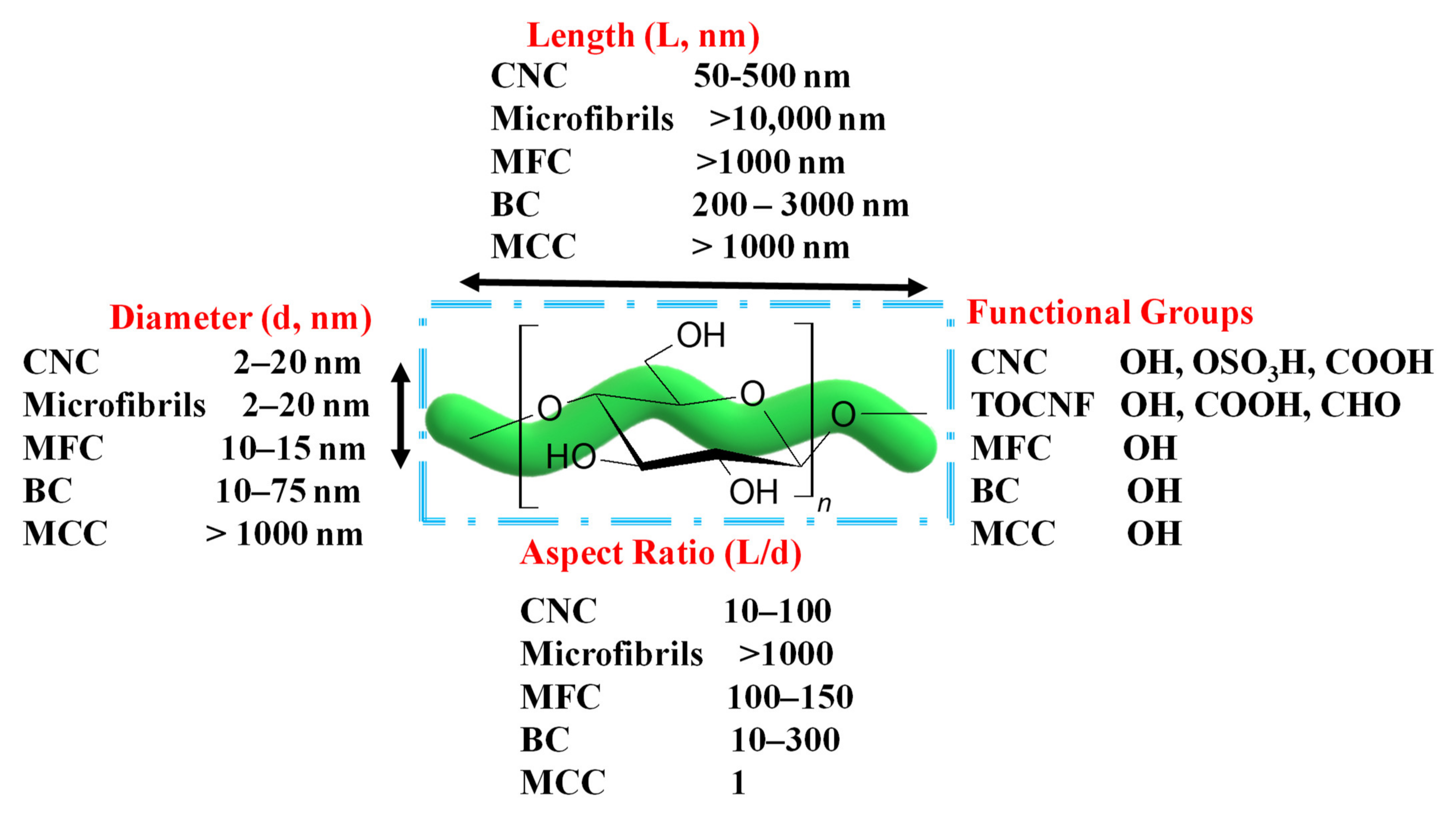

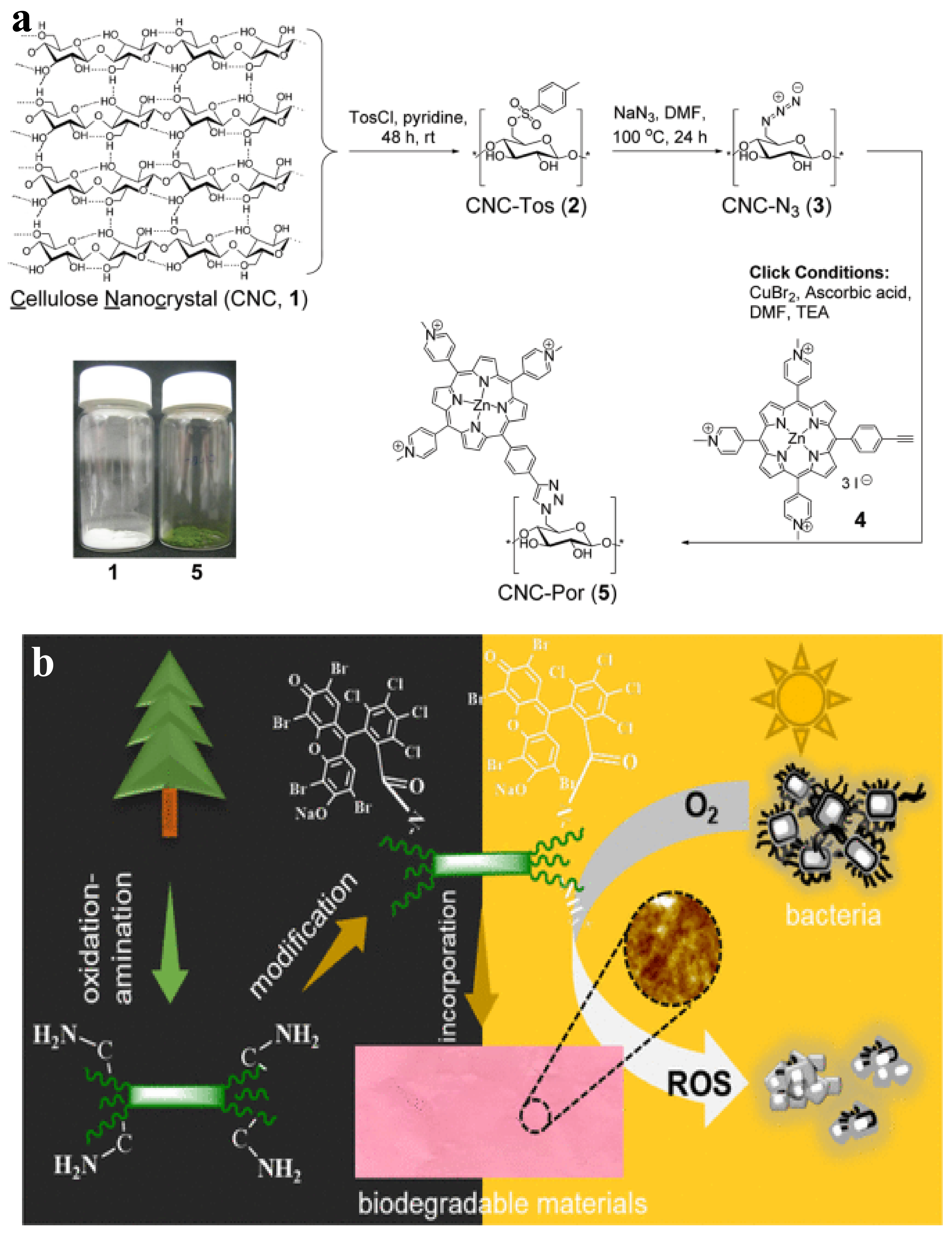

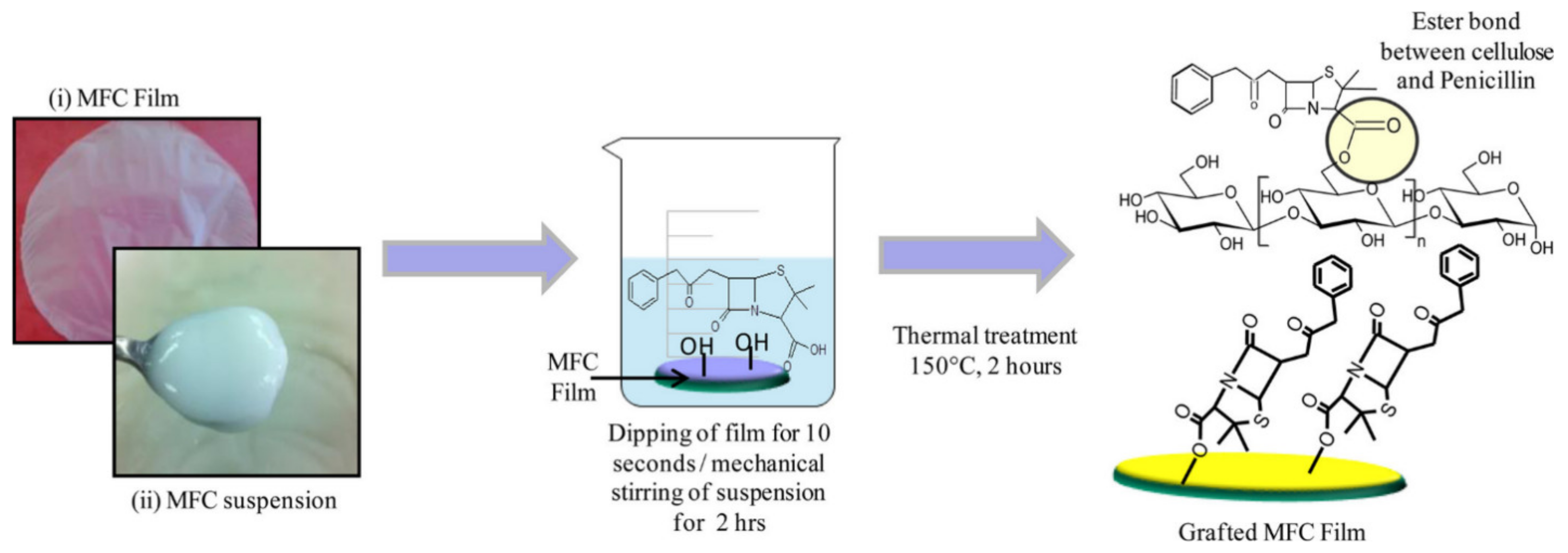


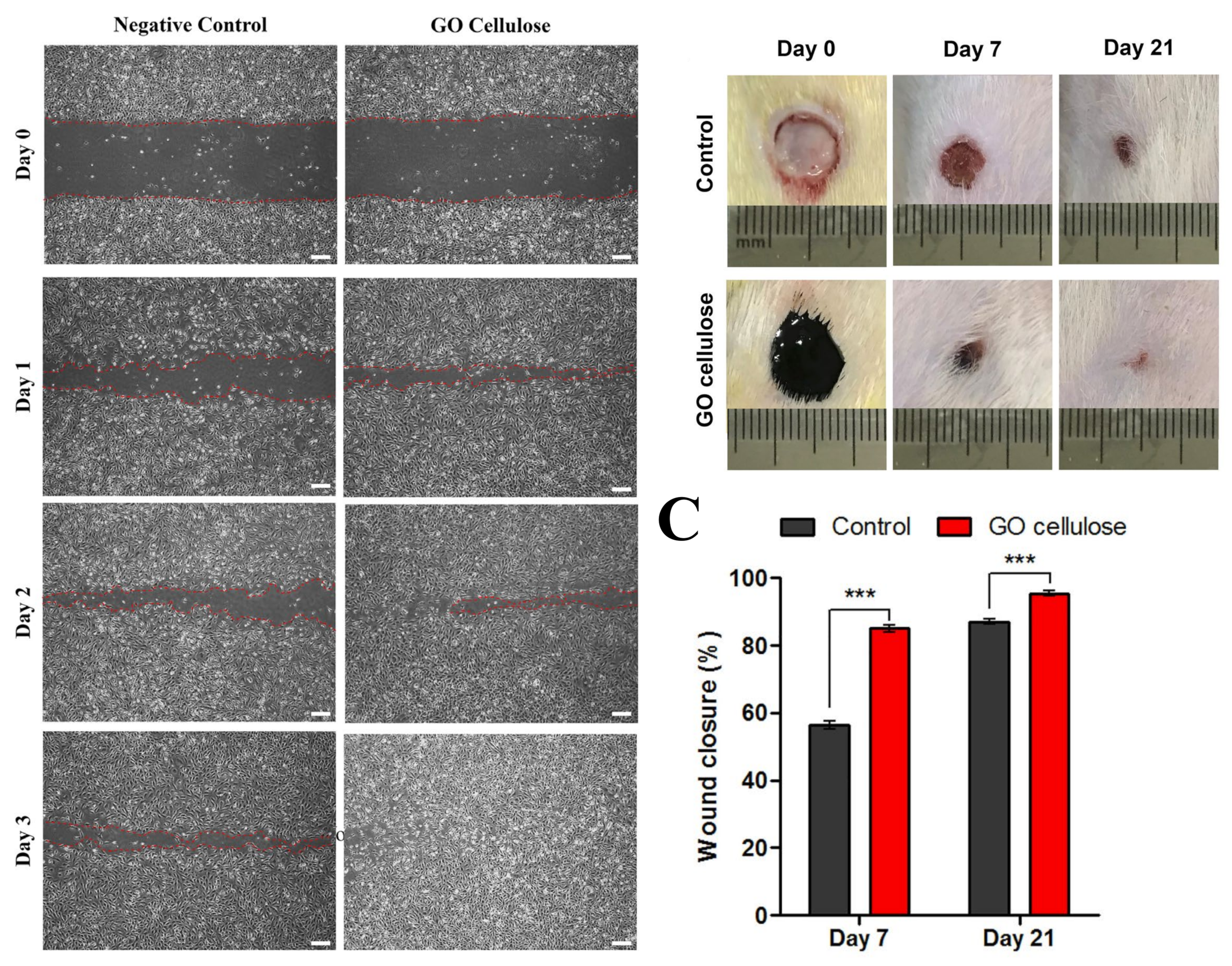


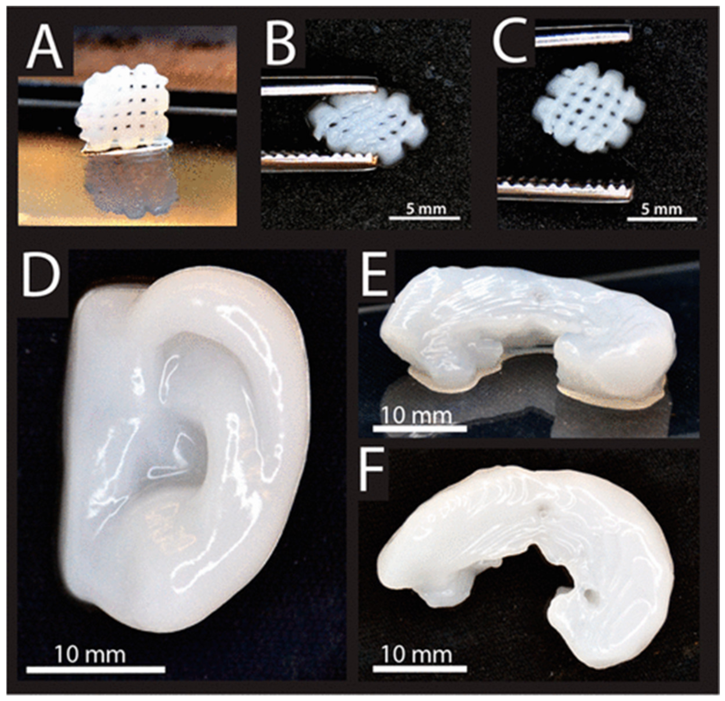
Publisher’s Note: MDPI stays neutral with regard to jurisdictional claims in published maps and institutional affiliations. |
© 2022 by the authors. Licensee MDPI, Basel, Switzerland. This article is an open access article distributed under the terms and conditions of the Creative Commons Attribution (CC BY) license (https://creativecommons.org/licenses/by/4.0/).