Article

Screening for Selective Anticancer Activity of Extracts from 59 Plant Species Collected in Southern Spain (Andalusia)

by
Víctor Jiménez-González
1,*,
Guillermo Benítez
2,
Julio Enrique Pastor
3,
Miguel López-Lázaro
1 and
José Manuel Calderón-Montaño
1,*
1
Department of Pharmacology, Faculty of Pharmacy, University of Seville, 41012 Seville, Spain
2
Department of Botany, Faculty of Pharmacy, University of Granada, 18071 Granada, Spain
3
Department of Vegetal Biology and Ecology, Faculty of Biology, University of Seville, 41012 Seville, Spain
*
Authors to whom correspondence should be addressed.
Pharmaceuticals 2026, 19(4), 616; https://doi.org/10.3390/ph19040616
Submission received: 1 March 2026 / Revised: 4 April 2026 / Accepted: 9 April 2026 / Published: 14 April 2026

Abstract

Background: Despite pharmacological advances, many cancer therapies provide only limited clinical benefits while often inducing significant toxicity. Therefore, the search for more effective and safer anticancer drugs remains an urgent priority. This study aimed to identify plant extracts from the Andalusian flora (Southern Spain) with selective anticancer potential. Methodology: A total of 67 extracts from 59 plant species were screened for selective cytotoxicity using A549 lung adenocarcinoma and HaCaT non-malignant cells. The most promising candidates, extracts from Thymelaea lanuginosa and Daphne oleoides, were further evaluated through fluorescence-based co-cultures, cell cycle analysis, and redox-mechanism assay. These extracts were also tested against a panel of cancer cells derived from different tissues (MDA-MB-231, T24, KATO-III, SK-OV-3, and MeWo). Results: Several extracts exhibited selective activity against A549 cancer cells, including extracts from Chamaeiris foetidissima (L.) Medik. (=Iris foetidissima L.), Daphne oleoides Schreb, Iberodes linifolia (L.) M. Serrano, R. Carbajal & S. Ortiz, Reseda media Lag., Saxifraga hirsuta L., Seseli montanum subsp. granatense (Willk.) C. Pardo, Thymelaea lanuginosa (Lam.), and Tordylium officinale L. The extracts from D. oleoides and T. lanuginosa were over 1000 times more active against lung cancer cells than non-malignant cells. These extracts induced a specific G1-phase arrest in A549 cells. Both extracts showed also selective activity against triple-negative breast cancer cells (MDA-MB-231) and bladder cancer cells (T24). Conclusions: These findings highlight Daphne and Thymelaea species as valuable sources for discovering novel selective anticancer agents. Future research should focus on bio-guided fractionation and in vivo validation to fully delineate their therapeutic potential.

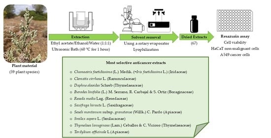
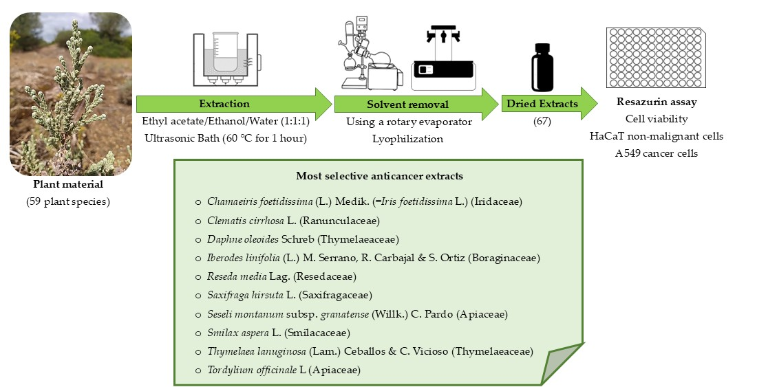
Graphical Abstract

1. Introduction

Globally, one in four deaths resulting from non-communicable illnesses is caused by cancer [1,2]. Improvements in diagnostic techniques, widespread early detection campaigns, and advancements in treatment have led to an increase in cancer survival rates. The 5-year survival rate after diagnosis has increased from an average of 49% in the 1970s to 69% during the 2014–2020 period. Early detection of cancer has been a major contributor to improved survival rates. At this stage, survival is high because tumors remain localized and can often be eliminated by surgery or radiotherapy. However, once cancer has spread to distant sites (metastasis), the main therapeutic approach relies on systemic drugs to reach disseminated cancer cells. Most of these drugs target highly proliferative cells, thereby affecting not only cancer cells but also healthy cells such as immune and epithelial cells. As a result, they frequently cause adverse effects including neutropenia, diarrhea, and severe fatigue. To minimize toxicity, such drugs must be administered at doses tolerable for patients, which are often lower than those required for complete eradication of cancer cells. To overcome these limitations, targeted therapies and immunotherapies have been developed in recent decades, aiming for greater selectivity against cancer. Targeted therapies act on specific proteins essential for tumor growth and survival, while immunotherapies activate the immune system to attack cancer cells. These strategies have improved survival in certain cancers, for example with the combination of tyrosine kinase inhibitors and immune checkpoint inhibitors in renal cancer [3]. Nevertheless, the proportion of patients diagnosed with metastatic disease who survive beyond five years remains very low [1,2]. For instance, survival rates for patients with liver, lung, esophageal, pancreatic, and urinary bladder cancers remain below 10% at the metastatic stage. These data highlight the limitations of current treatments. Indeed, a recent study reported that despite substantial economic investment in anticancer drug development, only a small fraction of drugs approved between 1995 and 2020 have provided meaningful clinical benefit for patients [4]. Therefore, the identification of novel anticancer agents capable of selectively targeting malignant cells is essential to achieve more effective and cost-efficient therapies.
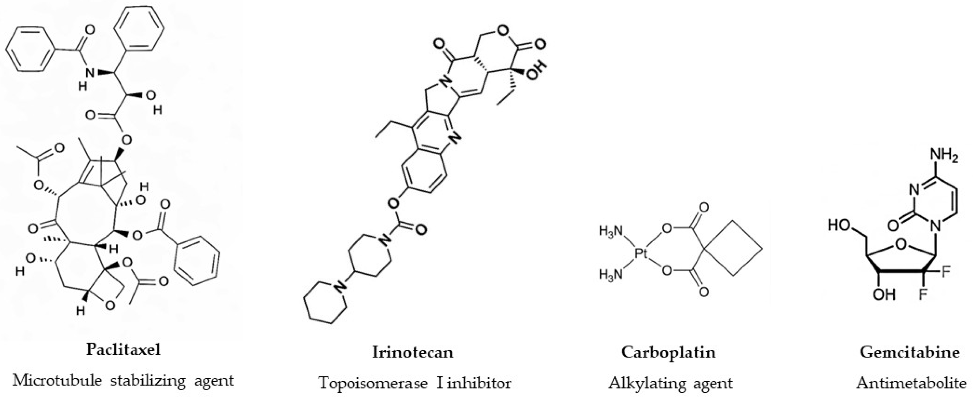
Nature has played a pivotal role in anticancer drug discovery, providing numerous clinically approved agents, either as unmodified compounds or as the basis for semisynthetic derivatives [5,6,7]. Notable examples of botanical oncology drugs include paclitaxel (extracted from Taxus brevifolia L.) and its derivative docetaxel. Paclitaxel is widely indicated for non-small cell lung cancer (NSCLC), Kaposi sarcoma, ovarian and breast cancers. Similarly, its semi-synthetic analogue, docetaxel, has become a cornerstone for treating prostate and mammary carcinomas. Etoposide (a semisynthetic compound derived from Podophyllum peltatum L.) is another plant-derived drug used for Hodgkin lymphoma, NSCLC, retinoblastoma, and Ewing sarcoma. Irinotecan (a derivative of camptothecin isolated from Camptotheca acuminata Decne.) is used for metastatic colorectal cancer. All these plant-derived drugs are included in the ‘WHO model list of essential medicines’ (23rd edition) [8] and continue to be investigated in combination with other agents in several clinical trials. For example; the combination of paclitaxel with pembrolizumab and carboplatin is under investigation for NSCLC patients in a Phase III trial at the University of Toronto [9]. Additionally, docetaxel is being studied with the CD40 agonist KK2269 in a trial for solid tumors sponsored by Kyowa Kirin [10]. A clinical trial in Shanghai (China) is evaluating efficacy and safety of a four-drug combination including etoposide for small cell lung cancer [11], while another at the Tang-Du Hospital (China) is investigating liposomal irinotecan combined with cisplatin/carboplatin for SCLC patient [12]. Figure 1 shows the chemical structures of four chemotherapeutic drugs broadly utilized in cancer treatment. These agents highlight the different sources of oncological leads, including one isolated directly from nature, one semi-synthetic derivative, and two fully synthetic compounds representing widely used chemical groups (alkylating agents and antimetabolites).
While modern drug discoveries (including molecular docking, structural modeling, and virtual screening) have revolutionized the field, they cannot substitute for the unparalleled chemical diversity provided by the natural world. Nature provides a level of structural complexity that remains indispensable for the development of new anticancer candidates. In fact, it is estimated that over 80% of plant species have yet to be investigated for their pharmacological properties, suggesting that nature still holds untapped potential for the identification of novel anticancer compounds [13,14]. Inspired by the successful historical large-scale screening programs that led to the discovery of landmark drugs such as paclitaxel [15], and given the rich biodiversity of Andalusia (southern part of the Iberian Peninsula) [16], we initiated a research project several years ago to screen the region’s flora for plant species with selective anticancer potential. This process presents significant logistical challenges, including coordinating sample collection with plant life cycles, preparing extracts rapidly to prevent the degradation of active compounds, and executing the subsequent in vitro assays. Consequently, the screening has been performed in stages; to date, we have evaluated and reported on more than 160 species [17,18,19], focusing primarily on Western Andalusia. The strategic objective of this stage is to provide a comprehensive database of bioactivity, including both active and inactive species, to prioritize candidates for future in-depth bio-guided fractionation and chemical characterization. By sharing these results openly, we aim to facilitate the efficient use of resources in natural product research, identifying which species truly justify the high costs of molecular isolation.
In the present work, we continue this effort by evaluating 67 extracts from 59 plant species collected across several regions of Andalusia. The selective anticancer potential of these plant extracts was assessed using human lung cancer cell line A549. The selection of lung cancer as the primary target for this screening is justified by its global impact, being the second most prevalent cancer and leading cause of oncological mortality worldwide. Data from GLOBOCAN 2022 underscores this urgency, reporting approximately 2.5 million new diagnoses and 1.8 million deaths attributed to this disease [1]. Consequently, the A549 cell line was utilized in the present work to ensure methodological continuity with the large-scale screening of the Andalusian flora initiated by our research group [17,18,19].

2. Results and Discussion

In the present study, we explored the selective cytotoxic potential of 67 extracts derived from 59 plant species collected in Andalusia (Southern Spain). The plant material evaluated in this study was collected from diverse locations across Andalusia (Tables S1 and S2). This geographic region is recognized as a Mediterranean biodiversity hotspot, characterized by a high rate of endemism and specific climatic conditions [16]. These environmental factors often act as drivers for the biosynthesis of unique secondary metabolites, making the Andalusian flora an exceptional, yet underexplored, reservoir for novel bioactive molecules.
First, the collected plant materials (comprising leaves, flowers, fruits, aerial parts or whole plant depending on the species) were processed and subjected to solvent extraction. Following the extraction process, the extraction yields for all the evaluated samples were calculated and are detailed in Table S2. The yields varied considerably, ranging from 0.3% to 16.6%. This broad range of extraction efficiency is expected and can be attributed to the diverse botanical origins of the samples, the specific plant parts utilized, and their inherent differences in structural matrix and phytochemical density. With this library of 60 extracts established, in vitro selective anticancer activity was evaluated using a dual-cell model: the A549 cell line, representing lung adenocarcinoma, and non-malignant HaCaT keratinocytes as a control for healthy tissue. The resazurin assay was employed to assess cell viability. The A549 cell line was selected to this in vitro screening due to the dominance of NSCLC, which constitutes nearly 85% of all lung cancers, and specifically the adenocarcinoma subtype, which accounts for four out of ten lung cancer cases globally [20]. To determine the selectivity of extracts, the HaCaT human keratinocyte line were used as healthy control [21]. While HaCaT cells originate from epidermal tissue and A549 cells from lung epithelium, both retain epithelial features, making them a suitable comparative model for evaluating selectivity. Furthermore, unlike other normal cells, which are often difficult to maintain due to their requirement for specialized culture media enriched with hormones and growth factors, HaCaT cells can be cultured under standard conditions like those used for cancer cell lines. This consistency minimizes confounding variables in comparative experiments. Normal cells typically undergo a limited number of divisions before senescence, making it challenging to obtain enough for high-throughput screening. Their slow proliferation rate also limits experimental scalability. In contrast, HaCaT cells are spontaneously immortalized, without the need for viral oncogenes such as SV40, which can integrate randomly into the genome and potentially introduce unpredictable phenotypic effects. HaCaT cells maintain stable growth characteristics. Importantly, HaCaT cells, despite being non-malignant, exhibit a high proliferation rate comparable to that of many cancer cell lines. This feature is particularly relevant in the context of cancer treatment, as many cytotoxic drugs preferentially target rapidly dividing cells, both cancer cells and healthy cells. The use of HaCaT cells thus provides a suitable model to evaluate the potential effects of anticancer agents on proliferative non-tumor cells, allowing for a more accurate assessment of therapeutic selectivity and safety [22,23].
The effects of extracts were evaluated by treating A549 and HaCaT cells with different concentrations of the extracts for three days. This extended incubation period was selected to ensure that both cell lines underwent several rounds of replication in the presence of the extracts, facilitating the evaluation of cumulative toxicity. The surviving cell population was then quantified using the resazurin assay. Cisplatin, an anticancer drug used in clinical, was used as positive control under the same experimental conditions. The scientific names of the plants are displayed in Table 1 in alphabetical order, along with botanical family, part of the plant used to elaborate the extract, voucher number, province of origin of the sample, IC50 values for each cell line, and selectivity indices. Figure 2 and Supplementary Figures S1–S5 display the concentration-effect curves obtained for all 60 extracts and cisplatin. A wide range of concentrations was used to represent the graphs and allow a fast and easy understanding of the results.
Several extracts (17, 32, 37, and 43) were identified as essentially non-toxic under our experimental conditions (Figures S2–S4). These extracts did not reach an IC50 threshold in any of the evaluated cell lines, maintaining high cell viability even at the maximum concentration tested (1000 µg/mL).
Other extracts showed cytotoxicity but not selectivity against lung cancer cells (for example, 2, 4, 5, 6, 9, 12, 28, 31, 47, 48 and 53) and even some others (1, 13, 26, 41 and 44) exhibited greater activity against non-malignant cells than against cancer cells (Figures S1–S4). The extract from the aerial part with flowers of Frankenia laevis L. (Frankeniaceae, 26), the extract from the aerial part with flowers of Helianthemum angustatum Pomel (Cistaceae, 30) and the extract from the whole plant of Scilla peruviana L. (Hyacinthaceae, 53) showed a remarkable cytotoxicity against the non-malignant HaCaT cells with IC50 values close to 20 µg/mL. The extract from the aerial part with flowers of Anemone palmata L. (Ranunculaceae, 3) and the extract from the aerial part with flowers of Rumex bucephalophorus L. (Polygonaceae, 47) were the most toxic extracts against HaCaT cells, with IC50 values 1.3 and 2.1 µg/mL, respectively. To the best of our knowledge, the toxicity of these specific species on human non-malignant cells has not been previously reported, although species within the same genera are known to exert cytotoxicity against various cancer cell lines. A previous study on extracts from Frankenia laevis L. reported selective cytotoxicity against human hepatocarcinoma HepG2 cells [24]; however, this selectivity profile was determined by comparing human cancer cells with non-malignant murine cells, rather than a human-derived healthy control. The presence of highly cytotoxic extracts within our local flora underscores the absolute necessity of conducting rigorous safety screenings using appropriate models.
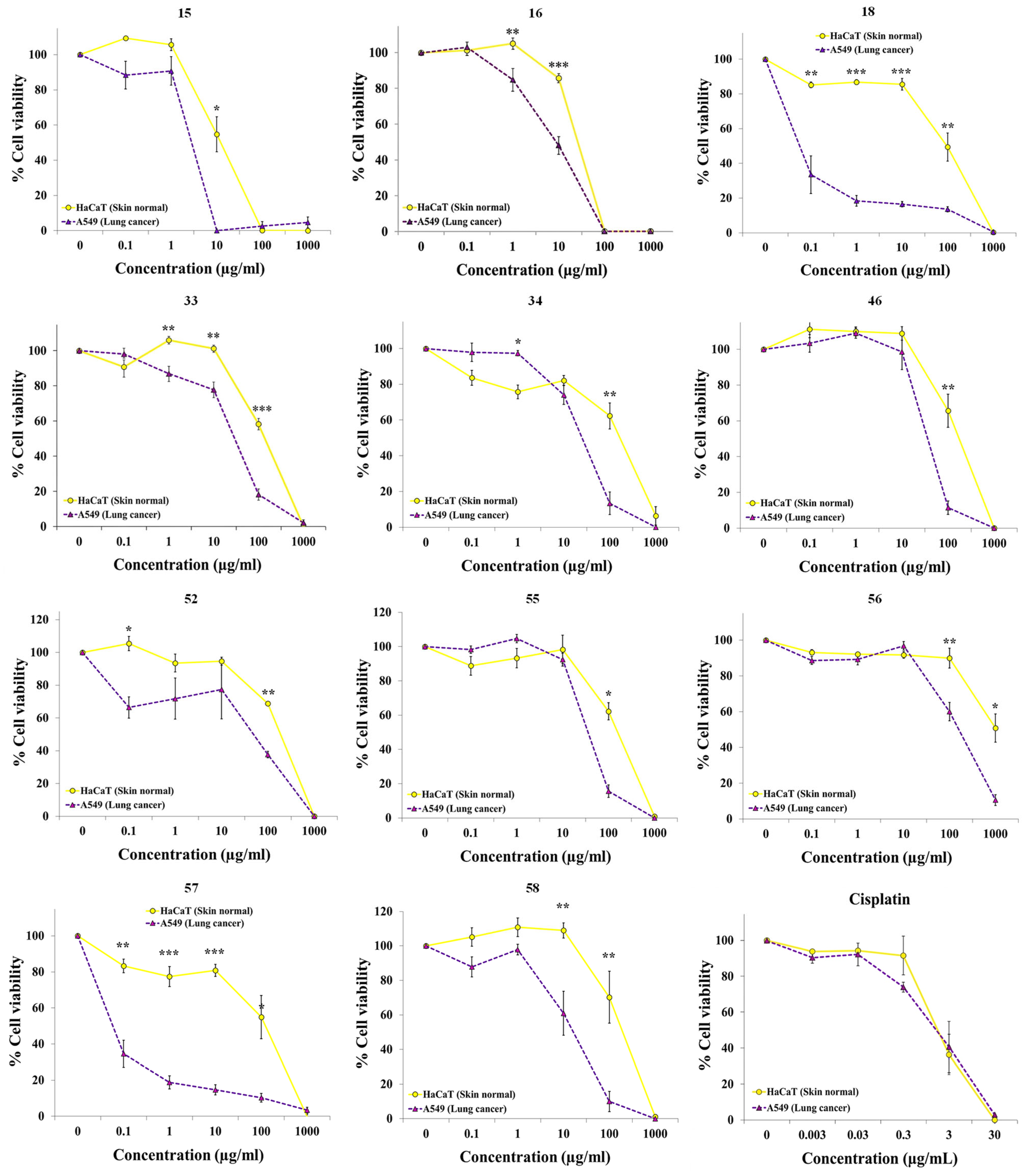
Our results show that some extracts exhibited selective cytotoxicity against the lung cancer cell line A549 (Figure 2, Figures S2 and S4). The extract from the flowers of Delphinium pentagynum Lam. (Ranunculaceae, 20), the extracts from flowers and from leaves of Dolichandra unguis-cati (L.) L.G.Lohmann (Bignoniaceae, 23 and 24), and the extract from flowers of Clematis flammula L. (Ranunculaceae, 16) exhibited modest selectivity, being approximately 2–4-fold more cytotoxic against lung cancer cells (Figure 2 and Figure S2). Ranunculaceae family is a well distributed family of plants worldwide, with around 50 genera, some of them widely distributed as Anemone, Delphinium, Clematis and Ranunculus [25]. Clematis genus comprises around 295 species [26] and its biological activity might be attributed to the presence of triterpenoids [27,28,29,30]. Some of these triterpenoids, as α-Hederin and D Rhamnose β-Hederin, have shown cytotoxic effects against breast cancer cell lines [31,32]. An ethanol extract from Clematis cirrhosa L. showed cytotoxicity against HT-29 colon cancer cell line, inducing cell cycle arrest and apoptosis [33]. Manghaslin and salvadoraside were the main bioactive phenolics identified as contributors to the observed activities. Boehmenan, a lignan extracted from Clematis armandi Franch, and different flavonoids from Clematis aethusifolia Turcz. were cytotoxic against A549 cells [34,35]. Some of these compounds or different combinations of them could be responsible for the effect observed with the extract from Clematis flammula L. in our experiments.
The extract from aerial parts with flowers of Iberodes linifolia (L.) M. Serrano, R. Carbajal & S. Ortiz (Boraginaceae, 33), the extract from the whole plant Reseda media Lag. (Resedaceae, 46), the extract from the whole plant Saxifraga hirsuta L. (Saxifragaceae, 52), the extract from fruits of Smilax aspera L. (Smilacaceae, 56) and the extract from the aerial parts of the endemic Seseli montanum subsp. granatense (Willk.) C. Pardo (Apiaceae, 55) were 5–7 times more active against A549 lung cancer cells than against HaCaT non-cancerous cells (Figure 2 and Table 1). To our knowledge, this is the first study to report cytotoxic activity within the genus Iberodes (33). The extract from Reseda media Lag. (46) was 5.5-fold times more selective to A549. Another plant from genus Reseda, R. lutea L., a very well-known mediterranean plant, was reported to have high cytotoxic effect on A549 (lung cancer) and A375 (melanoma) [36,37], an activity that has been attributed to the presence of isothiocyanates [36]. The observed activity of the extract from Saxifraga hirsuta (52) may be attributed to the presence of phenolic compounds, terpenes, or alkaloids. Several Saxifraga species are known to contain alkaloids and triterpenoids that have shown cytotoxic activity against five gastrointestinal cancer cell lines (BGC-823, GBC-SD, CCC-9810, HT-29, and HepG2) [38]. For instance, Liu et al. reported that a water-soluble extract from Saxifraga stolonifera inhibited the tumor growth on Lewis lung carcinoma-bearing mice [39]. The authors identified gallic acid, norbergenin, protocatechuic acid, and bergenin as four bioactive components within that extract. Different compounds described in literature or combination of them may participate in the activity found in Smilax aspera L. (56) fruits extract. For example, other species from this genus were reported to have cytotoxic phenylpropanoid glycosides, flavonoids, tannins, and saponins [40,41,42]. A549 lung cancer cells were 7 times more sensitive to the extract 55 (Seseli montanum subsp. granatense) than nonmalignant keratinocytes HaCaT. Lignans and a polyacetylenic fatty alcohol have previously been identified as components responsible for the cytotoxic activity in extracts from several Seseli species [43,44,45,46].
The extracts from aerial parts with fruits of Chamaeiris foetidissima (L.) Medik. (=Iris foetidissima L.) (Iridaceae, 34), the extract from aerial parts of Clematis cirrhosa L. (Ranunculaceae, 15) and the extract from aerial parts with flowers of Tordylium officinale L. (Apiaceae, 58) displayed very high selective activity against the cancer cells, showing greater selectivity than cisplatin and similar to that of gemcitabine, a drug also used in clinical practice (Figure 2 and Figure 3). We and other authors have reported that Iridaceae family, particularly Iris species, is a potential source of anticancer compounds [17,47,48]. In this work, our extract from Chamaeiris foetidissima (34) was almost 9 times more cytotoxic against the cancer cell lines. Phenolic acids, flavones, flavonols and triterpenoids can be involved in this activity [49,50]. As mentioned previously, triterpenoids, lignans and flavonoids may participate in the activity observed with extracts from the genus Clematis. The extract from Tordylium officinale L. (58) was almost 10-fold times more selective to A549 cancer cells. This could be attributed to the presence in this genus of cytotoxic coumarins [51].
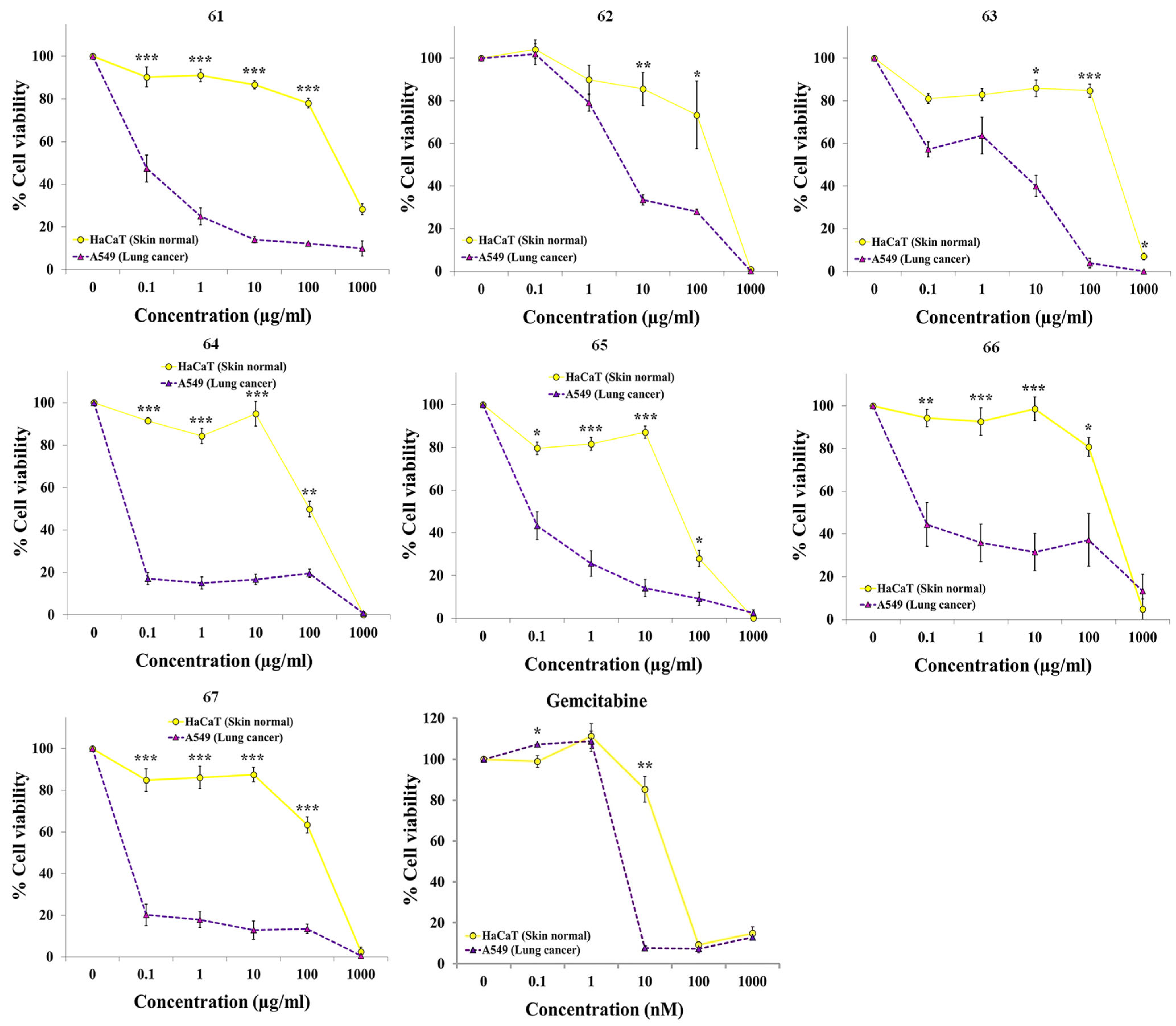
A particularly striking finding was that two extracts from the Thymelaeaceae family, 18 from the leaves of Daphne gnidium L. and 57 from the aerial parts of Thymelaea hirsuta (L.) Endl., displayed very high cytotoxicity against A549 cancer cells. Even at the lowest concentration tested (0.1 µg/mL), the viability of A549 cells was less than 40%, whereas the viability of non-malignant cells remained above 90% at concentrations up to 10 µg/mL (Figure 2). According to these results, the cytotoxic activity of both extracts was remarkably specific, showing over 1000-fold greater potency against the lung cancer cells compared to the non-cancerous cells. Previously, we reported that an extract from Daphne laureola L., another species belonging to Thymelaeaceae family, also showed high selectivity for lung cancer cells [19]. Therefore, we decided to further explore other plants from this family through targeted screening.
One species of Daphne and five species of Thymelaea were collected to continue this study (Tables S1 and S2). These species were selected based on their accessibility for collection in parks and nature reserves. Extracts were prepared according to our established methodology and subsequently assayed for selective anticancer activity using the resazurin assay. Although all tested extracts showed significant selective activity against A549 lung cancer cells (Figure 3 and Table 2), the extracts from Thymelaea elliptica (Boiss.) Endl. (62), Thymelaea granatensis (Pau) Lacaita (63) and Thymelaea lythroides Barratte & Murb. (65) were the least active and selective. Correspondingly, their IC50 values against A549 were the highest of the extracts, with values greater than 0.1 µg/mL. In contrast, the extracts from Thymelaea lanuginosa (Lam.) Ceballos & C.Vicioso (64) and Thymelaea tartonraira subsp. austroiberica Lambinon (66 and 67) were the most potent and selective against A549 cells. Interestingly, all extracts from both genera displayed greater selectivity than gemcitabine, as they remained non-cytotoxic to normal cells across a broader concentration range while retaining activity against cancer cells. Given that these are crude extracts, the observed activity suggests the presence of constituents with significant potency.
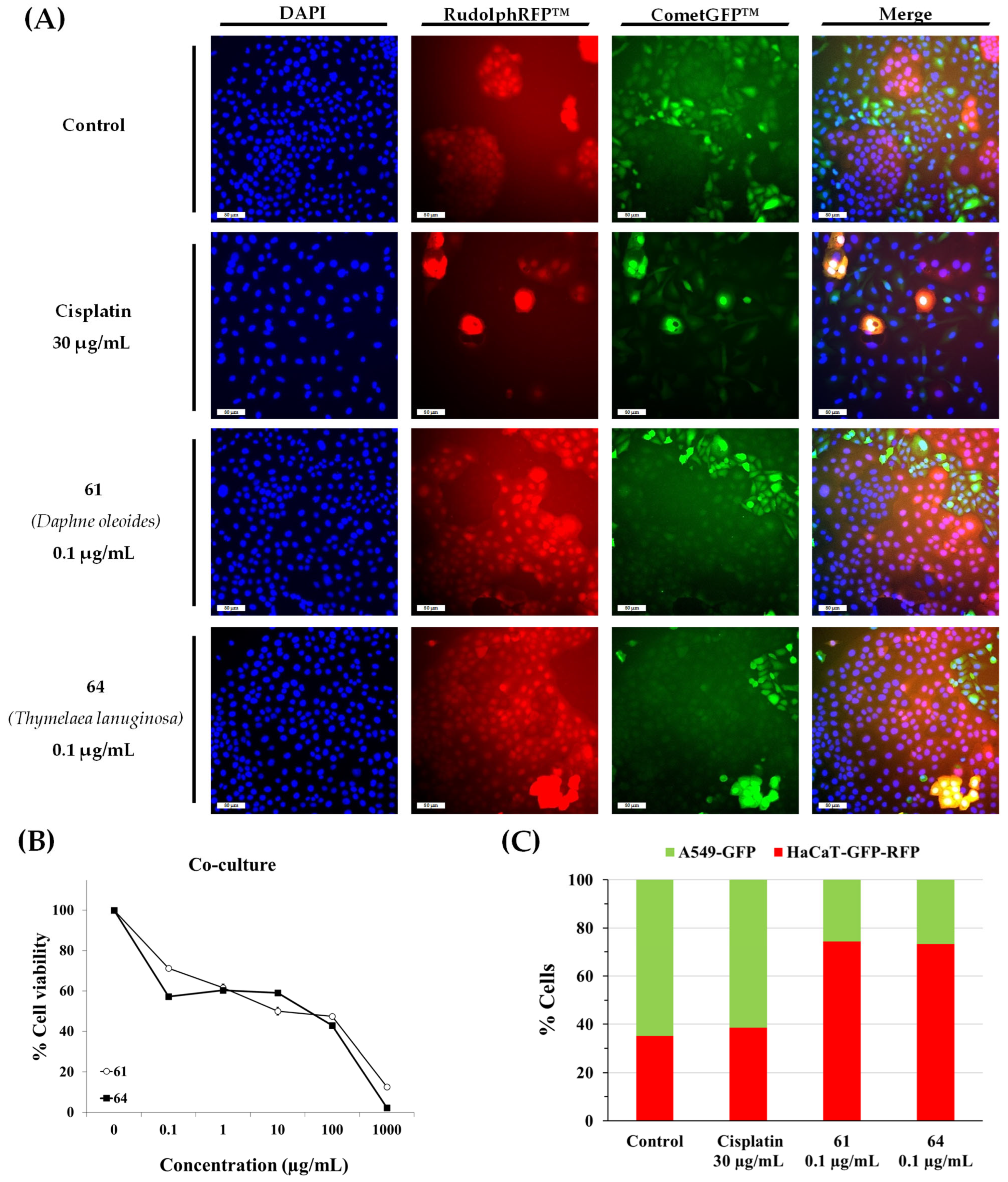
To continue the study, we selected the extracts from Daphne oleoides (61) and Thymelaea lanuginosa (64). The extract from D. oleoides showed less cytotoxicity against the non-malignant cell line than the extract from D. gnidium. The extract from T. lanuginosa was one of the most potent and selective and T. lanuginosa is the most unknown species. To validate the selective anticancer potential of the selected extracts, a co-culture model was established. Co-cultures were established by seeding A549-GFP and HaCaT-GFP-RFP cells together in the same wells at equal cell numbers (1:1 ratio) in 96-well plates. Following a 72 h exposure to cisplatin or extracts 61 and 64, viability was quantified with the resazurin assay. Finally, cells were fixed and analyzed using fluorescence microscopy (Figure 4). In untreated control groups, A549-GFP cells exhibited a higher proliferation rate, becoming the dominant population (64.8% vs. 35.2% for HaCaT-GFP-RFP). A comparable distribution was noted in cisplatin-treated samples (61.5% A549-GFP and 38.5% HaCaT-GFP-RFP); although the total cell density was reduced, the drug showed no preference for either cell type, affecting both lines to a similar extent. In contrast, the dominant cell population was reversed in the case of samples treated with both extracts. The cancer cell population decreased by more than half in the samples treated with 61 (25.7% A549-GFP versus 74.3% HaCaT-GFP-RFP). Similar results were observed in the samples treated with 64 (26.6% A549-GFP versus 73.4% HaCaT-GFP-RFP). These results suggest that both extracts have selective anticancer activity, reducing the A549 cancer cell population without significantly harming non-malignant HaCaT cells.
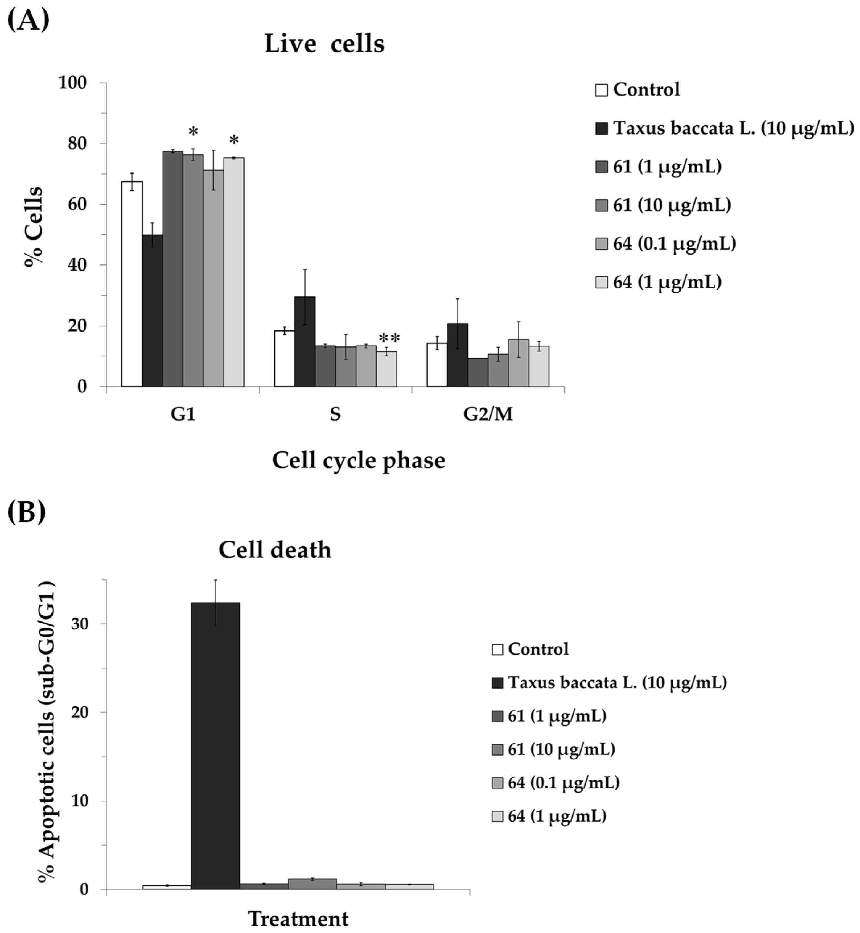
To investigate the mechanisms driving the growth-inhibitory effects of extracts 61 and 64, their impact on the cell cycle distribution of A549 cells was evaluated. An extract from the leaves of Taxus baccata L. (Taxaceae), was employed as a positive control due to its characteristic taxane content (mitotic inhibitor) [15,17]. A549 cells were exposed to extracts for 72 h and cell cycle distribution was determined by flow cytometry. As illustrated in Figure 5, T. baccata treatment led to a pronounced depletion of the G1 population, coupled with a significant accumulation of cells in the S and G2/M phases, alongside an increase in the sub-G0/G1 (apoptotic) fraction. In contrast, extracts 61 and 64 induced a distinct regulatory pattern, characterized by an expansion of the G1 phase population and a concomitant reduction in the S and G2/M proportions. Neither extract triggered a significant rise in the sub-G0/G1 population, suggesting that their antiproliferative activity is primarily mediated by cell cycle arrest rather than the induction of cytotoxicity. Furthermore, in contrast to the T. baccata extract, which exhibited a similar disruption of the cell cycle in both malignant and non-malignant cells, extracts 61 and 64 induced no significant alterations in the cell cycle profiles of HaCaT cells (Figure S6). This clear divergence in the response of healthy cells reinforces the highly selectivity of the studied extracts compared to conventional cytotoxic agents. Similar results have been observed for other species belonging to these genera or for their active constituents [52,53,54,55,56]. Sadeghi et al. [52] reported that the treatment of leukemia cells (K562) for 48 h with an extract of Daphne mucronata Royle increased the number of cells in G1 phase. In another study [55], an ethyl acetate extract of Daphne altaica Pall. increased the percentage of apoptotic cells and cell cycle arrest in esophageal carcinoma cells (Eca-109). An extract of Thymelaea hirsuta L. also induced cell cycle arrest and apoptosis in colorectal cancer cells [57]. Daphnane diterpenoids and flavonoids isolated from several Daphne species have been shown to induce cell cycle arrest through several mechanisms [53,54,56,58,59,60] including the regulation of the PI3K/Akt/mTOR signaling pathway, suppression of c-Myc expression, and upregulation of p21.
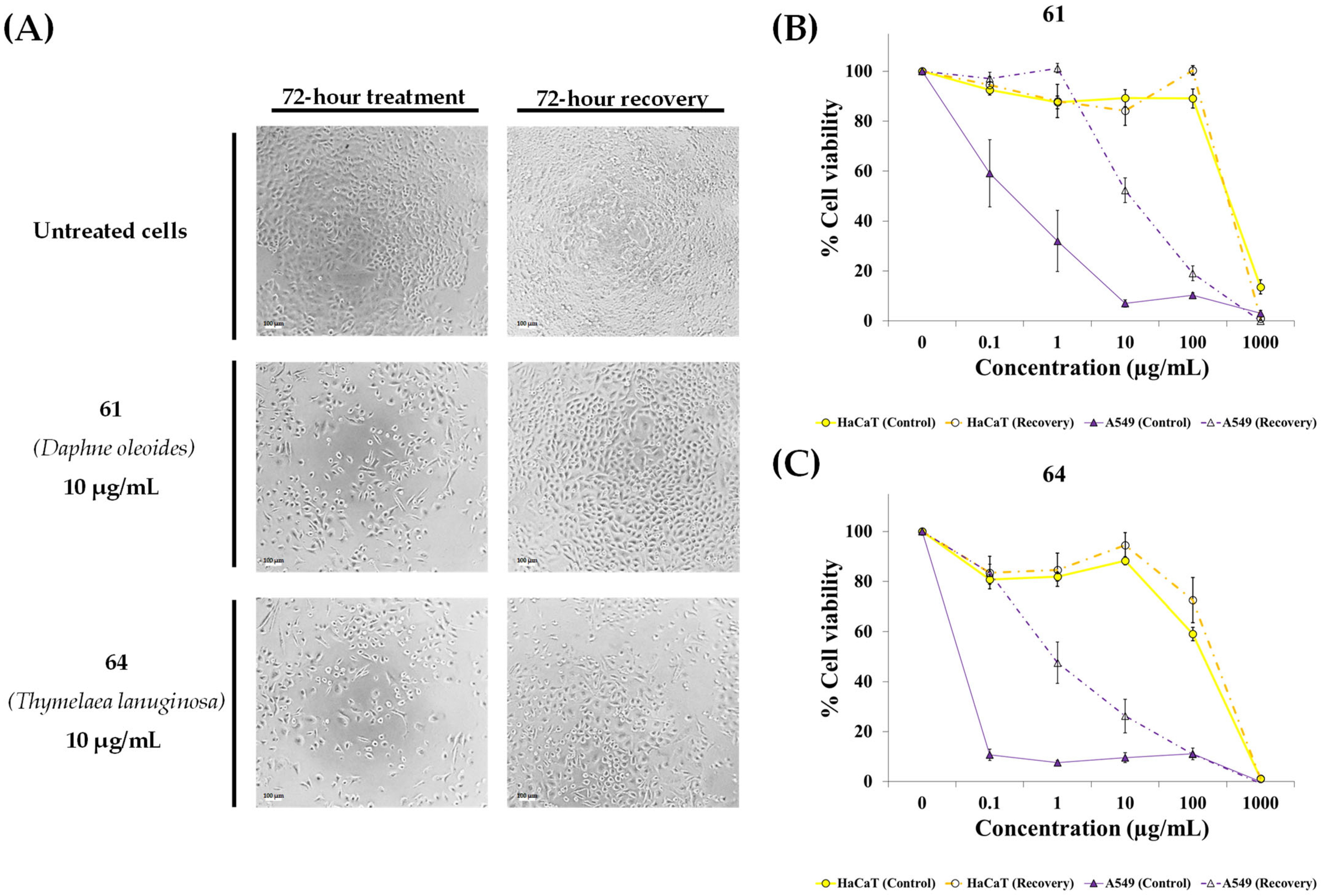
To further investigate the growth inhibition induced by extracts 61 and 64, a recovery assay was performed. A549 and HaCaT cells were treated for 72 h, and their cell viability was measured. Subsequently, the treatment was removed, and the cells were allowed to recover in fresh medium for an additional 72 h. Finally, cell viability was quantified again using resazurin assay. As shown in Figure 6, cells treated with concentrations below 10 µg/mL of extracts showed a significant recovery in metabolic activity and growth after the wash-out period. This evidence, combined with the lack of a sub-G0/G1 population in the flow cytometry data, suggest that these extracts may induce a reversible cell cycle arrest rather than cell death at these concentrations. The comparative analysis of the concentration-response curves reveals that the anticancer selectivity of these extracts is highly dose-dependent. A significant therapeutic window was observed in the ng/mL to low µg/mL range, where the extracts effectively induced a reversible growth arrest (cytostasis) specifically in the malignant A549 cells, without affecting the viability of the non-malignant HaCaT cells. This selectivity, however, disappears at the highest tested concentration (1000 µg/mL), where irreversible cytotoxicity occurs in both cell types. These results suggest that the most active bioactive constituents in these species may act as cytostatic agents. As the dosage increases, less potent secondary metabolites may reach a toxic threshold, leading to the observed lack of recovery at the highest concentrations.
To determinate whether extracts 61 and 64 were also active against other types of cancer, a panel of human cancer cell lines from different tissue origins was evaluated. This panel included bladder cancer (T24), ovarian carcinoma (SK-OV-3), melanoma (MeWo), triple-negative breast cancer (MDA-MB-231) and gastric cancer (KATO-III), assessed alongside the non-malignant HaCaT cells. Following a 72 h exposure to the extracts 61 and 64, or gemcitabine (positive control), cell viability was quantified with the resazurin assay (Table 3). Both extracts maintained substantial cytotoxicity against the breast and bladder cancer cell lines, albeit requiring higher half-maximal inhibitory concentrations (IC50 values ranging from 9.6 to 106.3 µg/mL). Both extracts achieved a selectivity index greater than 4.0 for the T24 and MDA-MB-231 cells, indicating a significant preference for these malignant cells over the non-cancerous HaCaT cells. Gemcitabine failed to exhibit any selectivity against MDA-MB-231 cells and reached a 4-fold selectivity for the T24 cells. However, the targeted selectivity was entirely lost in the MeWo melanoma cells. Furthermore, a divergent susceptibility profile was observed between the gastric and ovarian cancer cells lines: extract 61 exhibited preferential toxicity towards KATO-III over SK-OV-3, whereas extract 64 displayed the inverse trend. Collectively, these findings underscore that the therapeutic window of these extracts is highly dependent on the specific tumor type. The differential activity observed across theses cancer cell lines may be attributed to intrinsic differences in p53 status, redox homeostasis, epithelial–mesenchymal phenotype, and the predominance of distinct survival signaling pathways such as MAPK, PI3K/AKT, and c-MET.
The activity profile of the extracts is consistent with previously reported mechanisms for bioactive compounds from the Thymelaeaceae family. This family is composed of about 50 genera [61]. The main genera in this family are Daphne and Thymelaea. The genus Thymelaea is a monophyletic group considered a sister group to the genus Daphne [62]. Worldwide, Daphne and Thymelaea comprise approximately 92 and 34 accepted species, respectively [63,64]. The Thymelaeceae family is known for its potential cytotoxic activity, and plants within this family have been researched for their biological activities [65,66,67,68,69]. Some plants of both genera have been used as traditional remedies in the ethnopharmacology of China, Mediterranean, and North African countries [66,70]. For example, a decoction of the aerial parts of Thymelaea lythroides Barratte & Murb. has been traditionally used for the treatment of uterus cancer in Morocco [71]. Previous phytochemical studies of these genera have shown the presence of acrylates, coumarins, cromenes, cyclohexanones, diterpenenoids, esterols, flavonoids, lignans, phenolic compounds, sesquiterpenes [65,66,70,72,73,74,75]. Daphnane-type diterpenoids are the main biologically active constituents, exhibiting a wide range of pharmacological activities such as anticancer, antiinflammatory, and anti-HIV effects. There are five known groups of daphanane diterpenes: daphnetoxins, 12-hydroxydaphnetoxins, 1-alkydaphnanes, genkwanines, and resiniferonoids [65,72,73]. The most cytotoxic are those with a special orthoester group present in c-9, c-13, and c-14 [65,72,73]. While most daphnane diterpenes have been identified from the genus Daphne, the diversity of these compounds in the genus Thymelaea remains poorly understood [65,66,73,76]. For example, daphnane diterpenes isolated from the flowers of Daphne genkwa Siebold & Zucc. were highly cytotoxic against A549, with low activity against MRC-5 normal lung epithelial cells [77]. Yuanhuacine, one of these compounds, was also studied in H1993 lung carcinoma cells, affecting AMPK/mTORC2 signaling pathway and actin cytoskeleton organization [78]. Other daphnane diterpenes, such as yuanhadine, yuanhualine, yuanhuahine and yuanhuagine altered cell cycle in A549 cells by interfering with signaling pathways (Akt/mTOR, EGFR or Src pathways) [53,79]. Several extracts of different Daphne species or their constituents have also shown activity against other types of cancer [68,69,80,81,82,83,84,85,86]. For example, yuanhuatine selectively induces mitochondrial apoptosis in breast cancer cells (MCF-7) [85] and yuanhuacine induces cell cycle arrest in bladder cancer cells (T24T, a metastatic variant of the T24 cell line) by upregulating p21 protein expression levels [86].
Furthermore, it has been widely suggested that the selectivity of many phytochemicals against cancer cells may stem from the elevated basal levels of reactive oxygen species (ROS) characteristic of cancer cells, which renders them more susceptible to pro-oxidant stimuli [87,88,89]. To investigate whether this mechanism was responsible for the activity of extracts 61 and 64, we performed a rescue experiment using the antioxidants N-acetylcysteine (NAC) and catalase. NAC acts as a direct ROS scavenger and a precursor for glutathione synthesis, thereby mitigating intracellular oxidative stress, while enzyme catalase neutralizes hydrogen peroxide (H2O2). A549 were exposed to these extracts in the presence or absence of the antioxidants for 72 h and cell viability was quantified by the resazurin assay (Figure S7). While both antioxidants successfully rescued cell viability in the presence of H2O2 (positive control), they failed to mitigate the effects induced by extracts 61 and 64. These findings suggest that ROS generation may not play a dominant role in the observed selective activity of these extracts. Instead, they point toward a potentially more specific, ROS-independent anticancer mechanism.
The lack of protective effect of NAC and catalase, together with the cytotoxic activity observed for both extracts across different cancer cell lines, strongly suggests that their mechanism of action is not mediated by oxidative stress. As described above, daphnane-type diterpenoids present in Daphne species have been shown to affect multiple signaling pathways involved in cell proliferation [53,78,79,86]. The significant selectivity towards MDA-MB-231 (triple-negative breast cancer) and T24 (bladder cancer) suggests that the bioactive phytocompounds of these extracts may target non-hormonal signalling pathways, such as PI3K/AKT or RAS/MAPK signalling, upon which these aggressive phenotypes heavily rely. Furthermore, the differential sensitivity observed between KATO-III (gastric cancer) and SK-OV-3 (ovarian cancer) cells points to the possibility that the extracts may contain distinct bioactive compounds targeting different molecular pathways, such as c-MET-dependent signalling in gastric cancer cells or PI3K/AKT pathways in ovarian carcinoma. In contrast, the lack of selective activity in MeWo melanoma cells may reflect their intrinsic resistance to multiple cytotoxic mechanisms, including the high expression of ABC efflux transporters. Future phytochemical characterization and mechanistic studies will be essential to identify the precise molecular targets of these plants.

3. Materials and Methods

3.1. Plant Material

The plant material for this paper was collected by V. Jiménez-González between July 2018 and May 2022 in Cadiz, Córdoba, Granada, Jaén, Huelva, and Sevilla (Andalusia, Spain). Samples of vegetal origin (9–90 g) were collected following the guidelines for individual and environmental protection. We deposited voucher specimens for each plant in the herbarium of the University of Seville located at the Center for Research, Technology and Innovation (CITIUS II Celestino Mutis). Scientific name of species collected, plant part used, and voucher numbers are displayed in Table 1. The coordinates of collection are shown in Supplementary Table S1.

3.2. Preparation of the Extracts

Plant extracts were prepared within the first few hours following collection. Extraction was performed using a solvent mixture (100 to 200 mL of the mixture solvents: ethanol/ethyl acetate/water, 1:1:1 v/v/v) in an ultrasound water bath for 1 h at 60 °C. To ensure maximum efficiency, the ultrasound unit was operated at its maximum power output throughout the process.
After the extraction, solid particles, ethanol and ethyl acetate and water were sequentially eliminated by vacuum filtration, rotary evaporation and lyophilization respectively. After 72 h of lyophilization, the dried extracts were obtained. The final percentage yields (%) of the extracts prepared for our screening are summarized in Table S2.
Amber glass bottles were used to store the different extracts and preserved in a dark and cool room in the Department of Pharmacology of University of Seville. The first cell viability assay was performed in the next month after preparation to avoid any degradation of the compounds. A 100 mg/mL stock solution was prepared with DMSO, and the working solutions were prepared in the culture medium. The final DMSO concentration in all experimental groups was maintained below 1% (v/v), being non-toxic to the cells.

3.3. Drugs and Reagents

The following reagents were used: ECOSURF™ EH-9, resazurin, and ribonuclease A (Sigma Aldrich, St. Louis, MO, USA); gemcitabine (Pfizer S.L., Madrid, Spain); cisplatin and DAPI (Thermo Scientific Acros Organics, Waltham, MA, USA); and propidium iodide (Panreac Applichem, Darmstadt, Germany).

3.4. Cell Lines

The human cell lines A549 (non-small-cell lung cancer), KATO III (gastric cancer), MeWo (melanoma), SK-OV-3 (ovarian cancer), T24 (bladder cancer), and the non-malignant HaCaT skin cells [21] were all purchased from Cytion (Heidelberg, Germany). The MDA-MB-231 breast cancer line was provided by the American Type Culture Collection (ATCC, Manassas, VA, USA). Additionally, HaCaT-GFP-RFP and A549-GFP cells were utilized as previously detailed [17].
All cultures were maintained in Dulbecco’s Modified Eagle Medium (DMEM) containing L-glutamine, 4.5 g/L D-glucose, 10% fetal bovine serum, and 100 U/mL/100 µg/mL of penicillin/streptomycin. Standard culture conditions (37 °C, humidified atmosphere, 5% CO2) were applied. Unless specified, all culture consumables were obtained from Thermo Fisher Scientific or Biowest (Nuaillé, France).

3.5. Cell Viability Assay

The cytotoxic effect was evaluated using the resazurin assay. This colorimetric method relies on the metabolic capacity of living cells to convert the non-fluorescent blue dye (resazurin) into the pink, fluorescent product (resorufin) via redox reactions. Since this conversion is directly coupled to cellular metabolic activity, the resulting color intensity serves as a proxy for the number of viable cells. Briefly, cells were seeded at a density of 3000 and 5000 cells/well in 96-well plates. After a 24 h stabilization period, cells were treated for 72 h with varying concentrations of plant extracts or anticancer drugs. Post-treatment, a washing step with phosphate-buffered saline (PBS) was performed, followed by the addition of 150 μL of resazurin solution (20 μg/mL in medium). Following a 4–5 h incubation, the metabolic reduction of resazurin to resorufin was quantified by measuring absorbance at 540 and 620 nm using an iMark microplate reader (Bio-Rad Laboratories Inc., Hercules, CA, USA).
Cell viability was calculated as a percentage relative to untreated cells. Results represent the mean ± standard error of the mean (SEM) from at least three separate experiments. To determine the selectivity of the extracts, the selectivity index (S.I.) was calculated. The S.I. reflects the average of the individual IC50 ratios (non-malignant HaCaT/malignant A549) obtained in each independent experiment.

3.6. Co-Culture Assay

HaCaT-GFP-RFP and A549-GFP were co-cultured (3000 per well for each cell line) in 96-well plates. After a 24 h stabilization period, cells were treated for 72 h with varying concentrations of plant extracts or anticancer drug. Post-treatment, a washing step with phosphate-buffered saline (PBS) was performed, followed by the fixation with 70% cold ethanol and DNA staining with DAPI. Images were captured using a Nikon Eclipse Ti-E epifluorescence microscope (Nikon Europe B.V, Amstelveen, The Netherlands) at 20× magnification. Quantitative analysis of the co-culture was performed through manual cell counting to overcome the limitations of automated software in segmenting overlapping or clustered cells. For each well, between 150 and 350 cells were analyzed. The total cell population was determined by counting DAPI-stained nuclei. HaCaT-GFP-RFP cells were specifically identified by their red fluorescence (RFP). The A549-GFP subpopulation was subsequently calculated by subtracting the number of RFP-positive cells from the total DAPI-stained count. This manual approach ensured the accurate discrimination of individual cells within dense clusters.

3.7. Cell Cycle

For cell cycle analysis, cells were plated in 6-well plates at a density of 200,000 cells/well and incubated for 24 h to ensure attachment. Following medium renewal, treatments with plant extracts were applied for a 72 h period. Both the supernatant and trypsin-detached cells were subsequently harvested, centrifuged (300× g, 4 min, 4 °C), and rinsed twice using ice-cold PBS. Cell fixation was achieved by incubating the pellets in 70% ethanol at 4 °C for one hour. After an additional PBS wash, samples were treated with a staining cocktail (30 µg/mL propidium iodide, 0.1% Tween 20, 10 µg/mL ribonuclease A and 0.1% ECOSURF™ EH-9) for 60 min at 4 °C. DNA content and cell cycle distribution were then analyzed using a Beckman Coulter CYTOMICS FC500 flow cytometer (High Wycombe, UK).

3.8. Statistical Analysis

Statistical evaluations were performed using Microsoft Excel 365 (Microsoft Corp., Redmond, WA, USA). Data were analyzed using a paired, two-tailed Student’s t-test to determine differences between experimental groups. Statistical significance was defined by a p-value threshold of 0.05; results exceeding this value (p ≥ 0.05) were considered non-significant and are not marked with symbols. Significant differences are indicated in the figures as follows: * (p-value < 0.05), ** (p ≤ 0.01), and *** (p ≤ 0.001).

4. Conclusions

This study expanded a screening project aimed at identifying plant species from Andalusia with potential selective anticancer activity. Herein, we evaluated the selective anticancer activity of 59 plant species. Extracts from Chamaeiris foetidissima (L.) Medik. (Iridaceae), Clematis cirrhosa L. (Ranunculaceae), Daphne gnidium L. (Thymelaeaceae), Iberodes linifolia (L.) M.Serrano, R. Carbajal & S. Ortiz (Boraginaceae), Reseda media Lag. (Resedaceae), Saxifraga hirsuta L. (Saxifragaceae), Seseli montanum subsp. granatense (Willk.) C. Pardo (Apiaceae), Smilax aspera L. (Smilacaceae), Thymelaea hirsuta (L.) Endl. (Thymelaeaceae), and Tordylium officinale L. (Apiaceae) demonstrated significant selective cytotoxicity against A549 lung adenocarcinoma cells. The Thymelaeaceae family, particularly the genera Thymelaea and Daphne, emerged as the most promising candidates for further development. The extracts from D. gnidium, D. oleoides, T. hirsuta, T. lanuginosa and T. tartonraira were over 1000 times more active against A549 lung cancer cells than against HaCaT non-malignant cells.
A more in-depth study was carried out on the selective anticancer activity of the extracts from D. oleoides (the least cytotoxic against non-malignant cells) and T. lanuginosa (the most potent and selective against lung cancer cells). Our results in a co-culture model with A549-GFP and HaCaT-GFP-RFP cells confirm that the extracts from D. oleoides and T. lanuginosa possess a high degree of selectivity, effectively sparing non-malignant cells while targeting cancer cells. At low concentrations, the antiproliferative effect of these extracts may be mediated by a reversible arrest in the G1 phase of the cell cycle. Their antiproliferative effects are not mediated by the induction of acute oxidative stress, as confirmed by the lack of rescue effect with antioxidants (NAC and catalase).
In addition, the observed activity was not limited to lung cancer cells. The extracts showed activity in several cancer cell lines beyond lung cancer, though this activity was not always associated with selective cytotoxicity. They also exhibited selective cytotoxic effects against triple-negative breast (MDA-MB-231) and bladder (T24) cancer cells, but not to melanoma (MeWo) cells.
Despite these encouraging results, this study represents an initial screening approach aimed at identifying biologically active extracts rather than providing a comprehensive mechanistic characterization. A key limitation is the lack of phytochemical characterization, highlighting the need for bioassay-guided fractionation to isolate and identify the compounds responsible for the observed effects. Additionally, future research should expand the panel of both cancerous and non-malignant cell lines to allow a more accurate assessment of selectivity and therapeutic window. Finally, further molecular studies are required to elucidate the underlying signalling pathways, together with validation in in vivo tumor models to confirm the therapeutic potential of these plant species.

Supplementary Materials

The following supporting information can be downloaded at: https://www.mdpi.com/article/10.3390/ph19040616/s1, Table S1. Geographical coordinates of the collection sites for the botanical species evaluated in this study. Table S2. The extraction yields (%) obtained for each of the botanical extracts evaluated in the present study. Figure S1. Assessment of the differential cytotoxic effects of extracts 112 on human lung adenocarcinoma (A549) and non-tumorigenic cells (HaCaT). Figure S2. Assessment of the differential cytotoxic effects of extracts 1314, 17, 1927 on human lung adenocarcinoma (A549) and non-tumorigenic cells (HaCaT). Figure S3. Assessment of the differential cytotoxic effects of extracts 2832, 3541 on human lung adenocarcinoma (A549) and non-tumorigenic cells (HaCaT). Figure S4. Assessment of the differential cytotoxic effects of extracts 4245, 4751, 5354 and 59 on human lung adenocarcinoma (A549) and non-tumorigenic cells (HaCaT). Figure S5. Assessment of the differential cytotoxic effects of extract 60 on human lung adenocarcinoma (A549) and non-tumorigenic cells (HaCaT). Figure S6. Effects of extract 61 (Daphne oleoides), 64 (Thymelaea lanuginosa) and Taxus baccata on cell cycle distribution in HaCaT cells following a 72 h exposure. Figure S7. Assessment of the contribution of ROS generation to the cytotoxicity of the extract 61 (Daphne oleoides), 64 (Thymelaea lanuginosa) in A549 cells.

Author Contributions

Conceptualization, J.M.C.-M. and M.L.-L.; methodology, J.M.C.-M. and V.J.-G.; validation, J.M.C.-M. and V.J.-G.; formal analysis, J.M.C.-M. and V.J.-G.; investigation, J.M.C.-M. and V.J.-G.; data curation, J.E.P., G.B., J.M.C.-M. and V.J.-G.; writing—original draft preparation, V.J.-G. and J.M.C.-M.; writing—review and editing, J.M.C.-M., M.L.-L. and V.J.-G.; supervision, J.M.C.-M. and M.L.-L. All authors have read and agreed to the published version of the manuscript.

Funding

This research was partially funded by Junta de Andalucía (grant numbers 2019/CTS-657; and 2021/CTS-657) and the University of Seville through the “VI Plan Propio de Investigación y Transferencia” (grant numbers VIPPIT-2019-I.5, VIPPIT-2020-I.5, and VIPPIT-2021-I).

Institutional Review Board Statement

Not applicable.

Informed Consent Statement

Not applicable.

Data Availability Statement

The original contributions presented in this study are included in the article/Supplementary Materials. Further inquiries can be directed to the corresponding authors.

Acknowledgments

The authors express their gratitude to the Biology and Herbarium services at CITIUS (University of Seville) for their essential support in cell line maintenance and plant voucher preservation. They are also grateful to the University of Seville’s VI Plan Propio for the financial funding provided for this research. Special thanks are extended to the Territorial Delegations of Sustainable Development (Junta de Andalucía) in the provinces of Cadiz, Jaen, Cordoba, Huelva and Sevilla. Furthermore, we acknowledge the support from the Dirección General de Biodiversidad, Bosques y Desertificación del Ministerio for the necessary authorizations (ESNC112; ABSCH-IRCC-ES-258969-1). During the preparation of this manuscript, the authors used the AI language model Gemini 3.5 Pro (Google) to proofread the text for grammatical errors and to improve the overall readability and fluency of the English language. The authors reviewed and edited all AI-generated suggestions to ensure scientific accuracy and take full responsibility for the final content of the publication.

Conflicts of Interest

The authors declare no conflicts of interest.

References

  1. Bray, F.; Laversanne, M.; Sung, H.; Ferlay, J.; Siegel, R.L.; Soerjomataram, I.; Jemal, A. Global Cancer Statistics 2022: GLOBOCAN Estimates of Incidence and Mortality Worldwide for 36 Cancers in 185 Countries. CA Cancer J. Clin. 2024, 74, 229–263. [Google Scholar] [CrossRef] [Scilit] [PubMed]
  2. Siegel, R.L.; Giaquinto, A.N.; Jemal, A. Cancer Statistics, 2024. CA Cancer J. Clin. 2024, 74, 12–49. [Google Scholar] [CrossRef] [Scilit] [PubMed]
  3. Rose, T.L.; Kim, W.Y. Renal Cell Carcinoma: A Review. JAMA 2024, 332, 1001. [Google Scholar] [CrossRef] [Scilit] [PubMed]
  4. Brinkhuis, F.; Goettsch, W.G.; Mantel-Teeuwisse, A.K.; Bloem, L.T. Added Benefit and Revenues of Oncology Drugs Approved by the European Medicines Agency between 1995 and 2020: Retrospective Cohort Study. BMJ 2024, e077391. [Google Scholar] [CrossRef] [Scilit]
  5. Kingston, D.G.I.; Newman, D.J. The Search for Novel Drug Leads for Predominately Antitumor Therapies by Utilizing Mother Nature’s Pharmacophoric Libraries. Curr. Opin. Drug Discov. Dev. 2005, 8, 207–227. [Google Scholar]
  6. Newman, D.J.; Cragg, G.M. Natural Products as Sources of New Drugs over the Nearly Four Decades from 01/1981 to 09/2019. J. Nat. Prod. 2020, 83, 770–803. [Google Scholar] [CrossRef] [Scilit]
  7. Cragg, G.M.; Grothaus, P.G.; Newman, D.J. Impact of Natural Products on Developing New Anti-Cancer Agents. Chem. Rev. 2009, 109, 3012–3043. [Google Scholar] [CrossRef] [Scilit]
  8. World Health Organization. WHO Model List of Essential Medicines, 23rd List; WHO: Geneva, Switzerland, 2023; Available online: https://www.who.int/publications/i/item/WHO-MHP-HPS-EML-2023.01 (accessed on 4 May 2025).
  9. Clinicaltrials. Phase 3 Study of Pembrolizumab in Combination With Carboplatin/Taxane (Paclitaxel or Nab-Paclitaxel) Followed by Pembrolizumab with or Without Maintenance MK-2870 in the First-Line Treatment of Metastatic Squamous Non-Small Cell Lung Cancer 2024. Available online: https://clinicaltrials.gov/study/NCT06422143 (accessed on 4 May 2025).
  10. Clinicaltrials. A Phase 1, Multicenter, Open-Label, Dose-Escalation Study Evaluating the Safety and Tolerability of Intravenous KK2269 Monotherapy and Combination Therapy With Docetaxel in Adult Participants with Solid Tumors 2024. Available online: https://clinicaltrials.gov/study/NCT06266299 (accessed on 4 May 2025).
  11. Clinicaltrials. A Randomized, Controlled Phase II Clinical Study on the Efficacy and Safety of First-Line Treatment for Extensive-Stage Small Cell Lung Cancer Using a Combination Therapy of Trilaciclib, Envafolimab, Etoposide, and Carboplatin 2024. Available online: https://clinicaltrials.gov/study/NCT06698965 (accessed on 4 May 2025).
  12. Clinicaltrials. The Therapeutic Effect of Irinotecan Liposomes Combined with Cisplatin/Carboplatin for Platinum Sensitive Recurrent Small Cell Lung Cancer 2024. Available online: https://clinicaltrials.gov/study/NCT06467786 (accessed on 4 May 2025).
  13. Ulloa Ulloa, C.; Acevedo-Rodríguez, P.; Beck, S.; Belgrano, M.J.; Bernal, R.; Berry, P.E.; Brako, L.; Celis, M.; Davidse, G.; Forzza, R.C.; et al. An Integrated Assessment of the Vascular Plant Species of the Americas. Science 2017, 358, 1614–1617. [Google Scholar] [CrossRef] [Scilit]
  14. Lughadha, E.N.; Govaerts, R.; Belyaeva, I.; Black, N.; Lindon, H.; Allkin, R.; Magill, R.E.; Nicolson, N. Counting Counts: Revised Estimates of Numbers of Accepted Species of Flowering Plants, Seed Plants, Vascular Plants and Land Plants with a Review of Other Recent Estimates. Phytotaxa 2016, 272, 82. [Google Scholar] [CrossRef] [Scilit]
  15. American Chemical Society. National Historic Chemical Landmarks. Discovery of Camptothecin and Taxol®. Available online: http://www.acs.org/content/acs/en/education/whatischemistry/landmarks/camptothecintaxol.html (accessed on 4 May 2025).
  16. Cueto, M.; Melendo, M.; Gimenez, E.; Fuentes, J.; López-Carrique, E.; Blanca, G. First updated checklist of the vascular flora of Andalusia (S of Spain), one of the main biodiversity centres in the Mediterranean Basin. Phytotaxa 2018, 339, 1–95. [Google Scholar] [CrossRef] [Scilit]
  17. Jiménez-González, V.; Benítez, G.; Pastor, J.E.; López-Lázaro, M.; Calderón-Montaño, J.M. Evaluation of Anticancer Activity of 76 Plant Species Collected in Andalusia (Spain) against Lung Cancer Cells. Plants 2023, 12, 3275. [Google Scholar] [CrossRef] [Scilit] [PubMed]
  18. Calderón-Montaño, J.M.; Martínez-Sánchez, S.M.; Jiménez-González, V.; Burgos-Morón, E.; Guillén-Mancina, E.; Jiménez-Alonso, J.J.; Díaz-Ortega, P.; García, F.; Aparicio, A.; López-Lázaro, M. Screening for Selective Anticancer Activity of 65 Extracts of Plants Collected in Western Andalusia, Spain. Plants 2021, 10, 2193. [Google Scholar] [CrossRef] [Scilit] [PubMed]
  19. Calderón-Montaño, J.M.; Martínez-Sánchez, S.M.; Burgos-Morón, E.; Guillén-Mancina, E.; Jiménez-Alonso, J.J.; García, F.; Aparicio, A.; López-Lázaro, M. Screening for Selective Anticancer Activity of Plants from Grazalema Natural Park, Spain. Nat. Prod. Res. 2019, 33, 3454–3458. [Google Scholar] [CrossRef] [Scilit] [PubMed]
  20. American Cancer Society. What Is Lung Cancer? Available online: https://www.cancer.org/cancer/types/lung-cancer/about/what-is.html#:~:text=About%2080%25%20to%2085%25%20of,(outlooks)%20are%20often%20similar (accessed on 4 May 2025).
  21. Boukamp, P.; Petrussevska, R.T.; Breitkreutz, D.; Hornung, J.; Markham, A.; Fusenig, N.E. Normal Keratinization in a Spontaneously Immortalized Aneuploid Human Keratinocyte Cell Line. J. Cell Biol. 1988, 106, 761–771. [Google Scholar] [CrossRef] [Scilit]
  22. Pessina, A.; Raimondi, A.; Cerri, A.; Piccirillo, M.; Neri, M.G.; Croera, C.; Foti, P.; Berti, E. High Sensitivity of Human Epidermal Keratinocytes (HaCaT) to Topoisomerase Inhibitors. Cell Prolif. 2001, 34, 243–252. [Google Scholar] [CrossRef] [Scilit]
  23. Strzyga-Łach, P.; Kurpios-Piec, D.; Chrzanowska, A.; Szczepaniak, J.; Bielenica, A. 1,3-Disubstituted Thiourea Derivatives: Promising Candidates for Medicinal Applications with Enhanced Cytotoxic Effects on Cancer Cells. Eur. J. Pharmacol. 2024, 982, 176885. [Google Scholar] [CrossRef] [Scilit]
  24. Rodrigues, M.J.; Jekő, J.; Cziáky, Z.; Pereira, C.G.; Custódio, L. The Medicinal Halophyte Frankenia laevis L. (Sea Heath) Has In Vitro Antioxidant Activity, α-Glucosidase Inhibition, and Cytotoxicity towards Hepatocarcinoma Cells. Plants 2022, 11, 1353. [Google Scholar] [CrossRef] [Scilit]
  25. Ranunculaceae Juss. |Plants of the World Online| Kew Science. Available online: http://powo.science.kew.org/taxon/urn:lsid:ipni.org:names:30000277-2 (accessed on 4 May 2025).
  26. Clematis L. |Plants of the World Online| Kew Science. Available online: http://powo.science.kew.org/taxon/urn:lsid:ipni.org:names:329401-2 (accessed on 4 May 2025).
  27. Zhao, M.; Da-Wa, Z.-M.; Guo, D.-L.; Fang, D.-M.; Chen, X.-Z.; Xu, H.-X.; Gu, Y.-C.; Xia, B.; Chen, L.; Ding, L.-S.; et al. Cytotoxic Triterpenoid Saponins from Clematis tangutica. Phytochemistry 2016, 130, 228–237. [Google Scholar] [CrossRef] [Scilit]
  28. Dong, F.-Y.; Cui, G.-H.; Zhang, Y.-H.; Zhu, R.-N.; Wu, X.-J.; Sun, T.-T.; Wang, W. Clematomandshurica Saponin E, a New Triterpenoid Saponin from Clematis mandshurica. J. Asian Nat. Prod. Res. 2010, 12, 1061–1068. [Google Scholar] [CrossRef] [Scilit]
  29. Sati, O.P.; Uniyal, S.K.; Bahuguna, S.; Kikuchi, T. Clematoside-S, a Triterpenoid Saponin from the Roots of Clematis grata. Phytochemistry 1990, 29, 3676–3678. [Google Scholar] [CrossRef] [Scilit]
  30. Li, S.-G.; Huang, X.-J.; Li, M.-M.; Wang, M.; Feng, R.-B.; Zhang, W.; Li, Y.-L.; Wang, Y.; Ye, W.-C. Triterpenoid Saponins from the Roots of Clematis uncinata. Chem. Pharm. Bull. 2014, 62, 35–44. [Google Scholar] [CrossRef] [Scilit] [PubMed]
  31. Cheng, L.; Xia, T.-S.; Wang, Y.-F.; Zhou, W.; Liang, X.-Q.; Xue, J.-Q.; Shi, L.; Wang, Y.; Ding, Q.; Wang, M. The Anticancer Effect and Mechanism of α-Hederin on Breast Cancer Cells. Int. J. Oncol. 2014, 45, 757–763. [Google Scholar] [CrossRef] [Scilit] [PubMed]
  32. Cheng, L.; Xia, T.-S.; Wang, Y.-F.; Zhou, W.; Liang, X.-Q.; Xue, J.-Q.; Shi, L.; Wang, Y.; Ding, Q. The Apoptotic Effect of D Rhamnose β-Hederin, a Novel Oleanane-Type Triterpenoid Saponin on Breast Cancer Cells. PLoS ONE 2014, 9, e90848. [Google Scholar] [CrossRef] [Scilit] [PubMed]
  33. Alruwad, M.I.; Salah El Dine, R.; Gendy, A.M.; Saleh, A.M.; Khalaf, M.A.; El Hefnawy, H.M.; Sabry, M.M. Insights into Clematis cirrhosa L. Ethanol Extract: Cytotoxic Effects, LC-ESI-QTOF-MS/MS Chemical Profiling, Molecular Docking, and Acute Toxicity Study. Pharmaceuticals 2024, 17, 1347. [Google Scholar] [CrossRef] [Scilit]
  34. Pan, L.-L.; Wang, X.-L.; Zhang, Q.-Y.; Luo, X.-L.; Xu, P.; Liu, S.-Y.; Hu, J.-F.; Liu, X.-H. Boehmenan, a Lignan from the Chinese Medicinal Plant Clematis armandii, Induces Apoptosis in Lung Cancer Cells through Modulation of EGF-Dependent Pathways. Phytomedicine 2016, 23, 468–476. [Google Scholar] [CrossRef] [Scilit]
  35. Chang, Y.; Zhang, P.; Zhang, X.; Chen, J.; Rausch, W.-D.; Gula, A.; Bao, B. Cytotoxic Activities of Flavonoids from a Traditional Mongolian Medicinal Herb Clematis aethusifolia Turcz. Nat. Prod. Res. 2017, 31, 1223–1227. [Google Scholar] [CrossRef] [Scilit]
  36. Radulović, N.S.; Zlatković, D.B.; Ilić-Tomić, T.; Senerović, L.; Nikodinovic-Runic, J. Cytotoxic Effect of Reseda lutea L.: A Case of Forgotten Remedy. J. Ethnopharmacol. 2014, 153, 125–132. [Google Scholar] [CrossRef] [Scilit]
  37. Kiziltaş, H. Comprehensive Evaluation of Reseda lutea L. (Wild Mignonette) and 7 Isolated Flavonol Glycosides: Determination of Antioxidant Activity, Anti-Alzheimer, Antidiabetic and Cytotoxic Effects with In Vitro and in Silico Methods. Turk. J. Chem. 2022, 46, 1185–1198. [Google Scholar] [CrossRef] [Scilit]
  38. Liu, C.; Dang, J.; Wu, M. Drug Target Investigation of N-p-Coumaroyl-N’-Caffeoylputrescine, a Naturally-Occurring Alkaloid Derived from Saxifraga tangutica. Antioxidants 2024, 14, 12. [Google Scholar] [CrossRef] [Scilit]
  39. Liu, D.; Yang, P.; Zhang, Y.-Q. Water-Soluble Extract of Saxifraga stolonifera Has Anti-Tumor Effects on Lewis Lung Carcinoma-Bearing Mice. Bioorg. Med. Chem. Lett. 2016, 26, 4671–4678. [Google Scholar] [CrossRef] [Scilit]
  40. Zhang, C.; Feng, S.; Zhang, L.; Ren, Z. A New Cytotoxic Steroidal Saponin from the Rhizomes and Roots of Smilax scobinicaulis. Nat. Prod. Res. 2013, 27, 1255–1260. [Google Scholar] [CrossRef] [Scilit] [PubMed]
  41. Fu, S.; Yang, Y.; Liu, D.; Luo, Y.; Ye, X.; Liu, Y.; Chen, X.; Wang, S.; Wu, H.; Wang, Y.; et al. Flavonoids and Tannins from Smilax china L. Rhizome Induce Apoptosis Via Mitochondrial Pathway and MDM2-P53 Signaling in Human Lung Adenocarcinoma Cells. Am. J. Chin. Med. 2017, 45, 369–384. [Google Scholar] [CrossRef] [Scilit] [PubMed]
  42. Wang, W.-X.; Li, T.-X.; Ma, H.; Zhang, J.-F.; Jia, A.-Q. Tumoral Cytotoxic and Antioxidative Phenylpropanoid Glycosides in Smilax riparia A. DC. J. Ethnopharmacol. 2013, 149, 527–532. [Google Scholar] [CrossRef] [Scilit] [PubMed]
  43. Cinar, A.S.; Bakar-Ates, F.; Onder, A. Seseli Petraeum M. Bieb. (Apiaceae) Significantly Inhibited Cellular Growth of A549 Lung Cancer Cells through G0/G1 Cell Cycle Arrest. An. Acad. Bras. Ciênc. 2020, 92, e20191533. [Google Scholar] [CrossRef] [Scilit]
  44. Vučković, I.; Trajković, V.; Macura, S.; Tešević, V.; Janaćković, P.; Milosavljević, S. A Novel Cytotoxic Lignan from Seseli annuum L. Phytother. Res. 2007, 21, 790–792. [Google Scholar] [CrossRef] [Scilit]
  45. Vaglica, A.; Maggio, A.; Badalamenti, N.; Bruno, M.; Lauricella, M.; Occhipinti, C.; D’Anneo, A. Seseli tortuosum L. Subsp. Tortuosum Essential Oils and Their Principal Constituents as Anticancer Agents. Plants 2024, 13, 678. [Google Scholar] [CrossRef] [Scilit]
  46. Hu, C.; Chang, J.; Lee, K. Seselidiol, A New Cytotoxic Polyacetylene from Seseli mairei. J. Nat. Prod. 1990, 53, 932–935. [Google Scholar] [CrossRef] [Scilit]
  47. Khatib, S.; Faraloni, C.; Bouissane, L. Exploring the Use of Iris Species: Antioxidant Properties, Phytochemistry, Medicinal and Industrial Applications. Antioxidants 2022, 11, 526. [Google Scholar] [CrossRef] [Scilit]
  48. Mykhailenko, O.; Lesyk, R.; Finiuk, N.; Stoika, R.; Yushchenko, T.; Ocheretniuk, A. In Vitro Anticancer Activity Screening of Iridaceae Plant Extracts. J. Appl. Pharm. Sci. 2020, 10, 59–63. [Google Scholar] [CrossRef] [Scilit]
  49. Mykhailenko, O.; Korinek, M.; Ivanauskas, L.; Bezruk, I.; Myhal, A.; Petrikaitė, V.; El-Shazly, M.; Lin, G.-H.; Lin, C.-Y.; Yen, C.-H.; et al. Qualitative and Quantitative Analysis of Ukrainian Iris Species: A Fresh Look on Their Antioxidant Content and Biological Activities. Molecules 2020, 25, 4588. [Google Scholar] [CrossRef] [Scilit]
  50. Kostić, A.Ž.; Gašić, U.M.; Pešić, M.B.; Stanojević, S.P.; Barać, M.B.; Mačukanović-Jocić, M.P.; Avramov, S.N.; Tešić, Ž.L. Phytochemical Analysis and Total Antioxidant Capacity of Rhizome, Above-Ground Vegetative Parts and Flower of Three Iris Species. Chem. Biodivers. 2019, 16, e1800565. [Google Scholar] [CrossRef] [Scilit] [PubMed]
  51. Kofinas, C.; Chinou, I.; Loukis, A.; Harvala, C.; Roussakis, C.; Maillard, M.; Hostettmann, K. Cytotoxic Coumarins from the Aerial Parts of Tordylium apulum and Their Effects on a Non-Small-Cell Bronchial Carcinoma Line. Planta Med. 1998, 64, 174–176. [Google Scholar] [CrossRef] [Scilit] [PubMed]
  52. Sadeghi, H.; Yazdanparast, R. Anti-Tumor Activity and Cell Cycle Arrest of a New Diterpene Ester from Daphne mucronata Using K562 Cells. Iran. Biomed. J. 2003, 7, 127–131. [Google Scholar]
  53. Jo, S.-K.; Hong, J.-Y.; Park, H.J.; Lee, S.K. Anticancer Activity of Novel Daphnane Diterpenoids from Daphne genkwa through Cell-Cycle Arrest and Suppression of Akt/STAT/Src Signalings in Human Lung Cancer Cells. Biomol. Ther. 2012, 20, 513–519. [Google Scholar] [CrossRef] [Scilit]
  54. Huang, Y.-C.; Lee, P.-C.; Wang, J.J.; Hsu, Y.-C. Anticancer Effect and Mechanism of Hydroxygenkwanin in Oral Squamous Cell Carcinoma. Front. Oncol. 2019, 9, 911. [Google Scholar] [CrossRef] [Scilit]
  55. Kizaibek, M.; Wubuli, A.; Gu, Z.; Bahetjan, D.; Tursinbai, L.; Nurhamit, K.; Chen, B.; Wang, J.; Tahan, O.; Cao, P. Effects of an Ethyl Acetate Extract of Daphne altaica Stem Bark on the Cell Cycle, Apoptosis and Expression of PPARγ in Eca-109 Human Esophageal Carcinoma Cells. Mol. Med. Rep. 2020, 22, 1400–1408. [Google Scholar] [CrossRef] [Scilit]
  56. Pan, R.-R.; Zhang, C.-Y.; Li, Y.; Zhang, B.-B.; Zhao, L.; Ye, Y.; Song, Y.-N.; Zhang, M.; Tie, H.-Y.; Zhang, H.; et al. Daphnane Diterpenoids from Daphne genkwa Inhibit PI3K/Akt/mTOR Signaling and Induce Cell Cycle Arrest and Apoptosis in Human Colon Cancer Cells. J. Nat. Prod. 2020, 83, 1238–1248. [Google Scholar] [CrossRef] [Scilit]
  57. Toumi, I.; Yatouji, S.; Borie, N.; Remy, S.; Renault, J.-H.; Chazee, L.; Hammami, M.; Martiny, L.; Charpentier, E.; El Btaouri, H. Anti-Tumoral Effect of Thymelaea hirsuta L. Extracts in Colorectal Cancer Cells. Anticancer Agents Med. Chem. 2022, 22, 687–698. [Google Scholar] [CrossRef] [Scilit]
  58. Wang, X.; Wang, Y.; Liu, Z.; Zhao, H.; Yao, G.-D.; Liu, Q.; Song, S.-J. New Daphnane Diterpenoidal 1,3,4-Oxdiazole Derivatives as Potential Anti-Hepatoma Agents: Synthesis, Biological Evaluation and Molecular Modeling Studies. Bioorganic Chem. 2024, 145, 107208. [Google Scholar] [CrossRef] [Scilit]
  59. Xie, Q.; Fan, X.; Han, Y.; Wu, B.-X.; Zhu, B. Daphnoretin Arrests the Cell Cycle and Induces Apoptosis in Human Breast Cancer Cells. J. Nat. Prod. 2022, 85, 2332–2339. [Google Scholar] [CrossRef] [Scilit]
  60. Kang, H.; Lee, H.; Jee, D.J.; Shin, S.; Nah, S.; Yoon, S.Y.; Kim, J.W. PRDM1, a Tumor-Suppressor Gene, Is Induced by Genkwadaphnin in Human Colon Cancer SW620 Cells. J. Cell Biochem. 2016, 117, 172–179. [Google Scholar] [CrossRef] [Scilit]
  61. WFO Plant List|World Flora Online. Available online: https://wfoplantlist.org/taxon/wfo-7000000613-2024-12 (accessed on 14 April 2025).
  62. Galicia-Herbada, D. Origin and Diversification of Thymelaea (Thymelaeaceae): Inferences from a Phylogenetic Study Based on ITS (rDNA) Sequences. Plant Syst. Evol. 2006, 257, 159–187. [Google Scholar] [CrossRef] [Scilit]
  63. WFO Plant List|World Flora Online. Available online: https://wfoplantlist.org/taxon/wfo-4000010759-2024-12 (accessed on 14 April 2025).
  64. WFO Plant List|World Flora Online. Available online: https://wfoplantlist.org/taxon/wfo-4000038329-2024-12 (accessed on 14 April 2025).
  65. Hou, Z.; Yao, G.; Song, S. Daphnane-Type Diterpenes from Genus Daphne and Their Anti-Tumor Activity. Chin. Herb. Med. 2021, 13, 145–156. [Google Scholar] [CrossRef] [Scilit] [PubMed]
  66. Marmouzi, I.; Bouchmaa, N.; Kharbach, M.; Ezzat, S.M.; Merghany, R.M.; Berkiks, I.; El Jemli, M. Thymelaea Genus: Ethnopharmacology, Chemodiversity, and Bioactivities. S. Afr. J. Bot. 2021, 142, 175–192. [Google Scholar] [CrossRef] [Scilit]
  67. Chaabane, F.; Krifa, M.; Matera, E.; Loussaeif, A.; Dijoux-Franca, M.-G.; Ghedira, K.; Dumontet, C.; Chekir-Ghedira, L. In Vitro Antileukaemic Activity of Extracts from Daphne gnidium Leaves against Sensitive and Multidrug Resistant K562/R7 Cells. Tumor Biol. 2014, 35, 8991–8998. [Google Scholar] [CrossRef] [Scilit] [PubMed]
  68. Hu, Q.-F.; Mu, H.-X.; Huang, H.-T.; Lv, H.-Y.; Li, S.-L.; Tu, P.-F.; Li, G.-P. Secolignans, Neolignans and Phenylpropanoids from Daphne Feddei and Their Biological Activities. Chem. Pharm. Bull. 2011, 59, 1421–1424. [Google Scholar] [CrossRef] [Scilit]
  69. Huang, S.Z.; Zhang, X.J.; Li, X.Y.; Kong, L.M.; Jiang, H.Z.; Ma, Q.Y.; Liu, Y.Q.; Hu, J.M.; Zheng, Y.T.; Li, Y.; et al. Daphnane-Type Diterpene Esters with Cytotoxic and Anti-HIV-1 Activities from Daphne acutiloba Rehd. Phytochemistry 2012, 75, 99–107. [Google Scholar] [CrossRef] [Scilit]
  70. Moshiashvili, G.; Tabatadze, N.; Mshvildadze, V. The Genus Daphne: A Review of Its Traditional Uses, Phytochemistry and Pharmacology. Fitoterapia 2020, 143, 104540. [Google Scholar] [CrossRef] [Scilit]
  71. Kabbaj, F.Z.; Meddah, B.; Cherrah, Y. Ethnopharmacological Profile of Traditional Plants Used in Morocco by Cancer Patients as Herbal Therapeutics. Phytopharmacology 2012, 2, 243–256. [Google Scholar]
  72. Jin, Y.-X.; Shi, L.-L.; Zhang, D.-P.; Wei, H.-Y.; Si, Y.; Ma, G.-X.; Zhang, J. A Review on Daphnane-Type Diterpenoids and Their Bioactive Studies. Molecules 2019, 24, 1842. [Google Scholar] [CrossRef] [Scilit]
  73. Nie, Y.-W.; Li, Y.; Luo, L.; Zhang, C.-Y.; Fan, W.; Gu, W.-Y.; Shi, K.-R.; Zhai, X.-X.; Zhu, J.-Y. Phytochemistry and Pharmacological Activities of the Diterpenoids from the Genus Daphne. Molecules 2021, 26, 6598. [Google Scholar] [CrossRef] [Scilit] [PubMed]
  74. Zhao, L.; Zhang, Y.; Yin, Q.; Chen, G.; Li, W.; Li, N. Research Progress on the Toxicity of Toxic Traditional Herbals from Thymelaeaceae. J. Ethnopharmacol. 2024, 322, 117594. [Google Scholar] [CrossRef] [Scilit] [PubMed]
  75. Nocera, P.; Bajsa-Hirschel, J.; Masi, M.; Ross, S.A.; Cantrell, C.L.; Duke, S.O.; Surico, G.; Evidente, A. Secondary Metabolites of Thymelaea hirsuta, a Plant Collected from the Sicilian Island of Lampedusa. Nat. Prod. Res. 2021, 35, 3977–3984. [Google Scholar] [CrossRef] [Scilit] [PubMed]
  76. Miyamae, Y.; Villareal, M.O.; Abdrabbah, M.B.; Isoda, H.; Shigemori, H. Hirseins A and B, Daphnane Diterpenoids from Thymelaea hirsuta That Inhibit Melanogenesis in B16 Melanoma Cells. J. Nat. Prod. 2009, 72, 938–941. [Google Scholar] [CrossRef] [Scilit]
  77. Hong, J.-Y.; Nam, J.-W.; Seo, E.-K.; Lee, S.K. Daphnane Diterpene Esters with Anti-Proliferative Activities against Human Lung Cancer Cells from Daphne genkwa. Chem. Pharm. Bull. 2010, 58, 234–237. [Google Scholar] [CrossRef] [Scilit]
  78. Kang, J.I.; Hong, J.-Y.; Lee, H.-J.; Bae, S.Y.; Jung, C.; Park, H.J.; Lee, S.K. Anti-Tumor Activity of Yuanhuacine by Regulating AMPK/mTOR Signaling Pathway and Actin Cytoskeleton Organization in Non-Small Cell Lung Cancer Cells. PLoS ONE 2015, 10, 1–17. [Google Scholar] [CrossRef] [Scilit]
  79. Hong, J.-Y.; Chung, H.-J.; Lee, H.-J.; Park, H.J.; Lee, S.K. Growth Inhibition of Human Lung Cancer Cells via Down-Regulation of Epidermal Growth Factor Receptor Signaling by Yuanhuadine, a Daphnane Diterpene from Daphne genkwa. J. Nat. Prod. 2011, 74, 2102–2108. [Google Scholar] [CrossRef] [Scilit]
  80. Li, S.; Chou, G.; Hseu, Y.; Yang, H.; Kwan, H.; Yu, Z. Isolation of Anticancer Constituents from Flos Genkwa (Daphne genkwa Sieb.et Zucc.) through Bioassay-Guided Procedures. Chem. Cent. J. 2013, 7, 159. [Google Scholar] [CrossRef] [Scilit]
  81. Zhang, W.; Zhang, W.D.; Liu, R.H.; Shen, Y.H.; Zhang, C.; Cheng, H.S.; Fu, P.; Shan, L. Two New Chemical Constituents from Daphne odora Thunb. Var. marginata. Nat. Prod. Res. 2006, 20, 1290–1294. [Google Scholar] [CrossRef] [Scilit]
  82. Lu, Y.; Li, X.; Mu, H.; Huang, H.; Li, G.-P.; Hu, Q. Bioactive Phenylpropanoids from Daphne feddei. J. Braz. Chem. Soc. 2012, 23, 656–660. [Google Scholar] [CrossRef] [Scilit]
  83. Amir, G.; Miri, R.; Javidnia, K.; Davoudi, M. Study of Cytotoxic Activity of Daphne mucronata Royle Grown in Iran. Iran. J. Med. Sci. 2001, 26, 146–151. [Google Scholar]
  84. Kupchan, S.M.; Baxter, R.L. Mezerein: Antileukemic Principle Isolated from Daphne mezereum L. Science 1975, 187, 652–653. [Google Scholar] [CrossRef] [Scilit]
  85. Zhang, Y.-Y.; Shang, X.-Y.; Hou, X.-W.; Li, L.-Z.; Wang, W.; Hayashi, T.; Zhang, Y.; Yao, G.-D.; Song, S.-J. Yuanhuatine from Daphne genkwa Selectively Induces Mitochondrial Apoptosis in Estrogen Receptor α-Positive Breast Cancer Cells In Vitro. Planta Med. 2019, 85, 1275–1286. [Google Scholar] [CrossRef] [Scilit]
  86. Zhang, R.; Wang, Y.; Li, J.; Jin, H.; Song, S.; Huang, C. The Chinese Herb Isolate Yuanhuacine (YHL-14) Induces G2/M Arrest in Human Cancer Cells by Up-Regulating P21 Protein Expression through an P53 Protein-Independent Cascade. J. Biol. Chem. 2014, 289, 6394–6403. [Google Scholar] [CrossRef] [Scilit]
  87. Martin-Cordero, C.; Leon-Gonzalez, A.L.; Calderon-Montano, J.M.; Burgos-Moron, E.; Lopez-Lazaro, M. Pro-oxidant natural products as anticancer agents. Curr. Drug Targets 2012, 13, 1006–1028. [Google Scholar] [CrossRef] [Scilit]
  88. An, X.; Yu, W.; Liu, J.; Tang, D.; Yang, L.; Chen, X. Oxidative cell death in cancer: Mechanisms and therapeutic opportunities. Cell Death Dis. 2024, 15, 556. [Google Scholar] [CrossRef] [Scilit]
  89. Glorieux, C.; Liu, S.; Trachootham, D.; Huang, P. Targeting ROS in cancer: Rationale and strategies. Nat. Rev. Drug Discov. 2024, 23, 583–606. [Google Scholar] [CrossRef] [Scilit] [PubMed]
Figure 1. Chemical structures and classification of representative anticancer agents: Paclitaxel (a taxane diterpenoid isolated from Taxus species), irinotecan (a semi-synthetic derivative of camptothecin that acts as a topoisomerase I inhibitor), carboplatin (a second-generation inorganic platinum-based alkylating agent) and gemcitabine (a nucleoside analogue that functions as an antimetabolite).
Pharmaceuticals 19 00616 g001
Figure 2. Dose–response curves of selective anticancer extracts (15, 16, 18, 33, 34, 46, 52, 5558) and the anticancer drug cisplatin on A549 lung cancer cells and HaCaT non-malignant cells. Cell viability was quantified with the resazurin assay following a 72 h incubation period. Results are expressed as the mean ± SEM of three or more independent biological replicates. Statistical significance was assessed using a paired t-test (levels of significance: * p < 0.05, ** p < 0.01, *** p < 0.001).
Pharmaceuticals 19 00616 g002
Figure 3. Dose–response curves of plant extracts 6167 and the anticancer drug gemcitabine on A549 lung cancer cells and HaCaT non-malignant cells. Cell viability was quantified with the resazurin assay following a 72 h incubation period. Results are expressed as the mean ± SEM of three or more independent biological replicates. Statistical significance was assessed using a paired t-test (levels of significance: * p < 0.05, ** p < 0.01, *** p < 0.001).
Pharmaceuticals 19 00616 g003
Figure 4. Cytotoxic activity of plant extracts 61, 64 and cisplatin on HaCaT-GFP-RFP non-malignant cells and A549-GFP lung cancer cells. Following a 72 h treatment of the cocultured populations, cell viability was assessed with the resazurin assay. Subsequently, cell fixation and DAPI-mediated DNA staining were performed. Fluorescence imaging was conducted using a Nikon Eclipse Ti-E microscope at 20× magnification. (A) Illustrative photographs. Nuclei were stained with DAPI (blue fluorescence). The A549-GFP cells are identified by green fluorescence (CometGFP expression), while HaCaT-GFP-RFP cells exhibit dual-color labeling (green and red through the co-expression of CometGFP and RudolphRFP). (B) Cell Viability and (C) differential quantification of each cell type. Data are presented as mean ± SEM from at least two independent biological replicates.
Pharmaceuticals 19 00616 g004
Figure 5. Effects of extract 61 (Daphne oleoides), 64 (Thymelaea lanuginosa) and Taxus baccata on cell cycle distribution in A549 cells following a 72 h exposure. Post-treatment, DNA content was quantified via flow cytometry using propidium iodide staining. (A) Analysis of the cell-cycle profile in viable populations. (B) Quantification of the lethal fraction (dead cells), defined by a sub-G0/G1 DNA content (<2 N). Results are expressed as mean ± SEM of a minimum of two independent biological replicates. Statistical significance was determined by a paired t-test (* p < 0.05, ** p < 0.01) compared to untreated cells.
Pharmaceuticals 19 00616 g005
Figure 6. Effect of extracts 61 and 64 on the viability of A549 and HaCaT cells. Cells were treated for 72 h and allowed a 72 h recovery period after treatment removal. Cell viability was quantified with the resazurin assay. (A) Representative phase-contrast photographs captured at 10× magnification showing A549 cell density and morphology. (B,C) Cell viability profiles after 72 h of continuous treatment with extracts (solid lines) compared to a corresponding set of cells allowed to recover in drug-free medium for an additional 72 h (dashed lines). Data represent mean ± SEM from at least three independent biological replicates.
Pharmaceuticals 19 00616 g006
Table 1. Pharmacological and ethnobotanical profile of the 60 studied extracts, including species, family, plant part used, voucher numbers, collection sites, IC50 values and Selectivity Index (S.I.).
ExtractSpeciesFamilyPlant Part UsedVoucher Number (SEV)Collection SiteIC50
(Mean ± SEM, µg/mL)
S.I.
(Mean ± SEM)
A549
(Cancer)
HaCaT
(Non-Malignant)
1Acanthus mollis L.AcanthaceaeAerial part with flowers289720Cordoba>1000311.3 ± 81.00.4 ± 0.2
2Acanthus mollis L.AcanthaceaeFruits289720Cordoba335.7 ± 33.3275.5 ± 22.20.9 ± 0.1
3Anemone palmata L.RanunculaceaeAerial part with flowers290026Huelva0.8 ± 0.31.3 ± 0.61.5 ± 0.7
4Anogramma leptophylla (L.) LinkHemionitidaceaeAerial part289717Cordoba501.4 ± 172.2303.9 ± 23.20.7 ± 0.3
5Antirrhinum cirrhigerum (Welw. ex Ficalho) Rothm.VeronicaceaeAerial plant with flowers289239Sevilla640.7 ± 140.2528.9 ± 92.50.8 ± 0.2
6Asphodelus fistulosus L.AsphodelaceaeAerial parts with flowers288083Sevilla272.4 ± 23.5405.9 ± 23.11.0 ± 0.4
7Asphodelus ramosus L.AsphodelaceaeLeaves290005Sevilla201.8 ± 7.9263.1 ± 12.31.3 ± 0.1
8Asphodelus ramosus L.AsphodelaceaeFlowers290005Sevilla>1000>1000N.D.
9Ballota hirsuta Benth.LamiaceaeAerial part with flowers289718Cordoba369.0 ± 49.0336.8 ± 9.71.0 ± 0.1
10Bryonia cretica subsp. dioica (Jacq.) TutinCucurbitaceaeAerial part289999Sevilla>1000>1000N.D.
11Cardamine hirsuta L.BrassicaceaeWhole plant289998Sevilla225.7 ± 17.5260.0 ± 37.41.2 ± 0.2
12Carpobrotus edulis (L.) N.E.Br.AizoaceaeFlowers290024Huelva76.1 ± 19.444.1 ± 7.10.7 ± 0.2
13Carpobrotus edulis (L.) N.E.Br.AizoaceaeLeaves290024Huelva226.3 ± 30.0104.0 ± 32.90.4 ± 0.1
14Cerinthe major L.BoraginaceaeAerial parts with flowers288089Sevilla170.8 ± 48.2167.6 ± 52.41.5 ± 0.8
15Clematis cirrhosa L.RanunculaceaeAerial part290018Cádiz2.1 ± 0.713.3 ± 3.69.1 ± 7.3
16Clematis flammula L.RanunculaceaeFlowers289722Cordoba7.4 ± 2.128.1 ± 1.04.3 ± 0.7
17Crambre filiformis Jacq.BrassicaceaeAerial part with flowers289716Cordoba>1000>1000N.D.
18Daphne gnidium L.ThymelaeaceaeLeaves288067Sevilla<0.1 106.7 ± 32.4>1000
19Delphinium pentagynum Lam.RanunculaceaeAerial part with flowers289721Cordoba489.4 ± 76.0525.3 ± 40.31.2 ± 0.2
20Delphinium pentagynum Lam.RanunculaceaeFlowers289721Cordoba30.8 ± 4.8211.3 ± 29.83.7 ± 1.6
21Dioscorea communis (L.) Caddick & WilkinDioscoreaceaeAerial part290002Sevilla273.9 ± 55.3321.7 ± 29.51.4 ± 0.4
22Dipcadi serotinum (L.) Medik.HyacinthaceaeWhole plant289805Huelva347.9 ± 34.9261.4 ± 31.20.8 ± 0.1
23Dolichandra unguis-cati (L.) L.G.Lohmann *BignoniaceaeFlowers290029Sevilla246.3 ± 38.2542.2 ± 23.61.9 ± 0.8
24Dolichandra unguis-cati (L.) L.G.Lohmann *BignoniaceaeLeaves290029Sevilla86.7 ± 23.6249.8 ± 10.22.6 ± 1.1
25Foeniculum vulgare Mill.ApiaceaeAerial part289989Sevilla309.1 ± 32.1457.7 ± 112.91.4 ± 0.2
26Frankenia laevis L.FrankeniaceaeAerial part with flowers290022Cádiz92.9 ± 57.120.9 ± 3.40.5 ± 0.2
27Geranium purpureum Vill.Geraniaceae Aerial part289715Cordoba36.9 ± 4.161.7 ± 4.11.8 ± 0.2
28Gladiolus × byzantinus Mill.IridaceaeAerial parts with flowers289236Sevilla219.2 ± 44.7188.5 ± 15.21.0 ± 0.2
29Globularia spinosa L.GlobulariaceaeAerial part289794Jaén55.1 ± 25.958.7 ± 15.90.7 ± 0.4
30Helianthemum angustatum PomelCistaceaeAerial part with flowers289991Sevilla64.1 ± 25.024.8 ± 2.60.8 ± 0.3
31Helianthemum hirtum (L.) Mill.CistaceaeAerial part with flowers289992Sevilla88.5 ± 31.677.7 ± 15.10.9 ± 0.1
32Himantoglossum robertianum (Loisel.) P.DelforgeOrchidaceaeAerial part with flowers290001Sevilla>1000>1000N.D.
33Iberodes linifolia (L.) M.Serrano. R.Carbajal & S.OrtizBoraginaceaeAerial part with flowers289724Cordoba30.6 ± 3.5139.5 ± 14.45.0 ± 0.6
34Chamaeiris foetidissima (L.) Medik. (=Iris foetidissima L.)IridaceaeAerial part with fruits290023Cádiz25.2 ± 5.0188.2 ± 59.78.9 ± 4.3
35Jasione montana L.CampanulaceaeAerial part with flowers289719Cordoba491.3 ± 95.3462.6 ± 57.70.8 ± 0.3
36Lamarckia aurea (L.) MoenchPoaceaeWhole plant289243Sevilla100.5 ± 16.0118.4 ± 18.11.2 ± 0.1
37Lepidium didymum (L.) Sm.BrassicaceaeWhole plant289733Sevilla>1000>1000N.D.
38Linum appressum Caball.LinaceaeAerial part289795Jaén423.4 ± 53.3483.6 ± 74.01.3 ± 0.3
39Misopates calycinum (Lange) Rothm.VeronicaceaeWhole plant289238Sevilla>1000855.2 ± 179.0N.D.
40Nepeta tuberosa L.LamiaceaeAerial part with flowers289726Cordoba528.9 ± 114.7355.1 ± 15.00.8 ± 0.2
41Nepeta tuberosa L.LamiaceaeRoots289726Cordoba>1000415.0 ± 57.0N.D.
42Osyris alba L.SantalaceaeAerial part with flowers289713Cordoba246.6 ± 47.7273.6 ± 17.11.3 ± 0.3
43Parietaria hirsuta L.UrticaceaeWhole plant288079Sevilla>1000>1000N.D.
44Petrosedum forsterianum (Sm.) GrulichCrassulaceaeWhole plant290019Cádiz324.8 ± 9.7130.3 ± 43.70.5 ± 0.1
45Pteridium aquilinum (L.) KuhnHypolepidaceaeAerial part290007Huelva319.7 ± 14.9313.5 ± 22.00.7 ± 0.3
46Reseda media Lag.ResedaceaeWhole plant290014Huelva35.2 ± 7.0140.0 ± 55.75.5 ± 2.6
47Rumex bucephalophorus L.PolygonaceaeAerial part with flowers290025Huelva1.8 ± 0.62.1 ± 1.10.9 ± 0.3
48Rumex spinosus L.PolygonaceaeWhole plant288080Sevilla363.9 ± 27.1312.9 ± 4.10.9 ± 0.1
49Ruscus aculeatus L.RuscaceaeLeaves290009Huelva>1000>1000N.D.
50Ruscus aculeatus L.RuscaceaeFruits290009Huelva208.3 ± 26.4294.9 ± 23.91.5 ± 0.3
51Ruta montana (L.) L.RutaceaeAerial part with flowers290000Sevilla143.3 ± 39.0189.9 ± 22.22.2 ± 1.2
52Saxifraga hirsuta L.SaxifragaceaeWhole plant290003Sevilla37.9 ± 11.3187.8 ± 2.56.0 ± 1.4
53Scilla peruviana L.HyacinthaceaeWhole plant290021Cádiz24.5 ± 4.225.2 ± 2.01.2 ± 0.4
54Selaginella denticulata (L.) SpringSelaginellaceaeWhole plant290004Sevilla110.3 ± 36.1150.5 ± 29.21.7 ± 0.7
55Seseli montanum subsp. granatense (Willk.) C. PardoApiaceaeAerial part289796Jaén36.3 ± 3.9279.1 ± 46.97.5 ± 0.5
56Smilax aspera L.SmilacaceaeFruits290010Huelva163.9 ± 30.8>1000> 5.2
57Thymelaea hirsuta (L.) Endl.ThymelaeaceaeAerial parts288069Cádiz<0.1 134.7 ± 39.3>1000
58Tordylium officinale L.ApiaceaeAerial part with flowers289723Cordoba21.8 ± 8.9169.0 ± 62.99.9 ± 2.4
59Trachelium caeruleum L.CampanulaceaeLeaves289725Cordoba172.6 ± 50.8261.4 ± 39.41.8 ± 0.8
60Tradescantia pallida (Rose) D.R.Hunt *CommelinaceaeAerial parts288082Sevilla487.1 ± 113.4>1000N.D.
Cisplatin2.8 ± 1.6 2.3 ± 1.1 0.9 ± 0.1
(*) Non-native species of ornamental origin currently found in wild habitats. Bold numbers represent the specific extract identifier. A Voucher specimen was deposited in the Herbarium of the University of Seville (SEV) for each plant. The selectivity index reflects the average of the individual IC50 ratios (non-malignant HaCaT/malignant A549) obtained in each independent experiment; N.D.: not determined.
Table 2. Pharmacological and ethnobotanical profile of the studied Thymelaeaceae family, including species, plant part used, voucher numbers, collection sites, IC50 values and Selectivity Index (S.I).
ExtractSpeciesFamilyPlant Part UsedVoucher Number (SEV)Collection SiteIC50
(Mean ± SEM, µg/mL)
S.I.
(Mean ± SEM)
A549
(Cancer)
HaCaT
(Non-Malignant)
61Daphne oleoides Schreb.ThymelaeaceaeAerial part with flowers289192Cádiz<0.1371.2 ± 21.8>1000
62Thymelaea elliptica (Boiss.) Endl.ThymelaeaceaeAerial part with flowers289792Granada4.4 ± 0.4198.9 ± 73.144.3 ± 14.2
63Thymelaea granatensis (Pau) LacaitaThymelaeaceaeAerial part289797Jaén5.1 ± 2.4279.9 ± 15.797.5 ± 30.7
64Thymelaea lanuginosa (Lam.) Ceballos & C.ViciosoThymelaeaceaeAerial part289222Sevilla<0.1142.2 ± 56.3>1000
65Thymelaea lythroides Barratte & Murb.ThymelaeaceaeAerial part289798Sevilla0.5 ± 0.340.9 ± 4.3228.9 ± 108.8
66Thymelaea tartonraira subsp. austroiberica LambinonThymelaeaceaeAerial part289226Granada<0.1135.0 ± 54.7>1000
67Thymelaea tartonraira subsp. austroiberica LambinonThymelaeaceaeBark289226Granada<0.1163.9 ± 17.0>1000
Gemcitabine (nM)3.8 ± 0.228.8 ± 2.97.6 ± 0.9
Bold numbers represent the specific extract identifier. A Voucher specimen was deposited in the Herbarium of the University of Seville (SEV) for each plant. The selectivity index reflects the average of the individual IC50 ratios (non-malignant HaCaT/malignant A549) obtained in each independent experiment.
Table 3. IC50 values and selectivity indices (S.I.) of extract 61, 64 and gemcitabine on a panel of human cell lines.
Extract 61 (Mean ± SEM)Extract 64 (Mean ± SEM)Gemcitabine (Mean ± SEM)
Cell LineIC50
(µg/mL)
S.I.IC50
(µg/mL)
S.I.IC50
(ng/mL)
S.I.
HaCaT
(Non-malignant keratinocytes)
404.2 ± 82.0-179.1 ± 28.5-8.0 ± 0.1-
KATO-III
(Gastric cancer)
94.1 ± 5.73.6 ± 0.8406.8 ± 23.30.5 ± 0.16.0 ± 0.31.4 ± 0.1
MDA-MB-231
(Breast cancer)
51.0 ± 33.312.0 ± 6.39.6 ± 2.214.3 ± 2.8338.6 ± 27.00.0 ± 0.0
MeWo
(Melanoma)
235.3 ± 22.41.4 ± 0.4143.4 ± 25.51.3 ± 0.25.2 ± 0.31.6 ± 0.1
SK-OV-3
(Ovarian cancer)
>10000.2 ± 0.141.4 ± 3.12.6 ± 0.3387.0 ± 154.90.04 ± 0.0
T24
(Bladder cancer)
106.3 ± 16.44.4 ± 1.153.6 ± 21.45.0 ± 1.72.0 ± 0.14.0 ± 0.2
Disclaimer/Publisher’s Note: The statements, opinions and data contained in all publications are solely those of the individual author(s) and contributor(s) and not of MDPI and/or the editor(s). MDPI and/or the editor(s) disclaim responsibility for any injury to people or property resulting from any ideas, methods, instructions or products referred to in the content.