Review

Beyond Traditional Use: The Scientific Evidence for the Role of Astragali radix in Organ Protection via Modulating Oxidative Stress, Cell Death, and Immune Responses

1
State Key Laboratory of Integration and Innovation of Classic Formula and Modern Chinese Medicine, National Chinmedomics Research Center, National TCM Key Laboratory of Serum Pharmacochemistry, Metabolomics Laboratory, Department of Pharmaceutical Analysis, Heilongjiang University of Chinese Medicine, Heping Road 24, Harbin 150040, China
2
State Key Laboratory of Dampness Syndrome, The Second Affiliated Hospital Guangzhou University of Chinese Medicine, Dade Road 111, Guangzhou 510180, China
*
Authors to whom correspondence should be addressed.
Pharmaceuticals 2025, 18(10), 1448; https://doi.org/10.3390/ph18101448
Submission received: 15 August 2025 / Revised: 22 September 2025 / Accepted: 24 September 2025 / Published: 26 September 2025

Abstract

Astragali radix (AR) is a traditional Chinese herbal medicine derived from the roots of Astragalus membranaceus and A. mongholicus. AR exhibits a wide range of pharmacological activities, such as cardioprotective, hypoglycemic, antitumor, antiviral, and multi-organ restorative effects. Nearly 400 bioactive compounds have been identified in AR by phytochemical investigations, with astragalus polysaccharides (APS), astragalosides (I–IV), formononetin (FMN), and calycosin (CA) established as principal bioactive constituents. These components exhibit multifunctional mechanisms encompassing antioxidative stress, apoptotic suppression, autophagy regulation, anti-inflammation, and immune regulation, thereby exerting significant protective effects on critical organ systems such as the cardiovascular, renal, neural, hepatic, gastrointestinal, and immune systems. This review synthesized research over the past three decades, elucidating the organ protective mechanisms of AR through phytochemical profiling. Key findings demonstrated that FMN-mediated Nrf2 pathway activation could attenuate reactive oxygen species (ROS) generation, while astragaloside IV (AS-IV) could suppress endoplasmic reticulum stress by inactivating the PERK/ATF6/CHOP axis to ameliorate apoptosis. Additionally, comprehensive safety assessment and pharmacokinetic analysis also substantiated favorable bioavailability and toxicological profiles. To sum up, these findings provide a comprehensive theoretical basis and offer innovative strategies for preventing and treating complex diseases associated with multi-organ dysfunction, thereby facilitating future clinical applications.

1. Introduction

Astragali radix (AR) is a perennial herb native to East Asia, including China, Japan, Mongolia, and the Korean Peninsula, which has been applied for over two millennia. The earliest medicinal use of AR was documented in Shennong’s Classic of Materia Medica, where it was classified as a superior-grade herb with a sweet taste, warm nature, and affinity for the lung and spleen meridians, with traditional functions such as tonifying Qi, elevating Yang, promoting diuresis, resolving edema, and nourishing blood [1]. According to the 2020 Chinese Pharmacopoeia, its medicinal material is the dried roots of Astragalus membranaceus (Fisch.) Bge. var. mongholicus (Bge.) Hsiao or Astragalus membranaceus (Fisch.) Bge. (Fabaceae). Currently, A. membranaceus var. mongholicus serves as the primary source of astragalus medicinal materials and is considered to possess marginally superior quality relative to other variants [2]. AR has been extensively utilized for managing cancer, diabetes, liver protection, and viral infections, as well as cardiovascular and cerebrovascular diseases [3,4,5,6]. In the practice of traditional Chinese medicine (TCM), AR is frequently applied in combination with other herbs to enhance the therapeutic efficacy through synergy while minimizing adverse effects. Representative synergistic formulations include Danshen–AR pairing for diabetic nephropathy [7], AR–Licorice decoction for microbiota-mediated liver injury amelioration [8], and the Shenbai formula (AR/Ginseng/Bupleurum) for treating postviral myocarditis sequelae and reducing side effects of conventional therapy [9].
In general, the maintenance of physiological homeostasis relies primarily on the fundamental functional units constituted by tissues and organs. The systemic equilibrium may be disrupted by structural compromise or functional impairment, potentially precipitating life-threatening pathologies. Recently, non-communicable diseases (NCDs) represent the predominant global health burden, accounting for 71% of worldwide mortality [10]. Representative types include malignancies, diabetes mellitus, chronic kidney disease, and cardiovascular disorders. These conditions can induce progressive organ damage through distinct pathophysiological mechanisms. For instance, chronic hyperglycemia in diabetes may accelerate end-stage renal disease, blindness [11,12], cerebrovascular accidents, and peripheral neuropathy [13,14], as it may drive microvascular compromise that produces negative impact on the vascular endothelium, renal tissue, retinal networks, and neural structures. Notably, phytochemicals demonstrate considerable organ protective potential. Astragaloside (AS) IV can attenuate diabetic nephropathy progression through coordinated modulation of renal AMPK/PI3K/AKT signaling, intestinal microenvironment restoration, glucose–lipid metabolism regulation, and mitochondrial protection [15]. Concurrently, the flavonoid calycosin (CA) can ameliorate glucocorticoid-induced femoral head osteonecrosis through dual osteometabolic regulation—suppressing inflammatory cascades via suppressing the TLR4/NF-κB pathway while promoting osteogenic differentiation and angiogenesis [16]. Collectively, via multi-targeted, multi-pathway synergistic mechanisms, natural plants or their extracts have unique advantages in protecting damaged organs and tissues, highlighting their values as promising candidates for disease prevention and treatment in the future.
To date, examination on AR have discovered approximately 400 secondary metabolites, including polysaccharides [astragalus polysaccharides (APS) I, II, and III], saponins (AS I–IV), flavonoids (baicalin, and quercetin), amino acids, and coumarins [17]. Pharmacologically, these bioactive constituents exert therapeutic effects by modulating key signaling cascades such as AMPK/PI3K/AKT, Nrf2/ARE, mTOR, and Wnt/β-catenin. Key mechanisms encompass antioxidant activity, anti-inflammatory responses, anti-apoptotic actions, autophagy inhibition, immune regulation, etc. So far, there is an absence of systematic reviews on AR-mediated multi-organ protective mechanisms. Therefore, we carried out a systematic review through literature retrieval across English and Chinese databases of PubMed, Web of Science, Google Scholar, and CNKI through June 2025 using “Astragalus”, “biological activities”, “apoptosis”, “autophagy”, “organ injury” and “organ protection”. With the integration of current in vitro and in vivo evidence, our comprehensive analysis intends to identify novel therapeutic strategies for AR-mediated tissue/organ protective mechanisms, so as to facilitate the clinical translation of AR-based therapeutic agents in organ injury repair.

2. AR Extracts and Chemical Properties

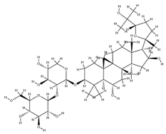
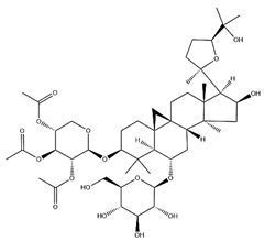
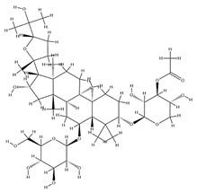
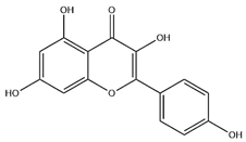
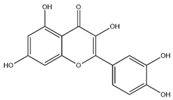
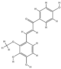
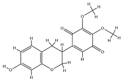
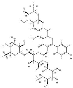
As mentioned above, we noted that there have been extensive phytochemical investigations over the past two decades, with the identification of over 400 bioactive constituents in AR, including polysaccharides, saponins, and flavonoids (Figure 1). These constituents—particularly astragalus polysaccharides (APS), AS, formononetin (FMN), and CA—have been recognized to be key therapeutic candidates given their protective effects on multiple organs through antioxidant, anti-inflammatory, antiapoptotic, and immunomodulatory activities. To gain a deeper mechanistic understanding of their organoprotective actions, these primary bioactive constituents were subjected to systematic characterization from the following perspectives of corresponding chemical structures, molecular weights, and extraction methodologies.

2.1. Polysaccharides

Acting as the principal bioactive macromolecules in AR, polysaccharides typically comprise high-molecular-weight polymers of glycosidically linked monosaccharides arranged in linear or branched configurations, with their structural architecture directly governing the pharmacological activity [18]. Heteropolysaccharides, glucans, and neutral/acidic polysaccharides constitute the predominant types among numerous polysaccharide classes isolated from AR. Among them, APS is generally isolated via water–alcohol extraction. Given their high polarity and aqueous solubility, crude APS at 10.97% efficiency can be yielded through hot water extraction at 75 °C followed by 80% ethanol precipitation [19]. According to compositional analysis via hot water extraction coupled with gas chromatography, the APS constituents were identified as l-rhamnose (l-Rha), d-xylose (d-Xyl), d-glucose (d-Glc), and d-galactose (d-Gal) in a 1:4:5:1.5 ratio. Structural characterization revealed a linear backbone in APS, consisting mainly of 1,3-linked b-D-Gal residues with the insertion of b-Glc, 1,6-linked a-Gal, 1,5-linked b-Xyl, 1,4-linked b-Gal, b-D-Gal, 1,2-linked a-Rha, and 1,2,4-linked a-Rha residues [20]. Chen et al. [21]. characterized two polysaccharides, designated APS-A1 and APS-B1, through infrared spectroscopy and nuclear magnetic resonance (NMR). Specifically, APS-A1, as a neutral polysaccharide, is composed of glucose, galactose, and arabinose, with a molar ratio of 52.3:1.0:1.3; while APS-B1 is a weakly acidic heteropolysaccharide composed of glucose, galactose, arabinose, xylose, rhamnose, and galacturonic acid, with a molar ratio of 75.2:17.3:19.4:1.0:1.1:1.3. Subsequently, this study provides an overview of other polysaccharides isolated and extracted from AR (Table 1).

2.2. Triterpenoid Saponins

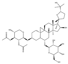
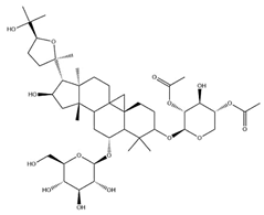
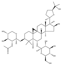
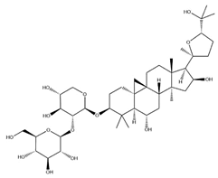
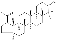
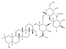
With pharmacologically significant components of AR, saponins have demonstrated clinically relevant activities of cardiovascular protection, liver protection, antidiabetes, and anti-inflammation [31]. Depending on their chemical configurations, these triterpenoid saponins are categorized into cycloartane-type and oleanane-type structural classes. Among them, cycloartane-type saponins (80% of the total saponin content) prominently include AS I–IV and isoastragalosides I–II [32]. Currently, over 170 distinct saponins have been identified with the use of advanced analytical techniques, particularly liquid chromatography–mass spectrometry (LC–MS) and NMR. High-performance thin-layer chromatography–mass spectrometry has quantified AS I-IV content in root extracts at 0.2–0.5 mg/g [33,34]. Meanwhile, modern purification methodologies yield AS-IV at 96.95% purity [35] by employing high-speed countercurrent chromatography with ethyl acetate/n-butanol/water (4.2:0.8:5, v/v) two-phase systems, an ideal approach for AS-IV extraction. Table 2 summarizes the structural characteristics of the characterized Astragalus triterpenoid saponins.

2.3. Flavonoids

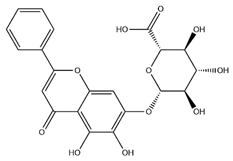
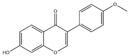
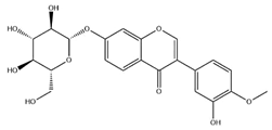
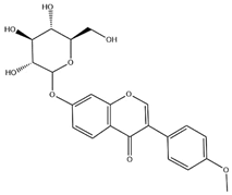
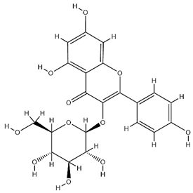
Flavonoids, categorized as flavonols, flavanones, anthocyanins, and isoflavones, constitute a prominent class of polyphenolic phytochemicals that are abundant in botanicals and human diets. These compounds exhibit significant anti-inflammatory, antiviral, antioxidant, and cardioprotective bioactivities [44]. Multiple flavonoid constituents in AR have been identified through phytochemical investigations. For instance, four flavonoid glycosides, i.e., 7-O-methylkaempferol 4′-O-β-D-galactopyranoside, hyperoside, isoquercitrin, and astragalin, were isolated in early studies employing chemical and spectroscopic methods [45]. Subsequently, two novel glycosides, i.e., kaempferol 7-methoxy-3-O-α-L-arabinosyl-(1→6)-β-D-galactopyranoside and kaempferol 3-O-α-L-rhamnopyranosyl-7-O-α-L-rhamnopyranosyl-(1→6)-β-D-galactopyranoside, were revealed by chromatographic study [45]. Moreover, Sheng et al. [46] isolated and identified flavonoid constituents from AR using methanol extraction, followed by UHPLC-MS/MS analysis. This facilitated the identification of 34 flavonoids, including well-known compounds (e.g., kaempferol, ononin, CA, and formonetin) that were confirmed in later research. Currently, water-to-methanol ratio optimization represents the predominant strategy for enhancing the extraction efficiency of flavonoids from AR [47]. Table 3 provides a comprehensive structural summary of Astragalus flavonoids characterized recently.

3. AR and Its Active Ingredients with Protective Effects on Multiple Organs and Tissues

AR primarily contains polysaccharides, saponins, and flavonoids according to modern phytochemical analyses. These constituents confer protective effects on multiple organ systems, including the intestinal tract, kidneys, joints, nervous system, cardiovascular system, liver, etc. These may be explained by diverse mechanisms, such as alleviating oxidative stress, modulating apoptosis pathways, regulating autophagy flux, attenuating immune responses, exerting anti-inflammatory effects, inhibiting ferroptosis and pyroptosis-mediated cell death, and displaying antifibrotic activity. This review continued to systematically summarize the evidence-based pharmacological characteristics and therapeutic applications of AR-derived compounds for specific organs, as shown in Table 2. Notably, a single active component (e.g., AS-IV) can target multiple organs (e.g., the heart, kidneys, and brain) via identical or distinct mechanisms, highlighting its multifunctionality. Conversely, there may be a formation of complex regulatory networks given that the protection of different organs frequently involves the coordinated regulation of multiple signaling pathways. Given the inherent complexity, it highlights the multicomponent, multitarget nature of AR, providing both a theoretical foundation and a significant research challenge for its clinical application in complex diseases.

3.1. Oxidative Stress

Oxidative stress refers to a pathological state arising from the imbalance between reactive oxygen species (ROS) and antioxidants that reduce ROS. This imbalance can trigger oxidative damage to biomolecules (e.g., lipids, proteins, and DNA) and cellular dysfunction [61]. Including both free radicals and non-free radicals, ROS are a class of oxygen-containing substances, such as superoxide anions (O2), hydrogen peroxide (H2O2), hydroxyl radicals (·OH), ozone (O3), and singlet oxygen (1O2). Under normal physiological conditions, free radicals are generated through pathways such as the mitochondrial electron transport chain, NADPH oxidase (NOX), and xanthine oxidase in a controlled manner. Subsequently, these yielded free radicals may be promptly eliminated by antioxidant enzymes, such as superoxide dismutase (SOD) and glutathione peroxidase (GPx), thus maintaining a dynamic equilibrium. However, ROS accumulate exponentially and exceed the clearance capacity in the context of physical exposure to external stimuli (e.g., toxins, radiation, or inflammation), thereby leading to oxidative stress [62]. Through the Fenton reaction, excessive ROS can generate highly toxic ·OH that may induce lipid peroxidation, and destroy membrane fluidity and integrity. With further impact on mitochondrial membrane potential collapse, endoplasmic reticulum (ER) stress, and calcium overload, this damage can ultimately activate several apoptotic pathways (e.g., the caspase cascade) and necrotic apoptosis, resulting in parenchymal cell death and organ dysfunction.
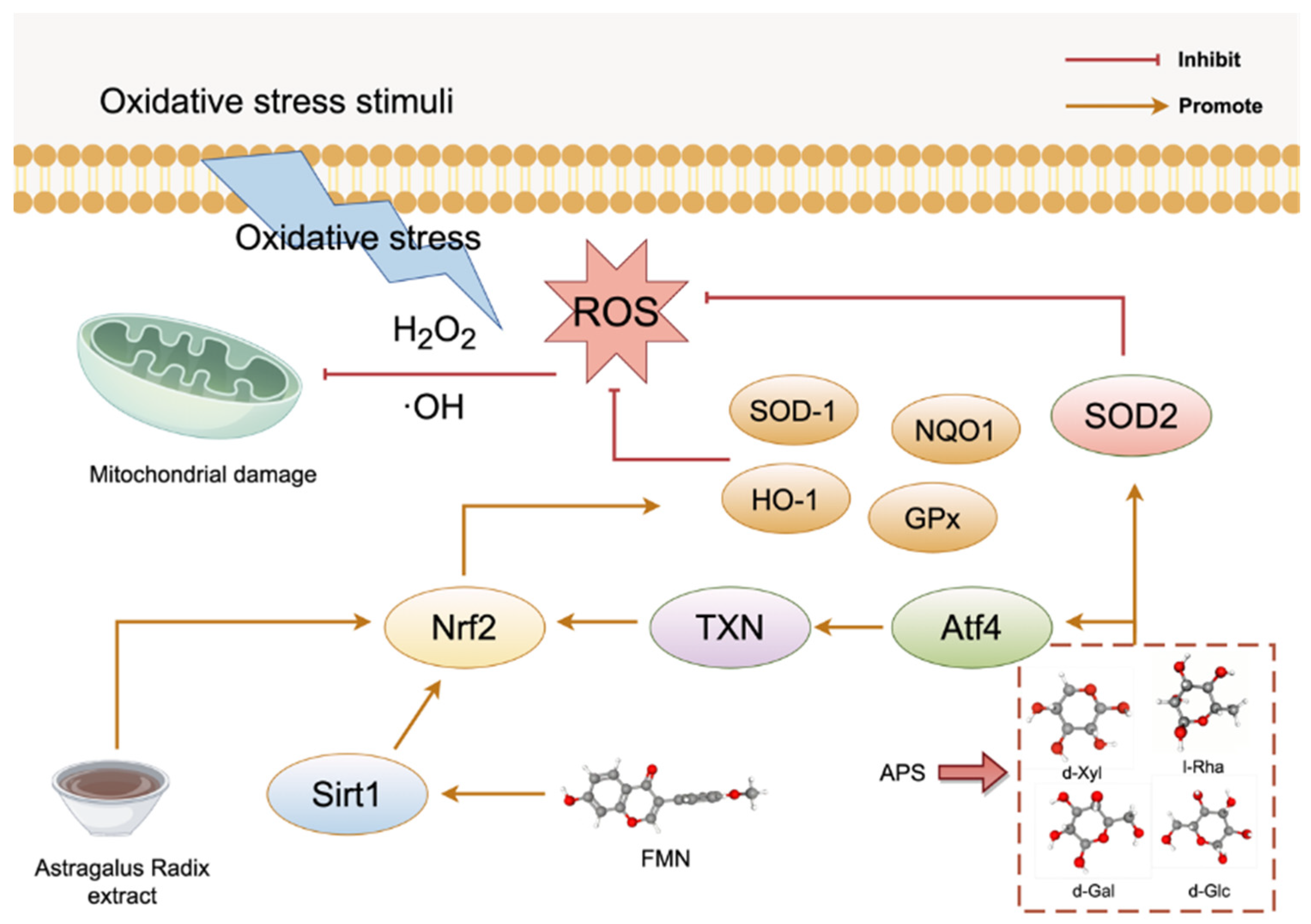
Numerous studies have extensively validated the antioxidant activity of AR, and have elucidated its involvement in antioxidant mechanisms. AR extracts exert antioxidant effects by reducing ROS release and activating antioxidant enzymes. For example, both lipopolysaccharide (LPS) from E. coli plus interferon-γ (IFN) inflammatory model and H2O2-induced oxidative stress model have revealed that AR extracts can effectively suppress the release of ROS. Furthermore, Nrf2 is a pivotal transcription factor antioxidant stress, plays a critical role in antioxidant responses [63]. AR extracts have been documented to exhibit significant antioxidant effects, as evidenced by the activation of Nrf2 and the substantial upregulation of two antioxidant enzymes of heme oxygenase-1 (HO-1) and NAD(P)H quinone oxidoreductase 1 (NQO1) [64]. The Nrf2/antioxidant response element (ARE) signaling pathway is a pivotal antioxidant signaling pathway within cells [65], which can be activated by silent information regulator 1 (Sirt 1). The flavonoid compound FMN present in AR has been reported to activate the Nrf2/ARE signaling pathway via upregulating Sirt1 expression, thereby enhancing the activity of antioxidant enzymes such as HO-1 and SOD-1 that are responsible for scavenging ROS. The result of this enhanced activity is a blockade of oxidative stress, which can improve the progression of diabetes and kidney disease [52]. In addition, AR-derived polysaccharides can reduce oxidative stress in osteoarthritis via a multifaceted process involving GCN2/TXN/NRF2. These proteins can activate the GCN2 kinase to sense oxidative stress and activate transcription factor (ATF) 4. ATF4 then upregulates the key antioxidant protein thioredoxin (TXN) to effectively suppress oxidative stress by scavenging ROS, thereby alleviating ROS-mediated DNA damage [66]. Figure 2 shows the specific factors and pathways involved in oxidative stress affected by the active components FMN and APS in AR.
Ji et al. [67] reported that baking AR at 260 °C for 30 min could elevate flavonoid concentration and provide neuroprotective effects through dual-pathway synergistic action. Roasting AR significantly increased the levels of catalase, HO-1, SOD-2, and glutathione peroxidase (GPX) by activating the Nrf2 pathway. It could further inhibit the release of ROS and offer neuroprotective effects by activating the brain-derived neurotrophic factor (BDNF) pathway and inhibiting neuronal apoptosis. It could also suppress the accumulation of beta-amyloid (Aβ)-induced neuronal oxidative damage and apoptosis.
Diabetic cardiomyopathy (DCM) refers to a myocardial metabolic dysfunction caused by the apoptosis of cardiac stem/progenitor cells (CSPCs) due to mitochondrial oxidative stress primarily induced by hyperglycemia-induced oxidative stress activation and elevated mitochondrial ROS levels [68]. Chen et al. [69] carried out an experiment with the use of Streptozotocin (STZ)-induced heterozygous (SOD-2+/−) knockout mice, where mitochondrial oxidative stress was stimulated with a 50% reduction in SOD-2 activity. The results revealed that the number of CSPCs decreased by 75–85% in mice with induced diabetes mellitus (DM) in the SOD-2+/− group, and the DM-SOD-2+/− group. Significantly intervention with APS led to 3.8- to 5.5-fold increase in the number of CSPCs. APS significantly reduced ROS production, as well as H2O2 and ·OH concentrations in CSPCs. Additionally, although the enzymatic and protein activities of SOD-2 in CSPCs were reduced in mice with DM, which were significantly increased after APS intervention. These findings indicate that APS specifically activates the SOD-2 pathway to inhibit mitochondrial ROS accumulation in diabetic hearts, highlighting a potential protective effect on CSPCs in diabetic patients. In addition, the production of ROS can be significantly influenced by glucose, lipids, succinate and other metabolic substrates [70]. For example, Jiang et al. found that AS-IV could alleviate myocardial ischemia–reperfusion injury by regulating succinate metabolism, inhibiting lysophospholipid accumulation, and activating the Nrf2/HO-1 antioxidant pathway [71]. In addition, CA was proven to reduce the levels of ROS and malondialdehyde (MDA) in myocardial tissue, enhance SOD activity, and mitigate oxidative stress-mediated myocardial cell necrosis in the isoproterenol-induced myocardial infarction model [72].
Therefore, AR enables the protection of the intestines [73], kidneys, joint cartilage, nervous system, heart [74,75], cardiovascular and skeletal muscles [76] by targeting oxidative stress-related signaling pathways and regulating the expression of related proteins, both upstream and downstream.

3.2. Antiapoptotic Effects

Apoptosis, similar to pyroptosis, necroptosis, and ferroptosis, is a genetically precisely regulated programmed cell death that is essential in maintaining tissue homeostasis, regulating normal development, and immune defense. In general, it is mainly mediated by the intrinsic mitochondrial pathway and the extrinsic death receptor pathway. Considering their differences in structural and functional characteristics, members of the Bcl-2 family can be classified into three categories, i.e., pro-survival proteins (e.g., Bcl-2, Bcl-XL, Bcl-w, Mcl-1, and A1); death effector proteins (e.g., Bax and Bak); and proapoptotic BH3-only proteins (e.g., Bid, Bim, Puma, Bad, and Noxa) [77]. In healthy cells, the death effectors Bax and Bak are bound by Bcl-2 proteins, whereas the proapoptotic BH3-only proteins are in an inactive state. Under the condition of cellular exposure to toxic stimuli, activated BH3-only proteins may release the bound Bax and Bak proteins, inducing mitochondrial outer membrane permeability and promoting the release of cytochrome c into the cytoplasm, in turn activating caspase-9 and effector caspases-3/7, ultimately resulting in cell disintegration [78]. Meanwhile, cytotoxic lymphocytes may produce death-inducing ligands in the extrinsic death receptor pathway. These ligands, involving FAS ligands, tumor necrosis factor (TNF), TNF-related apoptosis-inducing ligand (TRAIL), and TNF-α, can bind to specific death receptors on the target cell surface (FAS, DR4/DR5, and TNFR1, respectively), finally triggering the activation of caspase-8 directly [79].
Chk1 is a serine/threonine kinase that can modulate diverse cellular responses, relying primarily on its roles of controlling DNA damage, enforcing cell cycle checkpoints, and phosphorylating downstream substrates. Meanwhile, p53, a classical tumor suppressor, can activate its key downstream target p21, a cyclin-dependent kinase inhibitor. Both p53 and the p21 axis are central to coordinating the cellular response (e.g., cell cycle arrest and apoptosis) to DNA damage [80,81]. APS has been observed to attenuate paclitaxel-induced pro-apoptotic effects, which might be related to the reduced levels of phosphorylated H2A histone family member X (γH2AX; a DNA damage marker) and decreased PARP cleavage (an indicator of apoptosis execution). Furthermore, APS can upregulate the antiapoptotic proteins Bcl-XL and Mcl-1, and inhibits taxane-induced cell cycle arrest and apoptosis via downregulating the Chk1-p53-p21 signaling axis. This multifaceted mechanism underlies the ability of APS to selectively shield critical organs from paclitaxel toxicity. In addition, AS-IV (25–50 mg/kg) can suppress cell apoptosis through two pathways. On the one hand, it suppresses the extrinsic (death receptor) pathway by significantly downregulating the expression of Fas, Fas ligand (FasL), and Caspase-8 to inhibit the activation of downstream cleaved Caspase-3. Concurrently, it can inhibit the intrinsic (mitochondrial) pathway by remarkably heightening the Bcl-2/Bax ratio and reducing cytochrome c release from mitochondria [82]. In addition, AR extracts can retard oxidative stress-induced cell apoptosis by downregulating Bax expression, upregulating Bcl-2 expression, and restraining ROS production [83].
Simultaneously, AR also exhibits antitumor activity against diverse solid tumors. One of the major concerns in contemporary cancer research lies in overcoming chemoresistance and enhancing tumor sensitivity to therapeutic agents. In this context, dynamic and reversible posttranslational modification of O-GlcNAcylation has emerged as a critical regulator of cellular metabolism and signaling pathways, which facilitates rapid cellular adaptation to various stress stimuli [84]. O-GlcNAc transferase (OGT) and O-GlcNAcase (OGA) stand out as the two major enzymes regulating O-GlcNAcylation [85]. Specifically, hyper-O-GlcNAcylation, a posttranslational modification of proteins, occurs in the majority of malignant tumors, revealing positive correlation with tumorigenesis and progression [86]. Mechanistically, this modification engages in pathological crosstalk with ER stress across multiple cancers. ER stress propagates apoptosis through three distinct pathways: IRE1α, ATF6, and PERK, which independently induce apoptosis by activating apoptotic effectors, including caspase 12, c-Jun N-terminal kinase (JNK), ATF4, and the transcription factor C/EBP homologous protein (CHOP) [87]. For instance, APS impeded O-GlcNAc synthesis by decreasing OGT and augmenting OGA levels in doxorubicin (Dox)-treated Hep3B hepatocellular carcinoma (HCC) cell, leading to exacerbated Dox-induced ER stress, as evidenced by elevated CHOP expression. This study revealed that APS could promote apoptosis in Hep3B cells by downregulating Bcl-2 and upregulating Bax, Bim, and cleaved Caspase-3 through the CHOP-Bcl-2 pathway. Collectively, APS can increase the sensitivity of HCC cells to Dox [88]. Notably, Sun et al. [89] demonstrated that APS could specifically suppress the PERK-ATF6-CHOP ER stress axis to reduce cardiomyocyte apoptosis, thereby attenuating DCM. Moreover, peroxisome proliferator-activated receptor-γ coactivator 1 (PGC-1α) has been identified as a significant downstream target of Sirt1. FMN can modulate the homeostasis of mitochondrial dynamics (inhibiting fission/promoting fusion) through activating the Sirt1/PGC-1α pathway. This pathway works to restore mitochondrial function, attenuate ROS, normalize the Bcl-2/Bax ratio, and inhibit caspase-3-mediated renal tubular apoptosis [90].
In summary, AR exhibits potent antiapoptotic activity across multiple disease models, which is mediated mechanistically through coordinated regulation of both intrinsic (mitochondrial) and extrinsic (death receptor) apoptotic pathways. Altogether, APS shows promise in protecting the immune system, brain, liver, heart [91], kidneys, stomach [92], and joint cartilage by targeting key signaling pathways, such as OGT/OGA, PERK-ATF4-CHOP, and Chk1-p53-p21 [93].

3.3. Autophagy

Autophagy constitutes an evolutionarily conserved lysosomal degradation pathway that removes damaged organelles, misfolded proteins, and intracellular pathogens from the cytoplasm and recycles macromolecules into metabolic precursors to maintain eukaryotic cellular homeostasis [94]. This catabolic process begins with the formation of double-membrane autophagosomes that sequester targeted cell constituents and fuse with the lysosomess to form autolysosomes. Within these acidic compartments, lysosomal hydrolases can degrade encapsulated materials into reusable biomolecules (e.g., amino acids and nucleotides) to ensure energy homeostasis. Autophagy is precisely orchestrated by over 40 autophagy-related genes (ATG) through three core mechanistic stages. It can be subdivided into four stages: autophagy initiation, autophagosome formation, autophagolysosome fusion, and autolysosome degradation. To be specific, during initiation, Beclin-1 recruits VPS34 and PI3P to the autophagic precursor, thereby driving the formation of phagophores. ATG5 subsequently binds to ATG12 and recruits ATG16 to form the ATG5-ATG12-ATG16 complex, which can further polymerize on the surface of the phagophore membrane. During vesicle maturation, the developed complex synergistically promotes phagophore extension with LC3-II, which can further bind selectively to substrates awaiting degradation to guide the enclosure of materials by the autophagosome. At stages of terminal fusion and degradation, the V-SNARE subunits of the autophagosome membrane are anchored to the lysosome via VAMP proteins, whereas the STX17 subunit of the lysosome membrane forms the t-SNARE subunit with SNAP29. The assembly of these subunits to form a SNARE complex serves to mediate membrane fusion to facilitate the formation of autophagolysosomes. V-ATPase proton pumps then acidify these components, leading to the activation of cathepsin hydrolases that degrade contents into reusable metabolites, including amino acids and nucleotides, completing the autophagic flux that sustains cellular homeostasis [95].
The regulation of autophagy is orchestrated through integrated signaling–transcriptional networks with mTOR as the master inhibitory switch. Mechanistically, mTOR can competitively bind to ULK1 at the site of Ser757 to disrupt AMPK-ULK1 complex assembly, which may suppress the initiation of autophagy. Through negative feedback loops, the downstream ATG1 kinase complex can reciprocally regulate both autophagosome biogenesis and mTOR activity. The PI3K/Akt pathway, as an upstream regulator of mTOR, is inhibited during cellular stress or starvation to initiate autophagy [96]. Transcriptional control involves the MiT-TFE family (MITF/TFEB/TFE3/TFEC), which enables the activation of CLEAR-network genes for lysosomal biogenesis [97]; NF-κB, which possesses bidirectional regulatory function can be activated by ROS to induce autophagy under oxidative stress, and can also enhance autophagy through the mTOR pathway [98]; FOXO1/ATF4/HIF, which upregulates autophagy genes; and β-catenin, which suppresses core autophagic components via TCF4-mediated transcriptional repression. Collectively, all these findings may facilitate the establishment of a hierarchical control system that dynamically adjusts autophagy flux to metabolic and stress conditions [99].
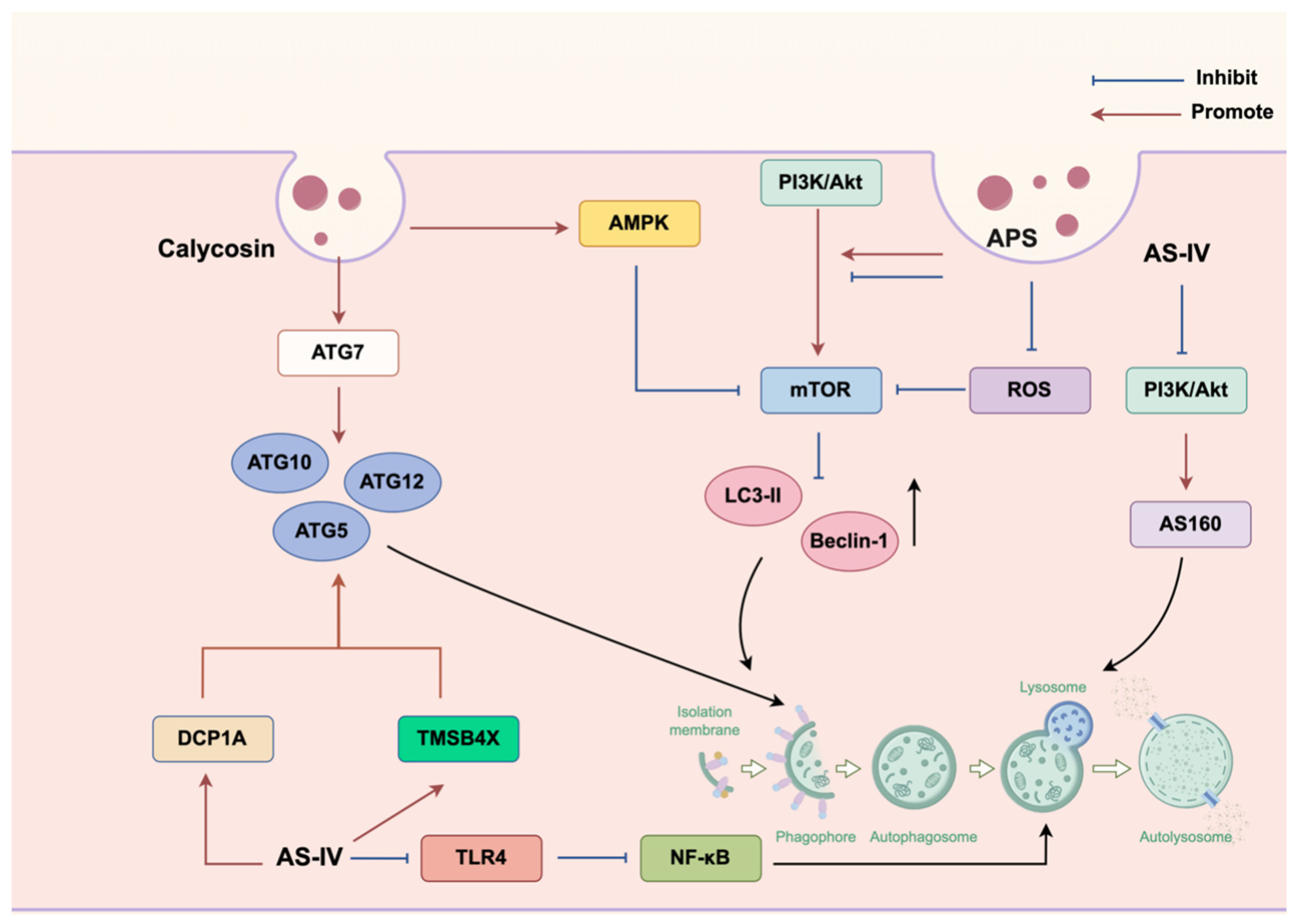
Substantial evidence has documented that multiple bioactive constituents in ARs that modulate cellular autophagy pathways constitute a primary molecular mechanism underlying their organ-protective effects. Core components (e.g., APS, AS, and isoflavones) can regulate autophagy flux by targeting distinct signaling nodes. Specifically, APS can enhance autophagy via activating the PI3K/Akt/mTOR pathway, demonstrating therapeutic potential in Parkinson’s disease (PD) model [100]. Similarly, CA can promote autophagic clearance in osteoporosis [101]. Conversely, fermented AS preparations may inactivate PI3K/Akt/mTOR signaling, activating podocyte autophagy to mitigate hyperglycemia-induced damage [102]. Notably, this bidirectional regulatory capacity stems from the context-dependent functionality of mTOR. In other words, the overactivation of mTOR requires suppression to restore autophagy during metabolic stress (e.g., hyperglycemia), whereas moderate mTOR activation can initiate protective autophagy during cellular differentiation or proteostasis imbalance. Meanwhile, AS can suppress TLR4/NF-κB signaling and attenuate inflammatory responses in RAW264.7 macrophages to induce cytoprotective autophagy. Fluorescence microscopy confirmed AS-mediated autophagosome accumulation and autophagic vacuole formation, with impaired autophagy exacerbating the release of inflammatory cytokines [103]. In addition, by reducing ROS generation and downregulating LC3-II/Beclin-1 expression through ROS–mTOR pathway inhibition, APS can mitigate cadmium (Cd)-induced autophagic flux imbalance in chicken embryo fibroblasts (CEFs), thereby preserving cell viability [104]. AS160 is an effector protein of AKT that regulates the activity of Rab14, a member of the Ras superfamily and a core regulatory factor in eukaryotic intracellular transport. Rab14 has been reported to affect the activity of autophagy by modulating autophagosome–lysosome fusion [105]. Significantly, AS-IV can ameliorate chronic glomerulonephritis by enhancing autophagic flux by inactivating the PI3K/AKT/AS160 signaling axis [106]. Mechanistically, AS can induce a cascade of alterations, such as suppressing PI3K phosphorylation, reducing phosphorylated AKT (p-AKT) and AS160 (p-AS160) levels, and downregulating Rab14 expression. Eventually, it can facilitate the relief of Rab14-mediated inhibition of autophagosome maturation, promoting autophagolysosome formation and ultimately augmenting autophagic activity. CA can upregulate the expression of ATG7—a critical autophagy execution gene, which can contribute to the restoration of the autophagic homeostasis and mitigation of Dox-induced cardiotoxicity in delayed cardiac injury model [107]. Concurrently, CA can activate AMPK/mTOR signaling to initiate early autophagy while promoting the assembly of the STX17-SNAP29-VAMP8 SNARE complex, eventually restoring autophagosome–lysosome fusion and autophagic flux through dual-phase regulation. This mechanism can inhibit vascular smooth muscle cell osteogenic differentiation to attenuate vascular calcification [108]. This study plotted relevant autophagy regulatory pathways, as depicted in Figure 3. A pioneering study demonstrated that AS-IV could suppress the progression of cervical cancer by activating the Atg7/Atg12/LC3 autophagy axis through targeting DCP1A and TMSB4X. Quantitative proteomic profiling via iTRAQ-PRM technology identified 32 differentially expressed proteins (16 upregulated; and 16 downregulated), with mRNA decapping enzyme 1A (DCP1A) and thymosin β-4 (TMSB4X) emerging as significantly upregulated autophagy regulators. Mechanistically, DCP1A can accelerate autophagosome maturation through the WDFY3/Atg12 pathway, whereas TMSB4X may modulate autophagy initiation via the Akt/Atg5/Atg12 cascade [109].
Collectively, astragalus-mediated modulation of key autophagy pathways—including the PI3K/AKT/mTOR, TLR4/NF-κB, ROS-mTOR, PI3K/AKT/AS160, AMPK/mTOR, PINK1-Parkin, DCP1A/WDFY3/Atg12, and TMSB4X/Akt/Atg5/Atg12 pathways—can inhibit the therapeutic efficacy against multiple pathologies, such as PD, diabetes mellitus, osteoporosis, renal disorders, cardiovascular diseases, and cervical carcinoma.

3.4. Immunoregulatory Effects

The immune system is a fascinating, highly coordinated self-defense network comprising multicomponents of immune organs, immune cells, and immune-active substances that execute immune surveillance, defensive responses, and homeostatic regulation collectively [110]. The multifaceted roles of the immune system encompasses the defense against pathogen invasion and the fight against infection, beyond the identification and elimination of harmful substances within the body. Moreover, it is pivotal in slowing aging, maintaining cellular homeostasis, and combatting neoplasms [111].
AR exhibit immunomodulatory properties by modulating key immune organs such as the bone marrow [112], thymus [113], and spleen [112,113]. Meanwhile, it can enhance the activity of diverse immune cells, such as macrophages [113], natural killer (NK) cells [114], dendritic cells [112], T lymphocytes [115], lymphocytes [116], and oligodendrocytes [117]. AR can also promote the release of immune mediators, like interleukins (ILs; IL-2, IL-6, and IL-10), TNF-α, IFN-γ, and immunoglobulins (IgG and IgM) [113,118,119]. Furthermore, the programmed cell death protein 1 (PD-1)/programmed death-ligand 1 (PD-L1) axis represents a critical immune checkpoint in cancer, where PD-L1 binding to PD-1 suppresses T-cell proliferation, activation, and cytokine secretion. One of the key bioactive components of AR, APS, has significant immunomodulatory potential and is relevant to this pathway.
APS can activate both CD4+ and CD8+ T cells and reduce PD-L1 expression, as demonstrated in prostate cancer models [115]. Furthermore, APS can reactivate exhausted CD8+ T cells, inducing the secretion of pro-inflammatory factors, including IFN-γ, chemokine (C-X-C motif) ligand (CXCL) 2, chemokine (C–C motif) ligand 5 (CCL5), and CXCL12, thereby enhancing antitumor immunity. Concurrently, APS can stimulate macrophages to secrete cytokines such as TNF-α, IL-6, and CXCL2, which contribute to remodeling the tumor microenvironment toward an immunostimulatory state. Importantly, APS can benefit the reversal of T-cell exhaustion and the potentiation of existing immunotherapies by inhibiting PD-L1 expression and disrupting the PD-1/PD-L1 interaction [115].
Demyelination is a common pathological process underlying multiple neurological disorders, which is characterized by myelin sheath damage that may disrupt neurological function [120]. Currently, immunosuppressants, hormones, and other immunomodulatory drugs are the primary therapeutic options for the treatment of demyelinating diseases. However, these approaches often result in incomplete neural repair owing to their failure of promoting myelin regeneration directly [121]. Mechanistically, myelin restoration depends mainly on the differentiation of oligodendrocyte precursor cells (OPCs) into mature, myelinating oligodendrocytes (OLs) [122]. Concurrently, the Wnt/β-catenin signaling pathway has been documented to fundamentally orchestrate OPC development and myelination [123,124]. Yuan et al. [117] demonstrated that AS-II specifically bound to the Pro253 and Ser257 residues of p75NTR via hydrogen bonds. This binding would further stabilize the spatial conformation of p75NTR, enhancing its resistance to proteolytic degradation and thermal stability. However, noticeably, AS-II failed to promote myelin regeneration or improve motor function in p75NTR-knockout mice, confirming p75NTR dependence. Mechanistically, AS-II binding can promote OPC differentiation into myelinating OLs as it enables the potentiation of the GSK3β-mediated destabilization of β-catenin. By targeting this endogenous repair mechanism through indirect immune modulation, AS-II represents a novel broad immunosuppression-independent therapeutic strategy for myelin regeneration.

3.5. Anti-Inflammatory Effects

Inflammation is our body’s innate protective response to injury that functions significantly in the pathogenesis of diverse diseases. Chronic inflammation refers to a prolonged release of pro-inflammatory factors (e.g., TNF-α, IL-1β, and IL-6), chemokines, ROS, and proteases from activated immune cells (e.g., macrophages, neutrophils, and T cells) and tissue-resident cells. Due to excessive degradation or deposition (fibrosis), this persistent inflammatory response may culminate in tissue damage, cellular dysfunction, and dysregulation of the extracellular matrix, resulting in the disruption of the structural and functional integrity of the involved organs. Macrophages are central effectors of innate immunity, which can mediate crucial host defense functions (e.g., phagocytosis, cytokine secretion, and antigen presentation) against invading pathogens. Meanwhile, monocyte chemotactic protein-1 (MCP-1) is a chemokine that recruits monocytes, a type of immune cells involved in inflammatory responses, to the site of an inflammatory response [125]. Pioneering work by Chen et al. [21] isolated and purified two homogeneous polysaccharides (APS-A1 and APS-B1) from AR. The two polysaccharides have been demonstrated to significantly downregulate the mRNA levels and suppress the protein expression of key pro-inflammatory mediators, including TNF-α, IL-6, MCP-1, and IL-1β, in macrophages. This reduction effectively curbs the excessive secretion of these cytokines and inflammatory mediators. This anti-inflammatory activity involves central signaling pathways of NF-κB and MAPK, two representative regulators of inflammatory mediator expression [126,127]. Chen et al. [21] reported that APS-A1 and APS-B1 inactivated NF-κB and suppressed the phosphorylation of key MAPK kinases (JNK, ERK, and IKKα/β), thereby synergistically attenuating inflammation through the dual MAPK/NF-κB signaling axis. Furthermore, the NLRP3 inflammasome is a well-characterized critical pattern recognition receptor comprising NLRP3, ASC, and procaspase-1, which may contribute to diverse inflammatory pathologies upon dysregulated activation. With the resultant activation of autocatalytic caspase-1, this process can mediate proteolytic maturation and the release of pro-inflammatory cytokines IL-1β and IL-18 [128]. In addition to its role in intestinal inflammation, AS-IV can directly inhibit the activation of the NLRP3 inflammasome, which is evidenced by significantly attenuated protein expression of NLRP3, ASC, caspase-1, and NF-κB in the intestinal mucosa. Concurrently, AS-IV was observed to reduce the release of downstream IL-1β and IL-18, conferring significant protection against indomethacin-induced intestinal inflammation in rats [129]. As primary immune sentinels, macrophages constitute the first line of host defense, exhibiting functional polarization into two main phenotypes of the classically activated (pro-inflammatory M1) and alternatively activated (anti-inflammatory M2) macrophages [130]. Upon activation, M1 macrophages produce substantial levels of pro-inflammatory mediators, including IL-1β, IL-6, TNF-α, IL-12, ROS, and inducible nitric oxide synthase (iNOS). Conversely, M2 macrophages, induced by IL-4 and IL-10, can secrete anti-inflammatory factors such as arginase-1 (Arg-1). In vitro experiments revealed that APS inhibited the differentiation of macrophages into the pro-inflammatory M1 type, reduced the expression of M1 marker CD86, and decreased the release of ROS and the pro-inflammatory factors TNF-α, IL-6, and IL-12. Moreover, polysaccharides directly promoted the conversion of macrophages to the M2 type, upregulated the expression of M2 markers CD206 and Arg-1, and increased the secretion of anti-inflammatory factors IL-4 and IL-10. Critically, a relevant experiment based on gene silencing technique confirmed that the Nrf2/HO-1 pathway could mediate APS-induced macrophage phenotype regulation [131]. In a carrageenan-induced mouse paw edema model, astragalin also significantly alleviated acute inflammatory responses through dual mechanisms of scavenging ROS and inhibiting the NF-κB-mediated inflammatory pathway. Its anti-inflammatory efficacy is comparable to that of indomethacin (10 mg/kg), a common clinically used anti-inflammatory drug. As a naturally derived component, astragalin offers a greater safety advantage over synthetic anti-inflammatory drugs [132]. In addition to its anti-inflammatory effects, FMN can alleviate psoriasis-like inflammation. Mechanistically, FMN can downregulate the expression of key inflammatory factors TNF-α and IL-6 by impeding the IFN signaling pathway [129].
Collectively, APS, AS-IV, and FM, serving as the bioactive constituents of AR, can mitigate inflammatory responses through a multitargeted mechanism. This involves the strategic regulation of key inflammatory mediators (e.g., TNF-α, IL-6, IL-1β, IL-18, and MCP-1) and the concerted modulation of pivotal signaling pathways (e.g., NF-κB, MAPK, the NLRP3 inflammasome, Nrf2/HO-1, and IFN). On the basis of the proposed integrated molecular intervention, AR extracts and compounds confer significant protective effects across multiple organ systems, including the immune system (via macrophage polarization modulation), intestines, the vasculature, the skin, and lungs, demonstrating its broad therapeutic potential in inflammation-driven pathologies.

3.6. Other Functions

AR and its bioactive constituents exhibit significant antitumor activity, including anti-proliferative and pro-apoptotic effects, across a variety of cancer types. In particular, ovarian cancer (OC), one of the most prevalent and lethal gynecological malignancies, is characterized by high recurrence rates, metastasis, and a 5-year survival rate of only 25% [133]. MicroRNAs (miRNAs) are a family of small noncoding RNAs that serve as either oncogenes or tumor suppressors, which play crucial roles in the development of OC. Guo et al. reported [134] that through the modulation of the miR-27a/FBXW7 pathway, APS could inhibit proliferation and migration while promoting apoptosis in OC cell lines (OV-90/SKOV-3). Peng et al. revealed [135] that APS overcame PARP inhibitor resistance in OC stem cells (OCSCs) by targeting PINK1/Parkin-mediated mitophagy. Furthermore, AR-derived triterpenoid saponins (total Astragalus saponins, TAS) can also induce ferroptosis in gastric cancer, in addition to OC. Ferroptosis, an iron-dependent form of lethal lipid peroxidation-driven regulated cell death [136], can be activated by TASs through the SIRT3 pathway. TAS can modulate SIRT3 expression to trigger the remodeling of proferroptotic proteins, manifesting as the upregulation of ACSL4 alongside downregulation of SLC7A11 and GPX4. This imbalance can further elevate the levels of intracellular ROS, Fe2+, MDA, and lactate dehydrogenase, ultimately leading to ferroptotic cell death [137].
Conversely, AS-IV exhibits potent antiferroptotic activity in the model of cisplatin-induced hepatotoxicity. AS-IV can inhibit ferroptosis and mitigate liver injury through the activation of the PPARα/FSP1 axis. In vivo, AS-IV significantly reduced the levels of hepatic lipid peroxidation markers (4-HNE and MDA) while increasing the levels of the anti-ferroptotic factors GPX4 and FSP1 and suppressing the pro-ferroptotic enzyme arachidonate 15-lipoxygenase. In vitro studies have confirmed the efficacy of AS-IV in reversing cisplatin-induced cell death and lipid ROS accumulation through this mechanism. Mechanistic investigations revealed that AS-IV can directly bind to and activates peroxisome proliferator-activated receptor alpha (PPARα), upregulating its downstream effector ferroptosis suppressor protein 1 (FSP1). Crucially, pretreatment with either GW6471 (the PPARα antagonist) or iFSP1 (the FSP1 inhibitor) could completely abolish AS-IV-mediated cytoprotection, confirming the essential role played by the PPARα/FSP1 axis in exerting its antiferroptotic activity [138].
Pyroptosis represents another programmed cell death integral to innate immunity, which is an important contributor to the pathogenesis of inflammatory diseases [139]. In the context of asthma, AS-IV can exert certain therapeutic effects through promoting pyroptosis in airway smooth muscle cells (ASMCs). Mechanistically, AS-IV can disrupt its interaction with the receptor for advanced glycation end products (RAGE) based on its inhibitory role in the expression and cytoplasmic translocation of high-mobility group box 1 (HMGB1. By inactivating the NF-κB signaling pathway, this blockade can further trigger the assembly and activation of NLRP3 inflammasomes. Caspase-1 activation then mediates pyroptotic cell death in ASMCs, accompanied by the release of pro-inflammatory cytokines IL-1β and IL-18 [140]. Furthermore, AS-IV is effective against cholestatic liver fibrosis, a condition characterized by pathological bile acid (BA) accumulation, including tauro-conjugated BAs. The farnesoid X receptor (FXR), a BA-activated nuclear receptor, can regulate BA homeostasis via downstream effectors (e.g., SHP, Cyp7a1, NTCP, and BSEP). Zang et al. [141] reported that ARs could improve fibrosis, liver damage, and inflammation in cholestatic liver fibrosis by targeting the BA-FXR pathway. Specifically, as a multitarget antifibrotic agent, AS can significantly suppress the expression of the profibrotic markers α-smooth muscle actin and collagen type I alpha 1 while concurrently attenuating pathological collagen deposition in the liver tissue.
Collectively, AR-mediated cytoprotection orchestrates multiple programmed cell death pathways (e.g., ferroptosis, and pyroptosis), antifibrotic actions, and strategic modulation of critical signaling axes, including but not limited to the miR-27a/FBXW7, PINK1/Parkin-mediated mitophagy, SIRT3-dependent regulation, PPARα/FSP1, HMGB1/RAGE/NF-κB, and tauro-conjugated BA-FXR pathways.

3.7. Multipathway Synergistic Effects

The bioactive constituents of AR extracts confer multiorgan protection through synergistic modulation of various interconnected pathways. For instance, in alcoholic liver injury (ALI) with oxidative stress as a major contributor, APS and AS-IV can activate the KEAP1/NRF2 antioxidant pathway, enhancing the activity of endogenous antioxidant enzymes [e.g., SOD and glutathione peroxidase (GSH-Px)]. Concurrently, these compounds can directly scavenge excessive ROS and inhibit oxidative stress-induced mitochondrial apoptosis, as exemplified by suppressed activation of Bax [142]. Meanwhile, oxidative stress-derived ROS can further promote hepatic inflammation by stimulating the release of pro-inflammatory cytokines (IL-1β, IL-6, and TNF-α) [143]. Significantly, APS and AS work to mitigate this inflammatory response by downregulating the TLR4/MyD88/NF-κB signaling axis, significantly reducing TNF-α and IL-1β levels. This anti-inflammatory action alleviates tissue damage and indirectly suppresses caspase-mediated apoptosis triggered by inflammation. Critically, APS and AS facilitate the establishment of a synergistic antioxidant–anti-inflammatory network. Furthermore, the activation of NRF2 can inhibit NF-κB phosphorylation, whereas the suppression of NF-κB can reduce ROS generation, creating a self-reinforcing cytoprotective loop that amplifies the hepatoprotective effects [142]. Cycloastragenol (CAG) is a AS-derived bioactive aglycone that exhibits multimodal neuroprotective effects against PD through coordinated regulation of oxidative stress, autophagy, inflammation, and immune responses [144]. Mechanistically, CAG can attenuate oxidative damage through suppressing phagocytosis-induced ROS generation by inhibiting microglial Scribble and Scrib-p22phox expression. Simultaneously, increased autophagic flux may promote the clearance of pathogenic α-synuclein (α-Syn), a hallmark protein in PD pathogenesis, and mitigate mitochondrial dysfunction. These mechanisms collectively inhibit NLRP3 inflammasome assembly via antioxidant/autophagy-mediated pathways, while increased autophagy directly degrades inflammasome components, ultimately reducing caspase-1-dependent gasdermin D cleavage and IL-1β/IL-18 release. Moreover, CAG can remodel the neuroimmune microenvironment by downregulating the expression of pro-inflammatory mediators (e.g., TNF-α, IL-6, and iNOS) and increasing the expression of neuroprotective markers (e.g., CD163), thereby driving microglial polarization toward a protective phenotype. Consequently, this phenotypic shift promotes the reduction of neuronal calcium overload, suppression of neuronal nitric oxide synthase activation, and inhibition of caspase-3-mediated apoptosis [144]. In addition, AR constituents confer renoprotection by modulating the PI3K/AKT/mTOR signaling axis, activating the NRF2/KEAP1/ARE antioxidant pathway, and suppressing TGF-β1/CTGF-driven fibrotic cascades, eventually leading to the preservation of the integrity of podocytes [145]. Table 4 lists the potential pharmacological effects of AR on different organs.

4. Safety Evaluation of AR Extract Components

AR, with a documented clinical history spanning nearly two millennia, is a cornerstone herb in TCM that possesses dual applications in medicinal and dietary contexts. As described in The Compendium of Materia Medica (Vol. 1, 2nd ed.), “AR, with edible stems, leaves, and flowers, is nontoxic.” With its inclusion in China’s “food-medicine homology” list, its modern regulatory recognition was affirmed in 2018, underscoring its safety profile as a natural product [147]. Pharmacological preparations—including aqueous decoctions and purified extracts (e.g., APS, AS)—demonstrate favorable safety within therapeutic dosages and a low incidence of adverse events [148]. Notably, a small-molecule compound isolated from AR, the telomerase activator TA-65, contains CAG as its primary bioactive aglycone. The neuroprotective efficacy of CAG, as evidenced by preclinical data, has been proven in murine models of neonatal hypoxic–ischemic brain damage (HIBD) [37]. In vivo studies demonstrated that TA-65 administration failed to increase telomere length in murine brains, coupled with no obvious effects on physiological parameters such as body weight, random blood glucose, hematological profiles, or hepatic/renal function markers. Moreover, TA-65 exhibited no significant modulatory effect on immune responses in HIBD models. In addition, chronic 24-week administration revealed no tumorigenic potential in major organs post-HIBD [149]. Furthermore, compound formulations, rather than isolated constituents, remain the major concern in current safety assessments of AR. Notably, a standardized AR-Panax notoginseng root extract blend indicated no genotoxicity in bacterial reverse mutation (Ames test) and murine bone marrow micronucleus assays. A 28-day repeated-dose (1200 mg/kg/day) oral toxicity study in rats revealed no mortality, target organ toxicity, or histopathological alterations, establishing a no-observed-adverse-effect level (NOAEL) of 1200 mg/kg/day [150]. Following single oral administration at 5000 mg/kg, the AR-containing polyherbal formulation HT042 (AR, Eleutherococcus senticosus, and Perilla frutescens) and its constituent botanicals exhibited no acute toxicity in Sprague–Dawley rats, with no observation of mortality or significant adverse effects. The LD50 exceeded 5000 mg/kg, satisfying the OECD Guideline 420 classification criteria for low-toxicity substances. In addition, in a 13-week subchronic toxicity assessment, no treatment-related mortality or clinically relevant toxicological alterations were induced through daily oral administration of HT042 at 4000 mg/kg/day (13-fold the clinical pediatric dose). Transient hematological variations (decreased red blood cell count and elevated reticulocytes/platelets) remained within the physiological range, whereas no correlative histopathological abnormalities were found in reversible organ weight increases (the spleen and kidneys). All these parameters were normalized during the 4-week recovery period, with the establishment of an NOAEL of 4000 mg/kg/day [151]. Altogether, these investigations confirm exceptional toxicological safety margins for AR-based formulations, maintaining biocompatibility at supratherapeutic exposures and supporting their clinical utility as functional ingredients.

5. Pharmacokinetic Study of AR

Pharmacokinetics (PK) offer a quantitative characterization of the absorption, distribution, metabolism, and excretion of xenobiotics within biological systems at the cellular, tissue, organ, and organismal levels. State-of-the-art analytical platforms, such as NMR, gas chromatography–mass spectrometry, and LC–MS, can quantify the temporal drug concentration profiles precisely. These methodologies can facilitate the establishment of critical parameters for clinical drug safety and efficacy given their roles in elucidating exposure–response relationships and mechanistic interactions in vivo. Critically, PK analysis provides a scientific basis for evaluating bioactive metabolite formation and potential toxicological implications [152].
PK profiling of AR bioactive constituents revealed the sustained-release characteristics underlying their therapeutic efficacy. For example, Xin Yu et al. [153] analyzed the plasma concentrations of six major components—calycosin-7-O-glucoside, ononin, methylnissolin-3-O-glucoside, AS–IV, and AS–II—in beagles following the oral administration of 4 g/kg aqueous extract using LC–MS. Complementary research by Jian Shi et al. [154] quantified eight analytes in rats after single-dose extract administration (4/16 g/kg). The panel included four parent compounds [calycosin-7-β-glucoside (CG), FMN, ononin, and AS-IV] and four phase II metabolites [calycosin-3′-glucuronide (C-3′-G), formononetin-7-O-glucuronide (F-7-G), calycosin-7-O-glucoside-3′-O-glucuronide (CG-3′-G), and formononetin-d-glucuronide (D-7-G)]. Consequently, rapid gastrointestinal absorption (Tmax < 1 h) was observed, with the highest exposure (Cmax > 1 μg/mL) observed in metabolite C-3′-G, followed by F-7-G. Collectively, saponins (notably AS-IV) and flavonoid metabolites exhibit prolonged systemic exposure through delayed absorption and extended elimination, revealing sustained pharmacological activities consistent with traditional dosing regimens. Another quantitative PK analysis revealed that AR aqueous extract significantly increased the bioavailability of six bioactive constituents: AS-I (LLOQ = 6.0 ng/mL), AS-II (9.6 ng/mL), AS-IV (11.8 ng/mL), calycosin-7-O-β-d glucoside (5.8 ng/mL), kaempferol (6.9 ng/mL), and FMN (9.4 ng/mL) compared with ultrafine powder (AP). Moreover, the administration of AP in the form of ultrafine powder outperformed the conventional intake of Huangqi water extract, which could increase the bioavailability of the compounds [155].
Combination therapy represents a cornerstone strategy for complex disease management. However, the presence of PK interactions [e.g., drug–drug interactions (DDIs) and herb–drug interactions [156] may potentiate therapeutic efficacy, induce cumulative toxicity, or compromise therapeutic outcomes. The cytochrome P450 (CYP) superfamily (notably CYP3A4, CYP2D6, and rodent-specific CYP2D1) remains central to these interactions, whose enzymatic activity governs xenobiotic metabolism and plasma concentration profiles. Thus, precise PK characterization elucidates metabolic interactions mediated by drug-metabolizing enzymes, revealing fundamental mechanisms of DDIs in PK [157]. Critically, AS-IV demonstrated inhibitory interactions with abemaciclib, a CDK4/6 inhibitor for treating breast cancer, upon coadministration [158]. For instance, through sampling from rats coadministered AS-IV and abemaciclib at various doses over 48 h, PK analysis of the plasma samples indicated that AS-IV dose-dependently potentiated systemic exposure to the CDK4/6 inhibitor. Specifically, AS-IV significantly increased the Cmax and AUC(0−t) of abemaciclib and eliminated t1/2. Table 5 summarizes the effects of AR and its extracts on PK parameters of various recent drugs. As a result, the increased concentration significantly reduced the activity of CYP3A4, indicating that AS-IV can enhance the therapeutic effect of abemaciclib by prolonging its in vivo half-life and reducing its clearance rate, thereby inhibiting its PK process. As demonstrated by the combination therapy addressing efficacy and safety concerns, AR injection can act synergistically with DOX, a broad-spectrum anthracycline antineoplastic agent limited by cardiotoxicity [159,160]. PK assessment via ultra-high-performance liquid chromatography with tandem mass spectrometry in male rats revealed that AR injection could obviously enhance the systemic DOX exposure, increasing the Cmax from 2090.01 to 5262.77 ng/mL and the AUC from 1190.23 to 3777.27 μg/L × h [161]. Complementary in vitro microsomal studies also demonstrated CYP450 isoform-specific modulation, namely, inhibition of CYP1A2, CYP2C9, CYP2E1, and CYP3A4 while enhancing CYP2D1. This targeted CYP regulation can attenuate DOX metabolism, concurrently reducing toxicity (e.g., cardiotoxicity) and enhancing antitumor efficacy through improved PK profiles. Thus, these findings validate its role in mitigating DOX-associated adverse effects while enhancing therapeutic outcomes.

6. Conclusions and Future Perspectives

This review comprehensively summarizes the advances in in vivo and in vitro studies on AR, focusing on its major bioactive components identified over decades (polysaccharides, saponins, and flavonoids, e.g., APS, FMN, and CA) and their key pharmacological activities (e.g., organ/tissue protection, metabolic regulation, antioxidant effects, anti-inflammation, apoptosis modulation, and immune regulation). These components exert potentially effective therapeutic effects in disease management through the mediation of multiple signaling pathways such as Nrf2/ARE, PERK/ATF6/PI3K/AKT, and HMGB1/RAGE/NF-κB. This review also clarifies the in vivo PK properties of AR by analyzing the metabolism of its bioactive components (saponins, flavonoids), defining their PK characteristics and metabolic pathways, as well as revealing the modulatory role of AR in the metabolism of other drugs through the CYP450 enzyme system.
Current concern of research on AR involves its major compound classes, such as polysaccharides, flavonoids, and saponins. However, at present, there is still a notable lag in the analysis of individual active components, particularly with regard to the APS. To date, existing studies have identified several high-abundance monomeric polysaccharides (e.g., APS-1, APS-2, AH-1, and AH-2), yet with a poor understanding of their protective effects on organs and tissues. Further in-depth investigations are necessitated to unveil the mechanisms of action and corresponding target pathways of these individual components. Furthermore, the present study did not further discuss the active components in the stems and leaves of AR, despite relevant analyses on them in the rhizomes. It should be acknowledged that the stems and leaves also exhibit similar pharmacological effects, while extensive research has demonstrated the significant therapeutic potential of AR in various diseases. Yin et al. [164] pointed out that AS derived from these traditionally discarded aerial parts could improve cognitive function, reduce cerebral infarction volume, and attenuate neuronal apoptosis in a D-galactose- and AlCl3-induced mouse model of memory impairment. In addition, novel triterpenoid saponins isolated from AR leaves could also regulate mitophagy via the PINK1-Parkin pathway [165]. Crucially, accumulating evidence has supported the neuroprotective effects of AS-IV and triterpenoid saponins present in the roots and leaves of AR-producing plants, underscoring the therapeutic value of these plants in neuroprotection and highlighting their potential for rational utilization of medicinal plant resources.
Notably, our review underlines the multiorgan protective effects of the core bioactive constituents of APS, AS, and flavonoids by synergistically regulating oxidative stress, apoptosis, autophagy, immune, and inflammatory pathways. Specifically, this study systematically clarifies the molecular mechanisms of the “multi-component, multi-target, multi-pathway” synergistic organoprotective effect unique to AR, providing a valuable basis for developing novel AR-derived organ protectants (e.g., anti-fibrotic drugs, therapies against ischemia–reperfusion injury, and chemotherapy-induced toxicity mitigators) and designing clinical strategies integrating these bioactive components with existing therapeutic agents (e.g., Dox, abemaciclib) to enhance efficacy and reduce toxicity. Nevertheless, in view of the above discoveries and research gaps or limitations, further studies are required to define the structure–activity relationships of specific AR monomers, elucidate the precise mechanisms of multi-component synergy, and evaluate the clinical translational potential of lead compounds. Future research should prioritize key lead molecules (e.g., AS-IV, specific APS fractions, and flavonoids such as FMN) and couple this focus with rigorous clinical trials. Through the proposed targeted approach, it may be feasible to accelerate the development of safe, AR-derived organ protective drugs with specific actions, optimize therapeutic regimens for AR-containing formulations, and ultimately provide more effective therapeutic options for diabetic complications, cardiovascular/cerebrovascular disorders, organ fibrosis, and cancer adjuvant therapy.

Author Contributions

Conceptualization, writing—original draft preparation, methodology, C.L.; methodology, H.L.; formal analysis, S.D.; investigation, L.Y.; data curation, Y.S.; writing—review and editing, supervision, investigation, and funding acquisition, X.W. and H.S.; visualization, L.K.; supervision, Y.G.; project administration, G.Y.; formal analysis, Y.H. All authors have read and agreed to the published version of the manuscript.

Funding

This work was supported by the National Natural Science Foundation of China (grants 82304709 and 32141005), the Heilongjiang University of Traditional Chinese Medicine (grant 201809), and the Key Research and Development Program of Heilongjiang Province (grant 2022ZX02C04).

Data Availability Statement

No new data were created or analyzed in this study. Data sharing is not applicable to this article.

Conflicts of Interest

The authors declare no conflicts of interest.

Abbreviations

ARAstragali radix
APSastragalus polysaccharides
FMNformononetin
CACalycosin
ROSreactive oxygen species
ASastragalosides; astragalus saponins
AS-IVastragaloside IV
IsoASisoastragalosides
SODsuperoxide dismutase
ATF4transcription factor 4
TXNthioredoxin
CSPCscardiac stem/progenitor cells
OGTO-GlcNAc transferase
OGAO-GlcNAcase
CHOPC/EBP homologous protein
ERendoplasmic reticulum
ATGautophagy-related genes
DOXdoxorubicin
ASMCsairway smooth muscle cells
CAGCycloastragenol
HIBDhypoxic–ischemic brain damage
NOAELno-observed-adverse-effect level
PKPharmacokinetics
DDIsdrug–drug interactions

References

  1. Xu, L.I.; Yu, X.; Chen, Q. Exploratory review on the effect of Astragalus mongholicus on signaling pathways. Front. Pharmacol. 2024, 15, 1510307. [Google Scholar] [CrossRef] [PubMed]
  2. Yang, Z.; Zhang, F.; Sun, C.; E, X.; He, Y. Investigation of Substance-Based Differences Among Mongolian Astragalus, Astragalus membranaceus, and Their Honey-Fried Varieties Based on Chemical Composition. Shizhen Guoji Guoyao 2021, 32, 2681–2684. [Google Scholar]
  3. Yu, J.; Ji, H.Y.; Liu, A.J. Alcohol-soluble polysaccharide from Astragalus membranaceus: Preparation, characteristics and antitumor activity. Int. J. Biol. Macromol. 2018, 118 Pt B, 2057–2064. [Google Scholar] [CrossRef] [PubMed]
  4. Sun, S.; Yang, S.; Zhang, N.; Yu, C.; Liu, J.; Feng, W.; Xu, W.; Mao, Y. Astragalus polysaccharides alleviates cardiac hypertrophy in diabetic cardiomyopathy via inhibiting the BMP10-mediated signaling pathway. Phytomedicine 2023, 109, 154543. [Google Scholar] [CrossRef]
  5. Wu, S.; Wen, F.; Zhong, X.; Du, W.; Chen, M.; Wang, J. Astragaloside IV ameliorate acute alcohol-induced liver injury in mice via modulating gut microbiota and regulating NLRP3/caspase-1 signaling pathway. Ann. Med. 2023, 55, 2216942. [Google Scholar] [CrossRef]
  6. Song, K.; Yu, J.Y.; Li, J.; Li, M.; Peng, L.Y.; Yi, P.F. Astragaloside IV Regulates cGAS-STING Signaling Pathway to Alleviate Immunosuppression Caused by PRRSV Infection. Viruses 2023, 15, 1586. [Google Scholar] [CrossRef]
  7. Shen, Z.; Cui, T.; Liu, Y.; Wu, S.; Han, C.; Li, J. Astragalus membranaceus and Salvia miltiorrhiza ameliorate diabetic kidney disease via the “gut-kidney axis”. Phytomedicine 2023, 121, 155129. [Google Scholar] [CrossRef]
  8. Zou, J.; Li, W.; Wang, G.; Fang, S.; Cai, J.; Wang, T.; Zhang, H.; Liu, P.; Wu, J.; Ma, Y. Hepatoprotective effects of Huangqi decoction (Astragali Radix and Glycyrrhizae Radix et Rhizoma) on cholestatic liver injury in mice: Involvement of alleviating intestinal microbiota dysbiosis. J. Ethnopharmacol. 2021, 267, 113544. [Google Scholar] [CrossRef]
  9. Song, H.; Ren, J.; Yang, L.; Sun, H.; Yan, G.; Han, Y.; Wang, X. Elucidation for the pharmacological effects and mechanism of Shen Bai formula in treating myocardial injury based on energy metabolism and serum metabolomic approaches. J. Ethnopharmacol. 2024, 323, 117670. [Google Scholar] [CrossRef]
  10. Martin, S.S.; Aday, A.W.; Allen, N.B.; Almarzooq, Z.I.; Anderson, C.A.M.; Arora, P.; Avery, C.L.; Baker-Smith, C.M.; Bansal, N.; Beaton, A.Z.; et al. 2025 Heart Disease and Stroke Statistics: A Report of US and Global Data From the American Heart Association. Circulation 2025, 151, e41–e660. [Google Scholar]
  11. Han, X.; Hu, S.; Yang, Q.; Sang, X.; Tang, D.; Cao, G. Paeoniflorin ameliorates airway inflammation and immune response in ovalbumin induced asthmatic mice: From oxidative stress to autophagy. Phytomedicine 2022, 96, 153835. [Google Scholar] [CrossRef]
  12. Muro, P.; Zhang, L.; Li, S.; Zhao, Z.; Jin, T.; Mao, F.; Mao, Z. The emerging role of oxidative stress in inflammatory bowel disease. Front. Endocrinol. 2024, 15, 1390351. [Google Scholar] [CrossRef]
  13. Wimmer, R.A.; Leopoldi, A.; Aichinger, M.; Wick, N.; Hantusch, B.; Novatchkova, M.; Taubenschmid, J.; Hämmerle, M.; Esk, C.; Bagley, J.A.; et al. Human blood vessel organoids as a model of diabetic vasculopathy. Nature 2019, 565, 505–510. [Google Scholar] [CrossRef] [PubMed]
  14. Beydag-Tasöz, B.S.; Yennek, S.; Grapin-Botton, A. Towards a better understanding of diabetes mellitus using organoid models. Nat. Rev. Endocrinol. 2023, 19, 232–248. [Google Scholar] [CrossRef] [PubMed]
  15. Gong, P.; Long, H.; Yang, Q.; Zhou, R.; Yang, W.J.; Chen, F.X.; Xie, J.W.; Zhao, Y.N.; Xu, H.B. Astragaloside IV alleviates diabetic nephropathy by modulating the gut-kidney axis and AMPK/PI3K/AKT pathway. Food Biosci. 2024, 62, 105448. [Google Scholar] [CrossRef]
  16. Zhu, D.; Yu, H.; Liu, P.; Yang, Q.; Chen, Y.; Luo, P.; Zhang, C.; Gao, Y. Calycosin modulates inflammation via suppressing TLR4/NF-κB pathway and promotes bone formation to ameliorate glucocorticoid-induced osteonecrosis of the femoral head in rat. Phytother. Res. 2021, 35, 2824–2835. [Google Scholar] [CrossRef]
  17. Liang, C.; Yao, Y.; Ding, H.; Li, X.; Li, Y.; Cai, T. Rapid classification and identification of chemical components of Astragali radix by UPLC-Q-TOF-M.S. Phytochem. Anal. 2022, 33, 943–960. [Google Scholar] [CrossRef]
  18. Zhao, T.; Yang, M.; Ma, L.; Liu, X.; Ding, Q.; Chai, G.; Lu, Y.; Wei, H.; Zhang, S.; Ding, C. Structural Modification and Biological Activity of Polysaccharides. Molecules 2023, 28, 5416. [Google Scholar] [CrossRef]
  19. Shang, H.; Wang, M.; Li, R.; Duan, M.; Wu, H.; Zhou, H. Extraction condition optimization and effects of drying methods on physicochemical properties and antioxidant activities of polysaccharides from Astragalus cicer L. Sci. Rep. 2018, 8, 3359. [Google Scholar] [CrossRef]
  20. Fu, J.; Huang, L.; Zhang, H.; Yang, S.; Chen, S. Structural features of a polysaccharide from Astragalus membranaceus (Fisch.) Bge. var. mongholicus (Bge.) Hsiao. J. Asian Nat. Prod. Res. 2013, 15, 687–692. [Google Scholar] [CrossRef]
  21. Chen, G.; Jiang, N.; Zheng, J.; Hu, H.; Yang, H.; Lin, A.; Hu, B.; Liu, H. Structural characterization and anti-inflammatory activity of polysaccharides from Astragalus membranaceus. Int. J. Biol. Macromol. 2023, 241, 124386. [Google Scholar] [CrossRef]
  22. Wang, S.C.; Shan, J.J.; Wang, Z.T.; Hu, Z.B. Isolation and Structural Analysis of an Acidic Polysaccharide from Astragalus membranaceus (Fisch.) Bunge. J. Integr. Plant Biol. 2006, 48, 1379–1384. [Google Scholar] [CrossRef]
  23. Kajimura, K.; Takagi, Y.; Miyano, K.; Sawabe, Y.; Mimura, M.; Sakagami, Y.; Yokoyama, H.; Yoneda, K. Polysaccharide of Astragali radix enhances IgM antibody production in aged mice. Biol. Pharm. Bull. 1997, 20, 1178–1182. [Google Scholar] [CrossRef] [PubMed]
  24. Liu, W.; Yu, X.; Xu, J.; Zhan, X.; Zhang, H.; Zhu, L. Separation, structural characterization, and bioactivity study of Astragalus polysaccharides. Food Ferment. Ind. 2020, 46, 50–56. [Google Scholar]
  25. Huang, Q.; Lv, G.; Li, Y.; Guo, J.; Wang, R. Research on Astragalus polysaccharides. Acta Pharm. Sin. 1982, 200–206. [Google Scholar] [CrossRef]
  26. Wang, S.; Peng, Y.; Zhuang, Y.; Wang, N.; Jin, J.; Zhan, Z. Purification, Structural Analysis and Cardio-Protective Activity of Polysaccharides from Radix Astragali. Molecules 2023, 28, 4167. [Google Scholar] [CrossRef] [PubMed]
  27. Yu, J.; Ji, H.; Dong, X.; Feng, Y.; Liu, A. Apoptosis of human gastric carcinoma MGC-803 cells induced by a novel Astragalus membranaceus polysaccharide via intrinsic mitochondrial pathways. Int. J. Biol. Macromol. 2019, 126, 811–819. [Google Scholar] [CrossRef] [PubMed]
  28. Liu, A.J.; Yu, J.; Ji, H.Y.; Zhang, H.C.; Zhang, Y.; Liu, H.P. Extraction of a Novel Cold-Water-Soluble Polysaccharide from Astragalus membranaceus and Its Antitumor and Immunological Activities. Molecules 2017, 23, 62. [Google Scholar] [CrossRef]
  29. Pu, X.; Ma, X.; Liu, L.; Ren, J.; Li, H.; Li, X.; Yu, S.; Zhang, W.; Fan, W. Structural characterization and antioxidant activity in vitro of polysaccharides from angelica and astragalus. Carbohydr. Polym. 2016, 137, 154–164. [Google Scholar] [CrossRef]
  30. Liu, Y.; Liu, W.; Li, J.; Tang, S.; Wang, M.; Huang, W.; Yao, W.; Gao, X. A polysaccharide extracted from Astragalus membranaceus residue improves cognitive dysfunction by altering gut microbiota in diabetic mice. Carbohydr. Polym. 2019, 205, 500–512. [Google Scholar] [CrossRef]
  31. Sheng, F.; Yang, S.; Li, M.; Wang, J.; Liu, L.; Zhang, L. Research Progress on the Anti-Cancer Effects of Astragalus membranaceus Saponins and Their Mechanisms of Action. Molecules 2024, 29, 3388. [Google Scholar] [CrossRef] [PubMed]
  32. Abd Elrahim Abd Elkader, H.T.; Essawy, A.E.; Al-Shami, A.S. Astragalus species: Phytochemistry, biological actions and molecular mechanisms underlying their potential neuroprotective effects on neurological diseases. Phytochemistry 2022, 202, 113293. [Google Scholar] [CrossRef] [PubMed]
  33. Li, H.; Zhao, Y.; Yang, W.; Zhang, Z. Characterization of astragaloside I-IV based on the separation of HPTLC from Pleurotus ostreatus cultivated with Astragalus. J. Food Sci. 2020, 85, 3183–3190. [Google Scholar] [CrossRef]
  34. Chu, C.; Cai, H.X.; Ren, M.T.; Liu, E.H.; Li, B.; Qi, L.W.; Li, P. Characterization of novel astragaloside malonates from Radix Astragali by HPLC with ESI quadrupole TOF MS. J. Sep. Sci. 2010, 33, 570–581. [Google Scholar] [CrossRef] [PubMed]
  35. Zhao, Y.D.; Su, Q.X.; Wang, N.L.; Pei, D.; Huang, X.Y. Separation of astragaloside IV from Astragalus membranaceus based on high-speed countercurrent chromatography in continuous injection mode. Phytochem. Anal. 2025, 36, 273–278. [Google Scholar] [CrossRef]
  36. Zhang, L.; Jiang, X.; Shi, J.; Zhang, J.; Shi, X.; Xie, Z.; Chen, G.; Zhang, H.; Mu, Y.; Chen, J.; et al. Isoastragaloside I attenuates cholestatic liver diseases by ameliorating liver injury, regulating bile acid metabolism and restoring intestinal barrier. J. Ethnopharmacol. 2024, 335, 118649. [Google Scholar] [CrossRef]
  37. Liu, P.; Zhao, H.; Luo, Y. Anti-Aging Implications of Astragalus Membranaceus (Huangqi): A Well-Known Chinese Tonic. Aging Dis. 2017, 8, 868–886. [Google Scholar] [CrossRef]
  38. Liang, Y.; Chen, B.; Liang, D.; Quan, X.; Gu, R.; Meng, Z.; Gan, H.; Wu, Z.; Sun, Y.; Liu, S.; et al. Pharmacological Effects of Astragaloside IV: A Review. Molecules 2023, 28, 6118. [Google Scholar] [CrossRef]
  39. Luo, L.F.; Qin, L.Y.; Wang, J.X.; Guan, P.; Wang, N.; Ji, E.S. Astragaloside IV Attenuates the Myocardial Injury Caused by Adriamycin by Inhibiting Autophagy. Front. Pharmacol. 2021, 12, 669782. [Google Scholar] [CrossRef]
  40. Liu, K.; Zhang, X.; Xie, L.; Deng, M.; Chen, H.; Song, J.; Long, J.; Li, X.; Luo, J. Lupeol and its derivatives as anticancer and anti-inflammatory agents: Molecular mechanisms and therapeutic efficacy. Pharmacol. Res. 2021, 164, 105373. [Google Scholar] [CrossRef]
  41. Chang, X.; Chen, X.; Guo, Y.; Gong, P.; Pei, S.; Wang, D.; Wang, P.; Wang, M.; Chen, F. Advances in Chemical Composition, Extraction Techniques, Analytical Methods, and Biological Activity of Astragali Radix. Molecules 2022, 27, 1058. [Google Scholar] [CrossRef] [PubMed]
  42. Yu, Q.T.; Qi, L.W.; Li, P.; Yi, L.; Zhao, J.; Bi, Z. Determination of seventeen main flavonoids and saponins in the medicinal plant Huang-qi (Radix astragali) by HPLC-DAD-ELSD. J. Sep. Sci. 2007, 30, 1292–1299. [Google Scholar] [CrossRef] [PubMed]
  43. Kitagawa, I.; Wang, H.; Yoshikawa, M. Saponin and Sapogenol. XXXVII. Chemical constituents of Astragali Radix, the root of Astragalus membranaceus Bunge. (4) Astragalosides VII and VIII. Chem. Pharm. Bull. 1983, 31, 716–722. [Google Scholar] [CrossRef]
  44. Shen, N.; Wang, T.; Gan, Q.; Liu, S.; Wang, L.; Jin, B. Plant flavonoids: Classification, distribution, biosynthesis, and antioxidant activity. Food Chem. 2022, 383, 132531. [Google Scholar] [CrossRef]
  45. Krasteva, I.; Platikanov, S.; Nikolov, S.; Kaloga, M. Flavonoids from Astragalus hamosus. Nat. Prod. Res. 2007, 21, 392–395. [Google Scholar] [CrossRef]
  46. Sheng, Z.; Jiang, Y.; Liu, J.; Yang, B. UHPLC-MS/MS Analysis on Flavonoids Composition in Astragalus membranaceus and Their Antioxidant Activity. Antioxidants 2021, 10, 1852. [Google Scholar] [CrossRef]
  47. Gencer, G.; Sarikurkcu, C.; Tepe, B. Unveiling the Phytochemical Diversity and Bioactivity of Astragalus melanophrurius: A First Report Integrating Experimental and In Silico Approaches. Pharmaceuticals 2025, 18, 103. [Google Scholar] [CrossRef]
  48. Altemimi, A.B.; Mohammed, M.J.; Yi-Chen, L.; Watson, D.G.; Lakhssassi, N.; Cacciola, F.; Ibrahim, S.A. Optimization of Ultrasonicated Kaempferol Extraction from Ocimum basilicum Using a Box-Behnken Design and Its Densitometric Validation. Foods 2020, 9, 1379. [Google Scholar] [CrossRef]
  49. Wang, H.; Ma, X.; Cheng, Q.; Wang, L.; Zhang, L. Deep Eutectic Solvent-Based Ultrahigh Pressure Extraction of Baicalin from Scutellaria baicalensis Georgi. Molecules 2018, 23, 3233. [Google Scholar] [CrossRef]
  50. Xing, C.; Cui, W.Q.; Zhang, Y.; Zou, X.S.; Hao, J.Y.; Zheng, S.D.; Wang, T.T.; Wang, X.Z.; Wu, T.; Liu, Y.Y.; et al. Ultrasound-assisted deep eutectic solvents extraction of glabridin and isoliquiritigenin from Glycyrrhiza glabra: Optimization, extraction mechanism and in vitro bioactivities. Ultrason. Sonochem. 2022, 83, 105946. [Google Scholar] [CrossRef]
  51. Pan, C.; Wang, H.; Shan, H.; Lü, H. Preparative Isolation and Purification of Calycosin and Formononetin from Astragali Radix using Hydrolytic Extraction Combined with High Speed Countercurrent Chromatography. J. Chromatogr. Sci. 2021, 59, 412–418. [Google Scholar] [CrossRef]
  52. Zhuang, K.; Jiang, X.; Liu, R.; Ye, C.; Wang, Y.; Wang, Y.; Quan, S.; Huang, H. Formononetin Activates the Nrf2/ARE Signaling Pathway Via Sirt1 to Improve Diabetic Renal Fibrosis. Front. Pharmacol. 2020, 11, 616378. [Google Scholar] [CrossRef] [PubMed]
  53. Yan, X.; Quan, S.; Guo, R.; Li, Z.; Bai, M.; Wang, B.; Su, P.; Xu, E.; Li, Y. Calycosin-7-O-β-D-glucoside downregulates mitophagy by mitigating mitochondrial fission to protect HT22 cells from oxygen-glucose deprivation/reperfusion-induced injury. Mol. Med. Rep. 2025, 31, 71. [Google Scholar] [CrossRef] [PubMed]
  54. Jiang, W.; Kan, H.; Li, P.; Liu, S.; Liu, Z. Screening and structural characterization of potential α-glucosidase inhibitors from Radix Astragali flavonoids extract by ultrafiltration LC-DAD-ESI-MSn. Anal. Methods 2014, 7, 123–128. [Google Scholar] [CrossRef]
  55. Fang, X.Y.; Li, J.Y.; Zhang, M.; Yang, L.; Wang, Y.Y.; Liu, X.; Zhang, J. Pharmacokinetic investigation on the mechanism of interaction of anti-breast cancer calycosin with albumin: In vitro. Arab. J. Chem. 2023, 16, 105175. [Google Scholar] [CrossRef]
  56. Wen, X.D.; Qi, L.W.; Li, P.; Bao, K.D.; Yan, X.W.; Yi, L.; Li, C.Y. Simultaneous determination of calycosin-7-O-beta-D-glucoside, ononin, astragaloside IV, astragaloside I and ferulic acid in rat plasma after oral administration of Danggui Buxue Tang extract for their pharmacokinetic studies by liquid chromatography-mass spectrometry. J. Chromatogr. B Analyt Technol. Biomed. Life Sci. 2008, 865, 99–105. [Google Scholar]
  57. Vesnina, A.; Milentyeva, I.; Minina, V.; Kozlova, O.; Asyakina, L. Evaluation of the In Vivo Anti-Atherosclerotic Activity of Quercetin Isolated from the Hairy Roots of Hedysarum neglectum Ledeb. Life 2023, 13, 1706. [Google Scholar] [CrossRef]
  58. Chen, Q.; Wang, J.; Sun, L.; Ba, B.; Shen, D. Mechanism of Astragalus membranaceus (Huangqi, HQ) for treatment of heart failure based on network pharmacology and molecular docking. J. Cell Mol. Med. 2024, 28, e18331. [Google Scholar] [CrossRef]
  59. Zhang, Y.; Chen, Z.; Chen, L.; Dong, Q.; Yang, D.H.; Zhang, Q.; Zeng, J.; Wang, Y.; Liu, X.; Cui, Y.; et al. Astragali radix (Huangqi): A time-honored nourishing herbal medicine. Chin. Med. 2024, 19, 119. [Google Scholar] [CrossRef]
  60. Yahara, S.; Kohjyouma, M.; Kohoda, H. Flavonoid glycosides and saponins from Astragalus shikokianus. Phytochemistry 2000, 53, 469–471. [Google Scholar] [CrossRef]
  61. Sies, H. Oxidative stress: A concept in redox biology and medicine. Redox Biol. 2015, 4, 180–183. [Google Scholar] [CrossRef] [PubMed]
  62. Shahzad, M.; Shabbir, A.; Wojcikowski, K.; Wohlmuth, H.; Gobe, G.C. The Antioxidant Effects of Radix Astragali (Astragalus membranaceus and Related Species) in Protecting Tissues from Injury and Disease. Curr. Drug Targets 2016, 17, 1331–1340. [Google Scholar] [CrossRef] [PubMed]
  63. Jaganjac, M.; Milkovic, L.; Zarkovic, N.; Zarkovic, K. Oxidative stress and regeneration. Free Radic. Biol. Med. 2022, 181, 154–165. [Google Scholar] [CrossRef] [PubMed]
  64. Adesso, S.; Russo, R.; Quaroni, A.; Autore, G.; Marzocco, S. Astragalus membranaceus Extract Attenuates Inflammation and Oxidative Stress in Intestinal Epithelial Cells via NF-κB Activation and Nrf2 Response. Int. J. Mol. Sci. 2018, 19, 800. [Google Scholar] [CrossRef]
  65. Hu, W.; Wang, J.; Guo, W.; Liu, Y.; Guo, Z.; Miao, Y.; Wang, D. Studies on characteristics and anti-diabetic and -nephritic effects of polysaccharides isolated from Paecilomyces hepiali fermentation mycelium in db/db mice. Carbohydr. Polym. 2020, 232, 115766. [Google Scholar] [CrossRef]
  66. Chen, K.; Qin, C.; Ji, C.; Yu, Y.; Wu, Y.; Xu, L.; Jiang, Y.; Zou, G. Astragalus polysaccharide alleviates oxidative stress and senescence in chondrocytes in osteoarthritis via GCN2/ATF4/TXN axis. Int. J. Biol. Macromol. 2025, 310 Pt 3, 143285. [Google Scholar] [CrossRef]
  67. Ji, Y.J.; Kang, M.H.; Han, S.H.; Kim, G.S.; Kim, H.D.; Jang, G.Y. Roasted Astragalus membranaceus Inhibits Aβ25-35-Induced Oxidative Stress in Neuronal Cells by Activating the Nrf2/HO-1 and AKT/CREB/BDNF Pathways. Antioxidants 2024, 13, 1311. [Google Scholar] [CrossRef]
  68. Giacco, F.; Brownlee, M. Oxidative stress and diabetic complications. Circ. Res. 2010, 107, 1058–1070. [Google Scholar] [CrossRef]
  69. Chen, W.; Ju, J.; Yang, Y.; Wang, H.; Chen, W.; Zhao, X.; Ye, H.; Zhang, Y. Astragalus polysaccharides protect cardiac stem and progenitor cells by the inhibition of oxidative stress-mediated apoptosis in diabetic hearts. Drug Des. Devel Ther. 2018, 12, 943–954. [Google Scholar] [CrossRef]
  70. Coolen, S.A.; Daykin, C.A.; van Duynhoven, J.P.; van Dorsten, F.A.; Wulfert, F.; Mathot, J.; Scheltinga, M.R.; Stroosma, O.; Vader, H.; Wijnen, M.H. Measurement of ischaemia-reperfusion in patients with intermittent claudication using NMR-based metabonomics. NMR Biomed. 2008, 21, 686–695. [Google Scholar] [CrossRef]
  71. Jiang, M.; Ni, J.; Cao, Y.; Xing, X.; Wu, Q.; Fan, G. Astragaloside IV Attenuates Myocardial Ischemia-Reperfusion Injury from Oxidative Stress by Regulating Succinate, Lysophospholipid Metabolism, and ROS Scavenging System. Oxidative Med. Cell. Longev. 2019, 2019, 9137654. [Google Scholar] [CrossRef]
  72. Huang, J.; Shen, H.; Jiang, M.; Huang, L.; Yuan, Y.; Wang, Q. Calycosin reduces infarct size, oxidative stress and preserve heart function in isoproterenol-induced myocardial infarction model. Pak. J. Pharm. Sci. 2020, 33, 1341–1347. [Google Scholar]
  73. Zhong, Y.; Liu, W.; Xiong, Y.; Li, Y.; Wan, Q.; Zhou, W.; Zhao, H.; Xiao, Q.; Liu, D. Astragaloside IV alleviates ulcerative colitis by regulating the balance of Th17/Treg cells. Phytomedicine 2022, 104, 154287. [Google Scholar] [CrossRef] [PubMed]
  74. Chen, J.; Xu, W.Y.; Gu, Y.; Tang, Y.X.; Xu, X.W.; Li, X.N.; Li, J.L. Inhibition of mtDNA-PRRs pathway-mediated sterile inflammation by astragalus polysaccharide protects against transport stress-induced cardiac injury in chicks. Poult. Sci. 2024, 103, 103638. [Google Scholar] [CrossRef] [PubMed]
  75. Wei, X.; Xin, J.; Chen, W.; Wang, J.; Lv, Y.; Wei, Y.; Li, Z.; Ding, Q.; Shen, Y.; Xu, X.; et al. Astragalus polysaccharide ameliorated complex factor-induced chronic fatigue syndrome by modulating the gut microbiota and metabolites in mice. Biomed. Pharmacother. 2023, 163, 114862. [Google Scholar] [CrossRef]
  76. Liu, H.; Wang, K.; Shang, T.; Cai, Z.; Lu, C.; Shen, M.; Yu, S.; Yao, X.; Shen, Y.; Chen, X.; et al. Astragaloside IV Improves Muscle Atrophy by Modulating the Activity of UPS and ALP via Suppressing Oxidative Stress and Inflammation in Denervated Mice. Mol. Neurobiol. 2025, 62, 4689–4704. [Google Scholar] [CrossRef] [PubMed]
  77. Yang, S.; Mao, Y.; Zhang, H.; Xu, Y.; An, J.; Huang, Z. The chemical biology of apoptosis: Revisited after 17 years. Eur. J. Med. Chem. 2019, 177, 63–75. [Google Scholar] [CrossRef]
  78. Moyer, A.; Tanaka, K.; Cheng, E.H. Apoptosis in Cancer Biology and Therapy. Annu. Rev. Pathol. 2025, 20, 303–328. [Google Scholar] [CrossRef]
  79. Grilo, A.L.; Mantalaris, A. Apoptosis: A mammalian cell bioprocessing perspective. Biotechnol. Adv. 2019, 37, 459–475. [Google Scholar] [CrossRef]
  80. Becker, M.S.; Schmezer, P.; Breuer, R.; Haas, S.F.; Essers, M.A.; Krammer, P.H.; Li-Weber, M. The traditional Chinese medical compound Rocaglamide protects nonmalignant primary cells from DNA damage-induced toxicity by inhibition of p53 expression. Cell Death Dis. 2014, 5, e1000. [Google Scholar] [CrossRef]
  81. Zeng, Q.Y.; Zeng, L.J.; Huang, Y.; Huang, Y.Q.; Zhu, Q.F.; Liao, Z.H. 8-60hIPP5(m)-induced G2/M cell cycle arrest involves activation of ATM/p53/p21(cip1/waf1) pathways and delayed cyclin B1 nuclear translocation. Asian Pac. J. Cancer Prev. 2014, 15, 4101–4107. [Google Scholar] [CrossRef]
  82. Bao, W.R.; Li, Z.P.; Zhang, Q.W.; Li, L.F.; Liu, H.B.; Ma, D.L.; Leung, C.H.; Lu, A.P.; Bian, Z.X.; Han, Q.B. Astragalus polysaccharide RAP Selectively Attenuates Paclitaxel-Induced Cytotoxicity Toward RAW 264.7 Cells by Reversing Cell Cycle Arrest and Apoptosis. Front. Pharmacol. 2018, 9, 1580. [Google Scholar] [CrossRef]
  83. Liu, X.; Wang, B.; Sun, Y.; Jia, Y.; Xu, Z. Astragalus root extract inhibits retinal cell apoptosis and repairs damaged retinal neovascularization in retinopathy of prematurity. Cell Cycle 2019, 18, 3147–3159. [Google Scholar] [CrossRef]
  84. Saha, A.; Bello, D.; Fernández-Tejada, A. Advances in chemical probing of protein O-GlcNAc glycosylation: Structural role and molecular mechanisms. Chem. Soc. Rev. 2021, 50, 10451–10485. [Google Scholar] [CrossRef]
  85. Fahie, K.; Papanicolaou, K.N.; Zachara, N.E. Integration of O-GlcNAc into Stress Response Pathways. Cells. 2022, 11, 3509. [Google Scholar] [CrossRef] [PubMed]
  86. de Queiroz, R.M.; Carvalho, E.; Dias, W.B. O-GlcNAcylation: The Sweet Side of the Cancer. Front. Oncol. 2014, 4, 132. [Google Scholar] [CrossRef] [PubMed]
  87. Lee, S.J.; Lee, D.E.; Choi, S.Y.; Kwon, O.S. OSMI-1 Enhances TRAIL-Induced Apoptosis through ER Stress and NF-κB Signaling in Colon Cancer Cells. Int. J. Mol. Sci. 2021, 22, 11073. [Google Scholar] [CrossRef] [PubMed]
  88. Li, M.; Duan, F.; Pan, Z.; Liu, X.; Lu, W.; Liang, C.; Fang, Z.; Peng, P.; Jia, D. Astragalus polysaccharide Promotes Doxorubicin-Induced Apoptosis by Reducing O-GlcNAcylation in Hepatocellular Carcinoma. Cells 2023, 12, 866. [Google Scholar] [CrossRef]
  89. Sun, S.; Yang, S.; An, N.; Wang, G.; Xu, Q.; Liu, J.; Mao, Y. Astragalus polysaccharides inhibits cardiomyocyte apoptosis during diabetic cardiomyopathy via the endoplasmic reticulum stress pathway. J. Ethnopharmacol. 2019, 238, 111857. [Google Scholar] [CrossRef]
  90. Huang, Q.; Chen, H.; Yin, K.; Shen, Y.; Lin, K.; Guo, X.; Zhang, X.; Wang, N.; Xin, W.; Xu, Y.; et al. Formononetin Attenuates Renal Tubular Injury and Mitochondrial Damage in Diabetic Nephropathy Partly via Regulating Sirt1/PGC-1α Pathway. Front. Pharmacol. 2022, 13, 901234. [Google Scholar] [CrossRef]
  91. Lai, P.F.; Mahendran, R.; Tsai, B.C.; Lu, C.Y.; Kuo, C.H.; Lin, K.H.; Lu, S.Y.; Wu, Y.L.; Chang, Y.M.; Kuo, W.W.; et al. Calycosin Enhances Heat Shock Related-Proteins in H9c2 Cells to Modulate Survival and Apoptosis against Heat Shock. Am. J. Chin. Med. 2024, 52, 1173–1193. [Google Scholar] [CrossRef] [PubMed]
  92. Hwang, S.T.; Kim, C.; Lee, J.H.; Chinnathambi, A.; Alharbi, S.A.; Shair, O.H.M.; Sethi, G.; Ahn, K.S. Cycloastragenol can negate constitutive STAT3 activation and promote paclitaxel-induced apoptosis in human gastric cancer cells. Phytomedicine 2019, 59, 152907. [Google Scholar] [CrossRef] [PubMed]
  93. Xu, J.; Yu, Y.; Chen, K.; Wang, Y.; Zhu, Y.; Zou, X.; Xu, X.; Jiang, Y. Astragalus polysaccharides ameliorate osteoarthritis via inhibiting apoptosis by regulating ROS-mediated ASK1/p38 MAPK signaling pathway targeting on TXN. Int. J. Biol. Macromol. 2024, 258 Pt 2, 129004. [Google Scholar] [CrossRef] [PubMed]
  94. Li, H.; Ye, Z.; Zheng, G.; Su, Z. Polysaccharides targeting autophagy to alleviate metabolic syndrome. Int. J. Biol. Macromol. 2024, 283 Pt 1, 137393. [Google Scholar] [CrossRef]
  95. Nakamura, S.; Yoshimori, T. New insights into autophagosome-lysosome fusion. J. Cell Sci. 2017, 130, 1209–1216. [Google Scholar] [CrossRef]
  96. Kim, Y.C.; Guan, K.-L. mTOR: Apharmacologic target for autophagy regulation. J. Clin. Investig. 2015, 125, 25–32. [Google Scholar] [CrossRef]
  97. La Spina, M.; Contreras, P.S.; Rissone, A.; Meena, N.K.; Jeong, E.; Martina, J.A. MiT/TFE Family of Transcription Factors: An Evolutionary Perspective. Front. Cell Dev. Biol. 2020, 8, 609683. [Google Scholar] [CrossRef]
  98. Park, A.; Koh, H.C. NF-κB/mTOR-mediated autophagy can regulate diquat-induced apoptosis. Arch Toxicol. 2019, 93, 1239–1253. Available online: https://pubmed.ncbi.nlm.nih.gov/30848314/ (accessed on 23 September 2025). [CrossRef]
  99. Vanhoutte, D.; Schips, T.G.; Vo, A.; Grimes, K.M.; Molkentin, J.D. Thbs1 induces lethal cardiac atrophy through PERK-ATF4 regulated autophagy. Nat. Commun. 2021, 12, 3928. [Google Scholar] [CrossRef]
  100. Tan, Y.; Yin, L.; Sun, Z.; Shao, S.; Chen, W.; Man, X.; Du, Y.; Chen, Y. Astragalus polysaccharide exerts anti-Parkinson via activating the PI3K/AKT/mTOR pathway to increase cellular autophagy level in vitro. Int. J. Biol. Macromol. 2020, 153, 349–356. [Google Scholar] [CrossRef]
  101. Xiang, S.; Luo, Y.; Liu, W.; Tang, C.; Zhu, T.; Tian, L.; Zheng, T.; Ling, L.; Jia, M.; Li, X. Calycosin alleviates ovariectomy-induced osteoporosis by promoting BMSCs autophagy via the PI3K/Akt/mTOR pathway. Naunyn Schmiedebergs Arch Pharmacol. 2025, 398, 11963–11974. [Google Scholar] [CrossRef]
  102. Yang, F.; Qu, Q.; Zhao, C.; Liu, X.; Yang, P.; Li, Z.; Han, L.; Shi, X. Paecilomyces cicadae-fermented Radix astragali activates podocyte autophagy by attenuating PI3K/AKT/mTOR pathways to protect against diabetic nephropathy in mice. Biomed. Pharmacother. 2020, 129, 110479. [Google Scholar] [CrossRef] [PubMed]
  103. Ying, Y.; Sun, C.B.; Zhang, S.Q.; Chen, B.J.; Yu, J.Z.; Liu, F.Y.; Wen, J.; Hou, J.; Han, S.S.; Yan, J.Y. Induction of autophagy via the TLR4/NF-κB signaling pathway by astragaloside IV contributes to the amelioration of inflammation in RAW264.7 cells. Biomed. Pharmacother. 2021, 137, 111271. [Google Scholar] [CrossRef] [PubMed]
  104. Shen, X.; Tang, Z.; Bai, Y.; Wan, M.; Yu, M.; Chen, J.; Li, G.; Zhang, R.; Ge, M. Astragalus Polysaccharide Protects Against Cadmium-Induced Autophagy Injury Through Reactive Oxygen Species (ROS) Pathway in Chicken Embryo Fibroblast. Biol. Trace Elem. Res. 2022, 200, 318–329. [Google Scholar] [CrossRef] [PubMed]
  105. Mauvezin, C.; Neufeld, T.P. Autophagosomes take the Klp98-A train. Small GTPases. 2017, 8, 16–19. [Google Scholar] [CrossRef]
  106. Lu, R.; Chen, J.; Liu, B.; Lin, H.; Bai, L.; Zhang, P.; Chen, D.; Li, H.; Li, J.; Zhou, Y.; et al. Protective role of Astragaloside IV in chronic glomerulonephritis by activating autophagy through PI3K/AKT/AS160 pathway. Phytother. Res. 2020, 34, 3236–3248. [Google Scholar] [CrossRef]
  107. Lu, X.; Lu, L.; Gao, L.; Wang, Y.; Wang, W. Calycosin attenuates doxorubicin-induced cardiotoxicity via autophagy regulation in zebrafish models. Biomed. Pharmacother. 2021, 137, 111375. [Google Scholar] [CrossRef]
  108. Zhou, Z.; Li, Y.; Jiang, W.; Wang, Z. Molecular Mechanism of Calycosin Inhibited Vascular Calcification. Nutrients 2023, 16, 99. [Google Scholar] [CrossRef]
  109. Xia, C.; He, Z.; Cai, Y. Quantitative proteomics analysis of differentially expressed proteins induced by astragaloside IV in cervical cancer cell invasion. Cell Mol. Biol. Lett. 2020, 25, 25. [Google Scholar] [CrossRef]
  110. Gray, J.I.; Farber, D.L. Tissue-Resident Immune Cells in Humans. Annu. Rev. Immunol. 2022, 40, 195–220. [Google Scholar] [CrossRef]
  111. Li, M.O.; Zhang, J.; Xu, Z.; Zhang, X.; Li, P.; Cornish, A.E. Multilayered Immunity by Tissue-Resident Lymphocytes in Cancer. Annu. Rev. Immunol. 2024, 42, 647–677. [Google Scholar] [CrossRef]
  112. Yang, L.; Han, X.; Yuan, J.; Xing, F.; Hu, Z.; Huang, F.; Wu, H.; Shi, H.; Zhang, T.; Wu, X. Early astragaloside IV administration attenuates experimental autoimmune encephalomyelitis in mice by suppressing the maturation and function of dendritic cells. Life Sci. 2020, 249, 117448. [Google Scholar] [CrossRef] [PubMed]
  113. Li, W.; Hu, X.; Wang, S.; Jiao, Z.; Sun, T.; Liu, T.; Song, K. Characterization and anti-tumor bioactivity of astragalus polysaccharides by immunomodulation. Int. J. Biol. Macromol. 2020, 145, 985–997. [Google Scholar] [CrossRef] [PubMed]
  114. Dou, B.; Li, S.; Wei, L.; Wang, L.; Zhu, S.; Wang, Z.; Ke, Z.; Chen, K.; Wang, Z. Astragaloside IV suppresses post-ischemic natural killer cell infiltration and activation in the brain: Involvement of histone deacetylase inhibition. Front. Med. 2021, 15, 79–90. [Google Scholar] [CrossRef] [PubMed]
  115. Wu, C.Y.; Yang, Y.H.; Lin, Y.S.; Shu, L.H.; Liu, H.T.; Lu, C.K.; Wu, Y.H.; Wu, Y.H. The effect and mechanism of astragalus polysaccharides on T cells and macrophages in inhibiting prostate cancer. Biomed. J. 2025, 48, 100741. [Google Scholar] [CrossRef]
  116. Zou, Y.; Li, S.; Chen, T.; Li, Z.; Gao, X.; Wang, Z. Astragaloside IV ameliorates peripheral immunosuppression induced by cerebral ischemia through inhibiting HPA axis. Int. Immunopharmacol. 2022, 105, 108569. [Google Scholar] [CrossRef]
  117. Yuan, J.; Tao, Y.; Wang, M.; Chen, Y.; Han, X.; Wu, H.; Shi, H.; Huang, F.; Wu, X. Astragaloside II, a natural saponin, facilitates remyelination in demyelination neurological diseases via p75NTR receptor mediated β-catenin/Id2/MBP signaling axis in oligodendrocyte precursor cells. J. Adv. Res. 2025. [Google Scholar] [CrossRef]
  118. Deng, Y.; Song, L.; Huang, J.; Zhou, W.; Liu, Y.; Lu, X.; Zhao, H.; Liu, D. Astragalus polysaccharides ameliorates experimental colitis by regulating memory B cells metabolism. Chem. Biol. Interact. 2024, 394, 110969. [Google Scholar] [CrossRef]
  119. Xu, H.T.; Zheng, Q.; Tai, Z.G.; Jiang, W.C.; Xie, S.Q.; Luo, Y.; Fei, X.Y.; Luo, Y.; Ma, X.; Kuai, L. Formononetin attenuates psoriasiform inflammation by regulating interferon signaling pathway. Phytomedicine 2024, 128, 155412. [Google Scholar] [CrossRef]
  120. Lassmann, H. Classification of demyelinating diseases at the interface between etiology and pathogenesis. Curr. Opin. Neurol. 2001, 14, 253–258. [Google Scholar] [CrossRef]
  121. Franklin, R.; Simons, M. CNS remyelination and inflammation: From basic mechanisms to therapeutic opportunities. Neuron 2022, 110, 3549–3565. [Google Scholar] [CrossRef] [PubMed]
  122. Emery, B.; Wood, T.L. Regulators of Oligodendrocyte Differentiation. Cold Spring Harb. Perspect. Biol. 2024, 16, a041358. [Google Scholar] [CrossRef] [PubMed]
  123. Ritala, J.F.; Lyne, S.B.; Sajanti, A.; Girard, R.; Koskimäki, J. Towards a comprehensive understanding of p75 neurotrophin receptor functions and interactions in the brain. Neural Regen. Res. 2022, 17, 701–704. [Google Scholar] [CrossRef]
  124. Chen, F.; Cheng, B.; Xu, X.; Yan, W.; Meng, Q.; Liu, J.; Yao, R.; Dong, F.; Liu, Y. High-intensity interval training stimulates remyelination via the Wnt/β-catenin pathway in cuprizone-induced demyelination mouse model. Neurol. Res. 2024, 46, 996–1007. [Google Scholar] [CrossRef] [PubMed]
  125. Bianconi, V.; Sahebkar, A.; Atkin, S.L.; Pirro, M. The regulation and importance of monocyte chemoattractant protein-1. Curr. Opin. Hematol. 2018, 25, 44–51. [Google Scholar] [CrossRef]
  126. Dong, N.; Li, X.; Xue, C.; Zhang, L.; Wang, C.; Xu, X.; Shan, A. Astragalus polysaccharides alleviates LPS-induced inflammation via the NF-κB/MAPK signaling pathway. J. Cell Physiol. 2020, 235, 5525–5540. [Google Scholar] [CrossRef]
  127. Wei, Y.; Qi, M.; Liu, C.; Li, L. Astragalus polysaccharide attenuates bleomycin-induced pulmonary fibrosis by inhibiting TLR4/NF-κB signaling pathway and regulating gut microbiota. Eur. J. Pharmacol. 2023, 944, 175594. [Google Scholar] [CrossRef]
  128. Jo, E.K.; Kim, J.K.; Shin, D.M.; Sasakawa, C. Molecular mechanisms regulating NLRP3 inflammasome activation. Cell Mol. Immunol. 2016, 13, 148–159. [Google Scholar] [CrossRef]
  129. Sun, Q.; Hu, M.; Yuan, C.; Ren, B.; Zhong, M.; Zhou, S.; Wang, X.; Gao, Q.; Zeng, M.; Cai, X.; et al. Astragaloside IV ameliorates indomethacin-induced intestinal inflammation in rats through inhibiting the activation of NLRP3 inflammasome. Int. Immunopharmacol. 2024, 135, 112281. [Google Scholar] [CrossRef]
  130. Shapouri-Moghaddam, A.; Mohammadian, S.; Vazini, H.; Taghadosi, M.; Esmaeili, S.A.; Mardani, F.; Seifi, B.; Mohammadi, A.; Afshari, J.T.; Sahebkar, A. Macrophage plasticity, polarization, and function in health and disease. J. Cell Physiol. 2018, 233, 6425–6440. [Google Scholar] [CrossRef]
  131. Sha, W.; Zhao, B.; Wei, H.; Yang, Y.; Yin, H.; Gao, J.; Zhao, W.; Kong, W.; Ge, G.; Lei, T. Astragalus polysaccharide ameliorates vascular endothelial dysfunction by stimulating macrophage M2 polarization via potentiating Nrf2/HO-1 signaling pathway. Phytomedicine 2023, 112, 154667. [Google Scholar] [CrossRef]
  132. Alblihed, M.A. Astragalin attenuates oxidative stress and acute inflammatory responses in carrageenan-induced paw edema in mice. Mol. Biol. Rep. 2020, 47, 6611–6620. [Google Scholar] [CrossRef]
  133. He, M.; Lai, Y.; Peng, H.; Tong, C. Role of Lymphadenectomy During Interval Debulking Surgery Performed After Neoadjuvant Chemotherapy in Patients with Advanced Ovarian Cancer. Front. Oncol. 2021, 11, 646135. [Google Scholar] [CrossRef] [PubMed]
  134. Guo, Y.; Zhang, Z.; Wang, Z.; Liu, G.; Liu, Y.; Wang, H. Astragalus polysaccharides inhibit ovarian cancer cell growth via microRNA-27a/FBXW7 signaling pathway. Biosci. Rep. 2020, 40, BSR20193396. [Google Scholar] [CrossRef] [PubMed]
  135. Peng, Q.; Yu, Y.; Ye, L.; Zhang, S.; Li, Y.; Hua, X.; Shen, S.; Hu, D.; Lu, W. Astragalus polysaccharides sensitize ovarian cancer stem cells to PARPi by inhibiting mitophagy via PINK1/Parkin signaling. iScience 2024, 27, 110376. [Google Scholar] [CrossRef] [PubMed]
  136. Jiang, X.; Stockwell, B.R.; Conrad, M. Ferroptosis: Mechanisms, biology and role in disease. Nat. Rev. Mol. Cell Biol. 2021, 22, 266–282. [Google Scholar] [CrossRef]
  137. Zou, Y.; Zhao, J.; Li, C.; Wang, R.; Jiang, X.; Zhu, Z.; Wang, Q.; Xiao, M. Total Astragalus saponins promote ferroptosis in gastric cancer cells by upregulating SIRT3. Transl. Cancer Res. 2025, 14, 1311–1322. [Google Scholar] [CrossRef]
  138. Guo, J.; Le, Y.; Yuan, A.; Liu, J.; Chen, H.; Qiu, J.; Wang, C.; Dou, X.; Yuan, X.; Lu, D. Astragaloside IV ameliorates cisplatin-induced liver injury by modulating ferroptosis-dependent pathways. J. Ethnopharmacol. 2024, 328, 118080. [Google Scholar] [CrossRef]
  139. Pan, L.; Chen, Y.; Jiang, Y.; Sun, Y.; Han, Y.; Wang, Y. Yanghe Pingchuan Granules Alleviate Airway Inflammation in Bronchial Asthma and Inhibit Pyroptosis by Blocking the TLR4/NF-κB/NRLP3 Signaling Pathway. Mediat. Inflamm. 2022, 2022, 6561048. [Google Scholar] [CrossRef]
  140. Zhang, H.; Zhang, J.; Pan, H.; Yang, K.; Hu, C. Astragaloside IV promotes the pyroptosis of airway smooth muscle cells in childhood asthma by suppressing HMGB1/RAGE axis to inactivate NF-κb pathway. Autoimmunity 2024, 57, 2387100. [Google Scholar] [CrossRef]
  141. Zhang, L.; Shi, J.; Shen, Q.; Fu, Y.; Qi, S.; Wu, J.; Chen, J.; Zhang, H.; Mu, Y.; Chen, G.; et al. Astragalus saponins protect against extrahepatic and intrahepatic cholestatic liver fibrosis models by activation of farnesoid X receptor. J. Ethnopharmacol. 2024, 318 Pt A, 116833. [Google Scholar] [CrossRef] [PubMed]
  142. Zhou, J.; Zhang, N.; Zhao, L.; Wu, W.; Zhang, L.; Zhou, F.; Li, J. Astragalus Polysaccharides and Saponins Alleviate Liver Injury and Regulate Gut Microbiota in Alcohol Liver Disease Mice. Foods 2021, 10, 2688. [Google Scholar] [CrossRef] [PubMed]
  143. Kamel, R.; El Morsy, E.M. Hepatoprotective effect of methylsulfonylmethane against carbon tetrachloride-induced acute liver injury in rats. Arch. Pharm. Res. 2013, 36, 1140–1148. [Google Scholar] [CrossRef] [PubMed]
  144. Feng, L.; Lo, H.; Zheng, J.; Weng, W.; Sun, Y.; Pan, X. Cycloastragenol reduces microglial NLRP3 inflammasome activation in Parkinson’s disease models by promoting autophagy and reducing Scrib-driven ROS. Phytomedicine 2024, 135, 156210. [Google Scholar] [CrossRef]
  145. Liu, M.; Di, Y.M.; May, B.; Zhang, A.L.; Zhang, L.; Chen, J.; Wang, R.; Liu, X.; Xue, C.C. Renal protective effects and mechanisms of Astragalus membranaceus for diabetic kidney disease in animal models: An updated systematic review and meta-analysis. Phytomedicine 2024, 129, 155646. [Google Scholar] [CrossRef]
  146. Chen, Z.; Ding, W.; Yang, X.; Lu, T.; Liu, Y. Isoliquiritigenin, a potential therapeutic agent for treatment of inflammation-associated diseases. J. Ethnopharmacol. 2024, 318 Pt B, 117059. [Google Scholar] [CrossRef]
  147. Tian, Y.; Ding, Y.P.; Shao, B.P.; Yang, J.; Wu, J.G. Interaction between homologous functional food Astragali Radix and intestinal flora. Zhongguo Zhong Yao Za Zhi 2020, 45, 2486–2492. [Google Scholar]
  148. Cheng, M.; Hu, J.; Zhao, Y.; Jiang, J.; Qi, R.; Chen, S.; Li, Y.; Zheng, H.; Liu, R.; Guo, Q.; et al. Efficacy and Safety of Astragalus-Containing Traditional Chinese Medicine Combined with Platinum-Based Chemotherapy in Advanced Gastric Cancer: A Systematic Review and Meta-Analysis. Front. Oncol. 2021, 11, 632168. [Google Scholar] [CrossRef]
  149. Li, J.; Fang, Z.; Shi, J.; Feng, Y.; Liu, H.; Chen, H. Neuroprotection and safety profile of Astragalus membranaceus extract TA-65 in neonatal hypoxic-ischemic brain damage by activating mitochondrial telomerase reverse transcriptase: Evidence from in vitro and in vivo studies. J. Ethnopharmacol. 2025, 351, 120073. [Google Scholar] [CrossRef]
  150. Murbach, T.S.; Glávits, R.; Endres, J.R.; Hirka, G.; Vértesi, A.; Béres, E.; Szakonyiné, I.P. Toxicological Evaluation of a Mixture of Astragalus membranaceus and Panax notoginseng Root Extracts (InnoSlim®). J. Toxicol. 2019, 2019, 5723851. [Google Scholar] [CrossRef]
  151. Song, J.; Lee, D.; Min, B.; Bae, J.S.; Chang, G.T.; Kim, H. Safety evaluation of Astragalus extract mixture HT042 and its constituent herbs in Sprague-Dawley rats. Phytomedicine 2017, 32, 59–67. [Google Scholar] [CrossRef]
  152. Kovacevic, K.D.; Gilbert, J.C.; Jilma, B. Pharmacokinetics, pharmacodynamics and safety of aptamers. Adv. Drug Deliv. Rev. 2018, 134, 36–50. [Google Scholar] [CrossRef]
  153. Yu, X.; Xia, K.; Wu, S.; Wang, Q.; Cheng, W.; Ji, C.; Yang, W.; Kang, C.; Yuan, Z.; Li, Y. Simultaneous determination and pharmacokinetic study of six components in beagle dog plasma by UPLC-MS/MS after oral administration of Astragalus membranaceus aqueous extract. Biomed. Chromatogr. 2022, 36, e5488. [Google Scholar] [CrossRef]
  154. Shi, J.; Zheng, L.; Lin, Z.; Hou, C.; Liu, W.; Yan, T.; Zhu, L.; Wang, Y.; Lu, L.; Liu, Z. Study of pharmacokinetic profiles and characteristics of active components and their metabolites in rat plasma following oral administration of the water extract of Astragali radix using UPLC-MS/MS. J. Ethnopharmacol. 2015, 169, 183–194. [Google Scholar] [CrossRef] [PubMed]
  155. Tao, Y.; Huang, S.; Yang, G.; Li, W.; Cai, B. A simple and sensitive LC-MS/MS approach for simultaneous quantification of six bioactive compounds in rats following oral administration of aqueous extract and ultrafine powder of Astragalus propinquus: Application to a comparative pharmacokinetic study. J. Chromatogr. B Analyt Technol. Biomed. Life Sci. 2018, 1096, 31–38. [Google Scholar] [CrossRef] [PubMed]
  156. Hinder, M. Pharmacodynamic Drug–Drug Interactions; Springer: Berlin/Heidelberg, Germany, 2011. [Google Scholar]
  157. Choi, M.K.; Song, I.S. Pharmacokinetic Drug-Drug Interactions and Herb-Drug Interactions. Pharmaceutics 2021, 13, 610. [Google Scholar] [CrossRef] [PubMed]
  158. Sun, S.; Liu, L.; Song, H.; Li, H. Pharmacokinetic study on the co-administration of abemaciclib and astragaloside IV in rats. Pharm. Biol. 2022, 60, 1944–1948. [Google Scholar] [CrossRef]
  159. Yu, J.; Wang, C.; Kong, Q.; Wu, X.; Lu, J.J.; Chen, X. Recent progress in doxorubicin-induced cardiotoxicity and protective potential of natural products. Phytomedicine 2018, 40, 125–139. [Google Scholar] [CrossRef]
  160. He, Z.H.; Shao, L.Q.; Xuan, L.Y.; Wang, C.G.; Wei, C.X.; Wang, Y.; Zhao, M. Effects of Astragalus membranaceus injection on cardiomyocyte apoptosis, endoplasmic reticulum stress and expression of connexin in cardiomyopathy rats induced by adriamycin. Chin. J. Appl. Physiol. 2018, 34, 5. [Google Scholar]
  161. Shi, W.; Liu, T.; Wang, K.; Mu, L.; Ji, L.; Li, Y.; Zhang, Y.; Ma, Q. Astragalus Injection Modulates the Pharmacokinetics of Doxorubicin and CYP450 Enzymes. Curr. Pharm. Des. 2025, 31, 3234–3246. [Google Scholar] [CrossRef]
  162. Du, W.; Hu, J.; Liang, J.; Yang, X.; Fang, B.; Ma, G. Effect of Astragali radix extract on pharmacokinetic behavior of dapagliflozin in healthy and type 2 diabetic rats. Front. Pharmacol. 2023, 14, 1214658. [Google Scholar] [CrossRef]
  163. Zhang, F.; Wei, Y.; Zhou, Y.; Wang, D.; Zhou, L.; Zhang, J.; Wu, X. Pharmacokinetics and hepatic uptake of gliquidone affected by Huangqi injection. Eur. J. Drug Metab. Pharmacokinet. 2014, 39, 255–261. [Google Scholar] [CrossRef]
  164. Yin, F.; Zhou, H.; Fang, Y.; Li, C.; He, Y.; Yu, L.; Wan, H.; Yang, J. Astragaloside IV alleviates ischemia reperfusion-induced apoptosis by inhibiting the activation of key factors in death receptor pathway and mitochondrial pathway. J. Ethnopharmacol. 2020, 248, 112319. [Google Scholar] [CrossRef]
  165. Cheng, Y.G.; Li, J.L.; Li, P.; Yang, S.Q.; Zang, Y.; Wang, Y.; Yu, Y.T.; Xie, X.; Li, H.F.; Hao, X.L.; et al. Neuroprotective triterpenoids from Astragalus membranaceus stems and leaves: Anti-inflammatory and anti-apoptotic mechanisms for memory improvement via in vivo and in vitro models. Bioorg. Chem. 2025, 160, 108492. [Google Scholar] [CrossRef]
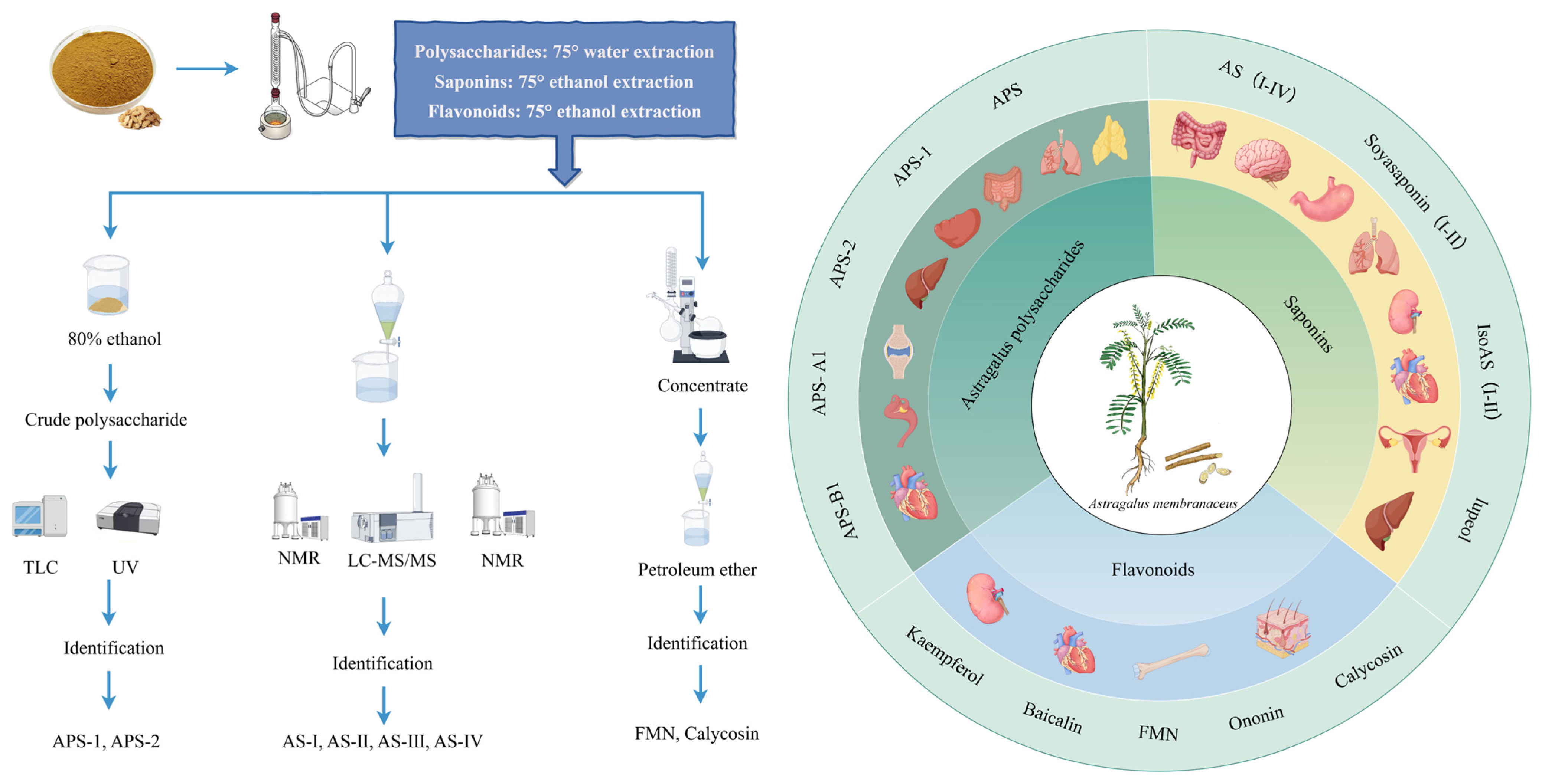
Figure 1. Process of extracting active ingredients from AR. This figure was drawn using figdraw (https://www.figdraw.com), export ID: AOORW8161e.
Pharmaceuticals 18 01448 g001
Figure 2. Molecular mechanism of the antioxidant action of AR. The APS constituents include l-rhamnose (l-Rha), d-xylose (d-Xyl), d-glucose (d-Glc), and d-galactose (d-Gal) in a 1:4:5:1.5 ratio. This figure was drawn using figdraw (https://www.figdraw.com), export ID: SROUT48444.
Pharmaceuticals 18 01448 g002
Figure 3. Molecular mechanisms of the anti-autophagy role exerted by AR. This figure was drawn using figdraw (https://www.figdraw.com), export ID: UPPUA29865.
Pharmaceuticals 18 01448 g003
Table 1. Polysaccharides extracted from AR.
SourceChemical NameCompositionQuality RatioMolecular Weight (Da)Classification Ref.
Astragalus polysaccharides (APS)APSl-Rha:d-Xyl:d-Glc:d-Gal1:4:5:1.53.01 × 105HeteropolysaccharidesWater-alcohol extraction[20]
APSID3Ara:Rha:Gal:Glc2:2:5:65.79 × 10⁵ [22]
F-8Rha:Rib:Fuc:Ara:Xyl:Man:Gal:Glc2:2:1: 2:6:2: 3:1002.2 × 104Neutral polysaccharide[23]
F-9Fuc:Xyl:Glc1:2:1001.2 × 104 [23]
APS-1Gal:Glc1:49.763.84 × 104Heteropolysaccharides[24]
APS-2Rha:Gal:Glc1:2.99:16.265.2 × 103Heteropolysaccharides[24]
AH-1Gly:Glc:Rha:Ara1:0.04:0.02:0.01Acidic polysaccharide[25]
AH-2Glc:Ara1:0.15[25]
APS-A1Glc:Gal:Ara52.3:1.0:1.32.62 × 106Neutral polysaccharide[21]
APS-B1Glc:Gal:Arae:Man:Rha:GalA75.2:17.3:19.4:1.0:1.1:1.34.95 × 106 Acidic polysaccharide[21]
APS2-IMan:Rha:GlcA:GalA:Glc:Gal:Xyl:Ara2.3:4.8:1.7:14.0:5.8:11.7:2.8:12.61.96 × 106Dextran[26]
APS3-IRha:GlcA:Glc:Gal:Ara0.8:2.3:0.8:2.3:4.13.91 × 106 Dextran[26]
APS4Rha:Ara:Xyl:Man:Gal12.1:0.3:0.6:1.0:1.0:1.71.5 × 106 [27]
cAMPs-1AFuc:Ara:Gal:Glc:Xyl0.01:0.06:0.20:1.00:0.061.23 × 104Water-soluble polysaccharides[28]
AAP-2ARha:Gal:Ara:Glc1:2.13:3.22:6.182.252 × 103Neutral polysaccharide[29]
AERP1Man:Rha:GalA:Glc:Gal:Ara1.00:2.59:12.15:2.60:3.07:4.542.01 × 106Acidic polysaccharide[30]
AERP22.11 × 103Dextran[30]
Table 2. Triterpenoid saponins extracted and isolated from AR.
SourceChemical NamesChemical Structure or CompositionMolecular FormulaMolecular Weight (Da)ClassificationExtraction MethodsRef.
Astragalus saponins (AS)AS-IPharmaceuticals 18 01448 i001C41H68O14869.04Triterpenoid saponins70% methanol extraction[33,34]
IsoAS-IPharmaceuticals 18 01448 i002C45H72O16869.02Triterpenoid saponins70% methanol extraction[33,34,36]
AS-IIPharmaceuticals 18 01448 i003C45H72O16827.13Triterpenoid saponins70% methanol extraction[33,37]
AS-IIIPharmaceuticals 18 01448 i004C41H68O14784.98Triterpenoid saponins70% methanol extraction[33,37]
AS-IVPharmaceuticals 18 01448 i005C41H68O14784.98Triterpenoid saponins70% methanol extraction[32,38,39]
lupeolPharmaceuticals 18 01448 i006C30H50O426.73Triterpenoid saponinsSupercritical CO2 extraction[40]
Soyasaponin IPharmaceuticals 18 01448 i007C48H78O18943.26Triterpenoid saponins45% methanol solid phase extraction[41]
Soyasaponin IIPharmaceuticals 18 01448 i008C47H76O17913.11Triterpenoid saponins45% methanol solid phase extraction[41]
Agroastragaloside IIIPharmaceuticals 18 01448 i009C51H82O211031.18Triterpenoid saponinsMethanol extraction[42]
Agroastragaloside IVPharmaceuticals 18 01448 i010C49H80O20989.14Triterpenoid saponinsMethanol extraction[42]
Acetylastragaloside IPharmaceuticals 18 01448 i011C47H74O17911.10Triterpenoid saponinsMethanol extraction[43]
IsoAS-IIPharmaceuticals 18 01448 i012C43H70O15827.02Triterpenoid saponinsMethanol extraction[43]
AS-VIIPharmaceuticals 18 01448 i013C47H78O19947.1Triterpenoid saponinsMethanol extraction[43]
Table 3. Flavonoids extracted and isolated from AR.
SourceChemical NamesChemical Structure or CompositionMolecular FormulaMolecular Weight (Da)ClassificationExtraction MethodsRef.
FlavonoidsKaempferolPharmaceuticals 18 01448 i014C15H10O6286.24flavonolUltrasound-assisted extraction (UAE)[48]
BaicalinPharmaceuticals 18 01448 i015C21H18O11446.36Flavonoid glycosidesMicrowave-assisted extraction (MAE)[49]
IsoliquiritigeninPharmaceuticals 18 01448 i016C15H12O4256.28ChalconeUAE[50]
FMNPharmaceuticals 18 01448 i017C16H12O4268.26isoflavoneHexane-ethyl acetate-ethanol-water two-phase solvent extraction system[51,52]
Calycosin-7-O-β-d glucosidePharmaceuticals 18 01448 i018C22H22O10446.40isoflavone70% ethanol extraction[53,54]
CAPharmaceuticals 18 01448 i019C16H12O5284.28isoflavone70% ethanol extraction[55]
OnoninPharmaceuticals 18 01448 i020C22H22O9430.40isoflavoneSolid phase extraction
(SPE)
[56]
QuercetinPharmaceuticals 18 01448 i021C15H10O7302.24flavonol50% ethanol extraction[57]
Licochalcone BPharmaceuticals 18 01448 i022C16H14O5286.28ChalconeEthanol extraction[58]
PendulonePharmaceuticals 18 01448 i023C17H16O6316.31IsoflavoneEthanol extraction[59,60]
Astrasikokioside IPharmaceuticals 18 01448 i024C39H50O23886.80Flavonoid glycosidesEthanol extraction[60]
IsoquercitrinPharmaceuticals 18 01448 i025C21H20O12464.38Flavonoid glycosides90% ethanol extraction[45]
AstragalinPharmaceuticals 18 01448 i026C21H20O11448.38Flavonoid glycosides90% ethanol extraction[45]
Table 4. List of potential pharmacological effects of AR on different organs. (Table Notes: ↑: increase; ↓: decrease).
ComponentsModelsMaximum Dose and Treatment Duration (In Vivo)OrgansEffectsMechanismsRef.
APSAβ25-35-induced HT22 mouse hippocampal neurons; Streptozotocin (STZ)-induced diabetic mouse model; and SOD-2+/− gene knockout mouse model2.0 g/kg; 10 weeksHeartAnti-oxidative stress; and apoptosis↑SOD-2 enzyme activity; ↓ROS production, and ↓apoptosis in CSPCs.[69]
APSChick transport stress (TS) model100 μL/animal; 8 h (oral)HeartAnti-oxidative stress; and immunoregulation↓mtDNA-PRRs pathway; ↑GSH, GPX, GST, SOD-2, and ↓MDA.[74]
APSDiabetic cardiomyopathy (DCM) rat model1 g/kg; 16 weeksHeartApoptosis↓PERK and ATF6 pathways in ER stress.[89]
CAH9c2 cardiomyocytes subjected to thermal shockHeartApoptosis↓p-JNK pathway, Fas/FasL apoptosis pathway, and ↑PI3K/Akt pathway.[91]
ASZebrafishHeartAutophagy↑atg7, LC3-II, and ↓p62.[107]
FMNHigh-glucose-induced glomerular mesangial cells (GMCs); and db/db diabetic mice25–50 mg/kg; 8 weeksKidneysAnti-oxidative stress; anti-inflammation; and anti-fibrosis↑Sirt1, and Nrf2/ARE pathway.[146]
FMNHigh-glucose-induced HK-2 human proximal tubule epithelial cell model; and STZ-induced diabetic nephropathy rat model20 mg/kg; 8 weeksKidneysApoptosisRegulation of Bcl-2/Bax balance, ↓caspase-3; as well as regulation of Sirt1/PGC-1α pathway and ↓ROS.[90]
Fermentation of AR with P. cicadaeHigh-glucose-induced podocytes; and STZ-induced mouse diabetic nephropathy model4.5 g/kg; 6 weeksKidneysAutophagy↓PI3K/AKT/mTOR pathway.[102]
ASLPS-induced human mesangial cell (HMC) model; and C-BSA-induced chronic glomerulonephritis (CGN) rat model20 mg/kg; 6 weeks KidneysAutophagy↓PI3K/AKT/AS160, ↑C3-II, Beclin1, and ↓p62.[106]
Astragalus injection/decoctionSTZ-induced T1D, spontaneous GK rat model (T2D)3–10 g/kg; 4–12 weeksKidneysAnti-oxidative stress; and anti-fibrosis↑NRF2/KEAP1 pathway, ↓TGF-β/Smad signaling, NF-κB activation, and regulation of the PI3K/AKT/mTOR pathway to protect podocytes.[145]
APSHep3B liver cancer cells; and Hep3B cell-bearing nude mouse model50 mg/kg; 28 daysLiverApoptosis↓O-GlcNAc synthesis, ↑Dox-induced ER stress, ↓Bcl-2, and ↑activated CHOP and Caspase-3.[88]
ASAML-12 cell ferroptosis model; and cisplatin-induced mouse liver injury model80 mg/kg; 9 daysLiverFerroptosis; anti-oxidative stress; and anti-inflammation↓Ferroptosis; ↑PPARα/FSP1 signaling pathway, ↓key markers of ferroptosis, and regulation of lipid peroxidation.[138]
ATSBiliary duct ligation (BDL) rat cholestatic liver fibrosis model; and DDC diet-induced mouse liver fibrosis model56 mg/kg; 4 weeksLiverAnti-fibrosis↑FXR, SHP, BSEP, NTCP expression, ↓Cyp7a1, and reduce serum/liver taurine-conjugated bile acids (BAs).[141]
APS, ASAlcoholic liver disease (ALD) mouse modelAPS: 600 mg/kg
AS: 100 mg/kg; 4 weeks
LiverAnti-oxidative stress; and anti-inflammation↑KEAP1/NRF2 antioxidant pathway, and ↓TLR4/MyD88/NF-κB inflammatory pathway.[142]
ASMiddle cerebral artery occlusion (MCAO) mouse model40 mg/kg; 3 daysSpleenImmunoregulationReduction of spleen atrophy and restoration of spleen NK/T/B-cell counts.[116]
APS4T1 breast cancer-bearing BALB/c mouse model200 mg/kg; 14 daysSpleen, thymusImmunoregulation; and apoptosisImprovement of thymus index and spleen index, and enhancement of macrophage phagocytic ability.[113]
ASCigarette smoke extract (CSE)-induced inflammatory model in RAW264.7 macrophagesLungsAutophagy; and anti-inflammation↓TLR4/NF-Κb pathway, ↑LC3-II, ↑ATG5, ATG7, Beclin1; and ↓p62.[103]
APSBleomycin (BLM)-induced model100 mg/kg; 28 daysLungsAnti-inflammation↓TLR4/NF-κB signaling pathway, TLR4/NF-κB pathway, TNF-α, IL-6, IL-1β, and TGF-β1.[127]
Cycloastragenol (CAG)Human gastric cancer SNU-1 and SNU-16 cellsStomachApoptosis↓STAT3 phosphorylation, JAK1/2, and Src kinase activity.[92]
ATSGastric cancer cells (SGC-7901)StomachFerroptosis; and apoptosis↑SIRT3 expression, ferroptosis, ↓SLC7A11/GPX4, and ↑ACSL4 protein.[137]
AR water extractIEC-6 intestinal epithelial cells; and mouse IBD modelIntestinesAnti-oxidative stress; and anti-inflammation↓NF-κB pathway, and ↑Nrf2 pathway.[64]
ASDSS-induced mouse model of ulcerative colitis100 mg/kg; 14 daysIntestinesAnti-oxidative stress; and immunoregulationRegulation of Th17/Treg immune balance, ↓MDA, and ↑SOD/GSH-Px.[73]
APSLPS-induced RAW264.7 macrophages; and collagen-induced arthritis (CIA) rat model200 mg/kg; 7 daysIntestinesAnti-inflammation↓TLR4/NF-κB pathway, p-p65, p-IκB levels, MAPK pathway, IL-6, IL-1β, and TNF-α.[126]
ASRat middle cerebral artery occlusion (MCAO) model20 mg/kg; 7 days BrainImmunoregulationReduction of NK cell activation receptor NKG2D expression and IFN-γ production, and reversal of NK cell deficiency in the spleen and blood.[114]
APSBV2 microglia; and chronic fatigue syndrome (CFS) mouse model800 mg/kg; 5 weeksGut; BrainAnti-oxidative stress; and anti-inflammation↑Nrf2 pathway; and ↓NF-κB pathway.[75]
APS6-OHDA-induced PC12 cellsBrain neuronsAutophagy↑PI3K/AKT/mTOR pathway.[100]
CAGα-Synuclein (α-Syn)-induced Parkinson’s disease (PD) mouse model and primary microglia125 mg/kg; 16 weeksCentral nervous system (Brain)Anti-oxidative stress; and autophagy; Anti-inflammation; and immunoregulation↓Scrib/NOX-ROS axis, inhibition of NLRP3, and ROS.[144]
Processed AR water extractAβ25-35-induced HT22 mouse hippocampal neuronsNervous systemAnti-oxidative stress; and apoptosis↑Nrf2 pathway, and AKT/CREB/BDNF pathway.[67]
APStBHP-induced mouse chondrocytes; and DMM surgery-induced osteoarthritis mouse modelJoint cartilageAnti-oxidative stress↑GCN2/ATF4/TXN axis.[66]
APSTBHP-induced chondrocyte apoptotic model; and DMM surgery-induced osteoarthritis mouse model200 mg/kg; 6 weeksJoint cartilageApoptosis↓Mitochondrial apoptosis pathway; and ↓ROS.[93]
ASSciatic nerve transection mouse model of muscle atrophy20 mg/kg; 14 daysSkeletal muscleAnti-oxidative stress; and anti-inflammation↑SOD1/GPX1, ↓ROS/NOX2/4, ↓NLRP3/IL-1β/IL-6/TNF-α, and ↓LC3II/BNIP3[76]
CAOvariectomized mice50 mg/kg; 12 weeks Bone tissueAutophagy↑PI3K/AKT/mTOR pathway.[101]
ASExperimental autoimmune encephalomyelitis model (EAE)20 mg/kg; 10 daysBone marrowImmunoregulation↓Maturation of splenic, bone marrow-derived DCs; ↓Th1/Th17 cell differentiation; ↓IL-6, and IL-12.[112]
APSTaxol-induced cytotoxicity in RAW 264.7 macrophages; and 4T1 tumor-bearing mouse model40 mg/kg; 6 weeksImmune systemImmunoregulation; and apoptosis↓G2/M phase arrest, P-H2A.X, PARP, ↑Bcl-XL, and Mcl-1.[82]
APSLPS-induced RAW264.7 macrophagesImmune systemAnti-inflammation↓NF-Κb, MAPK pathways; ↓ROS production (immune regulation); ↓NLRP3, iNOS, and COX-2 expression.[21]
ASRat vascular smooth muscle cellsVascular systemAutophagy↑AMPK/mTOR pathway, and STX17-SNAP29-VAMP8.[108]
APSLPS/high-glucose-induced THP-1 macrophages and HUVECs coculture; and STZ-induced T2DM rat model800 mg/kg; 8 weeksVascular endotheliumAnti-inflammation↑macrophage M2 polarization, Nrf2/HO-1 pathway, ↓ROS, VCAM-1, MCP-1, and ↓Bax/Bcl-2.[131]
APSOvarian cancer stem cells (OCSCs, 3AO/SKOV3 cells)OvariesAutophagy↓PINK1/Parkin pathway-mediated mitochondrial autophagy, and ↑TOMM20/COX IV, ↓PINK1 protein.[135]
APSOC cells (OV-90/SKOV3 cells)OvariesApoptosis↓EMT; ↓miR-27a expression; ↑FBXW7 protein, and ↑E-cadherin.[134]
ASSiHa cell nude mouse xenograft model50 mg/kg; 35 daysCervixAutophagyRegulation of the DCP1A/WDFY3/Atg12 and TMSB4X/Akt/Atg5/Atg12 pathways.[110]
ASPDGF-BB-induced human airway smooth muscle cells (ASMCs); and OVA-induced asthma mouse model100 mg/kg; 4 weeksRespiratory tractPyroptosisAlleviation of pulmonary inflammation; inhibition of HMGB1 cytoplasmic translocation, blockage of the HMGB1/RAG axis, inactivation of the NF-κB pathway, reduction of inflammatory factors, and ↑pyroptosis markers.[140]
APSMouse macrophages (RAW 264.7), and mouse spleen-derived CD4+/CD8+ T cellsProstateImmunoregulationRegulation of the PD-1/PD-L1 pathway, ↑T-cell secretion of IFN-γ, CXCL2, CCL5, TNF-α, IL-6, and CCL5.[115]
APSDSS-induced colitis mouse model200 mg/kg; 14 daysColonImmunoregulation↑Mitochondrial metabolism; ↑IgA+ MBCs, and ↓IgG+ MBCs.[118]
ASIndomethacin-induced SD rat enteritis model80 mg/kg; 3 daysSmall intestineAnti-inflammation↓NLRP3 inflammasome activation; ↓NF-κB4; ↓IL-1β, and IL-18.[129]
FMNHaCaT cells (psoriasis-like inflammation); and IMQ-induced psoriasis mouse modelSkinAnti-inflammation↓IFN-α/β/γ signaling pathway, CXCL9/10/11 and other chemokines, p-STAT1/3, IRF1 expression, TNF-α, IL-6, and IL-17 levels.[119]
APSCadmium (CdCl2) induced chicken embryo fibroblast (CEF) damage modelFibroblastsAutophagy; and anti-oxidative stress↓ROS, LC3-II, Beclin1, ↑SOD, and GSH-Px.[104]
ASOligodendrocyte precursor cells; Cuprizone (CPZ)-induced mouse demyelination model, and experimental autoimmune encephalomyelitis (EAE) mouse model50 mg/kg; 7 daysMyelin tissueImmunoregulationTargeting and binding to the p75NTR receptor, and ↓Wnt/β-catenin signaling pathway.[117]
Table 5. Effects of AR and AR extracts on the PK parameters of drugs.
MedicinesInteracting DrugsAnimal ModelsRoutes of Administration and DosagesPK Parameters of Ingredients Ref.
Cmax (ng/mL)AUC(0−t) (ng/mL·h)AUC(0−inf) (ng/mL·h)t1/2 (h)tmax (h)CL ((mL/h)/kg)MRT(0−t) (h)
Astragali radix extract (ARE)DapagliflozinMale SD ratsWith ARE (300 mg/kg for seven days, intragastrically administered)426.67 ± 58.924051.27 ± 952.614865.13 ± 1307.119.69 ± 3.471.50 ± 0.77228.25 ± 56.107.64 ± 0.70[162]
Astragalus InjectionDOXMale SD ratsWith Astragalus injection (4.25 mL/kg/day for 14 days, intraperitoneal injection)5262.77 ± 111.153777.27 ± 130.557141.76 ± 177.960.14 ± 0.0411.72 ± 1.221.09 ± 0.37[161]
AS-IVAbemaciclibMale Sprague Dawley ratsWith AS IV (50 mg/kg for 7d, orally administered163,000 ± 147,85036,380 ± 423038.24 ± 7.533.24 ± 1.150.49 ± 0.10720.57 ± 0.92[158]
With AS IV (100 mg/kg for 7d, orally administered1,698,170 ± 104,43043,810 ± 185051.59 ± 17.082.65 ± 0.770.34 ± 0.05920.87 ± 0.40[158]
With AS IV (150 mg/kg for 7d, orally administered23,080,500 ± 55,29066,140 ± 117066.17 ± 28.732.12 ± 0.840.19 ± 0.04221.76 ± 0.32[158]
Radix astragaliPioglitazoneMale Wistar ratsWith RA (28.35 g/kg for 7 days, orally administered)2334.0 ± 87,369.929945.3 ± 871,556.292.787 ± 0.081.17 ± 0.4236.55 ± 75.73[162]
Huangqi injection (HI)GliquidoneMale Wistar ratsWith HI (8 mL/kg for 5 min, intravenously)22.46 ± 3.61281.9 ± 12.42.46 ± 0.760.18 ± 0.04[163]
Disclaimer/Publisher’s Note: The statements, opinions and data contained in all publications are solely those of the individual author(s) and contributor(s) and not of MDPI and/or the editor(s). MDPI and/or the editor(s) disclaim responsibility for any injury to people or property resulting from any ideas, methods, instructions or products referred to in the content.