Article

Exploring the Antibacterial Potential of Artemisia judaica Compounds Targeting the Hydrolase/Antibiotic Protein in Klebsiella pneumoniae: In Vitro and In Silico Investigations

by
Fahdah Ayed Alshammari
Department of Biology, College of Science, Northern Border University, Arar 76312, Saudi Arabia
Pharmaceuticals 2024, 17(6), 667; https://doi.org/10.3390/ph17060667
Submission received: 28 March 2024 / Revised: 13 May 2024 / Accepted: 14 May 2024 / Published: 22 May 2024
(This article belongs to the Section Medicinal Chemistry)

Abstract

:
Carbapenem antibiotic resistance is an emerging medical concern. Bacteria that possess the Klebsiella pneumoniae carbapenemase (KPC) protein, an enzyme that catalyzes the degradation of carbapenem antibiotics, have exhibited remarkable resistance to traditional and even modern therapeutic approaches. This study aimed to identify potential natural drug candidates sourced from the leaves of Artemisia judaica (A. judaica). The phytoconstituents present in A. judaica dried leaves were extracted using ethanol 80%. A reasonable amount of the extract was used to identify these phytochemicals via gas chromatography/mass spectrometry (GC/MS). One hundred twenty-two bioactive compounds from A. judaica were identified and subjected to docking analysis against the target bacterial protein. Four compounds (PubChem CID: 6917974, 159099, 628694, and 482788) were selected based on favorable docking scores (−9, −7.8, −7.7, and −7.5 kcal/mol). This computational investigation highlights the potential of these four compounds as promising antibacterial candidates against the specific KPC protein. Additionally, in vitro antibacterial assays using A. judaica extracts were conducted. The minimum inhibitory concentration (MIC) against the bacterium K. pneumonia was 125 μg/mL. Well–disk diffusion tests exhibited inhibition zones ranging from 10.3 ± 0.5 mm to 17 ± 0.5 mm at different concentrations, and time–kill kinetics at 12 h indicated effective inhibition of bacterial growth by A. judaica leaf extracts. Our findings have revealed the pharmaceutical potential of Artemisia judaica as a natural source for drug candidates against carbapenem-resistant pathogens.

1. Introduction

The rise of carbapenem-resistant Enterobacteria represents a critical healthcare challenge, as it undermines the effectiveness of carbapenem antibiotics in the treatment of infections. This resistance poses significant difficulties in effectively managing patients and contributes to elevated rates of illness and mortality associated with bacterial infections [1]. One common resistance mechanism involves carbapenemase enzyme production [2]. The most commonly observed carbapenemases include OXA-48, KPC, NDM, and VIM types, although their prevalence varies significantly across countries and regions [3]. Klebsiella pneumoniae carbapenemase (KPC) is a class-A serine beta-lactamase discovered in the United States in 1996 [4]. In several areas, including India, the Mediterranean region, and some European countries, it has emerged as a common mechanism of carbapenem resistance among Enterobacteria [2,5]. Historically, KPC carbapenemases have demonstrated a broad spectrum of substrates, including penicillins, aztreonam, cephalosporins, and carbapenems. Notably, they exhibit resistance to commonly used beta-lactamase inhibitors such as sulbactam, clavulanic acid, and tazobactam [2,6,7,8].
The emergence of antibiotic-resistant strains has presented significant challenges in the management of bacterial infections, necessitating the development of novel therapeutic strategies. Discovering new antibiotics or alternative natural products, including plants, which have been used for centuries in traditional medicine, and improving the efficacy of existing ones through dosing regimens or synergy therapies are potential approaches [9]. Over the past three decades, extensive research has been conducted to investigate the antimicrobial properties of various plants, among which Artemisia has garnered significant attention [10]. Artemisia is a broad, diverse genus of plants that fall under the family Asteraceae, encompassing 200 to 400 species [11]. Artemisia species exhibit growth in temperate regions across both hemispheres, typically inhabiting arid or semiarid environments. Artemisia is a group of resilient herbaceous plants and shrubs renowned for their essential oils’ potent chemical components. These substances exhibit a range of biological actions, such as antioxidant, antibacterial, anticancer, digestive enzyme inhibitory, anti-inflammatory, anthelmintic, and numerous other properties [12].
Artemisia judaica L. is an aromatherapeutic plant abundant in Saint Katherine, Sinai, Egypt. Many bioactive compounds associated with plant secondary metabolites were found in the GC/MS analysis of Artemisia species. Some of these newly found phytochemical components show biological action [13]. Artemisia species are essential medical plants now attracting significant interest due to their bioactive compounds. The interest in them is primarily driven by their use in cosmetic products, manufacture of essential oils, chemical diversity, and biological functionality [13,14,15,16].
Due to its many advantageous qualities, such as its anti-inflammatory, antibacterial, and antidiabetic effects, it has long been used in traditional medicine. Furthermore, in different Arabic regions, including Saudi Arabia, it is frequently employed in folk medicine for addressing conditions such as fungal diseases, atherosclerosis, diabetes, cancer, arthritis, and inflammatory-related disorders, which are among the conditions that need attention [17,18,19]. Despite extensive research on the inhibitory effects of A. judaica, especially against specific organisms, there is a lack of information regarding its antibacterial properties against K. pneumoniae pathogens. Despite the use of A. judaica, its potential in Saudi Arabia remains largely unexplored, and comprehensive studies are scarce.
A global concern regarding antibiotic-resistant bacterial strains underscores the critical nature of this research. Although numerous investigations have been conducted into the antimicrobial properties of different plant species, the antibacterial potential of compounds found in A. judaica against Klebsiella pneumoniae has received relatively little attention. By filling this knowledge vacuum, the study intends to shed new light on the mechanisms by which these compounds operate. By integrating in vitro and in silico analyses, the research provides a holistic methodology for assessing the compounds’ antibacterial properties. The examination identified a cumulative sum of 110 constituents from the oils extracted from the entire A. judaica plant (including stems, foliage, and blossoms), and 81 constituents from the A. herba-alba plant. The oil of A. judaica contained β-eudesmol, hexadecanoic acid, spathulenol, eudesma-4 (15), 7-dien-1-β-ol, carvacrol, and thymol as its primary constituents. These constituents constitute 67.3% of the total composition of essential oils, equivalent to 96.7% of the oil itself [20].
Hence, the primary objective of this study was to investigate the antibacterial potential of compounds obtained from A. judaica, with a specific focus on their ability to target the hydrolase/antibiotic protein in Klebsiella pneumoniae. To accomplish this, a comprehensive approach was employed, combining various in vitro assays such as MIC, disk–well diffusion, and time–kill curve assays. Additionally, in silico investigations involved docking analysis and ADME assays. Through these experimental and computational methods, the effectiveness of A. judaica compounds against Klebsiella pneumoniae were assessed, aiming to gain valuable insights into their potential as antibacterial agents.

2. Results

2.1. Protein and Phytochemical Preparation

The study involved the extraction of chemical compounds from the plant Artemisia judaica using GC mass analysis, a widely used analytical technique (Figure 1). A total of 122 phytochemical compounds derived from the plant have been obtained and stored (Table S1). The compounds were synthesized and refined throughout the ligand production protocols and then transformed into the pdbqt file format for later assessment. The optimization and organization of the protein were conducted in preparation for the molecular docking approach using the AutoDock software PyRx-0.8 version. The compounds were then stored in the pdbqt file format in preparation for the docking. Table 1 presents a selection of four compounds based on their docking scores (kcal/mol) and corresponding details such as PubChem ID, chemical name, formula, and binding affinity.

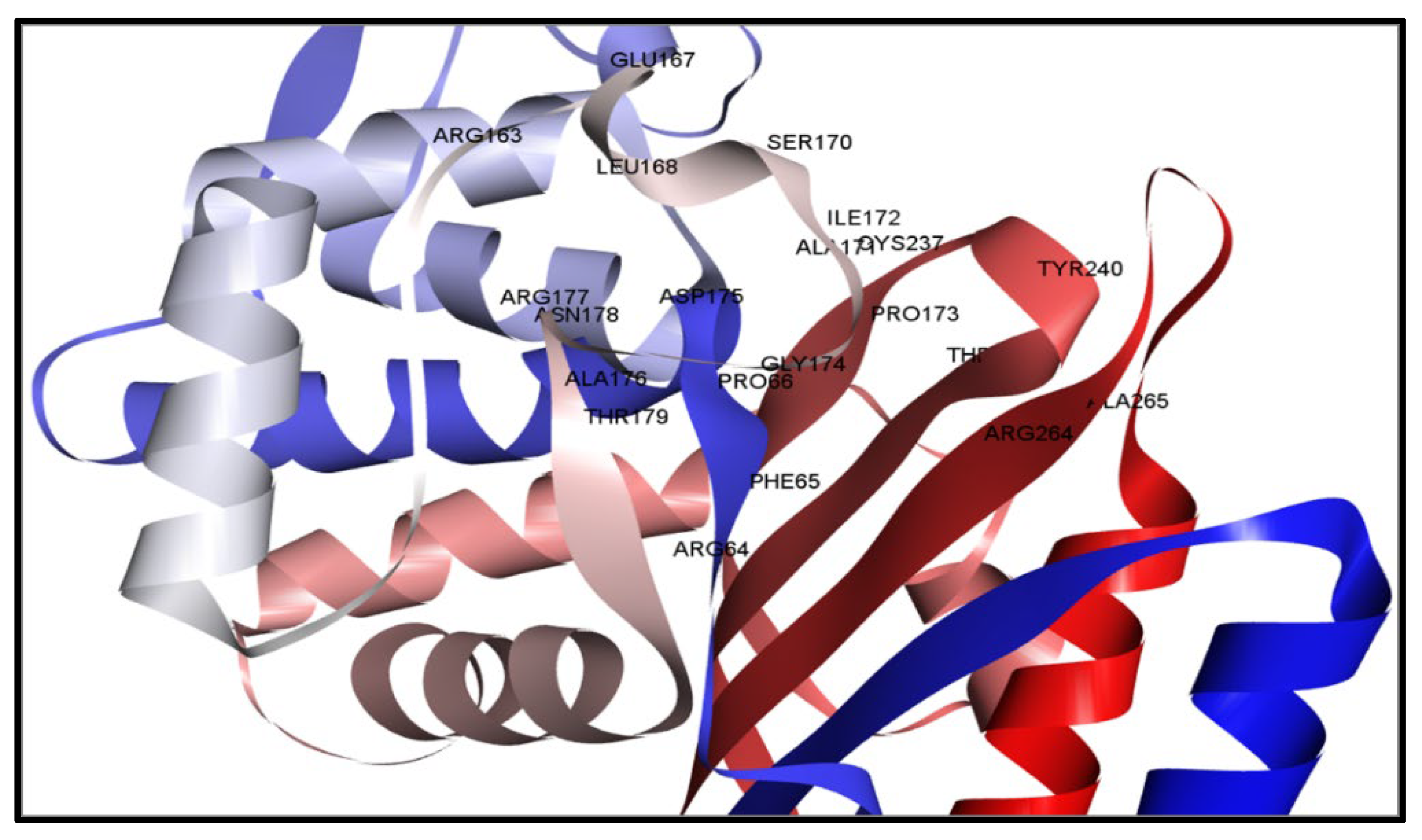
2.2. Active Position Detection and Generation of Receptor Grids

The configuration of many amino acid residues within a designated area gives rise to the active region of an enzyme, which facilitates transient contact with the substrate, generally referred to as the binding site. A chemical substrate can bind with the protein’s active site, leading to the catalysis of a process. Furthermore, it assists in stabilizing the intermediate stages of the process. In contrast, the concept of a binding site refers to a specific location on a protein or nucleic acid that has the potential to recognize a ligand and establish a robust binding association with the protein. The initial step in the research was to use the CASTpi service to find the protein’s active site (AS). After that, the active site’s mixed binding spot was found (Figure 2). By looking at the protein’s active site, it was easier to find the protein’s binding position residue (Figure 2). The study of the active site pocket identified the binding site locations at ARG64, PHE65, PRO66, ARG163, GLU167, LEU168, SER170, ALA171, ILE172, PRO173, GLY174, ASP175, ALA176, ARG177, ASN178, THR179, CYS237, TYR240, THR242, ARG264, and ALA265 residues. These positions are visually represented in Figure 2 using spherical shapes of varying colors, namely red, green, and orange. The server has discovered binding sites, which have been used to create a receptor grid in conducting a molecular docking simulation. The sizes of the grid box are X = 25Å, Y = 25Å, and Z = 43.54Å.

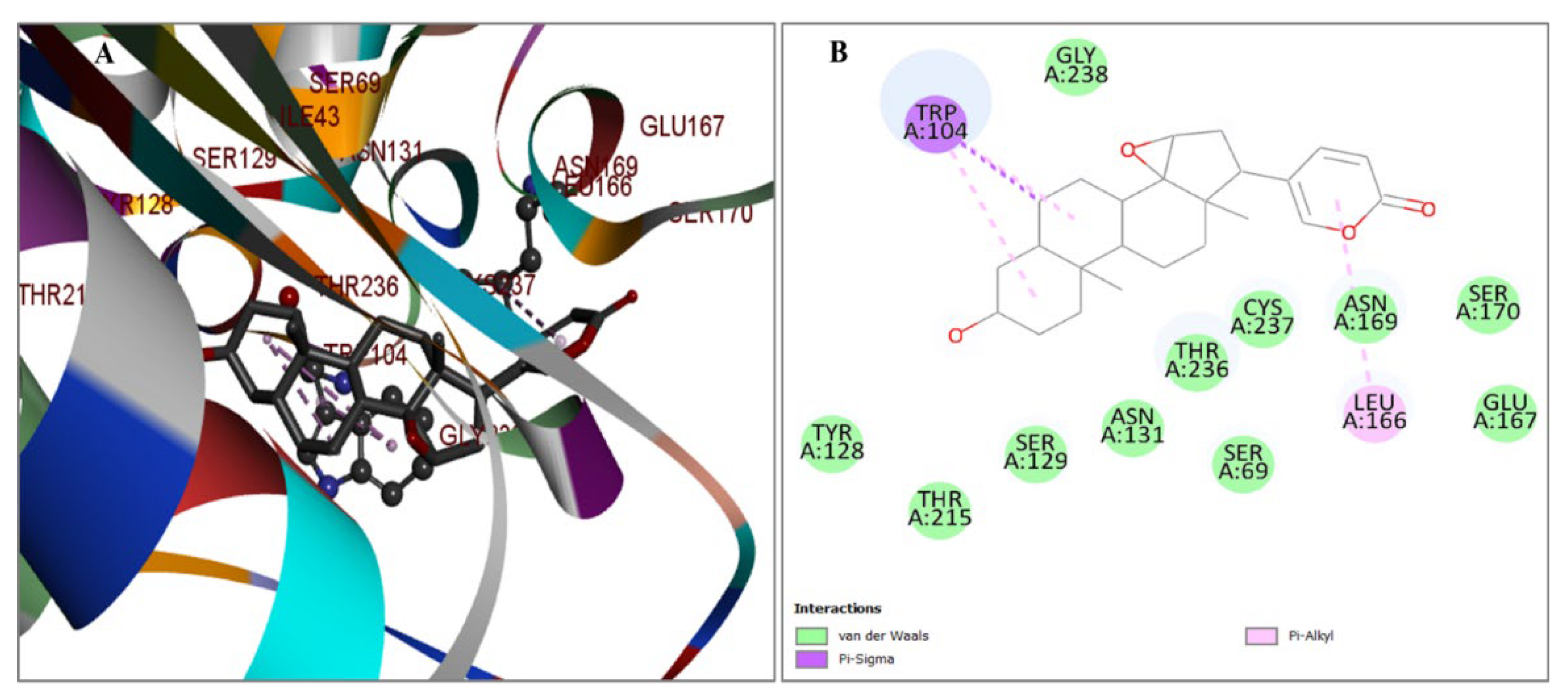
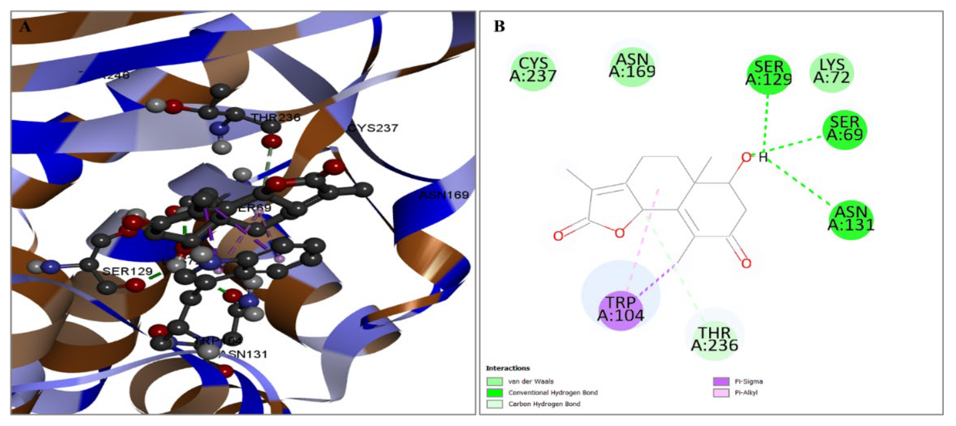
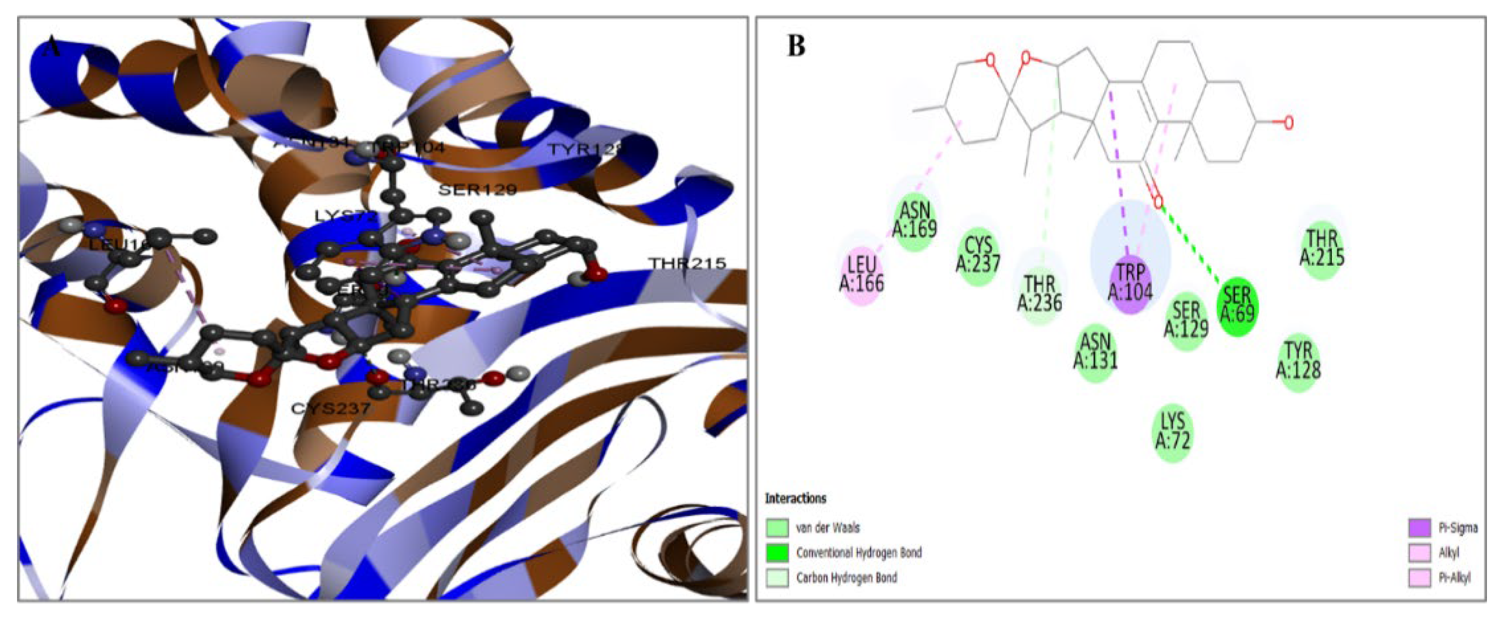
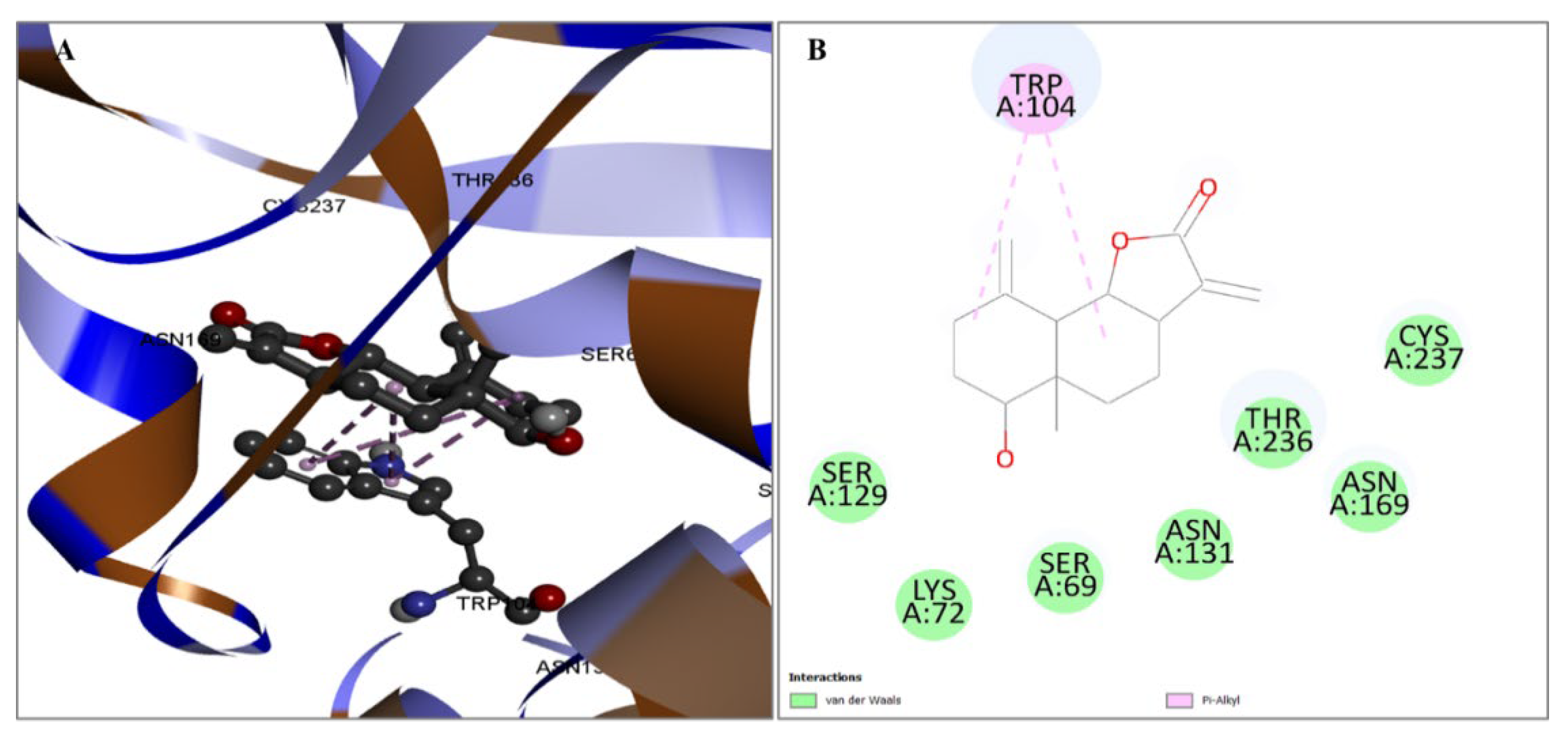
2.3. Evaluation of Protein and Ligand Interaction

Once the protein with the highest binding score was identified, a detailed analysis was undertaken to explore the intricate interactions between these substances. The research aimed to examine the utilization of the BIOVIA Discovery Studio Visualizer Tool 16.1.0, a software tool specifically designed for studying protein–ligand interactions, and the interaction between the four identified ligands and the target protein. The molecule CID6917974 has been shown to establish several hydrophobic interactions at the residue positions TRP104 and LEU166. The information in Figure 3 is documented in Table 2.
Figure 3. The interaction between the protein and the compound CID6917974. The two-dimensional bonding of the protein–ligand complex is depicted on the right (B), whereas the three-dimensional interaction is illustrated on the left (A). The chemical CID159099 was observed to establish numerous hydrogen and also carbon–hydrogen interactions with the preferred protein. The hydrogen bonding interactions occurring at the SER69, SER129, and ASN131 positions, together with the presence of single carbon–hydrogen bonds at the THR236 position, and TRP104 position hydrophobic bonds are depicted in Figure 4. The specific bond types are provided in Table 2.
Pharmaceuticals 17 00667 g003
The compound CID628694 demonstrated a substantial affinity for the goal protein in relation to binding. The molecule has two hydrogen bonding interactions at SER69 and THR236 sites. On the other hand, three hydrophobic interactions occur at LEU166 and TRP104 positions. The data presented in Figure 5 are also documented in Table 2.
Compound CID482788 demonstrated an important level of affinity for the target protein in terms of binding. The molecule has hydrophobic interactions at position TRP104. The data presented in Figure 6 are also documented in Table 2.

2.4. ADME Calculation

The assessment of pharmacokinetics (PK) properties, which encompasses the evaluation of pharmaceuticals, including their kinetics (movement), is a crucial element in developing drug and procedure designs. However, the focus of analysis mainly lies in the ADME characteristics. The physiochemical parameters included in this study comprise lipophilicity and water solubility. The discussion consists of pharmacokinetics, drug similarity, and medicinal chemistry, which have been presented. There is one potential explanation for the selection of optimal medication candidates. Before administering medicines in preclinical analyses, assessing pharmacophore characteristics may ascertain whether the molecule exhibits characteristics related to the control of xenobiotics. The pharmacophore characteristics of the four chosen drug-like compounds should be examined, which require determination by lipids, which should be analyzed using the SwissADME program. A solvent that lacks a net dipole moment does not exhibit significant polarity. Pharmacophore characteristics have ultimately shown favorable outcomes with the chemical in question showing efficacy and potential for medication development in scientific research. The pharmacokinetic characteristics of the four selected drugs are illustrated in Table 3.

2.5. Antibacterial Activity

2.5.1. MIC

The antibacterial effects of A. judaica extract against K. pneumoniae were examined in this research. The antibacterial activity of the extract was assessed by a combination of qualitative methods, such as well and disk diffusion assays, as well as a quantitative approach known as the minimum inhibitory concentration (MIC) test. The identification of bacterial proliferation was achieved by visually assessing the broth’s turbidity. The MIC of A. judaica extract against K. pneumoniae was 125 μg/mL (Figure 7). This value was observed at the fourth dilution.

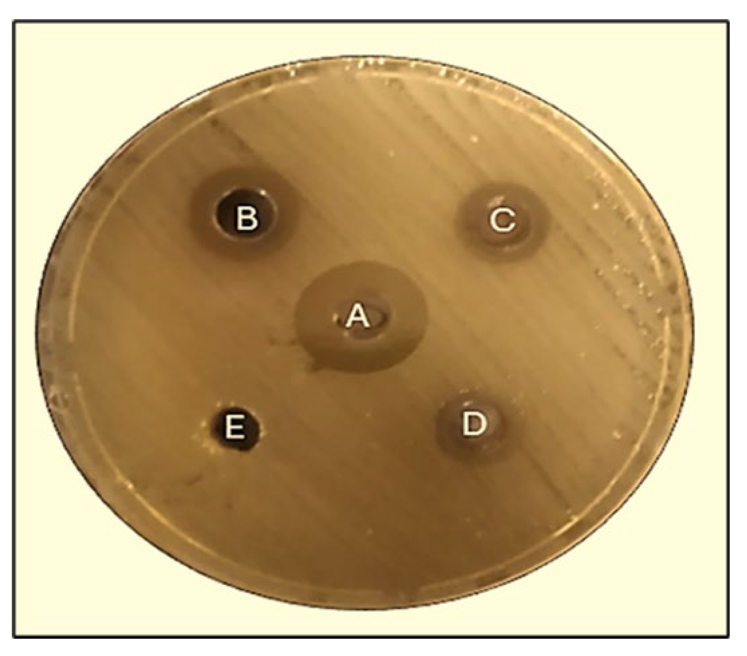
2.5.2. Well Diffusion Method

The inhibition zones measured by the well diffusion assay against the pathogenic bacterium K. pneumoniae were found to vary between 10.3 ± 0.5 mm and 17 ± 0.5 mm at 125, 250, and with sterile distilled water (SDW). In comparison, the effect of amoxicillin against K. pneumoniae was found to be 22.3 mm (Figure 8).

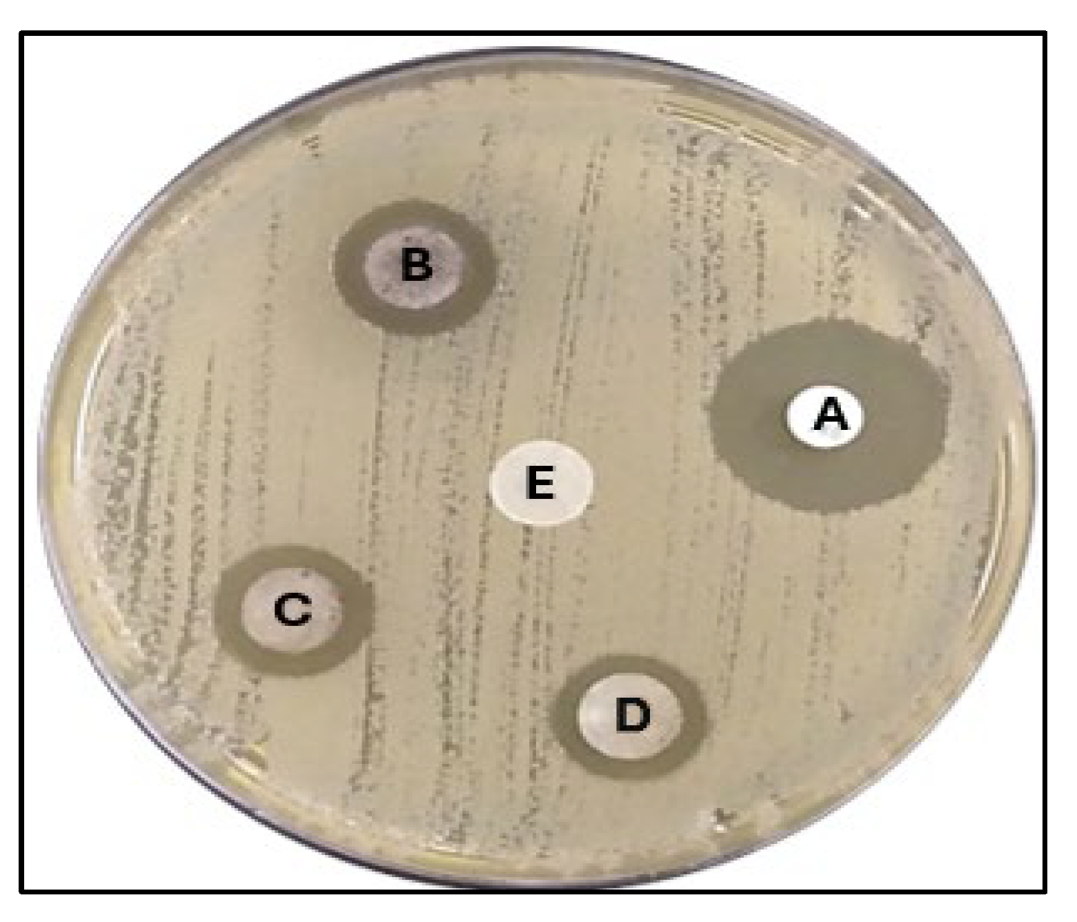
2.5.3. Disk Diffusion Method

The findings from the disk diffusion test demonstrated that the extract derived from A. judaica exhibited inhibitory effects on the proliferation of harmful bacteria, as depicted in Figure 9. The inhibition zones against K. pneumoniae varied between 11 ± 0.5 mm and 14 ± 0.5 mm at doses of 125, 250, and 500 µg/mL, respectively. As a positive control, oxacillin 1 µg (OXC 1) exhibited an effect of 23.3 mm against K. pneumoniae.

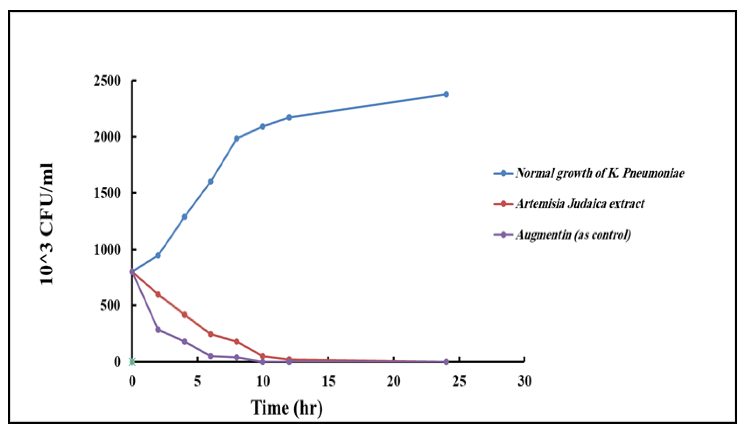
2.5.4. Time–Kill Curve

The time–kill curves of Artemisia judaica extract against K. pneumoniae were evaluated and associated with the control group using augmentin. The outcomes showed that the extract had a time-dependent antibacterial effect, gradually reducing the colony-forming units (CFUs) of K. pneumoniae over 24 h. Compared with the control group, the extract-treated group exhibited a decrease in CFUs at each time point (Figure 10). However, augmentin displayed a more rapid bactericidal effect, leading to the complete eradication of K. pneumoniae within 10 to 12 h.

3. Discussion

In the modern era, a multitude of research studies have extensively documented the remarkable efficacy of plant extracts in combating pathogenic bacteria that pose a threat to human health. These investigations have shed light on the potent antibacterial properties exhibited by diverse plant extracts [21,22,23]. In our investigation, we conducted both in silico and in vitro studies. A total of 122 trivial molecules were extracted from A. judaica using GC/MS analysis. It is important to note that of all the main chemicals identified in A. judaica, we showed only four docking scores and interactions using in silico methods. But in the crude extract, the docking score of some compounds was also high, such as of naphthalene (−7.2), nootkaton-11 (−7), stigmastero (−7), phthalic acid (−6.9), cis-sesquisabinene hydrate (−6.7), vulgarin (−6.7), (e)-isovalencenal (−6.5), methyl ester (−6.5), etc.
In contrast, while previous studies [17,24,25] have identified the phytoconstituents of A. judaica, it is important to note that these constituents can vary due to factors such as growing conditions, climatic conditions, cultivar, soil, plant age, and flowering state. Therefore, in this study, we analyzed the plant extract to examine its specific phytochemical composition. Through GC/MS analysis of the A. judaica extract obtained from its aerial parts, we detected 122 known compounds listed in S1 along with their chemical formulas. The phytochemical profiles of the extract revealed a high abundance of compounds in the aerial parts of A. judaica.
The utilization of computational or in silico methodologies is gaining significance in modern drug exploration attempts since they play a crucial role in promptly identifying promising drug candidates, often at a reduced expense compared to conventional techniques. Additionally, these methodologies reduce animal models in pharmacological investigations, facilitate the systematic development of innovative and secure drug candidates, repurpose pre-existing therapeutic agents, and aid medicinal chemists and pharmacologists in all drug exploration endeavors [26]. Therefore, identifying the possible protein of K. pneumoniae could serve as a viable target for rendering the protein associated with pathogenicity inactive. Given its distinctive characteristics, the crystal arrangement of the KPC-2 D179N variant (PDB: 8G2R) was selected as a prospective target for pharmaceutical intervention [27,28].
In this study, 122 compounds were subjected to docking using AutoDock Vina (Table S1). Four compounds were identified as promising candidates, and the most capable compounds were chosen for further simulation. Potential therapeutic candidates were found using various criteria (shown in Figure 2, Figure 3, Figure 4 and Figure 5). In addition, to ascertain the drug-like characteristics of the compounds, an analysis was conducted on their pharmacokinetic properties. The compounds also exhibited drug-like qualities, as shown in Table 3.
To evaluate the similarity of the identified phytoconstituents to established pharmaceuticals regarding their pharmacokinetics, toxicity, and physicochemical properties, a comparative study was conducted. The physicochemical parameters frequently examined in drug investigation encompass MW, LogS, HBA, HBD, nRTB, and LogP [29]. Wide-ranging studies have been conducted on these parameters to examine their impact on several pharmacokinetic factors, including absorption, bioavailability, permeability, and elimination, focusing on oral medications [30].
In vitro studies demonstrate the antibacterial efficacy of Artemisia judaica extract against K. pneumoniae, as evidenced by the observed inhibition zones and MIC values. Several studies have explored the antimicrobial properties of various Artemisia species, including Artemisia judaica, against different bacterial pathogens [18,31,32]. Some studies have reported similar findings, supporting the antibacterial efficacy of Artemisia judaica extract against pathogenic bacteria [33]. For example, a study on the antibacterial activity of Artemisia against a range of bacterial strains, including K. pneumoniae, was conducted and observed inhibition zones in agreement with the outcomes of the current study [34]. These findings suggest that Artemisia judaica extract possesses a broad-spectrum antibacterial effect. Variations could be due to changes in the extraction methods, plant sources, geographical locations, or even variations in the bacterial strains used in the studies. Furthermore, it is important to acknowledge that comparing findings across multiple studies might present difficulties owing to disparities in experimental parameters, including using distinct solvents, concentrations, and testing procedures. These factors can significantly impact the outcomes and make direct comparisons difficult. To further validate the findings of this study and establish the consistency of Artemisia judaica extract’s antibacterial efficacy against K. pneumoniae, additional studies involving larger sample sizes, standardized testing protocols, and rigorous statistical analysis are needed. Moreover, investigating the potential mechanisms of action and identifying the active compounds responsible for the observed antibacterial effects would provide valuable insights for future research and potential pharmaceutical applications.

4. Materials and Methods

4.1. Plant Material

The study comprised fresh leaves from Artemisia judaica in the research location of Jeddah, Saudi Arabia, in 2023. The Department of Biological Sciences, found within the Faculty of Science of King Abdulaziz University, has verified the genuineness of the plant. The A. judaica leaves were submerged in flowing tap water and washed with distilled water before processing. Subsequently, the plants were desiccated in a shaded place for 14 days at ambient temperature. After drying, the leaves were pulverized into a fine powder using an electric blender [35,36,37].

4.1.1. Preparation of Crude Extracts

The fresh leaves of A. judaica were dried and finely ground using an electric blender. The ground leaves were then placed in a glass flask with ethanol (80%) and incubated for two days. During this time, the flask was placed on a shaker (Shaker SHO 1-D) and agitated at a speed of 150 rpm for 48 h at room temperature. Subsequently, the mixture was filtered to separate the liquid extract. The extracted substance was made more potent by letting it evaporate at 55 °C and low pressure using a spinning evaporator (Buchi Rotavapor R-114 and Waterbath B-480). After concentration, the dry extract obtained was measured using an ADAM.0001g electronic balance, and the result was evaluated. Finally, in our study, the extraction yield of the raw plant material was determined to be 0.85 g. To ensure proper preservation, the desiccated extract was stored in a visually opaque glass container and kept refrigerated at a temperature of 4 °C until it was utilized for further analysis [38,39].

4.1.2. GS/MS Analysis

A Trace GC-TSQ mass spectrometer (Thermo Scientific, Austin, TX, USA) with a TG–5MS straight capillary column (30 m × 0.25 mm × 0.25 µm film thickness) was used to determine the chemicals in the samples. At first, the column oven was 50 °C. The temperature was raised by 5 °C every minute, then held for two minutes, until it hit 250 °C. Subsequently, the temperature was raised to 300 °C, maintaining a hold time of 2 min and a variable rate of 30 °C per minute. The temperature readings for the injector and MS transfer lines were consistently maintained at 270 °C and 260 °C, respectively. The carrier gas was helium, maintaining a consistent flow rate of 1 mL/min. The attenuated samples of 1 µL were autonomously delivered into the apparatus using the split mode of the Autosampler AS1300, in combination with gas chromatography (GC), with a solvent delay of 4 min. Full scan mode was utilized to acquire the EI mass spectra at 70 eV ionization voltages, encompassing a 50–650 m/z range. The temperature of the ion source was reduced to 200 °C. The ingredients were determined by a comparison study, where the mass spectra of the chemicals were compared to those acquired from the WILEY 09 and NIST 14 mass spectral databases [40].

4.2. Identifying and Preparing Proteins

The molecular targets associated with the Klebsiella pneumoniae gene were identified, and the Protein Data Bank (PDB) was used to obtain the X-ray crystallography structures of these proteins. The PDB structures contain water particles, cofactors, and metal ions [27]. However, these structures cannot provide detailed information on topologies, bond ordering, and formal atomic charges. The obtained PDB structures underwent the “prepare protein” procedure in Discovery Studio 4.0 software [41]. The isolation of the target proteins was achieved by removing water molecules, ligands, and other heteroatoms from the structure.

4.3. Detection of Active Sites

The receptor proteins’ binding sites were identified using the “receptor cavity technique” in Accelrys Discovery Studio 4.0 [42]. This study’s method helped identify the target receptor’s active areas. The identification process involved the analysis of the inhibitory qualities exhibited by the amino acid residues present inside the receptor’s binding region.

4.4. Model Enhancement and Confirmation

The protein’s 3-dimensional structure was uploaded to the internet platform “Galaxy Refine” on 20 January 2024. This was performed to enhance the organization of the protein. The quality of the utmost structure, energy score, and root mean square deviation (RMSD) value were determined utilizing the Galaxy Refine server [43]. The investigation required identifying the maximum and minimum root mean square deviation (RMSD) values. The ordinary interatomic distance and energy level were considered parameters while selecting the new structure. The PyMol v2.3.4 program displayed an image of the improved structure [44]. The fundamental value determines the standard deviation. The Z-score plot is available on the ProSA website at https://prosa.services.came.sbg.ac.at/prosa.php as of 25 January 2024.

4.5. Protein and Ligand Processing

Before docking, it is essential to fine-tune and verify the protein structures meticulously. The 3D framework of the protein was created using AutoDock Tools (ADT). The Gasteiger energy involves both the protein and the inclusion of non-polar hydrogen atoms. Moreover, it acts as a chelating agent for metallic ions and assists in removing cofactors from proteins. One hundred twenty-two chemicals from the medicinal plant Artemisia judaica were retrieved from the GC mass analysis. The compounds chosen for examination were sourced from a specified database and then underwent energy reduction utilizing the universal force field, which was considered specifically for the ligand molecules.

4.6. Finding Protein Active Positions and Generating Receptor Grids

The protein’s structural component has been uploaded to the CASTp 3.0 website, available at http://sts.bioe.uic.edu/. The proposal was completed on 27 January 2024. The web server recognized distinct regions within the protein and labeled the first one based on its surface area (SA). The identification and visualization of protein binding site residues were conducted using BIOVA Discovery Studio Visualizer Tool 16.1.0, facilitating the analysis of active pockets [45]. The receptor grids for molecular docking simulation were created using binding sites acquired from a web server, utilizing PyRx tools [46].

4.7. Simulation of Molecular Docking

Molecular docking was performed using PyRx-0.8 version software to conduct a simulated screening of the selected drugs. Virtual screening software has found several potential medications for different ailments [47]. This study integrates the docking capabilities of AutoDock along with AutoDock Vina into the Lamarckian genetic algorithm (LGA). Molecular docking interactions were conducted using the PyRx program AutoDock Vina [28]. BioVA Discovery Studio Visualizer Tools version 16.1.0.41 examines intricate key locations [48].

4.8. ADME Analysis

Evaluating a compound’s ADME characteristics is essential for developing potential medicinal qualities. Utilizing in silico methods to forecast the ADME properties of compounds in early drug development shows potential for decreasing the risk of failure in clinical trials. Various substances have been considered inappropriate for clinical trials and have not met market demand [49]. Therefore, ADME testing is a crucial element of the drug design process at its inception [50,51]. On 10 February 2024, the ADME web server was utilized to estimate the ADME features, including bioavailability, solubility profile, and gastrointestinal absorption of designated medications [52].

4.9. Bacterial Strain

The bacterial strain K. pneumoniae ATCC (13883), maintained in the department, was utilized as the bioreporter in this study. The isolate that had been kept was harvested from the glycerol stock and subsequently cultured in nutrient broth (NB) medium for further use. The initial culture was after that subjected to propagation under agitation conditions of 150 g at a temperature of 30 °C for a total duration of 24 h.

4.10. Antimicrobial Activity

4.10.1. Estimation of Minimum Inhibition Concentration (MIC)

The minimal inhibitory concentration (MIC) of the A. judaica extract was determined using 96-well microtiter plates and the micro-broth dilution method. The extract was systematically diluted over a concentration range of 1000 µg/mL to 0.0031 µg/mL and exposed to incubation with the microorganisms at 37 °C for 24 h. Following the incubation period, the Resazurin dye was introduced to evaluate bacterial proliferation. To ensure accuracy and reliability, control columns were included in the experiment to validate the obtained results [53].

4.10.2. Disk Diffusion Method

The bacterial strains were inoculated into Mueller–Hinton agar (MHA) media at 106 colony-forming units per milliliter (cfu/mL). The A. judaica extract was then impregnated with three different concentrations (125, 250, and 500 µg/mL), and a 7 mm paper strainer disk was placed on the agar after being impregnated. Subsequently, the plant extract was allowed to undergo diffusion into the medium for 30 min at ambient temperature. Oxacillin (OXA) was used as a positive control and sterile distilled water (SDW) was the negative control. A temperature of 37 °C was maintained for the incubation period of 24 h on the plates. The zone of inhibition was calculated using the mean and standard deviation (SD) of triplicate experiments.

4.10.3. Well Diffusion Method

The microbe was inoculated onto Mueller–Hinton agar (MHA) media at 106 colony-forming units per milliliter. A sterilized cork borer was used to create five holes in each culture plate. A positive control was prepared by adding 100 μL of amoxicillin, and a negative control was established using sterile distilled water (SDW). In the remaining three holes, 100 μL of Artemisia judaica extract was added at varying concentrations of 125, 250, and 500 µg/mL. For 24 h, the plates were placed in an incubator set at 37 °C. The zone of inhibition was quantified using the mean ± standard deviation (SD) of experiments conducted in triplicate.

4.10.4. Time–Kill Curve

The time–kill kinetics were assessed using Mueller–Hinton broth to determine the time–kill kinetics. Augmentin was employed as a control in this study. First, 500 µg/mL A. judaica extract and 10 µg/mL augmentin solutions were introduced into the wells to determine time–kill kinetics. Microorganisms were exposed to aerobic incubation at a temperature of 37 degrees Celsius. The experimental protocol involved subjecting the samples to incubation at different intervals (0, 2, 4, 6, 8, 10, 12, and 24 h) in the wells containing the plant extract and the antibiotic solution. A sterile loop collected samples from the cultures, which were then evenly spread on blood agar plates. Subsequently, the plates were subjected to incubation over 24 h at a temperature of approximately 37 °C. The quantification of bacterial clusters in the cultures was performed using a plate counter after the incubation period [54].

5. Conclusions

The present research conducted both in silico and in vitro studies to investigate the antibacterial prospects of an ethanolic crude extract obtained from Artemisia judaica collected in the Jeddah region of Saudi Arabia against K. pneumoniae. Unfortunately, effective therapeutics targeting K. pneumoniae infections are currently unavailable, leading to a high incidence of K. pneumoniae infections. Consequently, this research aimed to explore potential natural and potent antibacterial agents against K. pneumoniae. To achieve this, computer-aided drug design (CADD) methods were employed, involving molecular docking and simulation of ADMET methods. This study investigated the antibacterial properties of Artemisia judaica extract against K. pneumoniae using qualitative and quantitative methods. Through these techniques, four potential antibacterial candidates, namely resibufogenin (CID6917974), naphtho(1,2-b)furan-2,8(3H,4H)-dione, 3a,5,5a,6,7,9b-hexahydro-6-hydroxy-5a,9-dimethyl-3-methylene-,(3aS,5aR,6R,9bS) (CID159099), 16-hydroxy-5′,7,9,13-tetramethylspiro[5-oxapentacyclo[10.8.0.02,9.04,8.013,18]icos-1(12)-ene-6,2′-oxane]-11-one (CID628694), and reynosin (CID482788), were identified against K. pneumoniae.
The extract showed a minimum inhibitory concentration (MIC) of 125 μg/mL, inhibiting bacterial growth. In well and disk diffusion assays, the extract exhibited inhibition zones ranging from 10.3 ±0.5 mm to 17 ± 0.5 mm at different concentrations. Time–kill curve analysis demonstrated a gradual reduction in K. pneumoniae colony-forming units (CFUs) over 24 h, indicating a time-dependent antibacterial effect. These findings suggest the potential of Artemisia judaica extract as an alternative treatment against K. pneumoniae, warranting further investigation. However, further investigation, particularly in vivo studies, is recommended to evaluate the efficiency of these compounds against K. pneumoniae strains causing diseases.

Supplementary Materials

The following supporting information can be downloaded at: https://www.mdpi.com/article/10.3390/ph17060667/s1, Table S1: GC mass result of A. judaica.

Funding

The Deanship of Scientific Research at Northern Border University, Arar, KSA, funded this research work through project number NBU-FFR-2024-420-01.

Institutional Review Board Statement

Not applicable.

Informed Consent Statement

Not applicable.

Data Availability Statement

Data are contained within the article.

Acknowledgments

The author extends their appreciation to the Deanship of Scientific Research at Northern Border University, Arar, KSA for funding this research work through the project number NBU-FFR-2024-420-01.

Conflicts of Interest

The author declares no conflicts of interest.

References

  1. Tumbarello, M.; Viale, P.; Viscoli, C.; Trecarichi, E.M.; Tumietto, F.; Marchese, A.; Spanu, T.; Ambretti, S.; Ginocchio, F.; Cristini, F. Predictors of mortality in bloodstream infections caused by Klebsiella pneumoniae carbapenemase–producing K. pneumoniae: Importance of combination therapy. Clin. Infect. Dis. 2012, 55, 943–950. [Google Scholar] [CrossRef] [PubMed]
  2. Hobson, C.A.; Pierrat, G.; Tenaillon, O.; Bonacorsi, S.; Bercot, B.; Jaouen, E.; Jacquier, H.; Birgy, A. Klebsiella pneumoniae carbapenemase variants resistant to ceftazidime-avibactam: An evolutionary overview. Antimicrob. Agents Chemother. 2022, 66, e0044722. [Google Scholar] [CrossRef] [PubMed]
  3. Hansen, G.T. Continuous evolution: Perspective on the epidemiology of carbapenemase resistance among Enterobacterales and other Gram-negative bacteria. Infect. Dis. Ther. 2021, 10, 75–92. [Google Scholar] [CrossRef] [PubMed]
  4. Yigit, H.; Queenan, A.M.; Anderson, G.J.; Domenech-Sanchez, A.; Biddle, J.W.; Steward, C.D.; Alberti, S.; Bush, K.; Tenover, F.C. Novel carbapenem-hydrolyzing β-lactamase, KPC-1, from a carbapenem-resistant strain of Klebsiella pneumoniae. Antimicrob. Agents Chemother. 2001, 45, 1151–1161. [Google Scholar] [CrossRef] [PubMed]
  5. Nordmann, P.; Poirel, L. Epidemiology and diagnostics of carbapenem resistance in gram-negative bacteria. Clin. Infect. Dis. 2019, 69, S521–S528. [Google Scholar] [CrossRef] [PubMed]
  6. Tooke, C.L.; Hinchliffe, P.; Bragginton, E.C.; Colenso, C.K.; Hirvonen, V.H.; Takebayashi, Y.; Spencer, J. β-Lactamases and β-Lactamase Inhibitors in the 21st Century. J. Mol. Biol. 2019, 431, 3472–3500. [Google Scholar] [CrossRef] [PubMed]
  7. Zhanel, G.G.; Lawson, C.D.; Adam, H.; Schweizer, F.; Zelenitsky, S.; Lagacé-Wiens, P.R.; Denisuik, A.; Rubinstein, E.; Gin, A.S.; Hoban, D.J. Ceftazidime-avibactam: A novel cephalosporin/β-lactamase inhibitor combination. Drugs 2013, 73, 159–177. [Google Scholar] [CrossRef] [PubMed]
  8. Docquier, J.-D.; Mangani, S. An update on β-lactamase inhibitor discovery and development. Drug Resist. Updates 2018, 36, 13–29. [Google Scholar] [CrossRef]
  9. Jubair, N.; Rajagopal, M.; Chinnappan, S.; Abdullah, N.B.; Fatima, A. Review on the antibacterial mechanism of plant-derived compounds against multidrug-resistant bacteria (MDR). Evid.-Based Complement. Altern. Med. 2021, 2021, 3663315. [Google Scholar] [CrossRef]
  10. Hrytsyk, R.A.; Kutsyk, R.V.; Yurchyshyn, O.I.; Struk, O.A.; Kireev, I.V.; Grytsyk, A.R. The investigation of antimicrobial and antifungal activity of some Artemisia L. species. Pharmacia 2021, 68, 93–100. [Google Scholar] [CrossRef]
  11. Adewumi, O.A.; Singh, V.; Singh, G. Chemical composition, traditional uses and biological activities of artemisia species. J. Pharmacogn. Phytochem. 2020, 9, 1124–1140. [Google Scholar]
  12. Dhifi, W.; Bellili, S.; Jazi, S.; Bahloul, N.; Mnif, W. Essential oils’ chemical characterization and investigation of some biological activities: A critical review. Medicines 2016, 3, 25. [Google Scholar] [CrossRef]
  13. Salih, A.M.; Qahtan, A.A.; Al-Qurainy, F. Phytochemicals identification and bioactive compounds estimation of Artemisia Species grown in Saudia Arabia. Metabolites 2023, 13, 443. [Google Scholar] [CrossRef] [PubMed]
  14. Nigam, M.; Atanassova, M.; Mishra, A.P.; Pezzani, R.; Devkota, H.P.; Plygun, S.; Salehi, B.; Setzer, W.N.; Sharifi-Rad, J. Bioactive compounds and health benefits of Artemisia species. Nat. Prod. Commun. 2019, 14, 1934578X19850354. [Google Scholar]
  15. Petretto, G.L.; Chessa, M.; Piana, A.; Masia, M.D.; Foddai, M.; Mangano, G.; Culeddu, N.; Afifi, F.U.; Pintore, G. Chemical and biological study on the essential oil of Artemisia caerulescens L. ssp. densiflora (Viv.). Nat. Prod. Res. 2013, 27, 1709–1715. [Google Scholar] [CrossRef]
  16. Ekiert, H.; Klimek-Szczykutowicz, M.; Rzepiela, A.; Klin, P.; Szopa, A. Artemisia species with high biological values as a potential source of medicinal and cosmetic raw materials. Molecules 2022, 27, 6427. [Google Scholar] [CrossRef]
  17. Moharram, F.A.; Nagy, M.M.; El Dib, R.A.; El-Tantawy, M.M.; El Hossary, G.G.; El-Hosari, D.G. Pharmacological activity and flavonoids constituents of Artemisia judaica L. aerial parts. J. Ethnopharmacol. 2021, 270, 113777. [Google Scholar] [CrossRef] [PubMed]
  18. Ncibi, S.S.; Aymen, M.M.; Oraiby, M.E.; Almalki, J.A.; Khadashi, H.A.; Mobarki, A.A. Antioxidant, Antibacterial and Cytotoxic Activities of Artemisia judaica, Ruta graveolens and Suaeda monoica from Saudi Arabia. Biomed. Pharmacol. J. 2023, 16, 1451–1462. [Google Scholar] [CrossRef]
  19. Wang, K.; Zhou, M.; Gong, X.; Zhou, Y.; Chen, J.; Ma, J.; Zhang, P. Starch–protein interaction effects on lipid metabolism and gut microbes in host. Front. Nutr. 2022, 9, 1018026. [Google Scholar] [CrossRef]
  20. Al-Wahaibi, L.H.N.; Mahmood, A.; Khan, M.; Alkhathlan, H.Z. Comparative study on the essential oils of Artemisia judaica and A. herba-alba from Saudi Arabia. Arab. J. Chem. 2020, 13, 2053–2065. [Google Scholar] [CrossRef]
  21. Malik, M.A.; Albeladi, S.S.; Al-Maaqar, S.M.; Alshehri, A.A.; Al-Thabaiti, S.A.; Khan, I.; Kamli, M.R. Biosynthesis of Novel Ag-Cu Bimetallic Nanoparticles from Leaf Extract of Salvia officinalis and Their Antibacterial Activity. Life 2023, 13, 653. [Google Scholar] [CrossRef] [PubMed]
  22. Qaeed, M.A.; Hendi, A.; Obaid, A.S.; Thahe, A.A.; Osman, A.M.; Ismail, A.; Mindil, A.; Eid, A.A.; Aqlan, F.; Osman, N.M. The effect of different aqueous solutions ratios of Ocimum basilicum utilized in AgNPs synthesis on the inhibition of bacterial growth. Sci. Rep. 2023, 13, 5866. [Google Scholar] [CrossRef] [PubMed]
  23. Qaeed, M.A.; Hendi, A.; Thahe, A.A.; Al-Maaqar, S.M.; Osman, A.M.; Ismail, A.; Mindil, A.; Eid, A.A.; Aqlan, F.; Al-Nahari, E. Effect of Different Ratios of Mentha spicata Aqueous Solution Based on a Biosolvent on the Synthesis of AgNPs for Inhibiting Bacteria. J. Nanomater. 2023, 2023, 3599501. [Google Scholar] [CrossRef]
  24. Qanash, H.; Bazaid, A.S.; Aldarhami, A.; Alharbi, B.; Almashjary, M.N.; Hazzazi, M.S.; Felemban, H.R.; Abdelghany, T.M. Phytochemical characterization and efficacy of Artemisia judaica extract loaded chitosan nanoparticles as inhibitors of cancer proliferation and microbial growth. Polymers 2023, 15, 391. [Google Scholar] [CrossRef] [PubMed]
  25. Allam, H.; Benamar, H.; Ben Mansour, R.; Ksouri, R.; Bennaceur, M. Phenolic composition, antioxidant, and antibacterial activities of Artemisia judaica subsp. sahariensis. J. Herbs Spices Med. Plants 2019, 25, 347–362. [Google Scholar] [CrossRef]
  26. Brogi, S.; Ramalho, T.C.; Kuca, K.; Medina-Franco, J.L.; Valko, M. In silico methods for drug design and discovery. Front. Chem. 2020, 8, 612. [Google Scholar] [CrossRef] [PubMed]
  27. Alsenani, T.; Viviani, S.; Papp-Wallace, K.; Bonomo, R.; van den Akker, F. Exploring avibactam and relebactam inhibition of Klebsiella pneumoniae carbapenemase D179N variant: Role of the Ω loop-held deacylation water. Antimicrob. Agents Chemother. 2023, 67, e0035023. [Google Scholar] [CrossRef]
  28. Li, B.; Wang, W.; Zhao, L.; Li, M.; Yan, D.; Li, X.; Zhang, J.; Gao, Q.; Feng, Y.; Zheng, J. Aggregation-induced emission-based macrophage-like nanoparticles for targeted photothermal therapy and virus transmission blockage in Monkeypox. Adv. Mater. 2024, 36, 2305378. [Google Scholar] [CrossRef]
  29. Joshi, G.; Sindhu, J.; Thakur, S.; Rana, A.; Sharma, G.; Poduri, R. Recent efforts for drug identification from phytochemicals against SARS-CoV-2: Exploration of the chemical space to identify druggable leads. Food Chem. Toxicol. 2021, 152, 112160. [Google Scholar] [CrossRef]
  30. Egieyeh, S.A.; Oselusi, S.O.; Christoffels, A. Cheminformatic Characterization of Natural Antimicrob al Products for the Development of New Lead Compounds. 2021. Available online: https://repository.uwc.ac.za/handle/10566/6585 (accessed on 13 May 2024).
  31. El-Amier, Y.A.; Al Borki, A.E.-N.S.; Elagami, S.A. Potential of wild plant Artemisia judaica L. as sustainable source of antioxidant and antimicrobial compounds. J. Exp. Sci. 2019, 10, 4–8. [Google Scholar] [CrossRef]
  32. Mohammed, S.; Dekabo, A.; Hailu, T. Phytochemical analysis and anti-microbial activities of Artemisia spp. and rapid isolation methods of artemisinin. AMB Express 2022, 12, 17. [Google Scholar] [CrossRef] [PubMed]
  33. Younes, K.M.; Bin, M.K.; Unissa, R.; Almarshdi, A.A.; Alharbi, F.M.; Alenzi, S.S.; Albsher, B.N.; Abouzied, A.S. In-Vitro Evaluation of Anti-Microbial and Cytotoxic Activity of Artemisia judaica Leaves and Stem Extracts via Induction of Caspase Dependent Apoptosis. Indian J. Pharm. Educ. Res 2022, 56, s52–s57. [Google Scholar] [CrossRef]
  34. Rouhi, S.; Ramazanzadeh, R.; Mohammadi, S.; Abodollahi, A.; Shakib, P.; Mohammadi, B.; Ahmadi, A. Antibacterial effects of Artemisa aucheri leaf and Spirulina Blue-Green algae aqueous and alcoholic extracts on the multidrug-resistant Klebsiella pneumoniae isolated from the patients with pneumonia. Sci. J. Kurd. Univ. Med. Sci. 2020, 25, 124–139. [Google Scholar] [CrossRef]
  35. Gheith, I.; El-Mahmoudy, A. Assessment of the antimicrobial potential of the hydro-methanolic extract of Sidr (Ziziphus spina-christi) plant against selected pathogens in vitro. Life Sci. J. 2018, 15, 27–34. [Google Scholar]
  36. Abubakar, A.R.; Haque, M. Preparation of medicinal plants: Basic extraction and fractionation procedures for experimental purposes. J. Pharm. Bioallied Sci. 2020, 12, 1–10. [Google Scholar] [CrossRef] [PubMed]
  37. Makhawi, A.M.; Mustafa, M.I.; Uagoub, H.A. Phytochemical Screening and Antimicrobial Activity of Ziziphus spina-christi Stem Barks. bioRxiv 2020. bioRxiv:2020.02.24.963157. [Google Scholar]
  38. Kızıl, G.; Kızıl, M.; Yavuz, M.; Emen, S.; Hakimoğlu, F. Antioxidant Activities of Ethanol Extracts of Hypericum triquetrifolium. and Hypericum scabroides. Pharm. Biol. 2008, 46, 231–242. [Google Scholar] [CrossRef]
  39. Abdul Qadir, M.; Shahzadi, S.K.; Bashir, A.; Munir, A.; Shahzad, S. Evaluation of phenolic compounds and antioxidant and antimicrobial activities of some common herbs. Int. J. Anal. Chem. 2017, 2017, 3475738. [Google Scholar] [CrossRef] [PubMed]
  40. Abd El-Kareem, M.S.; Rabbih, M.A.E.F.; Selim, E.T.M.; Elsherbiny, E.A.E.-m.; El-Khateeb, A.Y. Application of GC/EIMS in combination with semi-empirical calculations for identification and investigation of some volatile components in basil essential oil. Int. J. Anal. Mass Spectrom. Chromatogr. 2016, 4, 14–25. [Google Scholar] [CrossRef]
  41. Billones, J.B.; Carrillo, M.C.O.; Organo, V.G.; Macalino, S.J.Y.; Sy, J.B.A.; Emnacen, I.A.; Clavio, N.A.B.; Concepcion, G.P. Toward antituberculosis drugs: In silico screening of synthetic compounds against Mycobacterium tuberculosis L,D-transpeptidase 2. Drug Des. Dev. Ther. 2016, 10, 1147–1157. [Google Scholar] [CrossRef]
  42. Audat, S.A.; Al-Shar’i, N.A.; Al-Oudat, B.A.; Bryant-Friedrich, A.; Bedi, M.F.; Zayed, A.L.; Al-Balas, Q.A. Identification of human leukotriene A4 hydrolase inhibitors using structure-based pharmacophore modeling and molecular docking. Molecules 2020, 25, 2871. [Google Scholar] [CrossRef] [PubMed]
  43. Heo, L.; Park, H.; Seok, C. GalaxyRefine: Protein structure refinement driven by side-chain repacking. Nucleic Acids Res. 2013, 41, W384–W388. [Google Scholar] [CrossRef] [PubMed]
  44. Zhu, W.; Wu, F.; Hu, J.; Wang, W.; Zhang, J.; Guo, G. Structural investigation of the interaction mechanism between chlorogenic acid and AMPA receptor via in silico approaches. Molecules 2022, 27, 3394. [Google Scholar] [CrossRef] [PubMed]
  45. Sumon, M.A.A.; Asseri, A.H.; Molla, M.H.R.; Aljahdali, M.O.; Hasan, M.R.; Rahman, M.A.; Hasan, M.T.; Sumon, T.A.; Gabr, M.H.; Islam, M.S. Identification of natural antiviral drug candidates against Tilapia Lake Virus: Computational drug design approaches. PLoS ONE 2023, 18, e0287944. [Google Scholar] [CrossRef] [PubMed]
  46. Molla, M.H.R.; Aljahdali, M.O.; Sumon, M.A.A.; Asseri, A.H.; Altayb, H.N.; Islam, M.S.; Alsaiari, A.A.; Opo, F.D.M.; Jahan, N.; Ahammad, F. Integrative ligand-based pharmacophore modeling, virtual screening, and molecular docking simulation approaches identified potential lead compounds against pancreatic cancer by targeting FAK1. Pharmaceuticals 2023, 16, 120. [Google Scholar] [CrossRef] [PubMed]
  47. Mun, C.S.; Hui, L.Y.; Sing, L.C.; Karunakaran, R.; Ravichandran, V. Multi-targeted molecular docking, pharmacokinetics, and drug-likeness evaluation of coumarin based compounds targeting proteins involved in development of COVID-19. Saudi J. Biol. Sci. 2022, 29, 103458. [Google Scholar] [CrossRef] [PubMed]
  48. Aljahdali, M.O.; Molla, M.H.R.; Ahammad, F. Compounds identified from marine mangrove plant (Avicennia alba) as potential antiviral drug candidates against WDSV, an in-silico approach. Mar. Drugs 2021, 19, 253. [Google Scholar] [CrossRef] [PubMed]
  49. Wu, F.; Zhou, Y.; Li, L.; Shen, X.; Chen, G.; Wang, X.; Liang, X.; Tan, M.; Huang, Z. Computational approaches in preclinical studies on drug discovery and development. Front. Chem. 2020, 8, 726. [Google Scholar] [CrossRef] [PubMed]
  50. Tibbitts, J.; Canter, D.; Graff, R.; Smith, A.; Khawli, L.A. Key factors influencing ADME properties of therapeutic proteins: A need for ADME characterization in drug discovery and development. MAbs 2016, 8, 229–245. [Google Scholar] [CrossRef]
  51. Shou, W.Z. Current status and future directions of high-throughput ADME screening in drug discovery. J. Pharm. Anal. 2020, 10, 201–208. [Google Scholar] [CrossRef]
  52. Daina, A.; Michielin, O.; Zoete, V. SwissADME: A free web tool to evaluate pharmacokinetics, drug-likeness and medicinal chemistry friendliness of small molecules. Sci. Rep. 2017, 7, 42717. [Google Scholar] [CrossRef] [PubMed]
  53. Al-Maaqar, S.M.; Al-Johny, B.O.; Al-Sharif, F.D.; Al-Shaeri, M.; AL-Kenani, N.; Hussain, M.B. Sensitivity of multidrug-resistant pathogenic bacteria to ethanolic extract of Ziziphus spina-christi L. (Sidr) leaves. Biosci. Res. 2022, 19, 1467–1475. [Google Scholar]
  54. Monica, C. District Laboratory Practice in Tropical Countries; Cambridge University Press: Cambridge, UK, 2006. [Google Scholar]
Figure 1. GC/MS chromatogram for the entire plant ethanolic extract of Artemisia judaica.
Pharmaceuticals 17 00667 g001
Figure 2. This image depicts the active site and the specific location where the protein forms a bond.
Pharmaceuticals 17 00667 g002
Figure 4. The relationship between the chemical CID159099 and the protein. The figure on the left depicts the 3D interaction (A), while the diagram on the right depicts the 2D contact between the protein and ligand compound (B).
Pharmaceuticals 17 00667 g004
Figure 5. The interaction with protein and compound CID628694 is illustrated. The three-dimensional interaction is depicted on the left (A), while the protein–ligand complex’s two-dimensional interaction is illustrated on the right (B).
Pharmaceuticals 17 00667 g005
Figure 6. The right side represents the 2D interaction of the protein–ligand complex (B), whereas the left side is the 3D interaction of CID482788 (A).
Pharmaceuticals 17 00667 g006
Figure 7. Resazurin dye was used to define A’s lowest inhibitory concentration (MIC). A. judaica extracts against K. pneumoniae. Rows A–C in the dataset correspond to triplicate samples of the bacterial strain. Columns 1 through 10 depict the sequential dilution of A. judaica extract with medium, whereas column C+ serves as a positive control consisting solely of cultured strains. Column C-, on the other hand, solely includes media, representing a lack of plate contamination throughout the preparation process.
Pharmaceuticals 17 00667 g007
Figure 8. Zones of inhibition (mm) of A. judaica against K. pneumoniae as determined by agar well diffusion. A, Amoxicillin; B, 500 µg/mL; C, 250 µg/mL; D, 125 µg/mL; E, Sterile distilled water (SDW).
Pharmaceuticals 17 00667 g008
Figure 9. Zones of inhibition (mm) of A. judaica against K. pneumoniae as determined by agar disk diffusion. Oxacillin (A), 500 µg/mL (B), 250 µg/mL (C), 125 µg/mL (D), and sterile distilled water (SDW) (E).
Pharmaceuticals 17 00667 g009
Figure 10. Time–kill curve for K. pneumoniae of Artemisia judaica extract and augmentin as control.
Pharmaceuticals 17 00667 g010
Table 1. A list of four compounds was chosen based on their docking of molecules score (kcal/mol) and PubChem ID, chemical name, formula, and binding affinity.
PubChem CIDChemical NameFormulaBinding Affinity (Kcal/mol)
6917974ResibufogeninC24H32O4−9.0
159099Naphtho(1,2-b)furan-2,8(3H,4H)-dione, 3a,5,5a,6,7,9b-hexahydro-6-hydroxy-5a,9-dimethyl-3-methylene-,(3aS,5aR,6R,9bS)C15H18O4−7.8
62869416-hydroxy-5′,7,9,13-tetramethylspiro[5-oxapentacyclo[10.8.0.02,9.04,8.013,18]icos-1(12)-ene-6,2′-oxane]-11-oneC27H40O4−7.7
482788ReynosinC15H20O3−7.5
Table 2. The chart of the connection and relationships of four designated phytochemicals with protein.
PubChem CIDResidueDistanceCategoryType
CID6917974TRP1043.69752HydrophobicPi-Sigma
TRP1044.65974HydrophobicPi-Alkyl
TRP1044.07224HydrophobicPi-Alkyl
TRP1045.46292HydrophobicPi-Alkyl
LEU1665.42357HydrophobicPi-Alkyl
CID159099SER692.35134Hydrogen BondConv-H-Bond
SER1292.06128Hydrogen BondConv-H-Bond
ASN1312.92787Hydrogen BondConv-H-Bond
THR2363.51664Hydrogen BondCarbon Hydrogen Bond
TRP1043.50246HydrophobicPi-Sigma
TRP1043.85502HydrophobicPi-Sigma
TRP1045.23469HydrophobicPi-Alkyl
TRP1044.48891HydrophobicPi-Alkyl
CID628694SER693.01252Hydrogen BondConv-H-Bond
THR2363.68071Hydrogen BondCarbon Hydrogen Bond
TRP1043.41301HydrophobicPi-Sigma
LEU1665.36347HydrophobicAlkyl
TRP1044.06557HydrophobicPi-Alkyl
TRP1045.18272HydrophobicPi-Alkyl
CID482788TRP1045.13792HydrophobicPi-Alkyl
TRP1044.22344HydrophobicPi-Alkyl
TRP1044.44737HydrophobicPi-Alkyl
TRP1045.06342HydrophobicPi-Alkyl
Table 3. The structures of ADME of the four drugs are included in the list of pharmacokinetics. The lists also show many physicochemical characteristics of those compounds and also include distinct physicochemical parameters of the substances.
Properties CID6917974CID159099CID628694CID482788
Phytochemical propertiesMW (g/M)384.51262.30428.60248.32
Heavy atoms28193118
Aromatic heavy atoms6000
Rotatable bond1000
Hydrogen bond acceptors4443
Hydrogen-bond donor1111
LipophilicityLog Po/w3.881.644.302.34
Water solubilityLog S (ESOL)−4.67
(moderately soluble)
−1.98
(soluble)
−5.14
(moderately soluble)
−2.66
(soluble)
PharmacokineticsGI absorptionHighHighHighHigh
Drug-likenessLipinskiYes
(0 violations)
Yes
(0 violations)
Yes
(0 violations)
Yes
(0 violations)
Medicinal chemistry synthesis accessibility 5.984.416.704.06
Disclaimer/Publisher’s Note: The statements, opinions and data contained in all publications are solely those of the individual author(s) and contributor(s) and not of MDPI and/or the editor(s). MDPI and/or the editor(s) disclaim responsibility for any injury to people or property resulting from any ideas, methods, instructions or products referred to in the content.

