Article

Antiproliferative and Carbonic Anhydrase II Inhibitory Potential of Chemical Constituents from Lycium shawii and Aloe vera: Evidence from In Silico Target Fishing and In Vitro Testing

1
Natural & Medical Sciences Research Center, University of Nizwa, P.O Box 33, 616 Birkat Al Mauz, Nizwa, Sultanate of Oman
2
HEJ Research Institute of Chemistry, University of Karachi, Karachi 75270, Pakistan
3
Department of Bioorganic Chemistry, Leibniz Institute of Plant Biochemistry, 06120 Halle, Germany
4
Department of Biological Sciences and Chemistry, University of Nizwa, P.O Box 33, 616 Birkat Al Mauz, Nizwa, Sultanate of Oman
5
Department of Chemistry, Abdul Wali Khan University Mardan, Mardan 23200, Pakistan
*
Author to whom correspondence should be addressed.
Both the authors contributed equally.
Pharmaceuticals 2020, 13(5), 94; https://doi.org/10.3390/ph13050094
Submission received: 31 March 2020 / Revised: 29 April 2020 / Accepted: 4 May 2020 / Published: 13 May 2020

Abstract

:
Lycium shawii Roem. & Schult and resin of Aloe vera (L.) BURM. F. are commonly used in Omani traditional medication against various ailments. Herein, their antiproliferative and antioxidant potential was explored. Bioassay-guided fractionation of the methanol extract of both plants led to the isolation of 14 known compounds, viz., 19 from L. shawii and 1020 from A. vera. Their structures were confirmed by combined spectroscopic techniques including 1D (1H and 13C) and 2D (HMBC, HSQC, COSY) nuclear magnetic resonance (NMR), and electrospray ionization-mass spectrometry (ESI-MS). The cytotoxic potential of isolates was tested against the triple-negative breast cancer cell line (MDA-MB-231). Compound 5 exhibited excellent antiproliferative activity in a range of 31 μM, followed by compounds 13, 7, and 12, which depicted IC50 values in the range of 35–60 μM, while 8, 6, and 9 also demonstrated IC50 values >72 μM. Subsequently, in silico target fishing was applied to predict the most potential cellular drug targets of the active compounds, using pharmacophore modeling and inverse molecular docking approach. The extensive in silico analysis suggests that our compounds may target carbonic anhydrase II (CA-II) to exert their anticancer activities. When tested on CA-II, compounds 5 (IC50 = 14.4 µM), 12 (IC50 = 23.3), and 2 (IC50 = 24.4 µM) showed excellent biological activities in vitro. Additionally, the ethyl acetate fraction of both plants showed promising antioxidant activity. Among the isolated compounds, 4 possesses the highest antioxidant (55 μM) activity followed by 14 (241 μM). The results indicated that compound 4 can be a promising candidate for antioxidant drugs, while compound 5 is a potential candidate for anticancer drugs.

1. Introduction

Cancer is one of the most dreadful diseases in the whole world. Due to this disease, nearly 8.2 million people died in 2012 and approximately 14.1 million new cases were reported [1]. Despite having advanced treatments in the world, the number of deaths is dramatically increasing annually. Secondary metabolites, obtained through bioassay guided isolation from medicinal plants, or their derivatives are major ingredients of anticancer drugs. Over 150 natural-product-derived drugs came on the market between 1981 and 2014 [2]. Evidence from clinical trials, in vivo animal studies, and tissue culture suggested that more than 20,000 natural products or secondary metabolites have the potential ability to reduce the development and severity of certain types of cancers [3]. The use of natural constituents for drug discovery is increasing day by day worldwide with growing interest in the development of healthcare systems [4].
Over the past few decades, with the nonstop developments in chemotherapy, the improvements in early detection, and the advances of personalized therapy, the survival rates of patients having breast cancer (BC) have dramatically increased. However, despite this development, BC still remains the foremost cause of cancer-related death for women worldwide [5,6], with 535,000 deaths (2016) in 195 countries across the world [7,8], and significant clinical challenges [9]. BC can be subdivided into four main molecular subtypes (luminal B, luminal A, triple-negative (TN), and Her2-enriched) on the basis of the expression of the progesterone receptor (PR), epidermal growth factor receptor 2 (ERBB2, also called HER2), and estrogen receptor (ER) [10]. Triple-negative breast cancer (TNBC), the most intense, critical, and fast-growing type of BC, does not express progesterone receptors (PR) or estrogen receptors (ER), and lacks human epidermal growth factor receptor 2 (HER2) [11,12,13,14]. Due to the lack of these receptors, common treatments (hormone therapy and drugs) that target ER, PR, and HER-2 are ineffective, thus, treatment options for TNBC are limited. In this scenario, cytotoxic chemotherapy is the mainstay treatment option. Although TNBC tends to respond well to initial chemotherapy in the earlier stages, it tends to recur more frequently than other breast cancers [15]. The treatment of TNBC (highly metastatic subtype) is still challenging due to the deficiency of targeted therapy. Therefore, new treatment modalities are urgently required to save human lives [16,17].
Carbonic anhydrases (CAs, EC 4.2.1.1) are zinc-containing metalloproteinases which reversibly catalyze the conversion of CO2 to bicarbonate (HCO3-) ions [18]. The control of acid–base homeostasis is crucial for normal cell growth and probably plays an important role in tumorigenesis [19,20]. The extracellular pH in tumors is more acidic than the intracellular pH [21,22]. To create the pH gradient between the outside and inside cell compartments, tumor cells increase ion transport proteins and CA enzymes [19,21,22,23,24]. Enzymatically active CA isozymes (11) were identified in mammals including four cytosolic (CA I, II, III, and VII); two mitochondrial (CA VA and VB); one secretory (CA VI); and four membrane-associated (CA IV, IX, XII, and XIV) [19]. The CA II is expressed in malignant brain tumors [25], renal cancer cell lines, and gastric and pancreatic carcinomas [26,27,28,29]. CA II inhibitors can be used as an adjunct to chemotherapy for such cancers.
A number of medicinal plants are reported to possess anticancer and antioxidant properties due to the presence of phenols, flavonoids, flavonoid glycosides, and tannins. Antioxidants are those constituents which delay, prevent, or remove oxidative stress, and, in turn, oxidative damage to a target cell caused by free radicals [30]. Their important role is to prevent damage to cellular constituents arising as a result of chemical and biological reactions involving free radicals [31]. The synthetic antioxidants are easier to process than natural antioxidants. However, limitations in the practice of synthetic antioxidants have been prescribed because of toxicity and health risks [32]. Hence, synthetic antioxidants can be replaced by safer natural antioxidants [33].
Lycium shawii Roem. & Schult, a native plant of the Arabian Peninsula including Oman, Egypt, Bahrain, Saudi Arabia, United Arab Emirates, Kuwait, and Qatar [34], has shown hypotensive, spermatatoxic, antiplasmodial, antioxidant, and antidiabetic potential. Moreover, it is commonly used in traditional medicines to treat jaundice, stomach, mouth sores, and coughs [35]. A. vera (L.) BURM. F. is currently exploited for the treatment of arthritis, Crohn’s disease, ulcerative colitis, asthma, ulcers, sores, cold, acne, and burns [36]. Traditionally, Aloe vera resin is used in the management of diabetes, obesity, and other infectious diseases [37]. Previous findings investigated beneficial effects of different parts (leaves and fruits) of L. shawii but, to the best of our knowledge, there is no report on the antioxidant activity of L. shawii stem extracts. The biological activities of the above plants encouraged our group to further examine the phytochemical composition and biological activities of L. shawii and A. vera resin which led to the isolation of 20 known compounds. The isolated compounds were scrutinized for their cytotoxic and antioxidant behavior, and the anticancer mechanism of active compounds was predicted by in silico target fishing.

2. Results and Discussion

2.1. Phytochemical Investigation

Phytochemical analysis of L. shawii led to the isolation of 9 compounds: dehydrocostus lactone (1), costunolide (2), lyciumate (3), catechin (4), aloe emodin (5), emodin (6), emodin-8-O-β-D-glucoside (7), aloe emodine 11-O-rhamnoside (8), and lyciumaside (9) [35]. Similarly, phytochemical investigation of A. vera resin provided 11 compounds including 10-hydroxy aloin A (10) [38], aloinoside B (11) [39], 7-demethylsiderin (12) [40], 6′-O-coumaroylaloesin (13) [41], feroxidin (14) [42], 3-(4-hydroxyphenyl)propanoic acid (15), methyl 3-(4-hydroxyphenyl)propionate (16) [43], 1-(2,4-dihydroxy6-methylphenyl)ethanone (17) [44], p-anisaldehyde (18), salicylaldehyde (19) [45], and p-cresol (20) [46]. All structures of the compounds were confirmed by combined spectroscopic techniques including 1D (1H and 13C) and 2D (HMBC, HSQC, COSY) nuclear magnetic resonance (NMR), and electrospray ionization-mass spectrometry (ESI-MS) (Figure S1). The structures of the compounds are shown in Figure 1.

2.2. Cytotoxic Activity

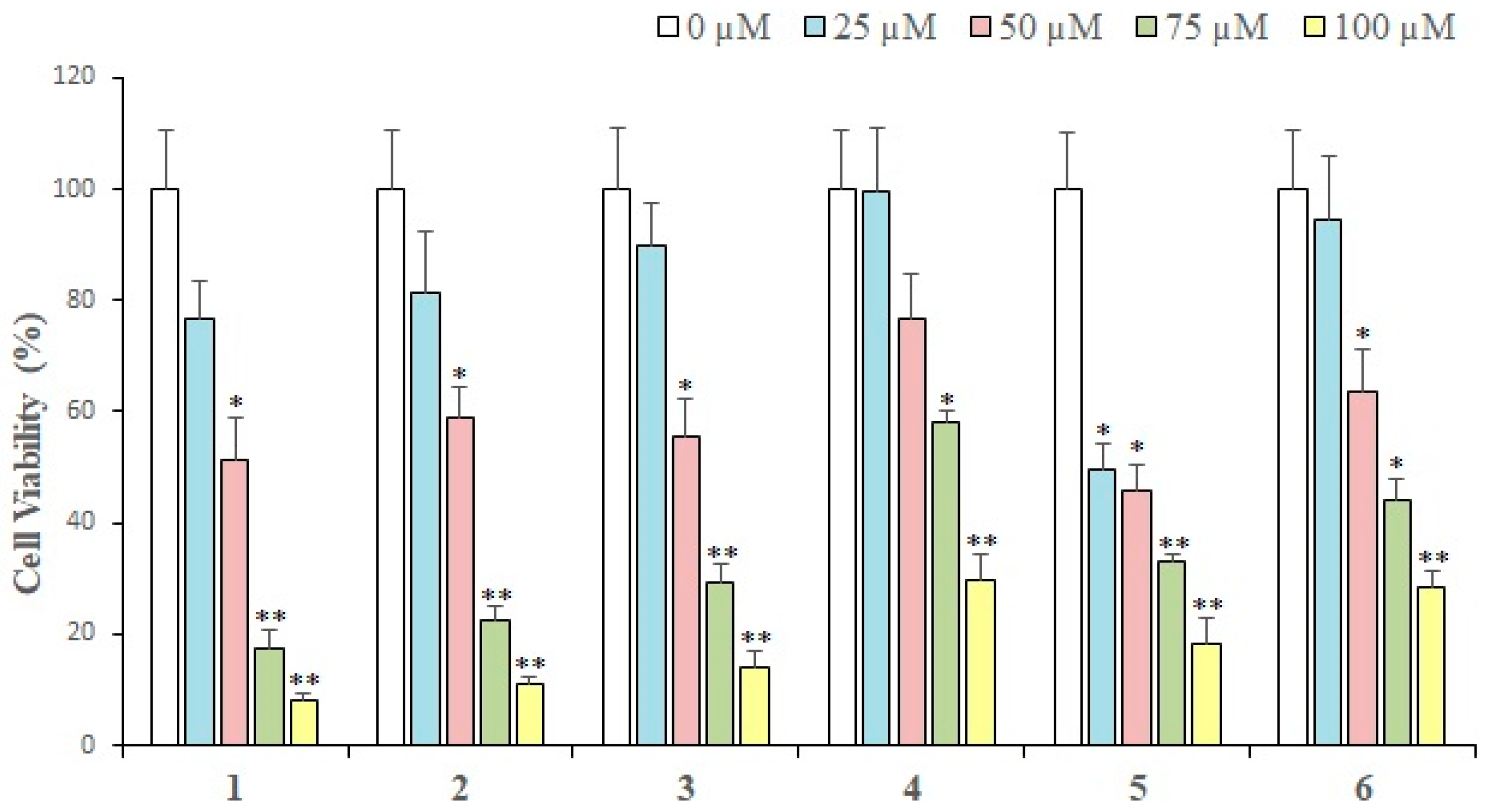
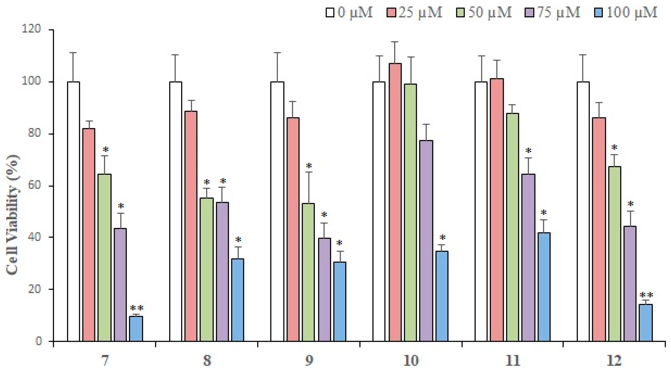
The in vitro cytotoxic activity of each compound isolated from L. shawii was examined against breast cancer cells. Results of an MTT assay showed that almost all the compounds exhibited a concentration-dependent growth inhibition of MDA-MB-231 breast cancer cells. At 25 µM (the lowest concentration tested), compound 5 showed maximum (50.53%) loss of cell viability as compared to the rest of the compounds. While 1 exhibited 76% cell viability, 2, 7, 9, 8, and 3 demonstrated 81–89% cell viability, respectively. Thus, at 25 µM, compound 5 showed a significant cytotoxic effect, while the rest of the compounds did not produce substantial antiproliferative activities. However, at 100 µM (the maximum concentration used to treat cancer cells), compound 1 exhibited maximal inhibition (92.1%) of cell proliferation, followed by compounds 7, 2, 3, 5, 6, 4, 9, and 8. Therefore, it is evident that at low concentrations (25 µM and 50 µM), compound 5 is the most active cytotoxic agent [47,48], while at concentrations of 75 µM and above, compound 1 is the most effective cytotoxic compound. Gaweesh et al. (2015) studied the cytotoxic activity of different fractions of L. shawii against HepG2, MCF7, and HCT116 cancer cells and reported that the CH2Cl2 fraction exhibits potent inhibition of the cell growth of MCF7 (breast cancer line) with IC50 value of 11 ± 0.195 μg/mL [49]. These active compounds might be potential sources of the anticancer activity of the extracts. Compound 9 was previously isolated by our group [35] which inhibited the cell proliferation in MDA-MB-231 breast cancer cells in a dose-dependent manner. At 25 µM, compound 9 did not inhibit cell growth significantly; however, at concentrations above 50 µM, 9 exhibited over 50% cell growth inhibition in MDA-MB-231 cells. At 100 µM, compound 9 showed approximately 70% loss of cell viability, which is considerable. The results are demonstrated in Figure 2 and Figure 3.
Compounds 1012 were isolated from Aloe vera resin and tested for their growth inhibitory potential against MDA-MB-231 cancer cells; 12 demonstrated the highest cytotoxic activities at three different concentrations (Figure 3). At 25 µM, 12 showed ~14.0% of cytotoxic effect, 11 exhibited no cell growth inhibition, while compound 10 depicted a growth-promoting effect on MDA-MB-231 cells. At 50 µM, compounds 1012 exhibited cytotoxic activity in the range of 67–99%, while at 75 µM, 12 showed 44%, while 11 and 10 depicted 64% and 77% cell viability, respectively, indicating that compound 12 is potent at this concentration. At 100 µM, 12 exhibited the highest activity, followed by 10 and 11. Based on the results, we can say that 1, 7, 2, 3, 12, and 5 were the most potential hits. The calculated IC50 values revealed that 5, 13, 7, and 12 are potent cytotoxic compounds with IC50 values ranging from 31 to 60 μM, while the IC50 values of 8, 6, and 9 were 72, 73, and 76 µM, respectively. The IC50 values of 112 are shown in Table 1.

2.3. Antioxidant Activity

The antioxidant activity of crude extracts/fractions and isolated compounds was tested using DPPH radical scavengers. The tests were performed at different concentrations to calculate the IC50 value. The ethyl acetate, BuOH, and CH2Cl2 fractions of L. shawii showed promising inhibitory potential of 76%, 72%, and 60%, respectively. The IC50 values for ethyl acetate, BuOH, and CH2Cl2 fractions of L. shawii were 378 ± 1.50, 650 ± 1.50, and 735 ± 2.00 μg/mL, respectively, indicating that the ethyl acetate fraction is most potent as compared to standard ascorbic acid (53 ± 1.32 μg/mL). The crude MeOH extract of L. shawii demonstrated mild activity (50 ± 1.50%), while aqueous and n-hexane fractions were inactive. According to Gaweesh et al. (2015) [49], the EtOAc fraction of aerial parts of L. shawii exhibited antioxidant activity with an IC50 value of 55.4 ± 3.48 μg/mL. The difference in activity may be due to the composition of the EtOAc fraction using only the stem instead of a mixture (leaves and stem). In the case of A. vera, only the ethyl acetate fraction exhibited moderate activity of 51%, while other fractions of this plant did not show promising results. The results are summarized in Table 2.
All the isolated compounds were screened in a DPPH radical scavenging assay in order to test their antioxidant potential. The results demonstrated that compound 4 exhibited potent antioxidant activity with an IC50 value of 55 ± 2.00 µM, followed by 14 (IC50 = 241 ± 1.50 µM), 6 (IC50 = 645 ± 1.50), and 13 (IC50 = 762 ± 2.00 µM) as compared to standard ascorbic acid. Compound 4 showed higher activity than 9 (IC50 = 30 µg/mL) reported by our group [35]. The results are tabulated in Table 3. It is a well-accepted notion that the presence and position of –OH groups in a molecule can increase or decrease the antioxidant activity [50]. Among diterpenes, compound 1 showed moderate activity which may be due to the presence of three exocyclic double bonds. Comparing anthraquinones, 6 possesses two –OH groups at the meta position of the right hand benzene ring, and one –OH at the C-8 position meta to methyl group at C-6. Compounds 5, 7, and 8 have similar basic skeletons with the absence of the meta –OH group which showed that the meta –OH groups play an important role in the antioxidant activity of anthraquinones. Comparing 4 and 14, 4 bears two –OH groups at the meta position of ring A, two –OH at ortho in ring B, one –OH at C-3 in ring C. Compound 14 has two –OH groups at the meta position of the benzene ring and one –OH at the C-3 position at cyclohexane. Due to the presence of two additional ortho (ring B)–OH groups, compounds showed higher DPPH radical scavenging activity. It is thus suggested that the chelating ability of the ortho and meta –OH groups in 4 played a greater role in the antioxidation property.
All the isolated compounds were screened in a DPPH radical scavenging assay in order to test their antioxidant potential. The results demonstrated that compound 4 exhibited potent antioxidant activity with an IC50 value of 55 ± 2.00 µM, followed by 14 (IC50 = 241 ± 1.50 µM), 6 (IC50 = 645 ± 1.50), and 13 (IC50 = 762 ± 2.00 µM) as compared to standard ascorbic acid. Compound 4 showed higher activity than 9 (IC50 = 30 µg/mL) reported by our group [35]. The results are tabulated in Table 3. It is a well-accepted notion that the presence and position of –OH groups in a molecule can increase or decrease the antioxidant activity [50]. Among diterpenes, compound 1 showed moderate activity which may be due to the presence of three exocyclic double bonds. Comparing anthraquinones, 6 possesses two –OH groups at the meta position of the right hand benzene ring, and one –OH at the C-8 position meta to methyl group at C-6. Compounds 5, 7, and 8 have similar basic skeletons with the absence of the meta –OH group which showed that the meta –OH groups play an important role in the antioxidant activity of anthraquinones. Comparing 4 and 14, 4 bears two –OH groups at the meta position of ring A, two –OH at ortho in ring B, one –OH at C-3 in ring C. Compound 14 has two –OH groups at the meta position of the benzene ring and one –OH at the C-3 position at cyclohexane. Due to the presence of two additional ortho (ring B) –OH groups, compounds showed higher DPPH radical scavenging activity. It is thus suggested that the chelating ability of the ortho and meta –OH groups in 4 played a greater role in the antioxidation property.

2.4. Human Intracellular Drug Targets

Compounds 13, 5, 7, and 12 were identified as the most potentially cytotoxic agents. We utilized cheminformatics techniques [51,52,53,54] to predict the most probable drug targets of these compounds. The kyoto encyclopedia of genes and genome (KEGG) database showed that seven major cancer drug targets are associated with triple-negative breast cancer including epidermal growth factor receptor (EGFR), proto-oncogene tyrosine-protein kinase Kit (c-KIT), insulin-like growth factor receptor 1 (IGFR1), notch receptor 1 (notch 1), phosphatidylinositol-3,4,5-trisphosphate-3-phosphatase (PTEN), phosphatidylinositol-4,5-bisphosphate 3-kinase (PI3K), and cyclin-dependent kinase 4 (CDK4).
The results of the Swiss Target Prediction server are summarized in Table S1 (supporting information) which revealed that compound 1 may target Cyp19A1 with 0.11 probability, which is not an anticancer drug target, while compound 2 depicted 0.09 to <0.05 probabilities for its predicted drug targets. Thus, the results for compound 2 were considered insignificant. Compounds 3 and 5 showed ≥0.10 and ~0.10 probabilities, respectively, for their predicted targets. Among the suggested targets, poly [ADP-ribose] polymerase (PARP)-1 is a potential drug target in TNBC, and carbonic anhydrase II (CA-II), CA-I, CA-XII, CA-IX, and estrogen receptor (ER) α and β are potential anticancer drug targets. However, ER is not expressed in TNBC, thus it was excluded from our study. Compound 7 showed 0.11 probability for its probable targets. Among the suggested targets, CA-II is a target of interest in cancer treatment. Compound 12 showed probabilities in the range of 0.09–0.04 for its suggested targets, in which CA-II was also included, however its probability was lower than 0.05. Based on these results and an extensive literature survey, PARP and CA-II were also included in our docking experiments.

2.5. Pharmacophore Modeling

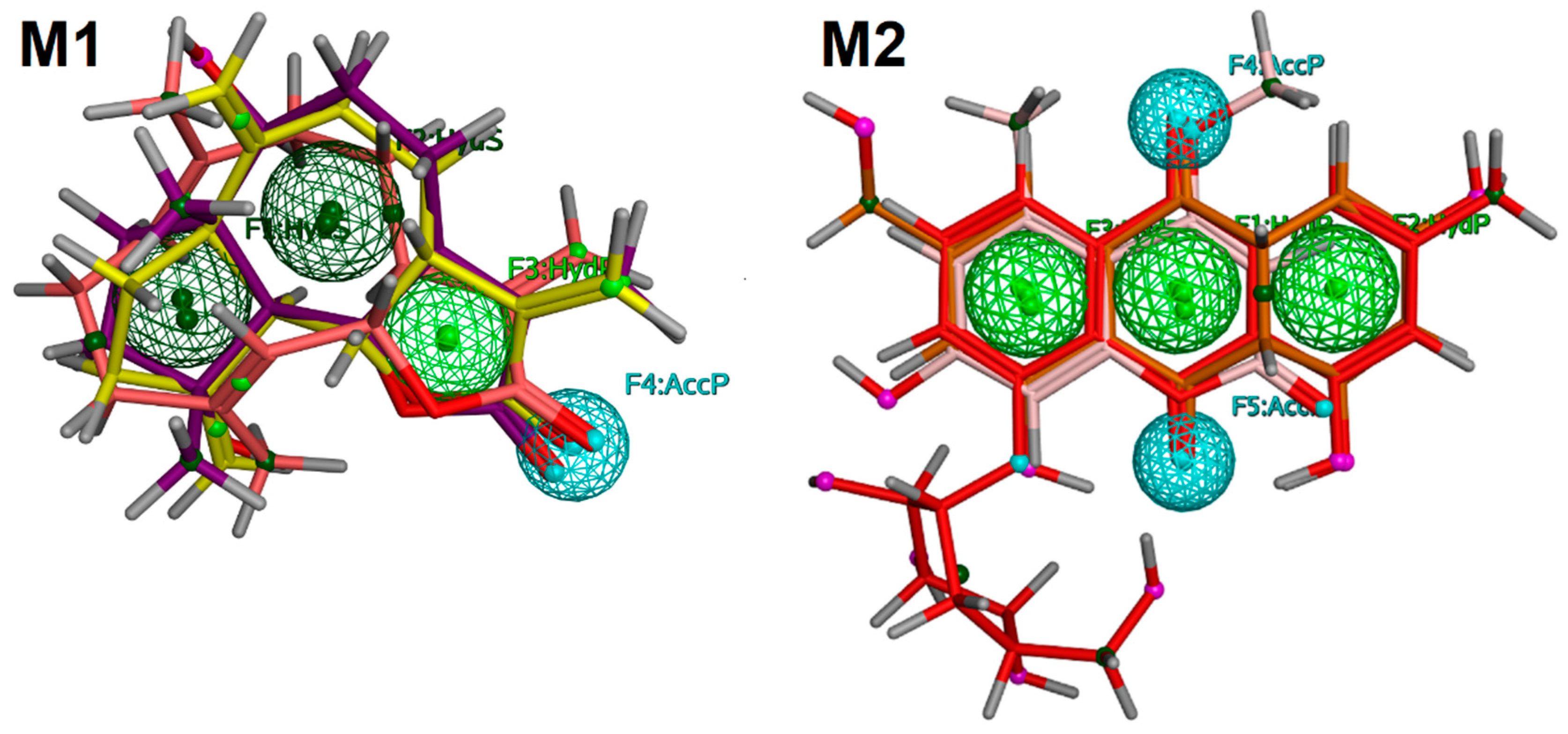
The Swiss Target Prediction (STP) server applies 2D-similarity searching to predict the target of the query compound. A number of compounds are present in this server with their actual biological activities and binding mechanism. In STP, 2D structures of the query compounds are matched with the 2D structures of the compounds present in its database and their probabilities are calculated and based on the calculated probabilities, and the target for the query compound is predicted. We also applied pharmacophore modeling to select the drug targets of our compounds with cytotoxic potential. It was hypothesized that the biological targets of those drugs that matched with the pharmacophore model can act as a target of our compounds. Two pharmacophore models, namely, M1 and M2, were generated by using compounds 13, 5, 7, and 12. M1 was generated by aligning the 3D structures of compounds 13, while M2 was created by aligning the 3D structures of compounds 5, 7, and 12. M1 possessed three hydrophobic (Hyd) and one H-Bond acceptor (AccP) feature while M2 contained three Hyd and two AccP features. The pharmacophore models are displayed in Figure 4.
A set of 62 drug molecules were screened from all the pharmacophore models. M1 retrieved five EGFR inhibitors (afatinib, dacomitinib, nazartinib, neratinib, pelitinib), three PI3K inhibitors (apitolisib, leniolisib, samotolisib), and one CDK4 inhibitor (Omacetaxine mepesuccinate). M2 identified six inhibitors of c-KIT (dovitinib lactate, midostaurin, ripretinib, semaxanib, toceranib, and sunitinib), one Notch 1 (crenigacestat), and two PI3K (dactolisib, samotolisib) inhibitors. This pharmacophore-based searching reflects that compounds 13 may target EGFR, PI3K, and CDK4 while compounds 5, 7, and 12 may bind with c-KIT, Notch 1, and PI3K. Subsequently, compounds 13, 5, 7, and 12 were subjected to structure-based inverse docking to further confirm the results of pharmacophore modeling.

2.6. Molecular Docking Studies

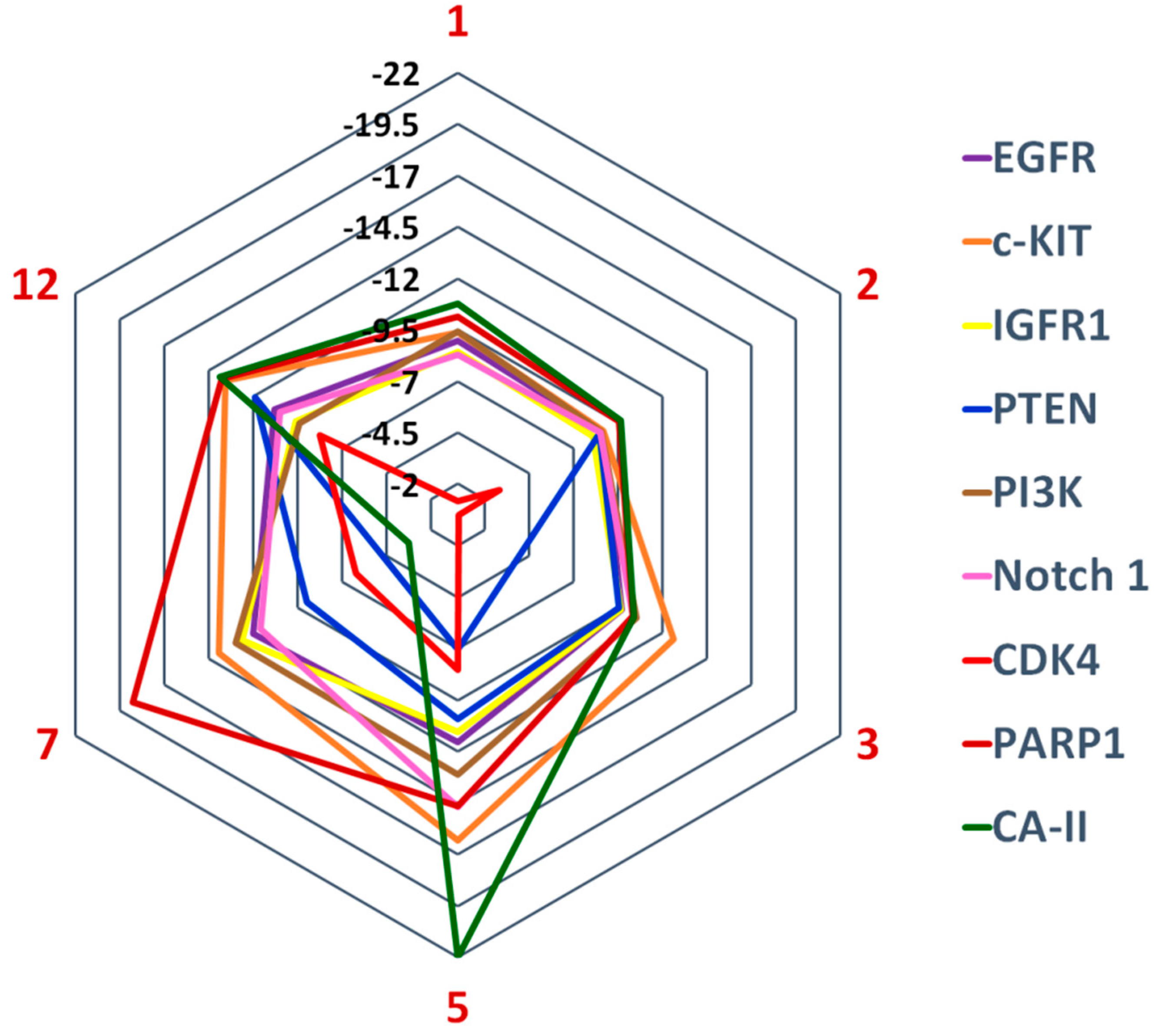
Compounds 13, 5, 7, and 12 were potential hits in our cell-based anticancer assay. Structure-based inverse docking analysis was carried out to predict their binding mechanism. Compounds were docked at the active site or ligand binding sites of the selected drug targets. The docking scores of the compounds suggests that CA-II is the most potential target for compounds 1 (−10.75), 2 (−9.64), 5 (−21.98), and 12 (−13.88), while c-KIT is the best target for compound 3 (−12.62). After CA-II, PARP-1 is an excellent candidate for 1, 2, 7, and 12, while PI3K and c-KIT are probable drug targets for compounds 3 and 5, respectively. Moreover, c-KIT is a good drug target for compounds 2, 7, and 12, while PARP1 is the fourth good target for compounds 3 and 5. PI3K was identified as the second most probable drug target for compound 3, while moderate for 1, 7, 2, and 5, and least for 12. The docking score suggests that the receptors EGFR, Notch 1, and IGFR have variable potency with these compounds, however, binding interactions suggest that compound 7 possesses good binding interactions with Notch 1, while compound 5 has higher binding interactions with EGFR. As per the docking scores, PTEN was the least favorable target for compounds 13, 5, and 7, while CDK4 was ranked as the worst target for all the compounds (Figure 5). The docking results are presented in Table S2.
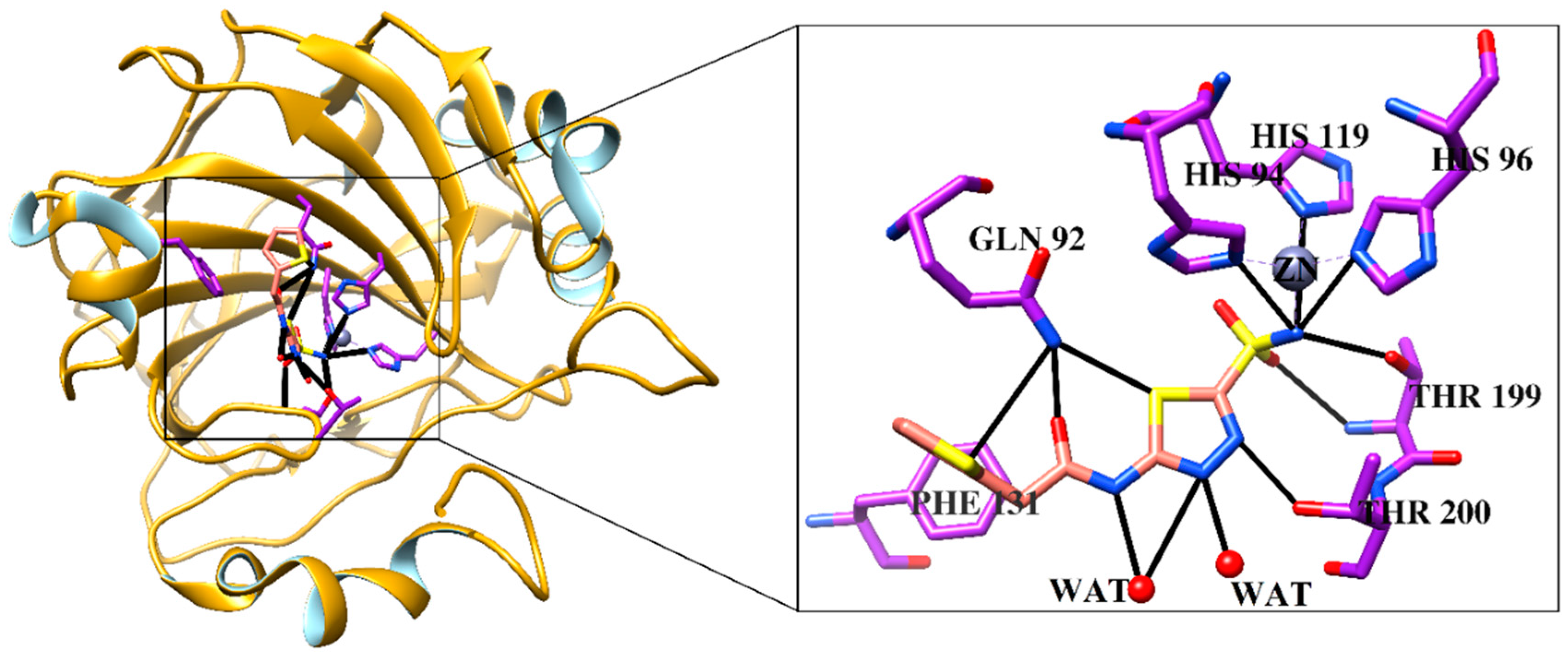
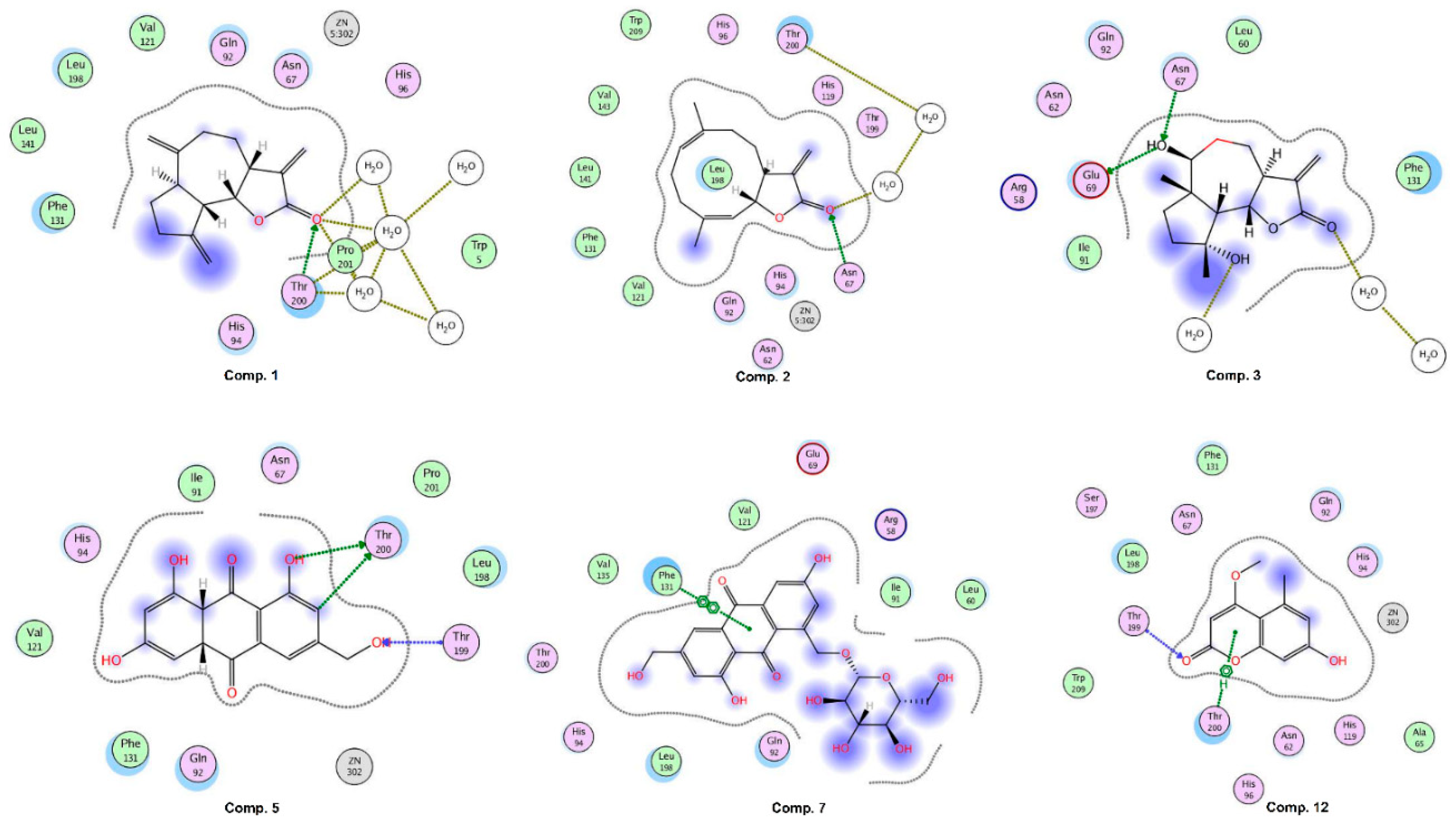
CA-II was exposed as the best target for compounds 1, 2, 5, and 7. Compound 1 binds with the docking score −10.75 and interacts with three water molecules and the side chain of Thr200 by H-bonding. Compound 2 interacts with the side chain of Asn67 and one water molecule via H-bonding. The –OH groups of compound 3 accept and donate H-bonds to the side chains of Asn67 and Glu69, respectively. Moreover, two water molecules provide H-bonding to the compound. The docked view of compound 5 depicts that its –OH groups donate H-bonds to the side chain of Thr200 and accepts H-bonds from the amino group of Thr199, while its ring mediates hydrophobic interactions with the side chain of Leu198. The docking score (−21.98) of 5 suggests that this compound possesses the highest binding potential for CA-II in silico. The side chain of Phe131 provides π-π interactions to the ring of compound 7. The docking score of 7 (−3.26) and its binding interactions suggest that this compound is the least active as compared to the rest of the compounds. The carbonyl moiety of compound 12 mediates bidentate interactions with the side/main chain amino groups of Thr199. Moreover, hydrophobic interaction was observed between the ring of the compound and the side chain of Thr199. The docking score of 12 (−13.88) is less than the docking score of compound 5, while higher than the docking scores of compounds 13 and 7. Some of the docked conformations of 12 showed that the compound may bind with the Zn atom present in the active site. The reference known drug, acetazolamide, interacts with ZN via metal–ligand interaction, and its carbonyl group also accepts H-bonds from the side chain of Thr199. The sulphate oxygen and ring nitrogen also mediate several water-mediated interactions. The active site of human CA-II is presented in Figure 6. The ligand (1GO), an acetazolamide derivative, complexed in the X-ray structure of CA-II, is also displayed, which interacts with ZN, His94, His96, His119, Gln92, Phe131, Thr199, Thr200, and two water molecules. Our compounds, except 12, do not bind with the zinc (ZN) present in the active site; however, they interact within the vicinity. The binding interactions of compounds are shown in Figure 7. The docking results are tabulated in supporting information Table S2.
After CA-II, PARP-1 was the second most probable target for compounds 1, 2, 7, and 12, while fourth for compounds 3 and 5. Among all the compounds, 7 (−18.74), 5 (−14.64), and 12 (−13.74) demonstrated higher binding scores than the niraparib (standard drug or positive control for PARP1, −13.18), while compounds 3 (−10.32), 1 (−10.14), 2 (−9.56) displayed lower scores than 7, 5, 12, and niraparib. At the active site of PARP-1, Glu988 and Ser904 provided H-bonds to compound 5, while Glu988 and a water molecule stabilized compound 7 via H-bonding. At the active site of c-KIT, compound 5 displayed the highest binding potential with docking score −16.31 as compared to the rest of the compounds, while compounds 7 (−13.91) and 12 (−13.56) displayed comparable docking scores. The binding modes also demonstrated the reason for higher binding scores. Compounds 5 and 7 had higher numbers of H-bonding and hydrophobic interactions as compared to 13 and 12. Moreover, compounds 5, 7, 12, and 3 depicted higher docking scores than the reference compound sunitinib, which is a drug complexed in the X-ray crystal structure of c-KIT (used in docking studies). Thr670, CYS673, LEU595, VAL603, and a water molecule (HOH289) played important roles in the stabilization of compounds 5, 7, and 12 in the active site. These residues provided hydrogen bonding and π–H interactions to the compounds.
Compounds 7 and 5 displayed higher binding scores than the positive control of EGFR (afatinib) and IGFR (ibutamoren mesylate). While compounds 5, 7, 12, and 3 scored better than crenigacestat (inhibitor of Notch 1), compounds 12, 5, and 7 were better than inhibitors of CDK4 (omacetaxine mepesuccinate), CA-II (acetazolamide) and PARP1 (niraparib). The docking results are tabulated in Table S1.

2.7. Inhibition of Carbonic Anhydrase II (CA-II) by Compounds 1, 2, 5, 7, and 12

Based on docking results, compounds 1, 2, 5, 7, and 12 were tested for their potential against CA-II in vitro. The quantity of compound 3 was not sufficient, thus it was not tested. The in vitro testing results showed that compound 5 is the most active compound with IC50 values of 14.4 µM, followed by compounds 12 and 2. Both the compounds (12 and 2) also showed very good activity in the range of 23 and 24 µM, respectively. The docking score also indicated that compounds 5 and 12 are the most active compounds. In docking, compounds 1 and 3 showed good binding potential after 5 and 12, however, in vitro results demonstrated that compound 1 is inactive. Compound 7 showed the least binding potential in silico and was found to be inactive against CA-II in vitro. The results are summarized in Table 4. The structure–activity relationship of the compounds is discussed in docking results. The experimental findings correlate well with the docking results.

3. Materials and Methods

This study consisted of extraction and isolation of bioactive compounds from L. shawii and A. vera and determination of their cytotoxic and antioxidant potential. The cytotoxic assay was conducted on TNBC cell lines, followed by in silico molecular targeting of active compounds. Pharmacophore modeling and molecular docking approaches were used in computational analysis. The computational results were validated by in vitro testing of active compounds against the most suitable predicted targets.

3.1. General Instrumentation

NMR spectra were recorded on an NMR spectrometer (BRUKER, Zürich, Switzerland) operating at 600 MHz with cryoprobe prodigy (150 MHz for 13C; chemical shift (δ) = ppm; coupling constants (J) = Hz). Infrared (IR) spectra were recorded on an ATR-Tensor 37 spectrophotometer, Bruker (Ettlingen, Baden-Württemberg, Germany). ESI-MS spectra were recorded on a mass spectrometer (Waters Quattro Premier XE, Waters, Milford, MA, USA). For thin-layer chromatography (TLC, silica gel 60F-254, Merck, Darmstadt, Hesse, Germany), precoated aluminum sheets were used. TLC plates were visualized under UV light at 254 and 366 nm and mostly sprayed with the ceric sulfate (Ce(SO4)2) reagent followed by heating with heating gun.

3.2. Plant Material and Identification

The whole plant materials of L. shawii and A. vera resin were purchased from market (Souq, Nizwa) and identified by the plant taxonomists Saif Al-Hatmi (Oman Botanical Garden, Muscat, Oman (OBGM)) and Syed Abdullah Gillani (Department of Biological Sciences and Chemistry (DBSC), University of Nizwa, Oman), respectively. Voucher specimens of A. vera (No. AFS-08/2016) and L. shawii (No: BSHR-05/2015) were deposited in the herbarium of OBGM and DBSC, respectively.

3.3. Extraction, Fractionation, and Isolation of Bioactive Compounds

The air-dried stem powder material of L. shawii was extracted with methanol for two weeks. The resulting methanol extract was suspended in distilled water (H2O) and successively partitioned into n-hexane, dichloromethane (CH2Cl2), ethyl acetate (EtOAc), n-butanol (n-BuOH), and aqueous (H2O) fractions. The n-hexane fraction was subjected to silica gel column chromatography (70–230 mesh; Merck) and produced two compounds, 1 and 2 [22]. Similarly, the EtOAc fraction was subjected to CC and eluted with an increasing polarity, viz., n-hexane–EtOAc, EtOAc, EtOAc–MeOH, and pure MeOH, to isolate seven compounds, 39 [35,55].
Similarly, the shade-dried powdered resin of A. vera was exhaustively extracted with MeOH (2 L) at room temperature (3 × 15 days). Evaporation of the MeOH in vacuo at 45 °C yielded a crude methanol extract, which after suspension in water was successively fractionated into n-hexane, CH2Cl2, EtOAc, and n-BuOH [36]. After taking TLC, CH2Cl2 and EtOAc fractions were combined. The combined material was subjected to CC and eluted with n-hexane, n-hexane–EtOAc, EtOAc, MeOH/EtOAc, and finally washed with 20% MeOH/EtOAc with 10% increments in polarity to afford several fractions which were subsequently subjected to further repeated CC with different concentrations of n-hexane-EtOAc-MeOH as eluent to obtain 11 compounds, 1020 [36].

3.4. Assay Protocol for Cytotoxic Activity

Cytotoxic activity was performed according to the previously described methods [56,57]. Breast cancer cell lines MDA-MB-231 were maintained in Dulbecco’s modified eagle medium (DMEM, Invitrogen, Carlsbad, CA, USA). The media was supplemented with 10% fetal bovine serum (FBS) and 1% antimycotic antibiotic (Invitrogen, Carlsbad, CA, USA). Cells were cultured in a 5% CO2-humidified atmosphere at 37 °C. Stock solution (5 mg/mL) of 3-(4,5-dimethylthiazol-2-yl)-2,5-diphenyltetrazolium bromide (MTT, Merck, Darmstadt, Hesse, Germany) was prepared in phosphate-buffered saline (PBS).
Cells were seeded at a density of 1 × 10⁴ cells per well in 96-well microtiter culture plates. After overnight incubation, normal growth medium was removed and replaced with either fresh medium (untreated control) or different concentrations of respective compounds in growth medium diluted from a 2 mM stock. After 24 h of incubation, MTT solution was added to each well (0.1 mg/mL in DMEM) and incubated further for 4 h at 37 °C. Upon termination, the supernatant was aspirated and the MTT formazan, formed by metabolically viable cells, was dissolved in a solubilization solution containing DMSO (100 սL) by mixing for 5 min on a gyratory shaker. The absorbance was measured at 540 nm (reference wavelength 690 nm) on an Ultra Multifunctional Microplate Reader (Bio-Rad, Hercules, California, USA). Absorbance of control (without treatment) was considered as 100% cell survival. Each treatment had three replicate wells [56]. The IC50 values of the active compounds were calculated using nonlinear regression through GraphPad Prism 4 software (La Jolla, CA, USA).

3.5. Assay Protocol for DPPH Radical Scavenging Activity

Free radical scavenging activity of the test fractions/compounds was determined by measuring the change in absorbance of DPPH (l, l-Diphenyl-2-picrylhydrazyl radical, Sigma-Aldrich, St Louis, MO, USA) at 515 nm by the microplate reader (SpectraMax M2, Molecular Devices, CA, USA) as previously described [57]. Ascorbic acid (Sigma-Aldrich, St Louis, MO, USA) was used as a standard with 90% inhibition and IC50 value of 53 µg/mL. The IC50 values of the tested compounds were calculated using nonlinear regression through GraphPad Prism 4 software (La Jolla, CA, USA).

3.6. In Silico Target Fishing

The coordinates of six compounds (13, 5, 7, and 12) were generated by ChemDraw Ultra 10 (PerkinElmer Inc.) [58] and converted into three-dimensional (3D) form using molecular operating environment (MOE version 2013.08) [59]. The SMILE formats of the compounds (13, 5, 7, and 12) were uploaded on the Swiss Target Prediction webserver (http://www.swisstargetprediction.ch/) to predict the intracellular drug targets of these compounds in humans. In addition, the Kyoto Encyclopedia of Genes and Genomes (KEGG) database (https://www.genome.jp/kegg/) was used to identify the probable targets of our active compounds in human triple-negative breast cancer (TNBC). The KEGG pathway (HSA05224 and H00031) showed that epidermal growth factor receptor (EGFR), proto-oncogene tyrosine-protein kinase Kit (c-KIT), insulin-like growth factor 1 receptor (IGF1R), and Notch receptor 1 (Notch 1) are particularly overexpressed in TNBC. Moreover, phosphatidylinositol-3,4,5-trisphosphate-3-phosphatase and dual-specificity protein phosphatase (PTEN), phosphatidylinositol-4,5-bisphosphate 3-kinase (PI3K), and G1/S-specific cyclin-D1 (CCND1 or CDK4) are found to be mutated in TNBC. Thus these targets were scrutinized in the molecular docking protocol.

3.7. Selection of Known Drugs

By extensive literature survey, 62 known anticancer drugs were selected and their 3D structures were taken from the PubChem database (https://pubchem.ncbi.nlm.nih.gov/), which was used as a library in pharmacophore searching. Moreover, during docking, those drugs were considered as positive control or reference compounds. The docking results of compounds 13, 5, 7, and 12 were compared with the docking scores of these drugs. The selected drugs for each target are tabulated in Table 5.

3.8. Pharmacophore Modeling

Pharmacophore modeling was performed by MOE-Pharmacophore editor using PPCH-ALL annotation scheme. PPCH-ALL has H-bond Donor (HBD), H-bond Acceptor (HBA) and their Projections, π vs. non-π H-bond Donor/Acceptor, General π vs. non-π Distinctions, Metal Ligator, Metal Ligator Projection, Cation, Anion, and Hydrophobe (HYD) terms. The pharmacophore models were generated by using 3D structures of active compounds 13, 5, 7, and 12. Compounds 13 were aligned and model 1 was generated, while model 2 was generated by aligning the compounds 5, 7, and 12. The common pharmacophoric features were selected in the aligned compounds.

3.9. Molecular Docking

The X-ray crystal structures of the target proteins including EGFR, c-KIT, IGF1R, Notch 1, PTEN, PI3K, CDK4, PARP1, and CA-II were retrieved from Research Collaboratory for Structural Bioinformatics Protein Data Bank (RCSB-PDB) (https://www.rcsb.org/). The data are summarized in Table 6. For docking, protein files were prepared by addition of protons, partial charges, and the removal of cocrystallized ligands and heteroatoms. The role of water molecules was deduced by visualizing the protein–ligand interactions. Only water molecules within the vicinity of 3.0 Å of active ligands were retained in the file during docking, otherwise they were removed from the protein structure. The structures of the compounds and selected drugs were energy minimized using MMFF94x forcefield and gradient: 0.05. During energy minimization, hydrogens were added, and partial charges were applied.
Molecular docking was performed by MOE using Triangle Matcher placement method, Rescoring1: London dG, Refinement: Forcefield, and Rescoring2: Affinity dG. As a default parameter, 30 docked conformations were selected to be saved for each compound after docking. After docking, we applied conformation sampling method to select the best docked orientation of compound. For this purpose, each conformation of all the docked compounds were visualized and based on the protein–ligand interactions, docking score, and rank, best conformation was selected for analysis. The images in 2D were captured through MOE ligand binding interaction while 3D images were taken by Chimera [60].

3.10. Carbonic Anhydrase II Inhibition

The total reaction volume of 200 µL included 20 µL of test compounds prepared in DMSO, followed by the addition of 140 µL of the HEPES–tris buffer, 20 µL of purified bovine erythrocyte CA-II (0.1 mg/mL) prepared in buffer, and 20 µL of a solution of 4-nitrophenyl acetate [70]. A 20 µL amount of test compound was incubated with the enzyme (EC 4.2.1.1, Sigma-Aldrich, St. Louis, MO, USA) for 15 min in a 96-well flat-bottom plate. The rate of product formation was monitored with the addition of 20 µL of 4-NPA as substrate, prepared in methanol at the final concentration of 0.7 mM, at 25 °C for 30 min with regular intervals of 1 min, by using microplate readers (Bio-Rad, Molecular Devices, CA, USA). HEPES-tris was used as a buffer for the reaction at the final concentration of 20 mM at pH 7.4. The percent inhibition was calculated by using the following formula:
% Inhibition = 100 − (OD test well/OD control) × 100

3.11. Statistical Analysis for Cytotoxic Activities

Results are expressed as mean ± SEM of at least three independent observations. Student’s t-test was used to statistically examine significant differences. Analysis of variance was performed using ANOVA. p-Values < 0.05 were considered statistically significant.

4. Conclusions

Twenty compounds (isolated from L. shawii and A. vera) were scrutinized for their anticancer potential in triple-negative breast cancer cell lines (MDA-MB-231). Among the tested compounds, compounds 5, 13, 7, and 12 were retrieved as most potential hits. We used extensive in silico target fishing techniques to predict their biological targets in the human genome. For this purpose, 2D-cheminformatic tools and 3D-pharmacophore modeling were used that suggested that carbonic anhydrase II (CA-II), poly [ADP-ribose] polymerase (PARP)-1, and proto-oncogene tyrosine-protein kinase Kit (c-KIT) can be possible targets for the active hits. The in silico findings were validated by in vitro testing of compounds on CA-II, which showed that 5, 12, and 2 are excellent inhibitors of CA-II. Moreover, antioxidant activities of compounds were examined, which demonstrated that compound 4 possesses the highest antioxidant potential. These results indicated that constituents of L. shawii and A. vera are promising drug candidates for triple-negative breast cancer and should be investigated in detail.

Supplementary Materials

The following are available online at https://www.mdpi.com/1424-8247/13/5/94/s1, Table S1: Human intra-cellular targets—swiss target prediction results. Table S2: Molecular docking scores and binding interactions of compounds 13, 5, 7, and 12 on the selected drug targets. Figure S1: 1H, 13C-NMR and ESI-MS spectra of the known compounds (120).

Author Contributions

N.U.R., K.R. and H.H. designed and performed all the experiments. H.Y.K., G.A. and M.K. performed the anticancer, antioxidant, and carbonic anhydrase II activities, respectively. S.A.H. and A.K. did molecular docking studies of all active compounds. N.U.R., S.A.H. and H.H. wrote the original draft of the manuscript. A.A.-H. supervised the project and assisted in reviewing and editing the manuscript. All authors have read and agreed to the published version of the manuscript.

Funding

The research was supported by The Oman Research Council (TRC) funded through the project (BFP/RGP/CBS/18/011).

Acknowledgments

The authors are grateful to the University of Nizwa, Oman for the generous support of this project.

Conflicts of Interest

The authors declare no conflict of interest.

References

  1. Roy, N.K.; Deka, A.; Bordoloi, D.; Mishra, S.; Kumar, A.P.; Sethi, G.; Kunnumakkara, A.B. The potential role of boswellic acids in cancer prevention and treatment. Cancer Lett. 2016, 377, 74–86. [Google Scholar] [CrossRef] [PubMed]
  2. Baskar, R.; Lee, K.A.; Yeo, R.; Yeoh, K.W. Cancer and radiation therapy: Current advances and future directions. Int. J. Med. Sci. 2012, 9, 193–199. [Google Scholar] [CrossRef] [PubMed] [Green Version]
  3. Newman, D.; Gordon, M.C. Natural products as sources of new drugs from 1981 to 2014. J. Nat. Prod. 2012, 79, 629–661. [Google Scholar] [CrossRef] [Green Version]
  4. Grover, J.K.; Yadav, S.; Vats, V. Medicinal plants of India with anti-diabetic potential. J. Ethnopharmacol. 2002, 81, 81–100. [Google Scholar] [CrossRef]
  5. Ferlay, J.; Soerjomataram, I.; Dikshit, R.; Eser, S.; Mathers, C.; Rebelo, M.; Parkin, D.M.; Forman, D.; Bray, F. Cancer incidence and mortality worldwide: Sources, methods and major patterns in GLOBOCAN 2012. Int. J. Cancer 2015, 136, E359–E386. [Google Scholar] [CrossRef] [PubMed]
  6. Ding, L.; Gu, H.; Xiong, X.; Ao, H.; Cao, J.; Lin, W.; Yu, M.; Lin, J.; Cui, Q. MicroRNAs involved in carcinogenesis, prognosis, therapeutic resistance and applications in human triple-negative breast cancer. Cells 2019, 8, 1492. [Google Scholar] [CrossRef] [Green Version]
  7. Fitzmaurice, C.; Akinyemiju, T.F.; Al Lami, F.H.; Alam, T.; Alizadeh-Navaei, R.; Allen, C.; Alsharif, U.; Alvis-Guzman, N.; Amini, E.; Anderson, B.O.; et al. Global, regional, and national cancer incidence, mortality, years of life lost, years lived with disability, and disability-adjusted life-years for 29 cancer groups, 1990 to 2016: A systematic analysis for the global burden of disease study. JAMA Oncol. 2018, 4, 1553–1568. [Google Scholar]
  8. Xiao, Y.; Humphries, B.; Yang, C.; Wang, Z. MiR-205 Dysregulations in Breast Cancer: The Complexity and Opportunities. Non-Coding RNA. 2019, 5, 53. [Google Scholar] [CrossRef] [Green Version]
  9. Carpenter, K.J.; Valfort, A.C.; Steinauer, N.; Chatterjee, A.; Abuirqeba, S.; Majidi, S.; Sengupta, M.; Di Paolo, R.J.; Shornick, L.P.; Zhang, J.; et al. LXR-inverse agonism stimulates immune-mediated tumor destruction by enhancing CD8 T-cell activity in triple negative breast cancer. Sci. Rep. 2019, 9, 1–8. [Google Scholar] [CrossRef]
  10. Alizart, M.; Saunus, J.; Cummings, M.; Lakhani, S.R. Molecular classification of breast carcinoma. Diagn. Histopathol. 2012, 18, 97–103. [Google Scholar] [CrossRef]
  11. Huynh, M.M.; Jayanthan, A.; Pambid, M.R.; Los, G.; Dunn, S.E. RSK2: A promising therapeutic target for the treatment of triple-negative breast cancer. Expert Opin. Ther. Targets 2020, 24, 1–5. [Google Scholar] [CrossRef] [PubMed] [Green Version]
  12. Perou, C.M.; Sørlie, T.; Eisen, M.B.; Van De Rijn, M.; Jeffrey, S.S.; Rees, C.A.; Pollack, J.R.; Ross, D.T.; Johnsen, H.; Akslen, L.A. Molecular portraits of human breast tumours. Nature 2000, 406, 747. [Google Scholar] [CrossRef] [PubMed]
  13. Rakha, E.A.; Reis-Filho, J.S.; Ellis, I.O. Basal-like breast cancer: A critical review. J. Clin. Oncol. 2008, 26, 2568–2581. [Google Scholar] [CrossRef] [PubMed]
  14. Huang, S.; Xu, W.; Hu, P.; Lakowski, T.M. Integrative analysis reveals subtype-specific regulatory determinants in triple negative breast cancer. Cancers 2019, 11, 507. [Google Scholar] [CrossRef] [Green Version]
  15. Wang, Y.; Qi, Y.X.; Qi, Z.; Tsang, S.Y. TRPC3 Regulates the proliferation and apoptosis resistance of triple negative breast cancer cells through the TRPC3/RASA4/MAPK pathway. Cancers 2019, 11, 558. [Google Scholar] [CrossRef] [Green Version]
  16. Wahba, H.A.; El-Hadaad, H.A. Current approaches in treatment of triple-negative breast cancer. Cancer Biol. Med. 2015, 12, 106. [Google Scholar]
  17. Mehanna, J.; Haddad, F.G.; Eid, R.; Lambertini, M.; Kouriem, H.R. Triple-negative breast cancer: Current perspective on the evolving therapeutic landscape. Int. J. Womens Health 2019, 11, 431. [Google Scholar] [CrossRef] [Green Version]
  18. Tripp, B.C.; Smith, K.; Ferry, J.G. Carbonic anhydrase: New insights for an ancient enzyme. J. Biol. Chem. 2001, 276, 48615–48618. [Google Scholar] [CrossRef] [Green Version]
  19. Chegwidden, W.R.; Dodgson, S.J.; Spencer, I.M. The roles of carbonic anhydrase in metabolism, cell growth and cancer in animals. In The Carbonic Anhydrases: New Horizons; Chegwidden, W.R., Carter, N.D., Edwards, Y.H., Eds.; Springer: Basel, Switzerland, 2000; pp. 343–361. [Google Scholar]
  20. Venta, P.J. Carbonic anhydrases in mammalian cell culture and tumors. In The Carbonic Anhydrases: Cellular Physiology and Molecular Genetics; Dodgson, S.J., Tashian, R.E., Gros, G., Carter, N.D., Eds.; Plenum Press: New York, NY, USA, 1991; pp. 71–78. [Google Scholar]
  21. Webb, S.D.; Sherratt, J.A.; Fish, R.G. Mathematical modelling of tumour acidity: Regulation of intracellular pH. J. Theor. Biol. 1999, 196, 237–250. [Google Scholar] [CrossRef]
  22. Gerweck, L.E. Tumor pH: Implications for treatment and novel drug design. Semin. Radiat. Oncol. 1998, 8, 176–182. [Google Scholar] [CrossRef]
  23. Montcourrier, P.; Silver, I.; Farnoud, R.; Bird, I.; Rochefort, H. Breast cancer cells have a high capacity to acidify extracellular milieu by a dual mechanism. Clin. Exp. Metastasis 1997, 15, 382–392. [Google Scholar] [CrossRef] [PubMed]
  24. Lee, A.H.; Tannock, I.F. Heterogeneity of intracellular pH and of mechanisms that regulate intracellular pH in populations of cultured cells. Cancer Res. 1998, 58, 1901–1908. [Google Scholar] [PubMed]
  25. Parkkila, A.K.; Herva, R.; Parkkila, S.; Rajaniemi, H. Immunohistochemical demonstration of human carbonic anhydrase isoenzymes I and II in brain tumours. Histochem. J. 1995, 27, 974–982. [Google Scholar] [CrossRef] [PubMed]
  26. Pastoreková, S.; Parkkila, S.; Parkkila, A.K.; Opavský, R.; Zelník, V.; Saarnio, J.; Pastorek, J. Carbonic anhydrase IX, MN/CA IX: Analysis of stomach complementary DNA sequence and expression in human and rat alimentary tracts. Gastroenterology 1997, 112, 398–408. [Google Scholar] [CrossRef]
  27. Frazier, M.L.; Lilly, B.J.; Wu, E.F.; Ota, T.; Hewett-Emmett, D. Carbonic anhydrase II gene expression in cell lines from human pancreatic adenocarcinoma. Pancreas 1990, 5, 507–514. [Google Scholar] [CrossRef]
  28. Parkkila, S.; Parkkila, A.K.; Juvonen, T.; Lehto, V.P.; Rajaniemi, H. Immunohistochemical demonstration of human carbonic anhydrase isoenzymes I and II in pancreatic tumours. Histochem. J. 1995, 27, 133–138. [Google Scholar]
  29. Parkkila, S.; Rajaniemi, H.; Parkkila, A.K.; Kivelä, J.; Waheed, A.; Pastoreková, S.; Pastorek, J.; Sly, W. Carbonic anhydrase inhibitor suppresses invasion of renal cancer cells in vitro. Proc. Natl. Acad. Sci. USA 2000, 97, 2220–2224. [Google Scholar] [CrossRef] [Green Version]
  30. Halliwell, B.; Gutteridge, J.M.C. Free Radicals in Biology and Medicine, 3rd ed.; Oxford University Press: Oxford, UK, 1998. [Google Scholar]
  31. Prochazkova, D.; Bousova, I.; Wilhelmova, N. Antioxidant and prooxidant properties of flavonoids. Fitoterapia 2011, 82, 513–523. [Google Scholar] [CrossRef]
  32. Li, Y.X.; Kim, S.K. Utilization of seaweed derived ingredients as potential antioxidants and functional ingredients in the food industry: An Overview. Food Sci. Biotechnol. 2011, 20, 1461–1466. [Google Scholar] [CrossRef]
  33. Mahdi-Pour, B.; Jothy, S.L.; Latha, L.Y.; Chen, Y.; Sasidharan, S. Antioxidant activity of methanol extracts of different parts of Lantana camara. Asian Pac. J. Trop. Biomed. 2012, 2, 960–965. [Google Scholar] [CrossRef] [Green Version]
  34. Ali, S.S.; El-Zawawy, N.A.; Al-Tohamy, R.; El-Sapagh, S.; Mustafa, A.M.; Sun, J. Lycium shawii Roem. & Schult.: A new bioactive antimicrobial and antioxidant agent to combat multi-drug/pan-drug resistant pathogens of wound burn infections. J. Tradit. Complement. Med. 2020, 10, 13–25. [Google Scholar] [PubMed]
  35. Rehman, N.U.; Hussain, H.; Al-Riyami, S.A.; Csuk, R.; Khiat, M.; Abbas, G.; Al-Rawahi, A.; Green, I.R.; Ahmed, I.; Al-Harrasi, A. Lyciumaside and lyciumate: A new diacylglycoside and sesquiterpene lactone from Lycium shawii. Helv. Chim. Acta 2016, 99, 632–635. [Google Scholar] [CrossRef]
  36. Rehman, N.U.; Al-Riyami, S.A.; Hussain, H.; Ali, A.; Khan, A.L.; Al-Harrasi, A. Secondary metabolites from the resins of Aloe vera and Commiphora mukul mitigate lipid peroxidation. Acta Pharmaceutica. 2019, 69, 433–441. [Google Scholar] [CrossRef] [PubMed] [Green Version]
  37. Taukoorah, U.; Mahomoodally, M.F. Crude Aloe vera gel shows antioxidant propensities and inhibits pancreatic lipase and glucose movement in vitro. Adv. Pharmacol. Sci. 2016, 2016, 3720850. [Google Scholar] [PubMed] [Green Version]
  38. Rauwald, H.W.; Lohse, K. Structure revision of 4-hydroxyaloin: 10-hydroxyaloins A and B as main in vitro-oxidation products of the diastereomeric aloins. Planta Med. 1992, 58, 259–262. [Google Scholar] [CrossRef] [PubMed]
  39. Gao, J.; Zhang, G.; Dai, R.; Bi, K. Isolation of aloinoside B and metabolism by rat intestinal Bacteria. Pharm. Biol. 2004, 42, 581–587. [Google Scholar] [CrossRef]
  40. Girol, C.G.; Fisch, K.M.; Heinekamp, T.; Günther, S.; Hüttel, W.; Piel, J.; Brakhage, A.A.; Müller, M. Regio- and stereoselective oxidative phenol coupling in Aspergillus nige. Angew. Chem. Int. Ed. Engl. 2012, 51, 9788–9791. [Google Scholar] [CrossRef]
  41. Heerden, F.R.; Viljoen, A.M.; Wyk, B.E. 6′-O-Coumaroylaloesin from Aloe castanea—A taxonomic marker for Aloe section Anguialoe. Phytochemistry 2000, 55, 117–120. [Google Scholar] [CrossRef]
  42. Speranza, G.; Manitto, P.; Monti, D.; Lianza, F. Feroxidin, a novel 1-methyltetralin derivative isolated from Cape aloe. Tetrahedron Lett. 1990, 31, 3077–3080. [Google Scholar] [CrossRef]
  43. Zakir, H.A.; Subbarao, G.V.; Pearse, S.J.; Gopalakrishnan, S.; Ito, O.; Ishikawa, T.; Kawano, N.; Nakahara, K.; Yoshihashi, T.; Ono, H.; et al. Detection, isolation and characterization of a root-exuded compound, methyl 3-(4-hydroxyphenyl) propionate, responsible for biological nitrification inhibition by sorghum (Sorghum bicolor). New Phytol. 2008, 180, 442–451. [Google Scholar] [CrossRef] [PubMed] [Green Version]
  44. Tabuchi, H.; Tajimi, A.T.; Ichihara, A. Phytotoxic metabolites isolated from Scolecotrichum graminis Fuckel. Biosci. Biotech. Biochem. 1994, 58, 1956–1959. [Google Scholar] [CrossRef]
  45. Harish, R.; Divakar, S.; Srivastava, A.; Shivanandappa, T. Isolation of antioxidant compounds from the methanolic extract of the roots of Decalepis hamiltonii (Wight and Arn.). J. Agric. Food Chem. 2005, 53, 7709–7714. [Google Scholar] [CrossRef] [PubMed]
  46. Poh, B.L.; Lim, C.H.; Tan, C.M.; Wong, W. 1H NMR study on the complexation of phenols with cyclotetrachromotropylene in aqueous solution. Tetrahedron 1993, 49, 7259–7266. [Google Scholar] [CrossRef]
  47. He, Z.H.; Huang, Y.Q.; Weng, S.F.; Tan, Y.R.; He, T.P.; Qin, Y.M.; Liang, N.C. Effect of Aloe emodin on invasion and metastasis of high metastatic breast cancer MDA-MB-231 cells. Zhong Yao Cai 2013, 36, 1481–1485. [Google Scholar] [PubMed]
  48. Huang, P.H.; Huang, C.Y.; Chen, M.C.; Lee, Y.T.; Yue, C.H.; Wang, H.Y.; Lin, H. Emodin and aloe-emodin suppress breast cancer cell proliferation through ERα inhibition. Evid. Based Complement. Alternat. Med. 2013, 2013, 376123. [Google Scholar] [CrossRef] [PubMed] [Green Version]
  49. Gaweesh, A.; Sengab, A.E.N.B.; Osman, H.M.H.S.M.; Abdou, A.M. Phytoconstituents, cytotoxic, antioxidant and hepatoprotective activities of the aerial parts of Lycium shawii R. growing in Egypt. Med. Aromat Plants 2015, 4, 1–7. [Google Scholar]
  50. Muhammad, A.; Tel-Çayan, G.; Öztürk, M.; Duru, M.E.; Nadeem, S.; Anis, I.; Ng, S.W.; Shah, M.R. Phytochemicals from and their antioxidant and anticholinesterase activities with structure-activity relationships. Pharm. Biol. 2016, 11, 1–7. [Google Scholar]
  51. Moumbock, A.F.; Li, J.; Mishra, P.; Gao, M.; Günther, S. Current computational methods for predicting protein interactions of natural products. Comput. Struct. Biotechnol. J. 2019, 17, 1367–1376. [Google Scholar] [CrossRef]
  52. Cereto-Massagué, A.; Ojeda, M.J.; Valls, C.; Mulero, M.; Pujadas, G.; Garcia-Vallve, S. Tools for in silico target fishing. Methods 2015, 71, 98–103. [Google Scholar] [CrossRef]
  53. Jenkins, J.L.; Bender, A.; Davies, J.W. In silico target fishing: Predicting biological targets from chemical structure. Drug Discov. Today Technol. 2006, 3, 413–421. [Google Scholar] [CrossRef]
  54. Akhoon, B.A.; Tiwari, H.; Nargotra, A. In Silico Drug Design Methods for Drug Repurposing. In In Silico Drug Design; Academic Press: Cambridge, MA, USA, 2019; pp. 47–84. [Google Scholar]
  55. Rehman, N.U.; Hussain, H.; Al-Riyami, S.A.; Green, I.R.; Al-Harrasi, A. Chemical constituents isolated from Lycium shawii and their chemotaxonomic significance. Rec. Nat. Prod. 2018, 12, 380–384. [Google Scholar] [CrossRef]
  56. Raees, M.A.; Hussain, H.; Al-Rawahi, A.; Csuk, R.; Muhammad, S.A.; Khan, H.Y.; Rehman, N.U.; Abbas, G.; Al-Broumi, M.A.; Green, I.R.; et al. Anti-proliferative and computational studies of two new pregnane glycosides from Desmidorchis flava. Bioorg. Chem. 2016, 67, 95–104. [Google Scholar] [CrossRef] [PubMed]
  57. Rehman, N.U.; Hussain, H.; Khan, H.Y.; Csuk, R.; Abbas, G.; Green, I.R.; Al-Harrasi, A. A norterpenoid and tripenoids from Commiphora mukul: Isolation and biological activity. Zeitschrift für Naturforschung B 2017, 72, 11–15. [Google Scholar] [CrossRef]
  58. Mills, N. ChemDraw Ultra 10.0 CambridgeSoft, 100 CambridgePark Drive, Cambridge, MA 02140. www.cambridgesoft.com. Commercial Price:  $1910 for download, $2150 for CD-ROM; Academic Price:  $710 for download, $800 for CD-ROM. J. Am. Chem. Soc. 2006, 128, 13649–13650. [Google Scholar] [CrossRef]
  59. Molecular Operating Environment (MOE) (2013.08); Chemical Computing Group ULC: Montreal, QC, Canada, 2018.
  60. Cisek, K.; Cooper, G.L.; Huseby, C.J.; Kuret, J. Structure and mechanism of action of tau aggregation inhibitors. Curr. Alzheimer Res. 2014, 11, 918–927. [Google Scholar] [CrossRef] [Green Version]
  61. Solca, F.; Dahl, G.; Zoephel, A.; Bader, G.; Sanderson, M.; Klein, C.; Kraemer, O.; Himmelsbach, F.; Haaksma, E.; Adolf, G.R. Target binding properties and cellular activity of afatinib (BIBW 2992), an irreversible ErbB family blocker. J. Pharmacol. Exp. Ther. 2012, 343, 342–350. [Google Scholar] [CrossRef]
  62. Gajiwala, K.S.; Wu, J.C.; Christensen, J.; Deshmukh, G.D.; Diehl, W.; DiNitto, J.P.; English, J.M.; Greig, M.J.; He, Y.A.; Jacques, S.L.; et al. KIT kinase mutants show unique mechanisms of drug resistance to imatinib and sunitinib in gastrointestinal stromal tumor patients. Proc. Natl. Acad. Sci. USA 2009, 106, 1542–1547. [Google Scholar] [CrossRef] [Green Version]
  63. Miller, L.M.; Mayer, S.C.; Berger, D.M.; Boschelli, D.H.; Boschelli, F.; Di, L.; Du, X.; Dutia, M.; Floyd, M.B.; Johnson, M.; et al. Lead identification to generate 3-cyanoquinoline inhibitors of insulin-like growth factor receptor (IGF-1R) for potential use in cancer treatment. Bioorg. Med. Chem. Lett. 2009, 19, 62–66. [Google Scholar] [CrossRef]
  64. Wu, Y.; Cain-Hom, C.; Choy, L.; Hagenbeek, T.J.; de Leon, G.P.; Chen, Y.; Finkle, D.; Venook, R.; Wu, X.; Ridgway, J.; et al. Therapeutic antibody targeting of individual Notch receptors. Nature 2010, 464, 1052–1057. [Google Scholar] [CrossRef]
  65. Lee, C.U.; Hahne, G.; Hanske, J.; Bange, T.; Bier, D.; Rademacher, C.; Hennig, S.; Grossmann, T.N. Redox modulation of PTEN phosphatase activity by hydrogen peroxide and bisperoxidovanadium complexes. Angew. Chem. Int. Ed. 2015, 54, 13796–13800. [Google Scholar] [CrossRef] [Green Version]
  66. Hoegenauer, K.; Soldermann, N.; Stauffer, F.; Furet, P.; Graveleau, N.; Smith, A.B.; Hebach, C.; Hollingworth, G.J.; Lewis, I.; Gutmann, S.; et al. Discovery and pharmacological characterization of novel quinazoline-based PI3K delta-selective inhibitors. ACS Med. Chem. Lett. 2016, 7, 762–767. [Google Scholar] [CrossRef] [PubMed]
  67. Day, P.J.; Cleasby, A.; Tickle, I.J.; O’Reilly, M.; Coyle, J.E.; Holding, F.P.; McMenamin, R.L.; Yon, J.; Chopra, R.; Lengauer, C.; et al. Crystal structure of human CDK4 in complex with a D-type cyclin. Proc. Natl. Acad. Sci. USA 2009, 106, 4166–4170. [Google Scholar] [CrossRef] [PubMed] [Green Version]
  68. Thorsell, A.G.; Ekblad, T.; Karlberg, T.; Löw, M.; Pinto, A.F.; Trésaugues, L.; Moche, M.; Cohen, M.S.; Schüler, H. Structural basis for potency and promiscuity in poly (ADP-ribose) polymerase (PARP) and tankyrase inhibitors. J. Med. Chem. 2016, 60, 1262–1271. [Google Scholar] [CrossRef] [PubMed]
  69. Biswas, S.; McKenna, R.; Supuran, C.T. Effect of incorporating a thiophene tail in the scaffold of acetazolamide on the inhibition of human carbonic anhydrase isoforms I, II, IX and XII. Bioorg. Med. Chem. Lett. 2013, 23, 5646–5649. [Google Scholar] [CrossRef] [PubMed]
  70. Shank, R.P.; Doose, D.R.; Streeter, A.J.; Bialer, M. Plasma and whole blood pharmacokinetics of topiramate: The role of carbonic anhydrase. Epilepsy Res. 2005, 63, 103–112. [Google Scholar] [CrossRef] [PubMed]
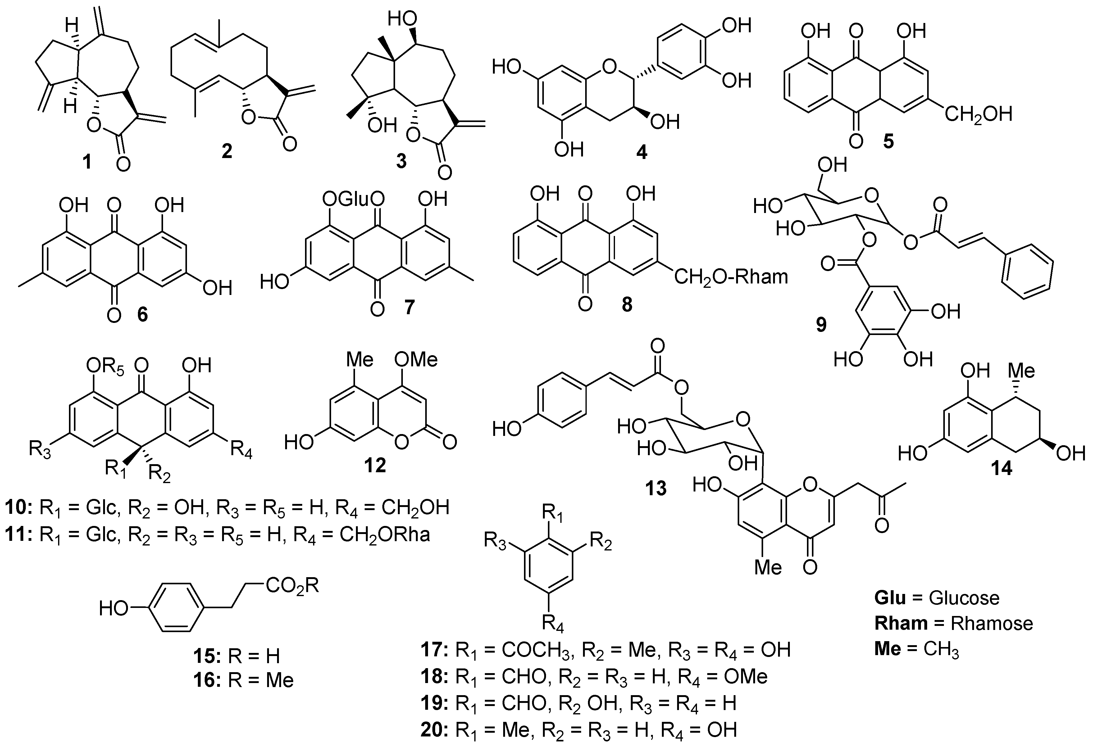
Figure 1. Structures of the compounds 19 isolated from L. shawii and 1020 isolated from A. vera resin.
Pharmaceuticals 13 00094 g001
Figure 2. The cytotoxic effects of compounds 16 (isolated from L. shawii) on MDA-MB-231 breast cancer cells. All results are expressed as mean ± SEM. * p < 0.05 and ** p < 0.01, compared to respective untreated controls.
Pharmaceuticals 13 00094 g002
Figure 3. The cytotoxic effects of compounds 79 (isolated from L. shawii) and 1012 (isolated from A. vera) on MDA-MB-231 breast cancer cells. All results are expressed as mean ± SEM. * p < 0.05 and ** p < 0.01, compared to respective untreated controls.
Pharmaceuticals 13 00094 g003
Figure 4. The depiction of pharmacophore models (M1 and M2). The hydrophobic (Hyd) features are shown in green spheres, hydrogen bond acceptor (AccP) features are displayed in cyan spheres. The compounds (shown in stick model) are aligned on their respective pharmacophore models.
Pharmaceuticals 13 00094 g004
Figure 5. Graphical presentation of predicted cellular targets of Compounds 13, 5, 7, and 12 based on docking scores.
Pharmaceuticals 13 00094 g005
Figure 6. The 3D structural topology of human carbonic anhydrase II is displayed in ribbon form complex with known inhibitor. The active site residues are shown in purple stick model, ligand is depicted in coral sticks, hydrogen bonds are displayed as black lines.
Pharmaceuticals 13 00094 g006
Figure 7. The binding interactions of compounds 13, 5, 7, and 12 at the active site of human carbonic anhydrase II. Side chain donors/acceptors are depicted in green arrows, backbone donors/acceptors are shown in blue arrows, solvent contact is presented in yellow dotted lines.
Pharmaceuticals 13 00094 g007
Table 1. The calculated IC50 values of compounds 112 in MDA-MB-231 breast cancer cell lines.
CompoundsIC50 (Mean ± SEM) (μM)
135.36 ± 2.56
242.08 ± 2.98
349.39 ± 3.73
4101.4 ± 7.09
531.36 ± 2.44
673.47 ± 5.89
757.32 ± 4.14
872.21 ± 6.29
976.9 ± 7.04
10142.8 ± 12.66
11140.4 ± 13.1
1260.09 ± 4.82
Doxorubicin (+ve control)3.31 ± 0.19
Highly active compounds are highlighted in bold.
Table 2. Antioxidant activity of different fractions of L. shawii and A. vera resin.
Antioxidant % Inhibition (IC50 ± SEM)
CodeL. shawiiCodeA. vera
BF72 (650 ± 1.50)MFNA
MF50EF51
WFNADF42
HFNABF32
EF76 (378 ± 1.50)WF35
DF60 (735 ± 2.00)HFNA
Ascorbic acid 90 (53 ± 1.32)
IC50 = μg/mL; concentration = 1 mg/mL; NA = not active; BF = n-butanol, MF = methanol, WF = aqueous, HF = hexane, EF = ethyl acetate, DF = dichloromethane.
Table 3. Antioxidant activity of the active compounds.
Numbering% Inhibition (1 mM)IC50 ± SEM (μM)
47855 ± 2.0
671645 ± 1.5
1373762 ± 2.0
1480241 ± 1.5
SEM = Standard Error Mean.
Table 4. The anti-CA-II activities of compounds 1, 2, 5, 7, and 12.
CompoundsDocking Score% InhibitionIC50 (µM) ± (SEM)
1−10.7533NA
2−9.6484.724.4
3−10.39NTNT
5−21.9886.314.4 ± 1.14
7−3.2637.5NA
12−13.8891.223.3 ± 1.63
SEM = Standard Error Mean; NA = Not Active; NT = Not Tested.
Table 5. Known drugs against the selected drug targets.
TargetDrugs
EGFRAfatinib, Canertinib dihydrochloride, Dacomitinib, Erlotinib, Gefitinib, Icotinib, Lapatinib, Lifirafenib, Masoprocol, Mavelertinib, Naquotinib, Nazartinib, Neratinib, Olmutinib, Osimertinib, Pelitinib, Rociletinib, Vandetanib, Varlitinib
c-KITAmuvatinib, Ancestim, Avapritinib, Cabozantinib, Dasatinib, Dovitinib lactate, Imatinib, Masitinib, Midostaurin, Motesanib, Nilotinib, Pazopanib, Regorafenib, Ripretinib, Semaxanib, Sorafenib, Sunitinib, Tandutinib, Toceranib, Vatalanib
IGFR1Ibutamoren mesylate, Linsitinib, Mecasermin, Mecasermin rinfabate, Toremifene
Notch 1Crenigacestat
PI3KApitolisib, Bimiralisib, Buparlisib, Dactolisib, Gedatolisib, Leniolisib, Omipalisib, Pictilisib, Samotolisib
CDK2Omacetaxine mepesuccinate
PARPOlaparib, Niraparib, Rucaparib, Talazoparib, Veliparib
CA-IIAcetazolamide
Table 6. The selected PDB structures for each anticancer drug target.
S #TargetPDB IDLigand IDResolution (Å)References
1EGFR2G5J0WN (Afatinib)2.8[61]
2c- Kit3G0EB49 (Sunitinib)1.6[62]
3IGFR13F5P741 (3-Cyanoquinoline)2.9[63]
4Notch 13L95Antibody FAB fragment2.19[64]
5PTEN5BZXVO4 (bisperoxovanadium complex)2.5[65]
6PI3K5ITD6CY (5-{4-[3-(4-acetylpiperazine-1-carbonyl)phenyl]quinazolin-6-yl}-2-methoxypyridine-3-carbonitrile)3.02[66]
7CDK42W9ZCyclin D2.45[67]
8PARP14R6E3JD (Niraparib)2.2[68]
9CA-II4IWZ1GO (acetazolamide derivative)1.598[69]