Article

Efficacy of Bacteriophages Against Staphylococcus aureus Isolates from Bovine Mastitis

1
Department of Bioprocess Engineering and Microbiology, University of Applied Sciences and Arts Hannover, D-30453 Hannover, Germany
2
Phage Technology Center GmbH, D-59199 Bönen, Germany
3
Section for Production, Nutrition and Health, Department of Veterinary and Animal Sciences, Faculty of Health and Medical Sciences, University of Copenhagen, DK-1870 Frederiksberg C, Denmark
*
Author to whom correspondence should be addressed.
Pharmaceuticals 2020, 13(3), 35; https://doi.org/10.3390/ph13030035
Submission received: 24 November 2019 / Revised: 5 February 2020 / Accepted: 24 February 2020 / Published: 26 February 2020
(This article belongs to the Special Issue Phage Therapy and Phage-Mediated Biological Control)

Abstract

:
The lytic efficacy of bacteriophages against Staphylococcus aureus isolates from bovine milk was investigated in vitro, regarding possible applications in the therapy of udder inflammation caused by bacterial infections (mastitis). The host range of sequenced, lytic bacteriophages was determined against a collection of 92 Staphylococcus (S.) aureus isolates. The isolates originated from quarter foremilk samples of clinical and subclinical mastitis cases. A spot test and a subsequent plaque assay were used to determine the phage host range. According to their host range, propagation and storage properties, three phages, STA1.ST29, EB1.ST11, and EB1.ST27, were selected for preparing a bacteriophage mixture (1:1:1), which was examined for its lytic activity against S. aureus in pasteurized and raw milk. It was found that almost two thirds of the isolates could be lysed by at least one of the tested phages. The bacteriophage mixture was able to reduce the S. aureus germ density in pasteurized milk and its reduction ability was maintained in raw milk, with only a moderate decrease compared to the results in pasteurized milk. The significant reduction ability of the phage mixture in raw milk promotes further in vivo investigation.

1. Introduction

Dealing with udder health disorders, including clinical and subclinical inflammation of the bovine mammary gland (mastitis), is one of the most important issues in the dairy industry. Mastitis leads to economic losses due to discarded milk, reduced milk production and a decrease in milk quality. Furthermore, it is a reason for culling chronically infected dairy cows, increasing costs due to additional labor, medication, and veterinary services and it impairs animal welfare [1,2,3].
Staphylococcus (S.) aureus is the most frequently isolated member of the contagious mastitis pathogens (major pathogen) [4,5,6]. There is a high risk of transmission of S. aureus between animals, especially during the milking process, and the pathogen has the ability to persist as a subclinical infection of the bovine udder. Important virulence factors of S. aureus-isolates enable their adhesion to epithelial cells, encapsulation, the formation of micro abscesses and the formation of biofilms, impeding treatment by antimicrobial agents and promoting a chronic progression [7,8]. Thus, an adequate treatment of S. aureus mastitis requires hygiene and special management routines, with three measures being identified for S. aureus intramammary infections (IMIs): a) separating the positively tested animals, b) culling the chronically infected animals and c) undertaking adequate hygiene and treatment measures [5,9]. Conventional treatment of S. aureus IMIs in the lactation and in the dry period requires the use of antimicrobial agents [10]. While dry cow therapy leads to cure rates of up to 78% for S. aureus mastitis [11,12], the bacteriological cure (BC) rates for treatment during lactation are considerably lower for S. aureus compared to other pathogens [4,13,14]. The reported cure rates for a lactation treatment of S. aureus mastitis vary between studies and depend on animal, pathogen and treatment factors as well as study design [5]. However, the cure rates for an antimicrobial treatment of S. aureus mastitis during lactation are generally considered unsatisfactory [13,14,15]. They decrease with high somatic cell counts (SCC), higher parity of cows, ß-lactamase-positive S. aureus infections and require an extended antimicrobial therapy to improve BC [4]. Linder et al. [14] investigated the BC rate of chronic subclinical S. aureus infections under treatment with marbofloxacin and cephalexin, and reported a remarkably low BC rate of 21.9% for treatment during lactation. This underlines a low accessibility for an antimicrobial treatment of S. aureus IMIs in the milking period and directs the attention to the development of resistance against the antimicrobial agents.
Although the resistance situation of antimicrobial agents for mastitis therapy in Germany is still moderate, there are studies describing the occurrence of resistant S. aureus isolates in dairy herds in addition to frequently occurring resistance to ß-lactam antibiotics [10,16,17]. The examination of Kreausukon et al. [17] reports the isolation of “Livestock-associated Methicillin resistant S. aureus” (LA-MRSA) isolates from bulk tank milk of German dairy herds with resistance especially against tetracycline, clindamycin and erythromycin. Spohr et al. [16] found a remarkable spread of MRSA of the spa-type t011 in three dairy herds in Southwest Germany. The fact that each application of antimicrobial agents promotes the emergence of resistance in bacterial isolates and an increasing awareness of resistance development, requires an overall reduction in the use of antimicrobial agents in human and veterinary medicine [2,18,19]. This emphasizes the need for alternative solutions for the treatment of bacterial infections, including conventional mastitis treatment. At this point, the therapeutic use of bacteriophages could be such an alternative for treating bacterial infections in animals as already successfully demonstrated in several animal models [20,21,22].
Bacteriophages (phages) are viruses that solely use prokaryotic cells for propagation. These highly host-specific agents occur ubiquitously in nature and can be isolated from the environment with comparatively little time and effort. After binding to specific structures on the host cell surface, the phage-genome is injected into the bacterial cell. Following replication of phage DNA or RNA and assembly of the viruses, phage-encoded enzymes lead to the lysis of the bacterial cell (lytically infecting phages) [23]. This results in the release of next generation phages and to an exponential growth of the phage population [24]. Thereby, the proliferation of phages occurs only as long as an adequately high bacterial concentration is present at the target site. This characteristic offers a possible advantage of so-called “self-limitation” for a therapeutic approach [22].
Bacteriophages can be grouped into four categories according to their infection strategies, based on the following criteria: a) does virion release occur (e.g., productive infection versus lysogeny of the cell when the phage genome is incorporated into the host cell genome (so-called prophage)), b) how does phage release occur (lytic or chronic), c) do the phage exhibit a certain degree of lysogenic cycles due to their genetics (distinction between temperate and non-temperate phage). For a detailed summary of these terms, see the 2016 article by Hobbs and Abedon [23]. A classification of phages according to their infection strategies, as advised by the authors, indicates I) lytic and non-temperate phages, II) chronic and non-temperate phages, III) lytic and temperate phages, and IV) chronic and temperate phages [23]. For therapeutic purposes, “professionally lytic” phages (category I), i.e., those that are obligately lytic, do not belong to the temperate phages and are not recent lytic mutants of the temperate phages, should be used [23,25].
Genes that code for toxins are important regarding the use of phages in therapy. These are potentially transferred by temperate bacteriophages (lysogenic conversion) [26]. The screening of phage genomes for the absence of lysogenic potential and toxin-coding genes are further steps that can be carried out before phages are used as therapeutic agents [25,27].
A number of studies already investigated the bactericidal activity of bacteriophages against S. aureus in milk, some of them with regard to food safety and the use of phages as biocontrol agents and others with regard to the use of phages as an alternative therapeutic agent to combat bacterial infections of the bovine udder. Milk represents a complex medium composed of different components like lipids, milk proteins (caseins, whey proteins) and an own bacterial flora [28]. Concerning this, the investigation of phages as an alternative treatment method for S. aureus mastitis in cattle requires an investigation of the bactericidal activity of phages in bovine milk. It was shown in former studies that a previous heat treatment (UHT > pasteurized) and the skimming of milk have a positive effect on the bactericidal activity of the phages. The authors assumed an inhibition of the ability of the phages to bind to their host cells in raw milk and mainly suggested lipids and whey proteins to be responsible for this [29,30]. The previous studies showed a decrease in phage titer or phage bactericidal activity in raw milk and referred this to an inhibition of the binding ability of the phage to the host cells in untreated raw milk, which the authors associated with a decrease of phage propagation in raw milk [29,31]. For untreated raw milk, however, previous investigations could show no reduction or merely a reduction in growth compared to an increasing germ density of the controls without phages, but no reduction in germ density compared to the initial values. We wanted to address this concern in the present study by examining the bactericidal activity of a mixture of newly isolated obligately lytic phages against S. aureus in milk.
In the present study, the efficacy of obligately lytic phages, specific for S. aureus, was investigated against S. aureus isolates from mastitis milk samples (host range). Further a phage mixture of three phages was examined in pasteurized and raw milk for its bactericidal activity against S. aureus, with regard to a prospective use in the treatment of bovine mastitis.

2. Results

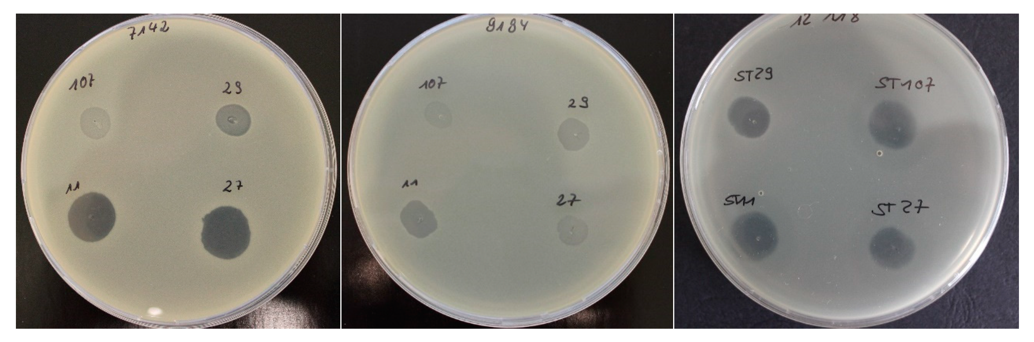
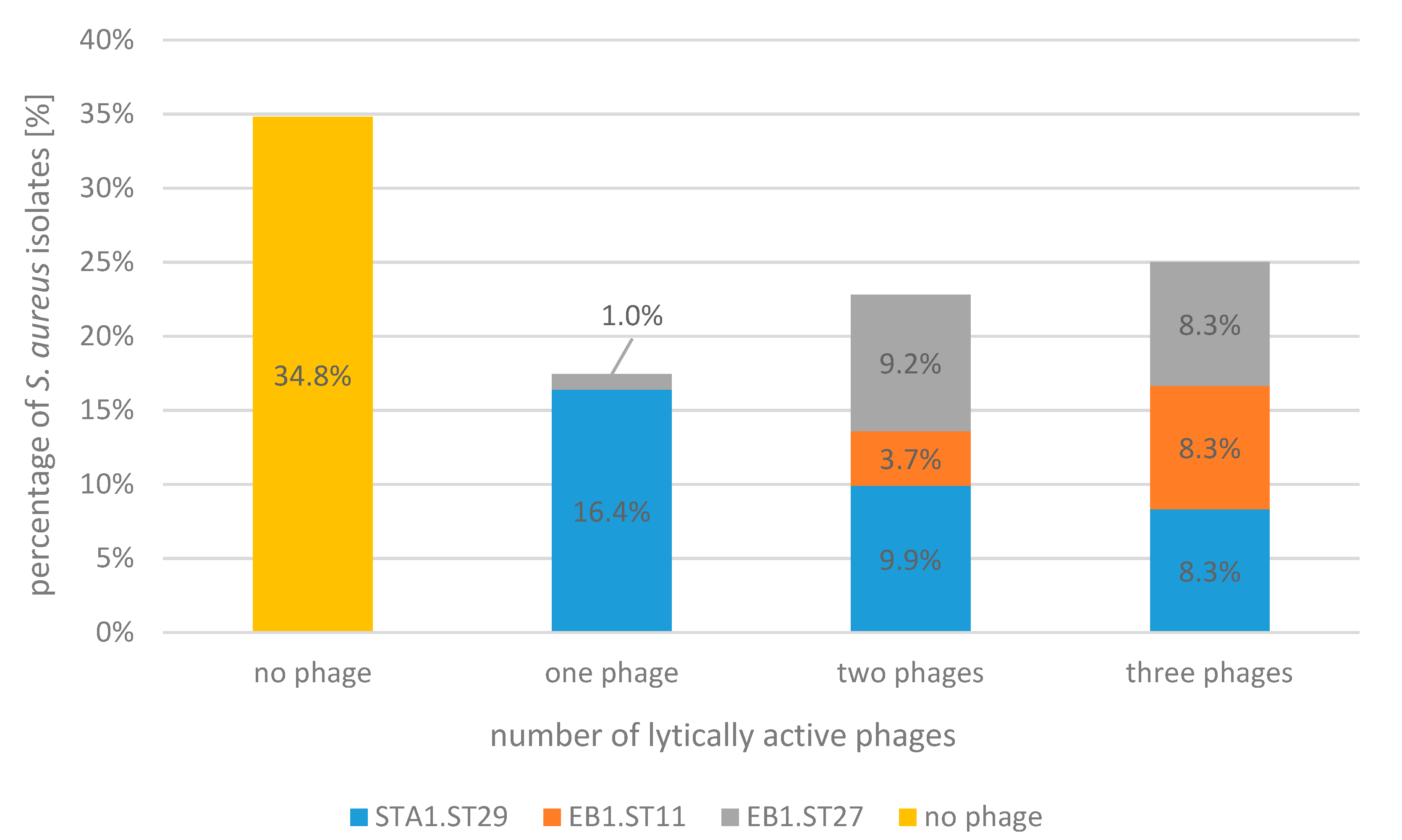
2.1. Bacteriophage Host Range: Spot Test and Plaque Assay

A spot test and a subsequent plaque assay were performed to determine the lytic efficacy (host range) of three phages (STA1.ST29, EB1.ST11, and EB1.ST27) against 92 S. aureus isolates from quarter foremilk samples of subclinical and clinical mastitis cases (Table 1). The subsequent plaque assay was performed for all phage-S. aureus combinations with a positive or unclear spot test to exclude false positive results. Figure 1 exemplary shows the interpretation of the spot test results.
The relative “efficiency of plating” (EOP) was calculated by dividing the phage concentration measured on the test strain by the phage concentration on the respective propagation strain. The results showed both, several isolates with a low relative EOP value, indicating a comparatively low efficiency compared to the propagation strain, and some isolates with a high EOP value, indicating a comparable efficiency of the phage for these isolates (Table 1).
The total of almost two-thirds (65.2%) of the tested isolates was lysed by at least one of the three phages (STA1.ST29, EB1.ST11, and EB1.ST27), while 17.4% of the S. aureus isolates were lysed by one phage, 22.8% by two phages and 25% were actually lysed by three phages. The percentage distribution of the individual phages in these categories is shown in Figure 2. A quantity of 34.8% of S. aureus isolates could not be lysed by the phages used in the current study.
The single phages, modified by the propagation on different S. aureus strains (restriction/modification) [32], showed differences in relation to their individual host range. Phage STA1.ST29 showed a comparatively broad host-range, lysing 63% of the S. aureus isolates, followed by phage EB1.ST27 with 44.6% and phage EB1.ST11 with 32.6%. An additional phage, STA1.ST107, was only examined in the spot test. STA1.ST107 showed a host spectrum very similar to EB1.ST11 and EB1.ST27 in the spot test but was excluded from further investigations due to a lack of stability and reproducibility, as the concentration (pfu/mL) of phage STA1.ST107 decreased more than 1-log unit within three months under storage at +6 °C and its propagation resulted in lower concentrations compared to the other phages.
The above results demonstrated a heterogeneous but broad host range of the individual phages and were used to compose a bacteriophage mixture for further examinations in milk containing the three phages STA1.ST29, EB1.ST11, and EB1.ST27 in equal quantities and with a final concentration of 6.0 × 10⁹ pfu/mL.
Spot test: 5 µL of each phage solution (1.0 × 10⁹ pfu/mL) were spotted onto the top layer (inoculated with the respective S. aureus isolate). Positive result: clear zone greater than or equal to the central area; unclear result: Turbid zone; negative result: no clear or turbid zone, but bacterial lawn.
Spot test results for S. aureus isolate 7142 (left) and the four tested phages: STA1.ST107: unclear; STA1.ST29: positive; EB1.ST11: positive; EB1.ST27: positive. Spot test results for S. aureus isolate 9194 (middle) and the four tested phages: all unclear. Spot test results for S. aureus isolate 12,118 (right) and the four tested phages: all positive. Positive and unclear results were further tested by plaque assay.
The percentage (%) of S. aureus isolates (n = 92) lysed by none of the phages, one of the three phages (STA1.ST29; EB1.ST11; EB1.ST27), two of the three phages, all three phages, based on the results of the spot test and the plaque assay. The differently colored parts of the bars represent the percentage (%) for the individual phages in each category, respectively.

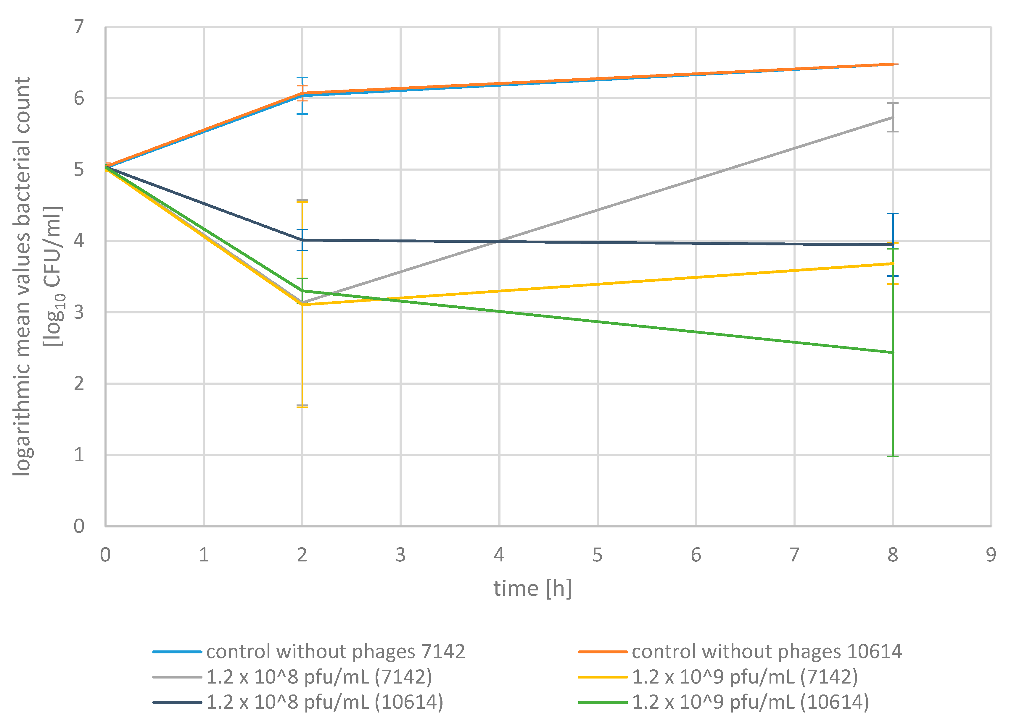
2.2. Phage Bactericidal Activity in Pasteurized Milk

We first investigated the ability of the phage mixture to reduce S. aureus in pasteurized milk to gain a stepwise approximation to the in vivo conditions (Figure 3). The pasteurized milk was inoculated with one of the two S. aureus isolates (7142; 10614) at 1.1 × 10⁵ cfu/mL, respectively. These two isolates from milk samples were selected according to their lysis by all three phages of the phage mixture. The mean values are presented for each strain. The final phage concentrations added to the S. aureus inoculated pasteurized milk were 1.2 × 10⁸ pfu/mL (for the addition of 0.1 mL phage mixture to 5.9 mL inoculated milk) or 1.2 × 10⁹ pfu/mL (for the addition of 1 mL phage mixture to 5 mL inoculated milk), respectively.
Adding the phage mixture at a volume of 0.1 mL and a final concentration of 1.2 × 10⁸ pfu/mL led to a reduction in the S. aureus germ density in pasteurized milk compared to the initial values of 98.9% for S. aureus 7142 and 92.1% for S. aureus 10,614 after two hours (p < 0.05). After eight hours, the reduction was 93.2% for S. aureus 10614, but no further reduction could be observed for S. aureus strain 7142 as the bacterial density slightly increased in the six-hour interval between the two measurements. However, the germ density remained below the value of the corresponding control cultures without phages after eight hours, with a difference of 84.7% (7142) and 99.8% (10614) (p < 0.05).
Adding the bacteriophage mixture at a volume of 1 mL and a final concentration of 1.2 × 10⁹ pfu/mL resulted in a reduction in bacterial density of 98.8% (7142) and 98.2% (10614) after two hours and of 95.5% (7142) and 99.8% (10614) after 8 h of incubation (p < 0.05). Adding the bacteriophages did not lead to an eradication of S. aureus at an initial S. aureus germ density of averagely 1.1 × 10⁵ cfu/mL. Nevertheless, the S. aureus germ density was 99.8% (7142) and 100% (10614) (>3 log units) lower compared to the control cultures without phages after eight hours using 1 mL phage mixture at a final concentration of 1.2 × 10⁹ pfu/mL (p < 0.05).
The addition of the phage mixture at a volume of 1 mL and a concentration of 1.2 × 10⁹ pfu/mL led to a better reduction of the S. aureus germ density for an incubation period of 8 h compared to the lower concentration and volume both, with respect to the initial value as well as to the respective value of the control culture.

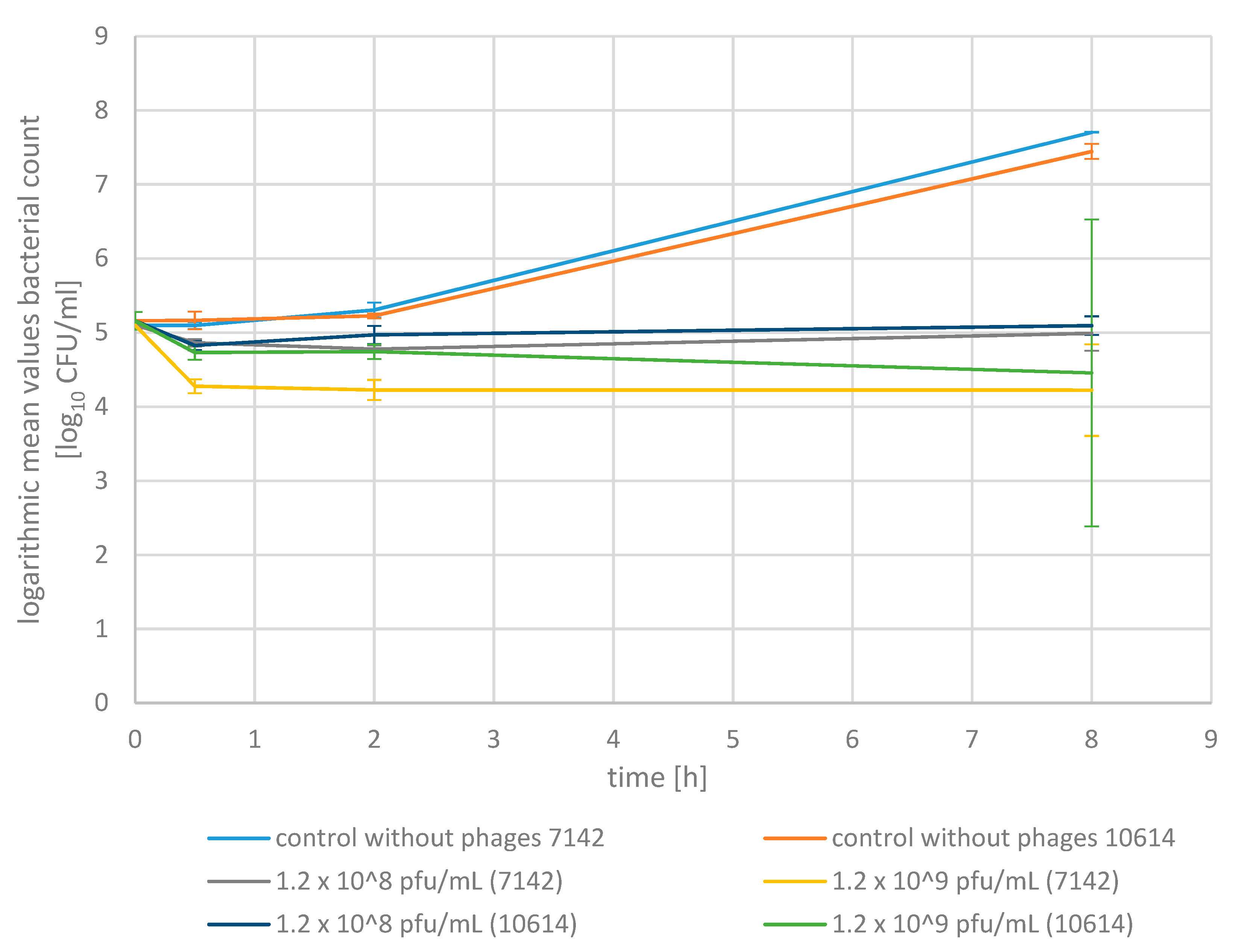
2.3. Phage Bactericidal Activity in Raw Milk
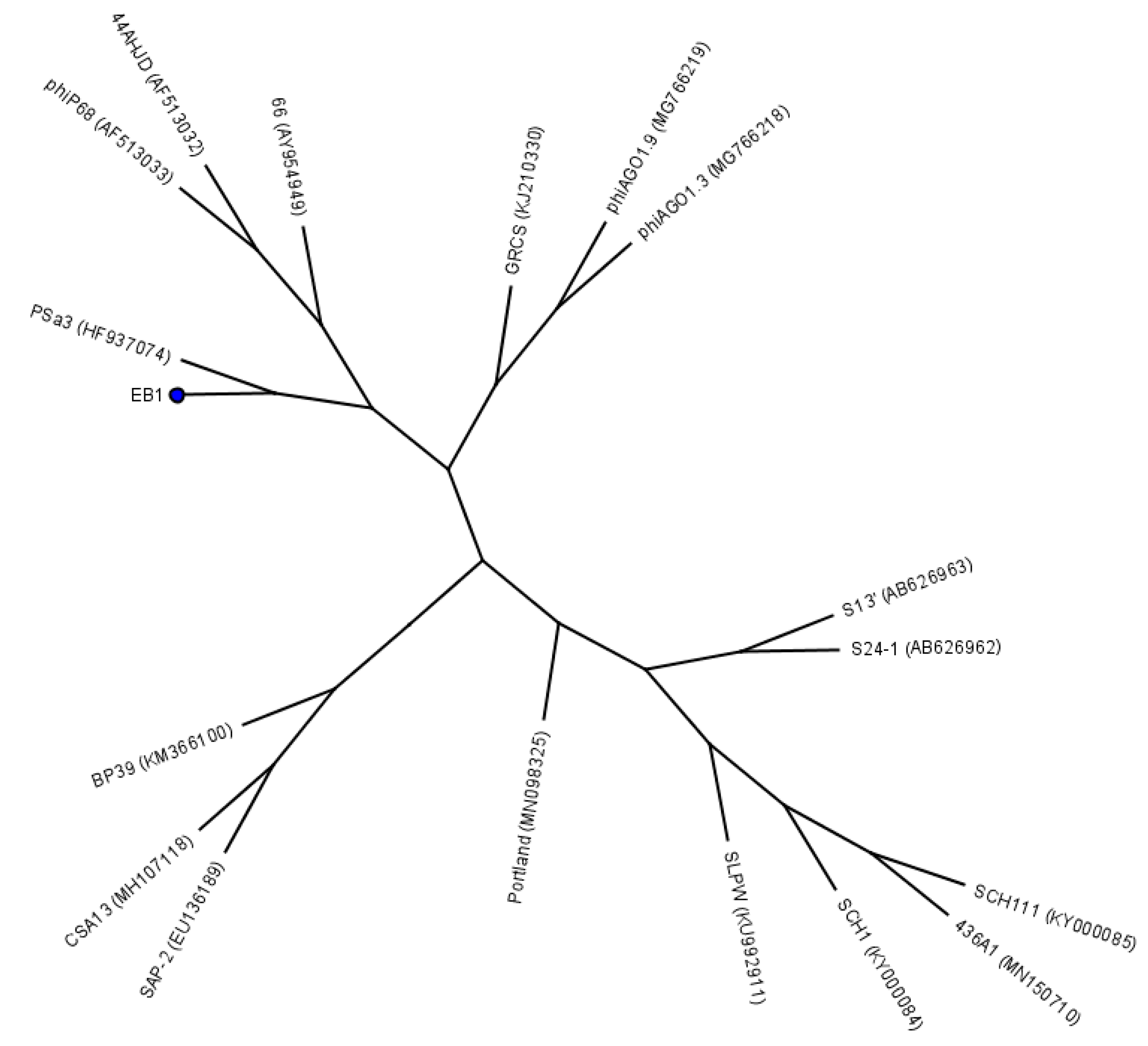
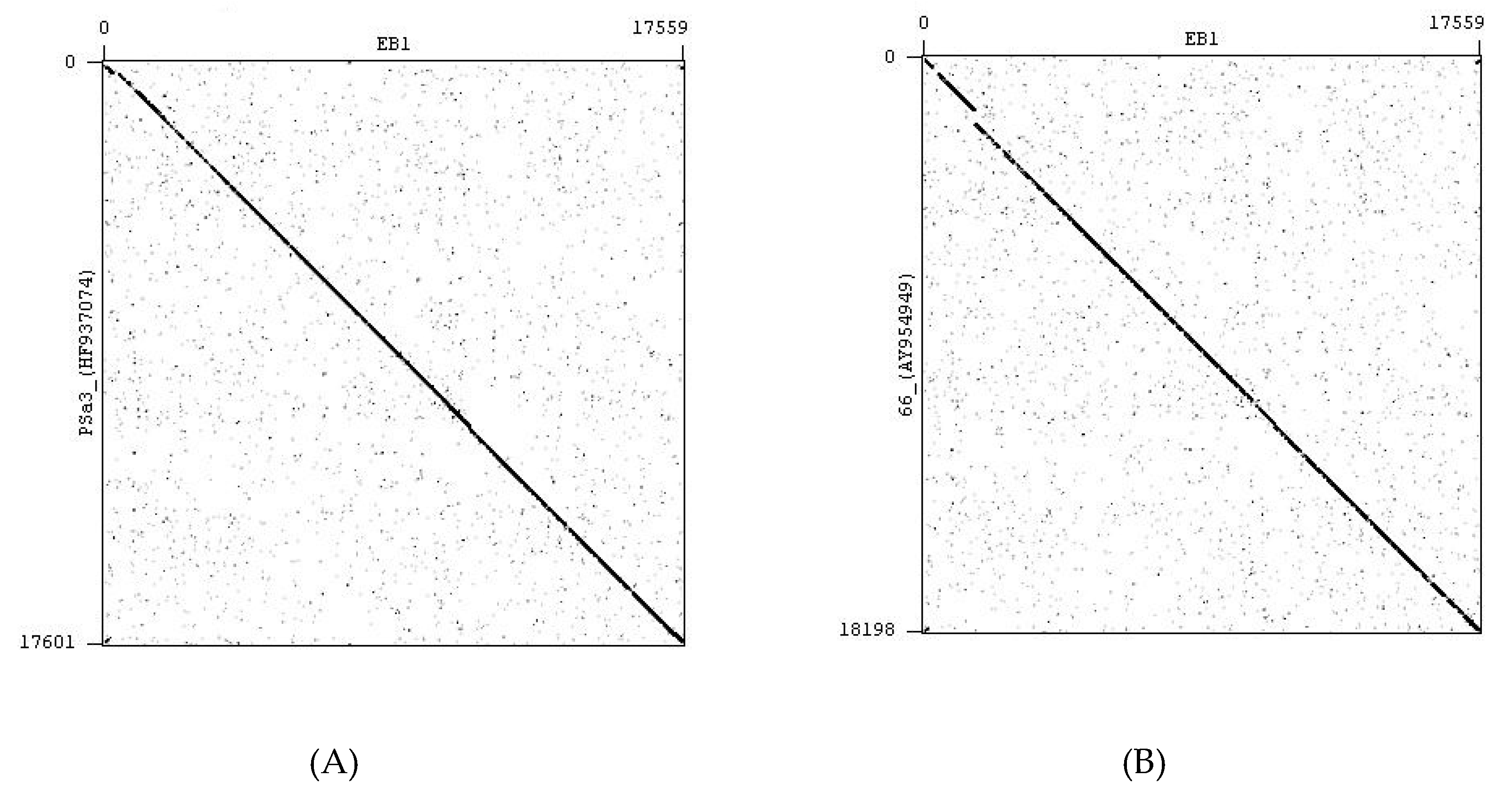
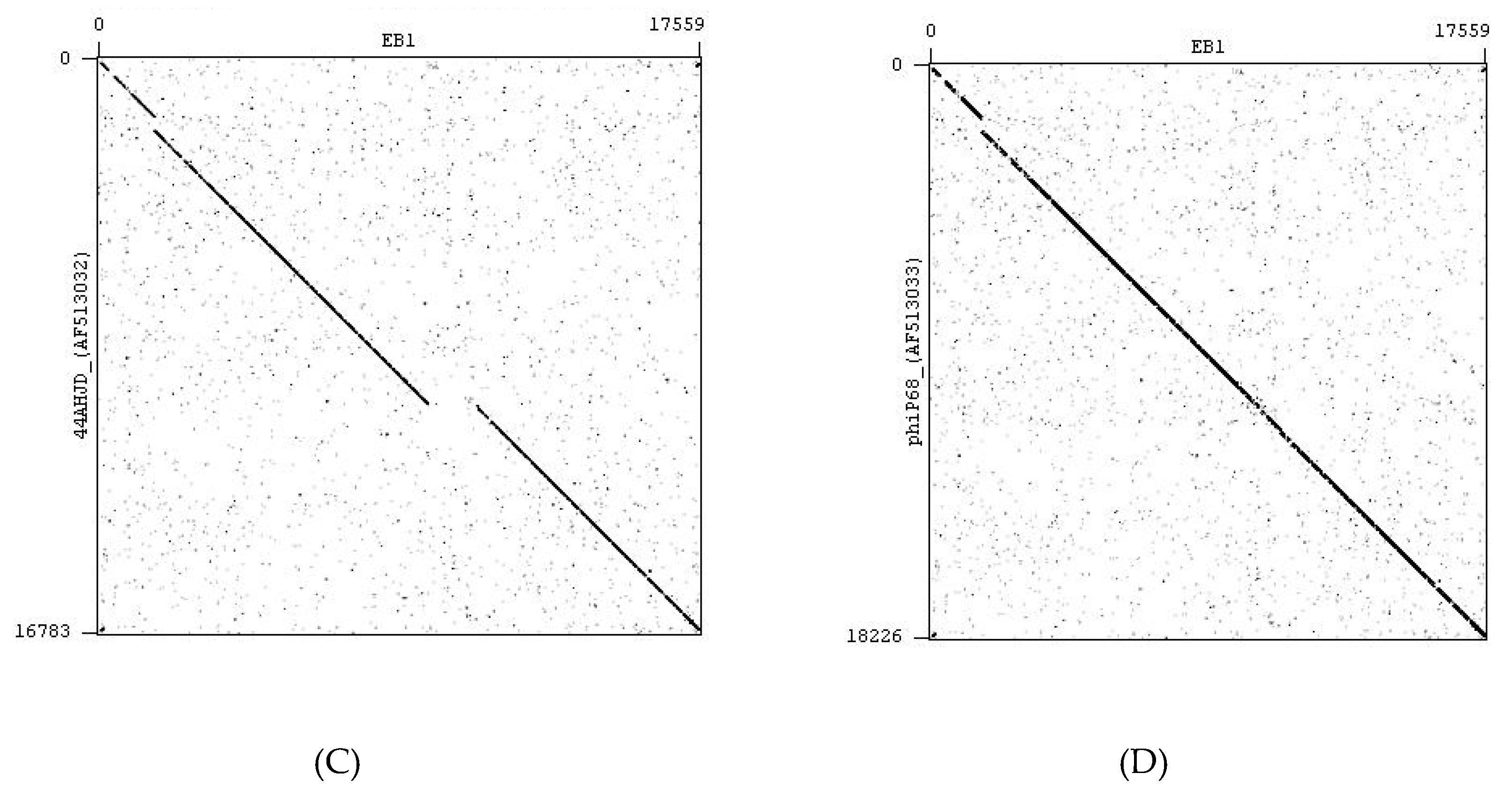
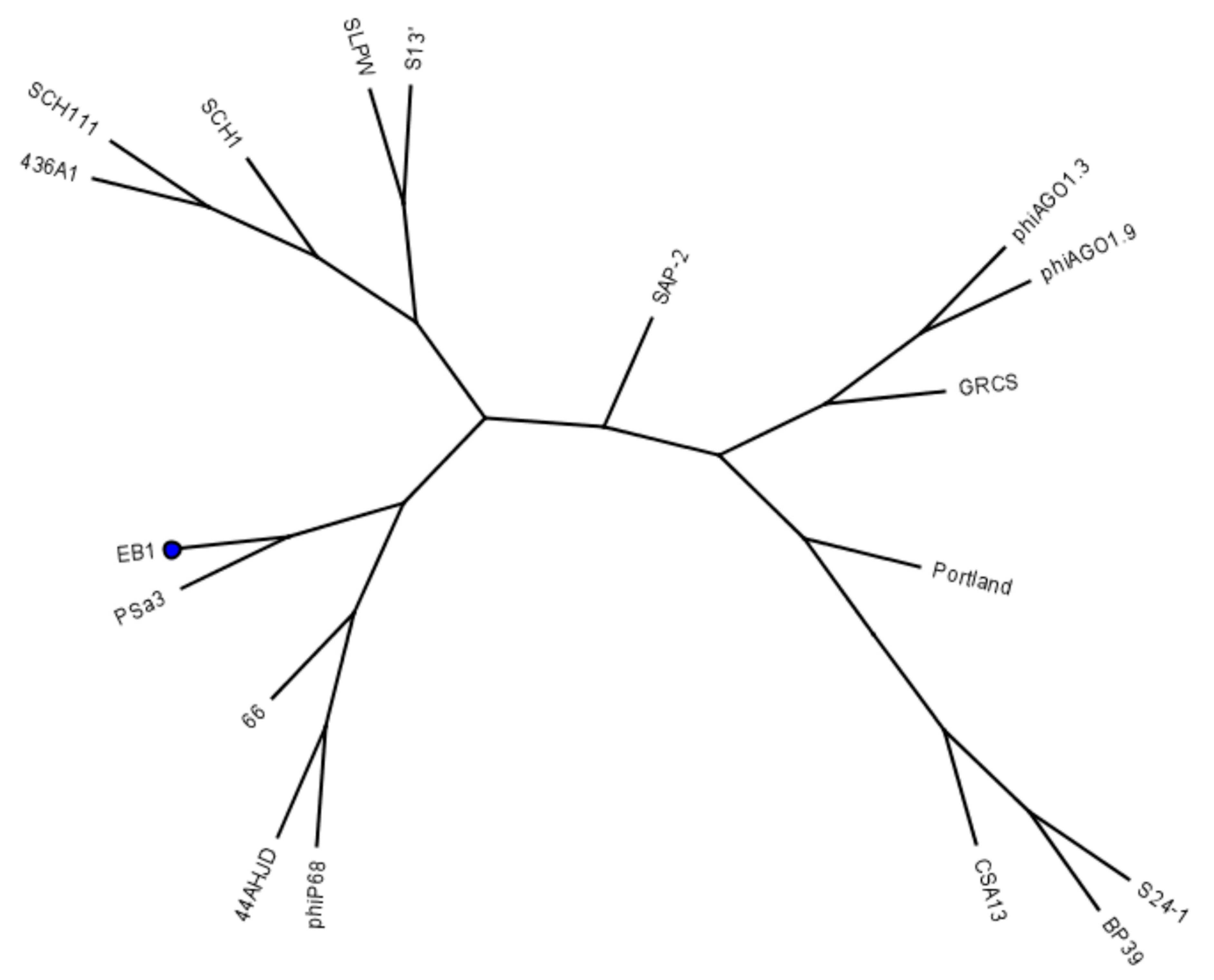
We further tested the efficacy of the bacteriophage mixture to reduce the S. aureus isolates in raw milk, as it represents the medium present in the udder (Figure 4). The methods used are the same as for the examination in pasteurized milk. Raw milk was inoculated at an initial value of 1.4 × 10⁵ cfu/mL with one of the two S. aureus isolates (7142 and 10614), respectively.
Adding the bacteriophage mixture at a volume of 1 mL and a final concentration of 1.2 × 10⁹ pfu/mL led to an average reduction in S. aureus germ density of 84.9% (7142) and 63.1% (10614) within 30 min, 86.6% (7142) and 62.0% (10614) after two hours and 86.6% (7142) and 80.5% (10614) after eight hours of incubation compared to the initial values (p < 0.05). When adding a lower volume of 0.1 mL at a ten-fold lower concentration of 1.2 × 10⁸ pfu/mL, a consistently lower reduction in the S. aureus germ density of 41.6% (7142) and 54.1% (10614) after 30 min, 52.0% (7142) and 36.3% after two hours and 21.8% (7142) and 14.8% (10614) after eight hours could be observed. Thus, the higher volume (1 mL) and final phage concentration (1.2 × 10⁹ pfu/mL) led to a higher reduction in S. aureus germ density in raw milk with a difference of averagely 62.2% after 8 h of incubation compared to the use of the lower volume (0.1 mL) and concentration (1.2 × 10⁸ pfu/mL) (p < 0.05).
As described before in pasteurized milk, applying the phage mixture did not lead to an eradication of S. aureus from raw milk in the case of the inoculation with an initial concentration of averagely 1.4 × 10⁵ cfu/mL. Nevertheless, the S. aureus germ density of samples treated with a volume of 1 mL and a concentration of 1.2 × 10⁹ pfu/mL was 100% (7142) and 99.9% (10614) (>3 log units) lower (p < 0.05) and at a lower volume (0.1 mL phage mixture) and a final concentration of 1.2 × 10⁸ pfu/mL, 99.8% (7142) and 99.6% (10614) lower (p < 0.05) compared to the control cultures without phages after eight hours of incubation at 37 °C.
There was also an improvement in reduction ability in raw milk by using a higher volume of phage mixture (1 mL) at a higher final concentration (1.2 × 10⁹ pfu/mL). The phage mixture led to a reduction in germ density with regard to the initial value and the respective values of the control without phages.

3. Discussion

Inadequate cure rates for an antimicrobial treatment during lactation as well as resistance to antimicrobial agents in S. aureus isolates have been described for S. aureus induced mastitis cases [14,16,17]. In order to overcome these issues, the aim of this study was to examine lytic bacteriophages in vitro for their ability to lyse S. aureus isolates from mastitis cases, with regard to the future use of phages in mastitis therapy.
In a first step, the host range of the bacteriophages (STA1.ST29; STA1.ST107; EB1.ST11; and EB1.ST27) was determined for a collection of 92 S. aureus isolates. The isolates were obtained from quarter foremilk samples of subclinical and clinical mastitis cases and were provided by the Department of Bioprocess Engineering and Microbiology, University of Applied Sciences and Arts Hannover (Germany). Since the isolates originated from 31 different dairy farms in Northern and Eastern Germany, it was assumed that they represented a cross-section of the population of S. aureus strains in this area. A spot test was performed in a first step for determining the host range of the phages on the collection of 92 S. aureus isolates. Since the clear zone in a spot test is only created by the inhibition of bacterial replication, a plaque assay was used to verify the spot test results by excluding false positive results due to so called “abortive infection”. This term describes the ability of a phage to kill the respective bacterial strain without a productive phage infection [33]. This extended investigation of lytic phage activity on a large S. aureus collection (n = 92) from mastitis milk samples differentiates the present study design from previous studies using smaller strain collections or merely performing a spot test for determining the host range and it is to our knowledge the first investigating phages against S. aureus isolates from German dairy herds.
The determination of the host range in the present study showed that almost two-thirds (65.2%) of the 92 S. aureus isolates could be lysed by at least one of the tested phages, while 47.8% were actually lysed by two or more of the phages under investigation. These results led to the preparation of a bacteriophage mixture to increase the host range and to minimize the potential risk of developing resistance to individual phages [24,27]. It has already been described that a bacteriophage mixture is significantly more efficient in lysing S. aureus isolates than a single phage [31].
The relative EOP is described in the literature as the concentration of the individual phage for the tested bacterial strain compared to the maximum phage concentration measured by plaque assay under different conditions [34]. In the case of our study, the maximum concentration to be achieved refers to the highest phage concentration measured for its propagation strain (ST11; ST27 or ST29). The relative “efficiency of plating” varies according to the different test isolates examined in the present study. There are some isolates with a comparatively high EOP value, which indicates an efficiency in phage replication comparable to that on the propagation strain. Several isolates have a low EOP value, which suggests that phage in situ replication is not as efficient or at least not as fast for a particular targeted strain other than the propagation strain.
The differences in the EOP of a phage in relation to the different test isolates are explained in the literature by the fact that the EOP depends on various factors such as the agar concentration in the top agar or the S. aureus germ density [34]. However, these factors were constant in our investigations and therefore had no influence on the different EOP values. In addition, the influence of specific host factors (masking by O-antigens; presence of restriction endonucleases) on the EOP is described and accordingly a difference of up to a factor of ten between the individual test isolates is not uncommon [34]. Further, a comparatively low ability to induce infection, possibly due to reduced specific structures on the host cell surface to which the phages bind, may increase the probability of delayed infection initiation [35].
A possible reason for the non-sensitivity of isolates (34.8%) might be the fact that the S. aureus phages in the current study belong to the families of Myo- (STA1) and Podoviridae (EB1). Thus, they represent only two of the three common S. aureus-bacteriophage families. This leads to the assumption that the isolation and additional application of a lytic phage from the third family (Siphoviridae) might result in an increased host range of a phage mixture.
All currently known staphylococcal phages require wall teichoic acid (WTA) for adsorption and infection [36,37]. WTA is an anionic glycopolymer that is linked to the peptidoglycan layer of most gram-positive bacteria [38]. The backbone of the WTA of most S. aureus strains contains 40–60 phosphodiester-linked polyribitol phosphate units, further decorated with three substitutions, D-alanine, α-GlcNAc or β-GlcNAc [39]. While Myoviridae, like phage K, bind both to the WTA backbone and to α-GlcNAc-modified WTA and thus have the broadest host range [36,39], Siphoviridae, like phi77, depend on GlcNAc-modified WTA and Podoviridae, like PS66, specifically depend on β-GlcNAc-modified WTA [37]. An optimal phage cocktail to fight S. aureus would thus include bacteriophages of all three families, with the idea that point mutations that eliminate the receptor of one bacteriophage might not automatically also lead to the exclusion of the other bacteriophages in the cocktail. However, a tag0 mutant as described in [36], entirely devoid of WTA, would be resistant against all bacteriophages of such an optimal cocktail. However, this might be of minor importance during practical applications. First, mutants devoid of WTA show a significant growth defect compared to wildtype strains and second, such mutants would be sensitive to ß-lactam antibiotics, as the MRSA resistance phenotype in S. aureus also depends on the presence of glycosylated WTA [40]. Thus, a combination of phage therapy and antibiotic treatment might lead to synergistic effects and would reduce the selection of both phage resistant and antibiotic resistant variants of S. aureus.
A direct isolation of lytic S. aureus-specific Siphoviridae from milk or sewage water generally proves difficult. Nonetheless, Garcia et al. [41] obtained two lytic phages (Siphoviridae) from their milk isolated temperate counterparts by DNA random deletion. O’Flaherty et al. [42] actually isolated two lytic Siphoviridae from farmyard slurry. Both achieved good results against S. aureus isolates [41,42].
Furthermore, we investigated the bactericidal activity of a phage mixture in pasteurized and raw milk for an approximation to the conditions prevailing in vivo. Milk represents a complex medium of polydisperse character composed of lipids, milk proteins (caseins, whey proteins), enzymes, lactose, minerals, trace elements, and vitamins [28]. Therefore, it was necessary to examine the lytic activity of the phages in bovine milk with regard to a future intramammary application in mastitis therapy. The two S. aureus isolates used for inoculating pasteurized and raw milk were selected based on their sensitivity to all three phages within the mixture, as determined in the first set of experiments. They represented only a small part of the total S. aureus spectrum tested, but were assumed to provide important information on the antibacterial potential of the bacteriophage mixture in milk. The bacteriophage mixture contained the phages STA1.ST29, EB1.ST11, and EB1.ST27 in equal quantities and with a concentration of 6.0 × 10⁹ pfu/mL. We investigated the influence of the phage mixture added in different volumes (0.1 mL; 1 mL) leading to different final concentrations (1.2 × 10⁸ pfu/mL; 1.2 × 10⁹ pfu/mL) and the influence of the incubation time on the reduction of the S. aureus germ density (cfu/mL) in milk. The trials were conducted with a view to an in vivo use and an application interval of eight hours (intermediate milking time), considered most practicable for treatment during lactation.
While the control without phages showed a steady increase of the bacterial count, adding the phage mixture at 1 mL and 1.2 × 10⁹ pfu/mL led to a significant reduction of the S. aureus germ density, both with respect to the control without phages as well as to the initial value, which has to be emphasized.
In pasteurized milk, an adequate reduction in the S. aureus germ density for an eight-hour incubation period (37 °C) was only achieved when a higher volume and concentration of bacteriophages (1 mL; 1.2 × 10⁹ pfu/mL) was used, with a reduction of averagely 97.7% after eight hours, compared to the initial germ density (p < 0.05). Compared to the values of the control culture at t8h, a reduction of up to 100% (>3 log units) was achieved after eight hours of incubation (p < 0.05).
Garcia et al. [31] achieved a similar reduction of 3.6 log units compared to the values of the control culture in pasteurized milk by using a phage mixture of two temperate phages after ten hours of incubation, but no significant reduction compared to the initial values. In the current study we used a comparably small number of three obligate lytic phages for the phage mixture, which exclusively enter the lytic propagation cycle, commonly lead to a faster lysis and should be preferred for a therapeutic use [25,27].
In raw milk, adding a higher volume and concentration of phage mixture (1 mL; 1.2 × 10⁹ pfu/mL), led to a higher reduction with a difference of 62.2% after 8 h compared to the use of a lower volume at a ten-fold lower concentration (0.1 mL; 1.2 × 10⁸ pfu/mL) (p < 0.05). The incubation period of 8 h led to a steady increase in the reduction of the S. aureus germ density for the use of a higher volume and concentration (1 mL; 1.2 × 10⁹ pfu/mL) with a difference of 10% compared to the reduction after 30 min (p < 0.05). In contrast, a decrease in the reduction ability over time for the lower volume and concentration (0.1 mL; 1.2 × 10⁸ pfu/mL) with a difference of 28.6% compared to the reduction of bacterial density after 30 min could be observed (p < 0.05). This shows that only the higher phage concentration was sufficient to adequately reduce the S. aureus germ density for an incubation period of 8 h in raw milk.
Adding a phage mixture in the present study with a volume of 1 mL at a concentration of 1.2 × 10⁹ pfu/mL to S. aureus-inoculated raw milk resulted in a reduction of averagely 83.6% after eight hours compared to the initial value, and to a reduction of averagely 100% after 8 hours compared to the control cultures at the same time. When considering these results, it has to be noted that the ability of the phage mixture to reduce the S. aureus germ density is maintained in raw milk with only a moderate decrease compared to pasteurized milk, with an initial S. aureus-inoculation of 1 × 10⁵ cfu/mL.
In contrast, other studies on the use of bacteriophages against S. aureus showed considerably lower or no reduction rates in raw milk and assumed that the ingredients interfere with the ability of the phages to bind to their host cells. In two different studies on the bactericidal activity of a single application of phage K, an inhibition of phage binding to its host cells in raw milk and whey was described [29,30]. Phage K is an exclusively lytic S. aureus specific phage with a broad host range belonging to the Myoviridae family [43], and is closely related to the STA1 bacteriophage studied here. Immunoglobulin activity, the clumping of S. aureus cells associated with fat globules, or whey proteins are suspected to be possible causes limiting phage K adsorption to the host cell surface in raw milk [29,30].
The design of the previous publications shows some divergences in comparison to the current study. In particular, only a solitary phage K was used in contrast to the application of a bacteriophage mixture as used in the present study. The combined use of several lytic phages offers advantages regarding the host range and therefore increases the bactericidal activity [31].
Regarding the application of a combined use of phages in the medium milk, a mixture of two temperate phages (104 to 105 pfu/mL) against S. aureus (102 cfu/mL) has been examined in a previous study [31]. These investigations were carried out in UHT milk, pasteurized milk and in semi-skimmed and unskimmed raw milk. The authors described significantly lower S. aureus numbers for the use of this mixture of temperate phages in raw milk after an incubation period of 11 h compared to the control without phages. Nevertheless, they found a considerable decline in the ability of the phages to reduce S. aureus in raw milk compared to the results obtained in pasteurized milk.
In contrast, the results of the present study show not only a relative reduction compared to the control without phages, but also an absolute reduction compared to the initial bacterial density; with only a slight decrease in bactericidal activity of phages (14.1% for absolute reduction) in raw milk compared to the results in pasteurized milk.
The use of higher initial concentrations of phages and S. aureus represent differences from the previous study. At the same time, the use of newly isolated obligate lytic phages could be a possible reason for the differences from the results of the previous study and thus represents an important factor with regard to the bactericidal activity of the bacteriophages used. Since obligate lytic phages generally lead to a faster lysis of the bacterium and have a lower lysogenic and transduction potential, this application offers a decisive advantage for an application in phage therapy. In further in vivo studies, a phage concentration as used in this study should also be applied.
In the present study, the continuous propagation of initially two genetically different phages (STA1 and EB1) on four different S. aureus strains (two isolated from mastitis samples) has resulted in four phages with different host ranges. This leads to the hypothesis that using S. aureus isolates from mastitis cases (ST 11 and ST 27) for the propagation of the phages (EB1.ST11 and EB1.ST27) in this study might be another reason for the bactericidal activity of the bacteriophage mixture in raw milk.
Utilizing a bacteriophage mixture in pasteurized milk and in raw milk in the present study, with an initial germ density of ~1.0 × 10⁵ cfu/mL, did not lead to a complete eradication of S. aureus. This observation is comparable to the results of another publication [31]. Garcia et al. [31] suggested that a total reduction in S. aureus in milk could not be achieved due to a low phage-cell ratio at the end of their investigation. In the same way, the stationary phase in the growth of S. aureus could also be responsible for the incomplete reduction, as the authors assume in their paper. Accordingly, low S. aureus cell densities and a stationary phase of the bacterial culture at the end of the incubation period could also have been the limiting factor in the present study. At the same time, this phage property also includes the advantage of so-called “self-limitation”, which is considered beneficial for a therapeutic approach by other authors [22].
However, the results of the present study already demonstrated that using the phage mixture at a higher volume of 1 mL and a higher final concentration of 1.2 × 10⁹ pfu/mL led to a higher reduction in S. aureus germ density in raw milk in vitro (p < 0.05). It is conceivable that the higher used volume led to a better distribution in vitro and consequently to an improved ability of the phages to target and bind to their host cells [44]. Further investigations on this issue should be conducted in vivo to determine a volume that results in an adequate dispersion in the udder. In order to adapt the application frequencies in vivo to an intermediate milking time of 8 h, our findings suggest that for field trials a calculation based on a concentration of 1.2 × 10⁹ pfu/mL would be necessary to achieve an appropriate bactericidal activity at a bacterial density of 1 × 10⁵ cfu/mL.
The shedding of S. aureus from infected mammary gland quarters varies depending on whether a subclinical or clinical infection is present and may increase after a stress event and shedding of 105 cfu/mL is not uncommon. The examined shedding of S. aureus in quarter-foremilk samples from subclinically infected mammary gland quarters after a stress event had a median value of 88 cfu/mL [45]. Only as an example, calculated for a subclinical infected udder quarter with an average S. aureus germ density of 100 cfu/mL, a minimum phage concentration of 1 × 10⁶ pfu/mL would be required. Assuming an increase in the milk volume between milking times to a total of 5 L/quarter of udder, an intra-mammary application every 8 h would require the injection of 5.0 × 10⁹ pfu/mL. If a volume of 10 mL phage solution is administered, it should have the concentration of 5.0 × 10⁸ pfu/mL.
Further investigations in vivo are necessary, as the conditions cannot be adequately reproduced in vitro due to the complex structure and functional characteristics of the bovine udder (immune response, dispersion, pharmacokinetic). They should first include a tolerability assessment for administering a phage mixture in the teat sinus of clinical healthy cows. Further investigations should be conducted to determine an adequate concentration and volume of phage solution for an intramammary administration. This should be followed by an examination of the bacteriological cure rates in comparison to an intramammary treatment with antimicrobial agents during lactation. Based on these in vivo examinations, an evaluation of the applicability and the economics of the phage mixture for mastitis therapy has to be performed.
Other studies, which investigated the use of phages in animal experiments already showed promising results [22,46,47]. Capparelli et al. [23] showed the activity of S. aureus bacteriophages against local and systemic S. aureus infections as well as intracellular and methicillin resistant strains of S. aureus (MRSA) in mice. Phage-treated mice neither showed a stimulated production of neutralizing antibodies nor did they suffer any side effects. These in vivo examinations point out that phage therapy in animals is able to generate good cure rates without adversely affecting animal health. Further studies examined the use of the single phage ΦSA012 or a mixture of two phages (Myo- and Podoviridae) isolated from sewage water against S. aureus isolated from bovine mastitis milk in a mouse model [46,47]. The authors reported reduced S. aureus proliferation and a decrease of the inflammatory response in the mouse mammals, which represents an important result with regard to the therapy of bovine intramammary infections (IMIs) using phages. However, the authors argue that a direct comparison between mouse mammals and bovine udders is not possible since their anatomy and physiology differ. The authors therefore indicate further investigations in bovine mammary glands [46,47]. In addition, there is the fact that the quantities of milk ingredients differ between animal species [48]. The investigation of the bactericidal activity of phages in raw milk from cattle, as described in the present study, is therefore an essential basis concerning the future use in the therapy of bovine mastitis, as it represents the medium in the bovine udder.
The number of in vivo studies on the use of phages for S. aureus mastitis treatment in cattle is quite low [42,49]. O’Flaherty et al. [42] administered three S. aureus phages intramammarily for tolerability assessment and found no increased somatic cell count (SCC) and thus no increased immune response to a relatively high phage concentration of 108 pfu/mL. Furthermore, they proposed the use of phages in special formulations as teat-dips or teat-washes, as they had already successfully examined phages in the form of hand-wash solutions against S. aureus in hospitals in a previous study [50]. This underlines the additional possibility of using phages prophylactically in the case of bacterial colonization of the bovine udder and udder skin. Gill et al. [49] investigated the single use of bacteriophage K for treating subclinical S. aureus mastitis in the lactation period. A cure rate of three of 18 quarters (16.7%) was achieved, whereas in the control group, zero of 20 quarters were cured. However, these results were not statistically significant. Intramammary infusion of the phage into quarters already infected with S. aureus had in contrast to the application in healthy quarters no adverse effects, as no increase in the somatic cell count was observed.
The phage mixture of the present study led to a considerable reduction of the S. aureus germ density in raw milk, which might lead to improved results in vivo compared to the previous examinations. In their study, Gill et al. [49] administered an amount of 10 mL intramammary infusion of phage K (1.25 × 10¹¹ pfu/mL) once per day for five days. In relation to the findings of our study, it would be worth considering administering a combination of several obligately lytic phages in form of a phage mixture to increase the host range and to fulfill safety requirements in phage therapy [25,27,31]. At the same time, the exemplary calculation based on our results shows that even a lower concentration (5 × 10⁸ pfu/mL) at a comparably high volume (10 mL) might be sufficient for the in vivo application of a phage mixture. A practicable eight-hour application interval (intermediate milking period) could be used in further in vivo trials.
The results of the present examinations indicate that bacteriophage therapy could be a promising measure in mastitis treatment. Indeed, it might be a future alternative in order to improve the cure rates of S. aureus mastitis during lactation, to reduce the number of antimicrobial agents and to counteract the development of antimicrobial resistance in S. aureus. Further investigations need to focus to a greater extent on studying obligately lytic phage mixtures in vivo for S. aureus mastitis therapy. The results of the present study offer an incentive to do so, as the use of a lytic bacteriophage mixture in raw milk led to significant and promising results against S. aureus isolates of mastitis cases.

4. Materials and Methods

4.1. S. aureus Strains and Culture Methods

The S. aureus isolates (n = 92) for the host range determination of phages were provided by the Department of Bioprocess Engineering and Microbiology, University of Applied Sciences and Arts Hannover (Germany). The isolates were obtained from quarter foremilk samples of clinical and subclinical bovine mastitis cases collected on 31 different farms in North and East Germany in order to obtain an adequate reproduction of the strain variety in the field. All S. aureus isolates were screened nuc gene positive. The isolates were stored at −80 °C, after a 24 h-culture of the respective strain in brain heart broth, with the addition of 20% glycerine, and, before use, incubated on esculin blood agar (Oxoid Deutschland GmbH, Wesel, Germany) for 24 h at 37 °C. Directly before every trial, an overnight culture of the respective strain was incubated in Luria-Bertani (LB) broth (trypton/pepton from casein: 1 g/100 mL; yeast extract, micro-granulated: 0.5 g/100 mL; sodium chloride (NaCl): 0.5 g/100 mL; Carl Roth GmbH & Co. KG, Karlsruhe, Germany) at 37 °C. LB broth was used for bacterial growth in suspension, phage spot test and double-layer agar (DLA) technique. Broth was supplemented with agar (Agar Agar; Carl Roth GmbH & Co. KG, Karlsruhe, Germany) at concentrations of 1.5% for bottom and 0.5% for top agar. Sterile dextrose was added to the top agar at 1.1% before use.
The S. aureus isolates ST11, ST27, ST29, and ST107, isolated and provided by PTC GmbH, Bönen, Germany, were used for routine phage propagation (Table 2). Phage propagation strains ST11 and ST27 originated from mastitis milk samples.

4.2. Bacteriophages

Bacteriophages EB1.ST11, EB1.ST27, STA1.ST29, and STA1.ST107, isolated and provided by the PTC GmbH, Bönen, Germany, are obligately lytic phages belonging to the order of Caudovirales and represent the families of Myoviridae (STA1) and Podoviridae (EB1), as their genome sequence analyses showed.
Phage EB1 was isolated from pig manure (Table 2) and is closely related to several members of the genus Rosenblumvirus, namely, PSa3 (93.5% genome identity), which was isolated and characterized by Kraushaar et al. [51], phiP68 (89.3% genome identity) and 44AHJD (82.5% genome identity) from the collection of the Felix d’Herelle Reference Center for Bacterial Viruses, Quebec, Canada [52], as well as phage 66 from the National Collection of Type Cultures (London) [53]. The genomic similarity between EB1 and other known bacteriophages of Rosenblumvirus is visually demonstrated by a tree, which was constructed with the help of the Geneious (version 4.8) tree builder using the UPGMA tree build method and the HKY Genetic Distance Model (Figure 5). The local genomic similarities between EB1 and a group of phiP68-like phages are illustrated as dotplots (Figure 6), which were created by Gepard version 1.40 [54].
The phages phiP68, 44AHDJ and 66 are known to adsorb to S. aureus wall teichoic acid (WTA) [38] modified with β-GlcNAc [37]. The phiP68-encoded ORF17 was found to be a virion-associated muralytic enzyme [55] and a receptor binding protein [56]. EB1 encodes a protein, showing 96.5% identity to the phiP68-encoded ORF17. The 648 amino acids long protein has only 23 amino acid substitutions from its phiP68 counterpart and the majority of them represent changes for structurally similar amino acids. Homologs of ORF17, encoded by other phiP68-like bacteriophages, demonstrate a close evolutionary relationship, forming a separate group within the phylogenetic tree (Figure 7).
Phage STA1 was isolated in a wastewater facility and is closely related to phage K (89.1% genome identity) [43] and other members of the genus Kayvirus.
The two genetically distinct phages were grown on different host strains, which leads to epigenetic modifications that may result in a different host range [32]. All bacteriophages used in this study are available upon request from the PTC GmbH, Bönen, Germany.

4.3. Bacteriophage Propagation

Phages were routinely propagated by inoculating LB-broth with 1% of an overnight culture of the respective S. aureus propagation strain (either ST11; ST27; ST29; or ST107). The initial value of the optical density at 600 nm (OD600) was determined by a photometer (SPEKOL®1500, Analytik Jena AG, Jena, Germany). Data were collected every 30 min until an OD600 of 0.3 was reached. To the exponentially growing S. aureus cells, 1% of a phage stock solution with a concentration >1.5 × 10¹⁰ pfu/mL (EB1.ST11), >1.3 × 10⁹ pfu/mL (EB1.ST27), >1.6 × 10⁹ pfu/mL (STA1.ST29), >2.0 × 10⁹ pfu/mL (STA1.ST107) was added. The OD600 was measured further at half-hourly intervals. Chloroform was added at 1 ‰ (one per mil) when a decrease in the OD600 was observed and the solution was then centrifuged at 10,000× g for 20 min at room temperature. The supernatant was removed and sterile filtered (0.45 µm-pore-size filter, Minisart®NML Plus/NY Plus; Sartorius AG, Göttingen, Germany) before phage plaque formation (pfu/mL) was determined by using a double-layer-agar-technique, as described below. The sterile filtered phage solution was stored at +6 °C in the dark until further use and the phage concentration (pfu/mL) was routinely determined.

4.4. Spot Test

A spot test was used to investigate the lytic activity of the phages EB1.ST11, EB1.ST27, STA1.ST29, and STA1.ST107 against a collection of 92 S. aureus isolates from mastitis cases to determine their host range. Briefly, 100 µL of a bacterial overnight culture were added to 5 mL of top agar (0.5% agar), supplemented with sterile dextrose before use. The solution was mixed by a Vortex Mixer (Vortex Genie®2; Scientific Industries Inc., Bohemia, NY, USA) and poured onto a petri dish (Ø 94 × 16 mm) prepared with 10 mL of LB-bottom agar. The petri dish was gently swirled and dried at room temperature before 5 µL of a phage solution (1.0 × 10⁹ pfu/mL) were spotted onto the top layer. The plates were allowed to dry at room temperature before being incubated inverted at 37 °C for 18 h. The spot test was considered positive (+) if a clear zone appeared at the site of application, and considered negative (−) if no clear zone appeared. If only a turbid spot could be seen, it was defined as an unclear result (+/−) (Table 1). In cases of positive (+) and unclear (+/−) spot test results, a plaque assay was performed as a second step to verify lytic phage activity for the respective S. aureus isolates.

4.5. Plaque Assay

Spot test results were verified by a plaque assay, using the double-layer-agar-technique, modified to the one described by Sambrook and Russel (Table 2) [57]. This method was used to exclude false positive spot test results [33,34]. Briefly, 100 µL of a serial dilution of phage solution (Ringer’s solution; Merck KGaA, Darmstadt, Germany) were added to 100 µL of a bacterial overnight culture of the respective S. aureus strain. After incubation for 5 min at room temperature, 5 mL of top agar (0.5% agar) were added, mixed by a Vortex Mixer and poured onto a petri dish (Ø 94 × 16 mm) prepared with 10 mL of LB-bottom agar. The petri dish was gently swirled and dried at room temperature for 30 min. Inverted plates were incubated at 37 °C for 18 h. Plaque forming units per milliliter (pfu/mL) were determined and the relative “efficiency of plating” (EOP) was calculated by dividing the phage concentration (pfu/mL) for the test strain by the phage concentration for the respective propagation strain (ST29; ST11; or ST27) [34]. Thus, the spot test results were confirmed by the active formation of a plaque by the phage in a lawn of the target bacterial strain.
A bacteriophage mixture was prepared to investigate the potential of a combined use of different phages against S. aureus in milk. The phage mixture was prepared of the three phages EB1.ST11, EB1.ST27, and STA1.ST29. These phages were selected for their host range determined by spot test and plaque assay, for a high concentration of phages after propagation (>1.0 × 10⁹ pfu/mL) and for an adequate storage stability at +6 °C. The latter was defined in terms of a decrease in concentration (pfu/mL) less than one log unit over a period of three months.

4.6. Phage Bactericidal Activity in Pasteurized Milk

The bactericidal activity of the phage mixture against S. aureus isolates from mastitis cases was investigated in milk in order to achieve a gradual adaptation to the conditions in vivo. The two S. aureus isolates used for inoculation originated from different dairy farms and had shown good results in the previous assays with regard to their sensitivity towards all three phages in the mixture.
Pasteurized milk was obtained by heating raw milk (<20,000 SCC/mL and quarter) to 63 °C for 30 min. The raw milk had been tested previously for the absence of S. aureus and antibiotic residues by direct plating and a Brilliant Black Reduction Test (Delvotest®BR Brilliant, DSM; MILKU Tierhygiene GmbH, Bovenden, Germany). The pasteurized milk was inoculated with S. aureus isolates 7142 or 10,614 at concentrations of 1.1 × 10⁵ cfu/mL, respectively.
We added either 0.1 mL phage mixture to 5.9 mL inoculated milk or 1 mL phage mixture to 5 mL inoculated milk. The final phage concentrations were 1.2 × 10⁸ pfu/mL or 1.2 × 10⁹ pfu/mL for the different added volumes. Assays were mixed by a Vortex Mixer before incubating at 37 °C without shaking and samples were taken at the beginning of the trial (t0h), after two hours (t2h) and after eight hours (t8h). The two-hour incubation time was related to the average replication time of the phages determined in the propagation trial, whereas the eight-hour incubation time referred to an intermediate milking period at milking-frequency of three times per day. It was used to indicate whether administering a phage mixture only once per milking time (eight hours) would lead to an adequate reduction. For examining the S. aureus germ density (cfu/mL), diluted suspensions (Ringer’s solution; Merck KGaA, Darmstadt, Germany) were plated by the spatula method with 100 µL sample material [58]. Briefly, this was carried out by plating serial dilutions (Ringer’s solution, Merck KGaA, Darmstadt, Germany) and subsequent calculation of colony forming units per milliliter (cfu/mL) after incubation at 37 °C for 24 h. Five measurements were collected for each data point. Pasteurized milk inoculated with S. aureus (7142 or 10614) was used as control culture without phages, S. aureus-free pasteurized milk as negative control.

4.7. Phage Bactericidal Activity in Raw Milk

The investigation of the phage bactericidal activity in raw milk was carried out as described above in pasteurized milk and was used to obtain a further adaption to in vivo conditions. Raw milk was tested previously for the absence of S. aureus and antibiotic residues. Raw milk was inoculated with the S. aureus isolates 7142 or 10614 at concentrations of 1.4 × 10⁵ cfu/mL. Furthermore, we added either 0.1 mL phage mixture to 5.9 mL inoculated milk or 1 mL phage mixture to 5 mL inoculated milk, with a final phage concentration of 1.2 × 10⁸ pfu/mL or 1.2 × 10⁹ pfu/mL, respectively. The preparations were mixed by a Vortex Mixer before incubating at 37 °C without shaking and examined for their S. aureus germ density at the beginning (t0h), after 30 min (t30min), after two hours (t2h) and after eight hours (t8h) by the spatula method with 100 µL sample material [58]. Trials were carried out in quintuplicate, including control cultures without phages (S. aureus inoculated raw milk) and negative control (non-inoculated raw milk).

4.8. Statistical Analysis

To obtain statistically relevant data for the examinations in milk, five measurements were collected for each data point. A statistical calculation was performed to investigate the influence of the different concentrations, achieved by different added volumes, and the incubation time on the S. aureus germ density. Logarithmic transformations of cfu (log10) were applied to approximate normal distribution and were analyzed by ANOVA using SPSS 25.0 (IBM SPSS 25.0.0.0., Armonk, USA). P values <0.05 were considered significant.

5. Conclusions

We determined the host range of obligately lytic bacteriophages against S. aureus isolates from clinical and subclinical mastitis cases. The examinations identified bacteriophages with a broad host range. In a second step, we examined the bactericidal activity of a three-component bacteriophage mixture for the reduction of S. aureus in pasteurized and raw milk. The results showed a significant reduction of the S. aureus germ density in pasteurized milk and a slightly lower but still significant reduction in raw milk after eight hours of incubation at 37 °C. The reduction in bacterial density was shown both in comparison to the initial values and to the values of the controls without phages. The results are promising and support further investigations into the use of bacteriophages in the treatment of bovine S. aureus mastitis.

Author Contributions

Conceptualization, I.T., V.K.; Funding acquisition, V.K.; Investigation, I.T.; Methodology, I.T., H.L., V.K.; Visualization, I.T., T.L.; Supervision, V.K..; Project administration, V.K.; Writing—original draft preparation, I.T.; Writing—review and editing, I.T., H.L., T.L., V.K.; All authors have read and agreed to the published version of the manuscript.

Funding

This research was funded by the Steinbeis Research Center Milk Science (Kirchlengern), Germany, grant number 32018.

Acknowledgments

The PTC (Phage Technology Center) GmbH, Bönen, Germany, provided the bacteriophages used in this study. The authors would like to thank the laboratory personnel of the Microbiology Work Group of the University of Applied Sciences and Arts Hannover. Isabel Titze is the recipient of a Hanns Seidel Foundation Fellowship for talented doctoral students, founded by the German Federal Ministry of Education and Research (BMBF).

Conflicts of Interest

The authors declare no conflict of interest.

References

  1. Seegers, H.; Fourichon, C.; Beaudeau, F. Production effects related to mastitis and mastitis economics in dairy cattle herds. Vet. Res. 2003, 34, 475–491. [Google Scholar] [CrossRef] [PubMed] [Green Version]
  2. IDF. Economic Consequences of Mastitis; Bulletin No 394; International Dairy Federation: Brussels, Belgium, 2005. [Google Scholar]
  3. Halasa, T.; Huijps, K.; Osteras, O.; Hogeveen, H. Economic effects of bovine mastitis and mastitis management: A review. Vet. Q. 2007, 29, 18–31. [Google Scholar] [CrossRef]
  4. Sol, J.; Sampimon, O.C.; Barkema, H.W.; Schukken, Y.H. Factors Associated with Cure after Therapy of Clinical Mastitis Caused by Staphylococcus aureus. J. Dairy Sci. 2000, 83, 278–284. [Google Scholar] [CrossRef]
  5. Barkema, H.; Schukken, Y.; Zadoks, R. Invited review: The role of cow, pathogen, and treatment regimen in the therapeutic success of bovine Staphylococcus aureus mastitis. J. Dairy Sci. 2006, 89, 1877–1895. [Google Scholar] [CrossRef] [Green Version]
  6. Tenhagen, B.-A.; Köster, G.; Wallmann, J.; Heuwieser, W. Prevalence of mastitis pathogens and their resistance against antimicrobial agents in dairy cows in Brandenburg, Germany. J. Dairy Sci. 2006, 89, 2542–2551. [Google Scholar] [CrossRef] [Green Version]
  7. Grieger, A.-S.; Zoche-Golob, V.; Paduch, J.-H.; Hoedemaker, M.; Krömker, V. Rezidivierende klinische Mastitiden bei Milchkühen–Bedeutung und Ursachen. Tierärztliche Prax. Ausg. G Großtiere Nutztiere 2014, 42, 156–162. [Google Scholar] [CrossRef]
  8. Schönborn, S.; Krömker, V. Detection of the biofilm component polysaccharide intercellular adhesin in Staphylococcus aureus infected cow udders. Vet. Microbiol. 2016, 196, 126–128. [Google Scholar] [CrossRef]
  9. GVA. Guidelines for Combating Bovine Mastitis as a Stock Problem, 5th ed.; German Veterinary Association: Gießen, Germany, 2012. [Google Scholar]
  10. GERMAP. GERMAP 2015–Bericht Über den Antibiotikaverbrauch und die Verbreitung von Antibiotikaresistenzen in der Human-und Veterinärmedizin in Deutschland; Bundesamt für Verbraucherschutz: Berlin, Germany, 2016; ISBN 978-3-9818383-0-5.
  11. Dingwell, R.T.; Leslie, K.E.; Duffield, T.F.; Schukken, Y.H.; DesCoteaux, L.; Keefe, G.P.; Bagg, R. Efficacy of intramammary tilmicosin and risk factors for cure of Staphylococcus aureus infection in the dry period. J. Dairy Sci. 2003, 86, 159–168. [Google Scholar] [CrossRef]
  12. Nickerson, S.C.; Owens, W.E.; Fox, L.K.; Scheifinger, C.C.; Shryock, T.R.; Spike, T.E. Comparison of tilmicosin and cephapirin as therapeutics for Staphylococcus aureus mastitis at dry-off. J. Dairy Sci. 1999, 82, 696–703. [Google Scholar] [CrossRef]
  13. Steele, N.; McDougall, S. Effect of prolonged duration therapy of subclinical mastitis in lactating dairy cows using penethamate hydriodide. N. Z. Vet. J. 2014, 62, 38–46. [Google Scholar] [CrossRef]
  14. Linder, M.; Paduch, J.H.; Grieger, A.S.; Mansion-de Vries, E.; Knorr, N.; Zinke, C.; Krömker, V. Heilungsraten chronischer subklinischer Staphylococcus aureus-Mastitiden nach antibiotischer Therapie bei laktierenden Milchkühen. Cure rates of chronic subclinical Staphylococcus aureus mastitis in lactating dairy cows after antibiotic therapy. Berl. Münch. Tierärztl. Wschr. 2013, 6, 291–296. [Google Scholar] [CrossRef]
  15. Owens, W.; Nickerson, S.; Ray, C. Efficacy of parenterally or intramammarily administered tilmicosin or ceftiofur against Staphylococcus aureus mastitis during lactation. J. Dairy Sci. 1999, 82, 645–647. [Google Scholar] [CrossRef]
  16. Spohr, M.; Rau, J.; Friedrich, A.; Klittich, G.; Fetsch, A.; Guerra, B.; Hammerl, J.A.; Tenhagen, B.-A. Methicillin-Resistant Staphylococcus aureus (MRSA) in Three Dairy Herds in Southwest Germany. Zoonoses Public Health 2011, 58, 252–261. [Google Scholar] [CrossRef]
  17. Kreausukon, K.; Fetsch, A.; Kraushaar, B.; Alt, K.; Muller, K.; Kromker, V.; Zessin, K.H.; Kasbohrer, A.; Tenhagen, B.A. Prevalence, antimicrobial resistance, and molecular characterization of methicillin-resistant Staphylococcus aureus from bulk tank milk of dairy herds. J. Dairy Sci. 2012, 95, 4382–4388. [Google Scholar] [CrossRef] [PubMed] [Green Version]
  18. Wise, R.; Hart, T.; Cars, O.; Streulens, M.; Helmuth, R.; Huovinen, P.; Sprenger, M. Antimicrobial resistance. BMJ 1998, 317, 609–610. [Google Scholar] [CrossRef] [Green Version]
  19. WHO. Antimicrobial Resistance: Global Report on Surveillance; World Health Organization: Geneva, Switzerland, 2014. [Google Scholar]
  20. Matsuzaki, S.; Yasuda, M.; Nishikawa, H.; Kuroda, M.; Ujihara, T.; Shuin, T.; Shen, Y.; Jin, Z.; Fujimoto, S.; Nasimuzzaman, M.D.; et al. Experimental Protection of Mice against Lethal Staphylococcus aureus Infection by Novel Bacteriophage ϕMR11. J. Infect. Dis. 2003, 187, 613–624. [Google Scholar] [CrossRef] [Green Version]
  21. Wills, Q.F.; Kerrigan, C.; Soothill, J.S. Experimental bacteriophage protection against Staphylococcus aureus abscesses in a rabbit model. Antimicrob. Agents Chemother. 2005, 49, 1220–1221. [Google Scholar] [CrossRef] [PubMed] [Green Version]
  22. Capparelli, R.; Parlato, M.; Borriello, G.; Salvatore, P.; Iannelli, D. Experimental phage therapy against Staphylococcus aureus in mice. Antimicrob. Agents Chemother. 2007, 51, 2765–2773. [Google Scholar] [CrossRef] [Green Version]
  23. Hobbs, Z.; Abedon, S.T. Diversity of phage infection types and associated terminology: The problem with ‘Lytic or lysogenic’. FEMS Microbiol. Lett. 2016, 363. [Google Scholar] [CrossRef] [Green Version]
  24. Carlton, R.M. Phage therapy: Past history and future prospects. Arch. Immunol. Ther. Exp. Engl. Ed. 1999, 47, 267–274. [Google Scholar] [CrossRef] [Green Version]
  25. Hyman, P. Phages for Phage Therapy: Isolation, Characterization, and Host Range Breadth. Pharmaceuticals 2019, 12, 35. [Google Scholar] [CrossRef] [PubMed] [Green Version]
  26. Brüssow, H.; Canchaya, C.; Hardt, W.-D. Phages and the evolution of bacterial pathogens: From genomic rearrangements to lysogenic conversion. Microbiol. Mol. Biol. Rev. 2004, 68, 560–602. [Google Scholar] [CrossRef] [PubMed] [Green Version]
  27. Sulakvelidze, A.; Alavidze, Z.; Morris, J.G., Jr. Bacteriophage therapy. Antimicrob. Agents Chemother. 2001, 45, 649–659. [Google Scholar] [CrossRef] [PubMed] [Green Version]
  28. Krömker, V. Kurzes Lehrbuch Milchkunde und Milchhygiene, 1st ed.; Stuttgart: Parey, Germany, 2007; pp. 10–12. [Google Scholar]
  29. O’Flaherty, S.; Coffey, A.; Meaney, W.J.; Fitzgerald, G.F.; Ross, R.P. Inhibition of bacteriophage K proliferation on Staphylococcus aureus in raw bovine milk. Lett. Appl. Microbiol. 2005, 41, 274–279. [Google Scholar] [CrossRef] [PubMed]
  30. Gill, J.J.; Sabour, P.M.; Leslie, K.E.; Griffiths, M.W. Bovine whey proteins inhibit the interaction of Staphylococcus aureus and bacteriophage K. J. Appl. Microbiol. 2006, 101, 377–386. [Google Scholar] [CrossRef]
  31. Garcia, P.; Madera, C.; Martinez, B.; Rodriguez, A.; Evaristo Suarez, J. Prevalence of bacteriophages infecting Staphylococcus aureus in dairy samples and their potential as biocontrol agents. J. Dairy Sci. 2009, 92, 3019–3026. [Google Scholar] [CrossRef]
  32. Arber, W. Host-controlled modification of bacteriophage. Annu. Rev. Microbiol. 1965, 19, 365–378. [Google Scholar] [CrossRef]
  33. Abedon, S.T. Lysis from without. Bacteriophage 2011, 1, 46–49. [Google Scholar] [CrossRef]
  34. Kutter, E. Phage host range and efficiency of plating. Methods Mol. Biol. 2009, 501, 141–149. [Google Scholar] [CrossRef]
  35. Scholl, D.; Adhya, S.; Merril, C. Escherichia coli K1′s capsule is a barrier to bacteriophage T7. Appl. Environ. Microbiol. 2005, 71, 4872–4874. [Google Scholar] [CrossRef] [Green Version]
  36. Xia, G.; Corrigan, R.M.; Winstel, V.; Goerke, C.; Grundling, A.; Peschel, A. Wall teichoic Acid-dependent adsorption of staphylococcal siphovirus and myovirus. J. Bacteriol. 2011, 193, 4006–4009. [Google Scholar] [CrossRef] [PubMed] [Green Version]
  37. Li, X.; Gerlach, D.; Du, X.; Larsen, J.; Stegger, M.; Kühner, P.; Peschel, A.; Xia, G.; Winstel, V. An accessory wall teichoic acid glycosyltransferase protects Staphylococcus aureus from the lytic activity of Podoviridae. Sci. Rep. 2015, 5, 17219. [Google Scholar] [CrossRef] [PubMed]
  38. Brown, S.; Santa Maria, J.P., Jr.; Walker, S. Wall teichoic acids of gram-positive bacteria. Annu. Rev. Microbiol. 2013, 67, 313–336. [Google Scholar] [CrossRef] [Green Version]
  39. Takeuchi, I.; Osada, K.; Azam, A.H.; Asakawa, H.; Miyanaga, K.; Tanji, Y. The Presence of Two Receptor-Binding Proteins Contributes to the Wide Host Range of Staphylococcal Twort-Like Phages. Appl. Environ. Microbiol. 2016, 82, 5763–5774. [Google Scholar] [CrossRef] [PubMed] [Green Version]
  40. Brown, S.; Xia, G.; Luhachack, L.G.; Campbell, J.; Meredith, T.C.; Chen, C.; Winstel, V.; Gekeler, C.; Irazoqui, J.E.; Peschel, A. Methicillin resistance in Staphylococcus aureus requires glycosylated wall teichoic acids. Proc. Natl. Acad. Sci. USA 2012, 109, 18909–18914. [Google Scholar] [CrossRef] [PubMed] [Green Version]
  41. Garcia, P.; Martinez, B.; Obeso, J.M.; Lavigne, R.; Lurz, R.; Rodriguez, A. Functional genomic analysis of two Staphylococcus aureus phages isolated from the dairy environment. Appl. Environ. Microbiol. 2009, 75, 7663–7673. [Google Scholar] [CrossRef] [Green Version]
  42. O’Flaherty, S.; Ross, R.P.; Flynn, J.; Meaney, W.J.; Fitzgerald, G.F.; Coffey, A. Isolation and characterization of two anti-staphylococcal bacteriophages specific for pathogenic Staphylococcus aureus associated with bovine infections. Lett. Appl. Microbiol. 2005, 41, 482–486. [Google Scholar] [CrossRef]
  43. O’Flaherty, S.; Coffey, A.; Edwards, R.; Meaney, W.; Fitzgerald, G.F.; Ross, R.P. Genome of Staphylococcal Phage K: A New Lineage of Myoviridae Infecting Gram-Positive Bacteria with a Low G+C Content. J. Bacteriol. 2004, 186, 2862–2871. [Google Scholar] [CrossRef] [Green Version]
  44. Harris, L.G.; Foster, S.J.; Richards, R.G. An introduction to staphylococcus aureus, and techniques for identifying and quantifying S. aureus adhesins in relation to adhesion to biomaterials: Review. Eur. Cells Mater. 2002, 4, 39–60. [Google Scholar] [CrossRef]
  45. Krömker, V.F.J.; Klocke, D. Shedding patterns of Staphylococcus aureus in quarter foremilk samples of cows with known S. aureus infections. Tieraerztliche Prax. Ausg. Grosstiere Nutztiere 2008, 36, 389–392. [Google Scholar]
  46. Iwano, H.; Inoue, Y.; Takasago, T.; Kobayashi, H.; Furusawa, T.; Taniguchi, K.; Fujiki, J.; Yokota, H.; Usui, M.; Tanji, Y. Bacteriophage ΦSA012 has a broad host range against Staphylococcus aureus and effective lytic capacity in a mouse mastitis model. Biology 2018, 7, 8. [Google Scholar] [CrossRef] [PubMed] [Green Version]
  47. Geng, H.; Zou, W.; Zhang, M.; Xu, L.; Liu, F.; Li, X.; Wang, L.; Xu, Y. Evaluation of phage therapy in the treatment of Staphylococcus aureus-induced mastitis in mice. Folia Microbiol. 2019, 1–13. [Google Scholar] [CrossRef] [PubMed]
  48. Barłowska, J.; Szwajkowska, M.; Litwińczuk, Z.; Król, J. Nutritional value and technological suitability of milk from various animal species used for dairy production. Compr. Rev. Food Sci. Food Saf. 2011, 10, 291–302. [Google Scholar] [CrossRef]
  49. Gill, J.J.; Pacan, J.C.; Carson, M.E.; Leslie, K.E.; Griffiths, M.W.; Sabour, P.M. Efficacy and pharmacokinetics of bacteriophage therapy in treatment of subclinical Staphylococcus aureus mastitis in lactating dairy cattle. Antimicrob. Agents Chemother. 2006, 50, 2912–2918. [Google Scholar] [CrossRef] [PubMed] [Green Version]
  50. O’Flaherty, S.; Ross, R.P.; Meaney, W.; Fitzgerald, G.F.; Elbreki, M.F.; Coffey, A. Potential of the polyvalent anti-Staphylococcus bacteriophage K for control of antibiotic-resistant staphylococci from hospitals. Appl. Environ. Microbiol. 2005, 71, 1836–1842. [Google Scholar] [CrossRef] [Green Version]
  51. Kraushaar, B.; Thanh, M.D.; Hammerl, J.A.; Reetz, J.; Fetsch, A.; Hertwig, S. Isolation and characterization of phages with lytic activity against methicillin-resistant Staphylococcus aureus strains belonging to clonal complex 398. Arch. Virol. 2013, 158, 2341–2350. [Google Scholar] [CrossRef] [PubMed]
  52. Vybiral, D.; Takáč, M.; Loessner, M.; Witte, A.; von Ahsen, U.; Bläsi, U. Complete nucleotide sequence and molecular characterization of two lytic Staphylococcus aureus phages: 44AHJD and P68. FEMS Microbiol. Lett. 2003, 219, 275–283. [Google Scholar] [CrossRef] [Green Version]
  53. Kwan, T.; Liu, J.; DuBow, M.; Gros, P.; Pelletier, J. The complete genomes and proteomes of 27 Staphylococcus aureus bacteriophages. Proc. Natl. Acad. Sci. USA 2005, 102, 5174–5179. [Google Scholar] [CrossRef] [Green Version]
  54. Krumsiek, J.; Arnold, R.; Rattei, T. Gepard: A rapid and sensitive tool for creating dotplots on genome scale. Bioinformatics 2007, 23, 1026–1028. [Google Scholar] [CrossRef] [Green Version]
  55. Takáč, M.; Bläsi, U. Phage P68 virion-associated protein 17 displays activity against clinical isolates of Staphylococcus aureus. Antimicrob. Agents Chemother. 2005, 49, 2934–2940. [Google Scholar] [CrossRef] [Green Version]
  56. Hrebík, D.; Štveráková, D.; Škubník, K.; Füzik, T.; Pantůček, R.; Plevka, P. Structure and genome ejection mechanism of Staphylococcus aureus phage P68. Sci. Adv. 2019, 5, eaaw7414. [Google Scholar] [CrossRef] [PubMed] [Green Version]
  57. Sambrook, J.; Russell, D. Molecular Cloning: A Laboratory Manual, 3rd ed.; Cold Spring Harbor Laboratory Press: Cold Spring Harbor, NY, USA, 2001. [Google Scholar]
  58. German Institute for Standardization (Deutsches Institut für Normung). Microbiological Milk Testing—Determination of the Bacterial Count–Part 5: Spatula Method; DIN 10192-5:1995-05; German Institute for Standardization: Berlin, Germany, 1995. [Google Scholar]
Figure 1. Interpretation of the spot test results.
Pharmaceuticals 13 00035 g001
Figure 2. Distribution of the S. aureus isolates.
Pharmaceuticals 13 00035 g002
Figure 3. Bactericidal activity of a phage mixture in pasteurized milk with standard deviations. The mean germ density (log10 cfu/mL) of the two S. aureus isolates 7142 and 10614, respectively, for the control culture without phages (blue: 7142; orange: 10614), the assays with the addition of 0.1 mL phage mixture at a final concentration of 1.2 × 10⁸ pfu/mL (grey: 7142; violet:10614) and 1 mL phage mixture at a final concentration of 1.2 × 10⁹ pfu/mL (yellow: 7142; green: 10614). Inoculation with S. aureus at a concentration of averagely 1.1 × 10⁵ cfu/mL. Dilution series were limited at 3.0 × 10⁶ cfu/mL for the investigation in pasteurized milk.
Pharmaceuticals 13 00035 g003
Figure 4. Bactericidal activity of a phage mixture in raw milk with standard deviations. The mean germ density (log10 cfu/mL) of the two S. aureus isolates 7142 and 10,614, respectively, for the control culture without phages (blue: 7142; orange: 10,614), the assays with the addition of 0.1 mL phage mixture at a final concentration of 1.2 × 10⁸ pfu/mL (grey: 7142; violet:10614) and 1 mL phage mixture at a final concentration of 1.2 × 10⁹ pfu/mL (yellow: 7142; green: 10614). Inoculation with S. aureus at a concentration of averagely 1.4 × 10⁵ cfu/mL.
Pharmaceuticals 13 00035 g004
Figure 5. UPGMA tree constructed by the Geneious tree builder (software version 4.8) on the basis of a multiple genome alignment of 18 S. aureus phages belonging to the Podoviridae family. The position of the phage EB1 is marked by a dot.
Pharmaceuticals 13 00035 g005
Figure 6. Local comparison of two phage genomic sequences (dotplot) calculated by Gepard software version 1.40: A—comparison of genomic sequences of phages EB1 and PSa3; B—comparison of genomic sequences of phages EB1 and 66; C—comparison of genomic sequences of phages EB1 and 44AHDJ; D—comparison of genomic sequences of phages EB1 and phiP68.
Pharmaceuticals 13 00035 g006aPharmaceuticals 13 00035 g006b
Figure 7. UPGMA tree constructed by the Geneious tree builder (software verion 4.8) on the basis of a multiple protein alignment of 18 receptor binding proteins (homologs of phiP68-encoded ORF17) present in the genomes of S. aureus phages belonging to the Podoviridae family. The position of the receptor binding protein encoded by the phage EB1 is marked by a dot.
Pharmaceuticals 13 00035 g007
Table 1. Host range of the phages: Results of a spot test and a plaque assay (relative efficiency of plating (EOP)).
Spot test/relative EOP
STA1.ST29 EB1.ST11 EB1.ST27
S. aureus isolatesspot test aEOP b,cspot test aEOP b,dspot test aEOP b,e
5208+/− +6.67 × 10⁻⁵+4.93 × 10⁻⁵
5209+/− +1.20 × 10⁻⁴+8.42 × 10⁻⁶
5210+3.88 × 10⁻⁵+5.70 × 10⁻⁴+/−
5213+/− +/−
5214+6.95 × 10⁻²+1.15 × 10⁻⁷+1.76 × 10⁻⁵
5219+/−
5220+/−
5221+/− +/−
5222+/− +/−
5223+/− +/−
5224+/− +/−
5225+/− +/−
5226+/− +/−
5228+/− +/−
5233+/− +/−
5235+/− +/−
7135+9.07 × 10⁻³+5.97 × 10⁻⁹+1.24 × 10⁻⁶
7142+1.23 × 10⁻²+7.13 × 10⁻⁴+1.79 × 10⁻²
8046+4.05 × 10⁻¹+2.74 × 10⁻⁶+5.86 × 10⁻⁵
9184+5.20 × 10⁻¹+1.58 × 10⁻⁶+7.57 × 10⁻⁵
9185+/− +/−
9192+/− +/−
9194+/− +/− +/−
9210+/− +2.32 × 10⁻⁴+2.74 × 10⁻⁴
9220+/− +/− +/−
9226+/− +/−
9234+/− +/−
9240+/− +/−
9242+/− +/−
9264+1.95 × 10⁻¹+2.53 × 10⁻⁴+4.70 × 10⁻⁵
9267+1.81 × 10⁻¹+/− +5.59 × 10⁻⁶
9271+/− +/−
9276+/− +/−
9833+1.12 × 10⁻¹+/− +9.87 × 10⁻⁸
9854+9.51 × 10⁻³+/− +/−
9858+1.43 × 100+/− +6.58 × 10⁻⁷
9899+1.55 × 10⁻²+/− +/−
9931+3.05 × 10⁻²+/− +/−
9996+1.33 × 10⁻²+/− +3.29 × 10⁻⁷
9999+6.90 × 10⁻²+9.18 × 10⁻⁸+1.72 × 10⁻⁶
10048+1.16 × 10⁻²+/− +/−
10141+1.34 × 10⁻²+/− +3.29 × 10⁻⁷
10154+1.32 × 10⁻²+/− +6.58 × 10⁻⁷
10170+3.78 × 10⁻³+1.05 × 10⁻⁷+5.26 × 10⁻⁶
10172+/−
10201+9.76 × 10⁻³+/− +3.29 × 10⁻⁷
10237+1.57 × 10⁻²+/− +/−
10366+1.23 × 10⁻²+/− +/−
10451+/−
10455+2.93 × 10⁻⁴+/−
10490+5.44 × 10⁻⁴
10538+4.39 × 10⁻³+/− +/−
10574+1.03 × 10⁻² +2.39 × 10⁻⁵
10614+3.93 × 10⁻²+1.34 × 10⁻³+3.85 × 10⁻³
10621+5.56 × 10⁻² +5.92 × 10⁻⁵
10644+2.40 × 10⁻² +/−
10645+2.03 × 10⁻¹ +2.39 × 10⁻⁵
10647+1.46 × 10⁻⁴ +1.43 × 10⁻²
10649+7.44 × 10⁻⁶+2.57 × 10⁻⁷+1.54 × 10⁻⁴
10678+/−
10682+/−
10693+1.46 × 10⁻⁴+2.28 × 10⁻⁶+5.17 × 10⁻⁵
10754+6.37 × 10⁻⁶+1.22 × 10⁻⁶+1.66 × 10⁻⁵
10811+8.54 × 10⁻³+/− +6.58 × 10⁻⁷
10856+4.88 × 10⁻⁵+/− +7.89 × 10⁻⁴
10934+1.68 × 10⁻³+/−
10939+9.76 × 10⁻⁵+/− +/−
10940+3.41 × 10⁻⁴
12101+2.80 × 10⁻³+/−
12104+1.00 × 10⁻²+5.95 × 10⁻⁶+/−
12105+1.06 × 10⁻²+1.19 × 10⁻⁴+2.39 × 10⁻³
12108+/− +7.50 × 10⁻⁶
12109+1.30 × 10⁻²+1.34 × 10⁻⁵+9.54 × 10⁻⁵
12110+4.32 × 10⁻⁴+3.73 × 10⁻⁵+3.29 × 10⁻⁶
12111+/− +/−
12112+/− +/−
12113+1.93 × 10⁻²+3.27 × 10⁻⁵+6.91 × 10⁻⁷
12114+9.76 × 10⁻⁴+8.65 × 10⁻⁷
12115+6.34 × 10⁻⁴+2.49 × 10⁻⁶+5.92 × 10⁻⁶
12116+3.27 × 10⁻²+/− +
12117+1.52 × 10⁻²+1.25 × 10⁻⁷+1.02 × 10⁻⁵
12118+2.63 × 10⁻²+7.49 × 10⁻⁶+1.91 × 10⁻⁵
12119+8.88 × 10⁻⁵+2.11 × 10⁻⁷+1.61 × 10⁻⁶
12120+/− +/−
12121+/− +/− +/−
12122+2.03 × 10⁻¹+9.01 × 10⁻⁵+3.81 × 10⁻³
12123+8.56 × 10⁻³+6.33 × 10⁻⁶+4.27 × 10⁻⁴
12124+/− +/−
12125+2.46 × 10⁻³+6.54 × 10⁻³+4.64 × 10⁻²
12126+6.39 × 10⁻³+1.58 × 10⁻⁶+3.03 × 10⁻⁶
12128+/− +/−
12134+2.78 × 10⁻³
a Spot test results: (+) sensitive for phage; (−) not sensitive for phage; (+/−) unclear result. b EOP: relative “efficiency of plating”; relative to the highest phage concentration (pfu/mL) obtained for the respective propagation strain (c ST29: 4.1 × 10⁹ pfu/mL, d: ST11: 4.7 × 10¹⁰ pfu/mL and e ST27: 1.5 × 10¹⁰ pfu/mL). Calculated by dividing the phage concentration measured on the test strain by the phage concentration on the respective propagation strain.
Table 2. Bacteriophages and propagation strains.
PhagePhage OriginDetailsPropagation Strain
(S. aureus)
Strain Origin
STA1.ST29sewage waterMyovirus;
related to phage K
ST29human isolate
STA1.ST107sewage waterMyovirus;
related to phage K
ST107pig farm
EB1.ST11pig manurePodovirus
related to phage PSa3
ST11mastitis milk sample
EB1.ST27pig manurePodovirus
related to phage PSa3
ST27mastitis milk sample
All phages were isolated and sequenced by PTC GmbH, Bönen, Germany.

