
Effect of Nanoparticles on Modified Screen Printed Inhibition Superoxide Dismutase Electrodes for Aluminum


Abstract
:1. Introduction
2. Materials and Methods
2.1. Reagents
2.2. Equipment
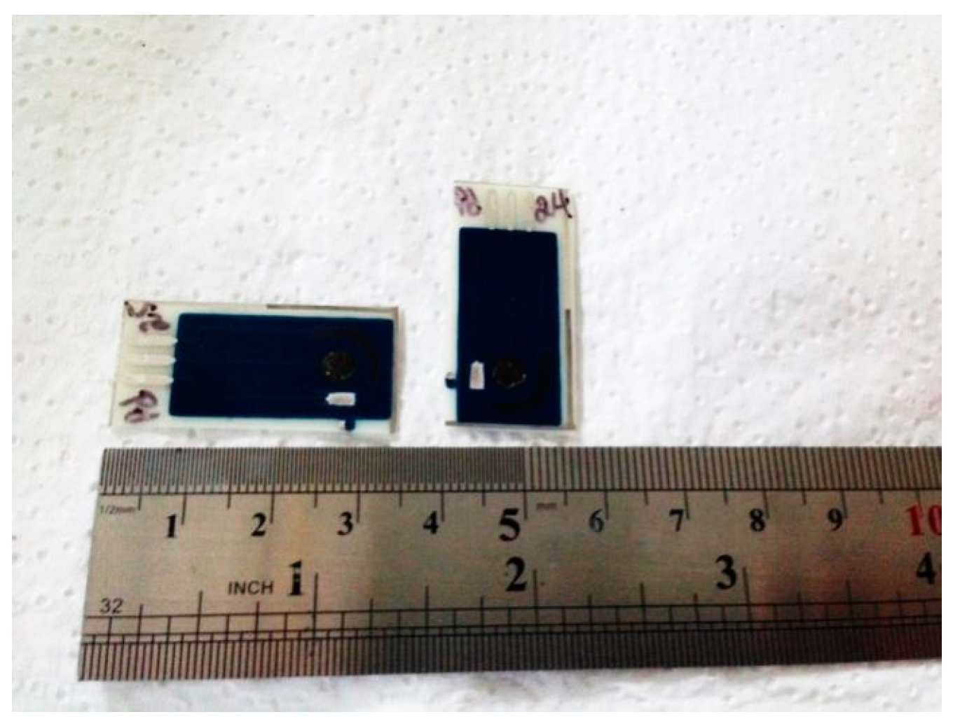
SPCTTFEs Construction
2.3. Nanoparticles Electrodeposition Methods
2.4. SOD Enzyme Immobilization onto AuNPs//SPCTTF Es
3. Results
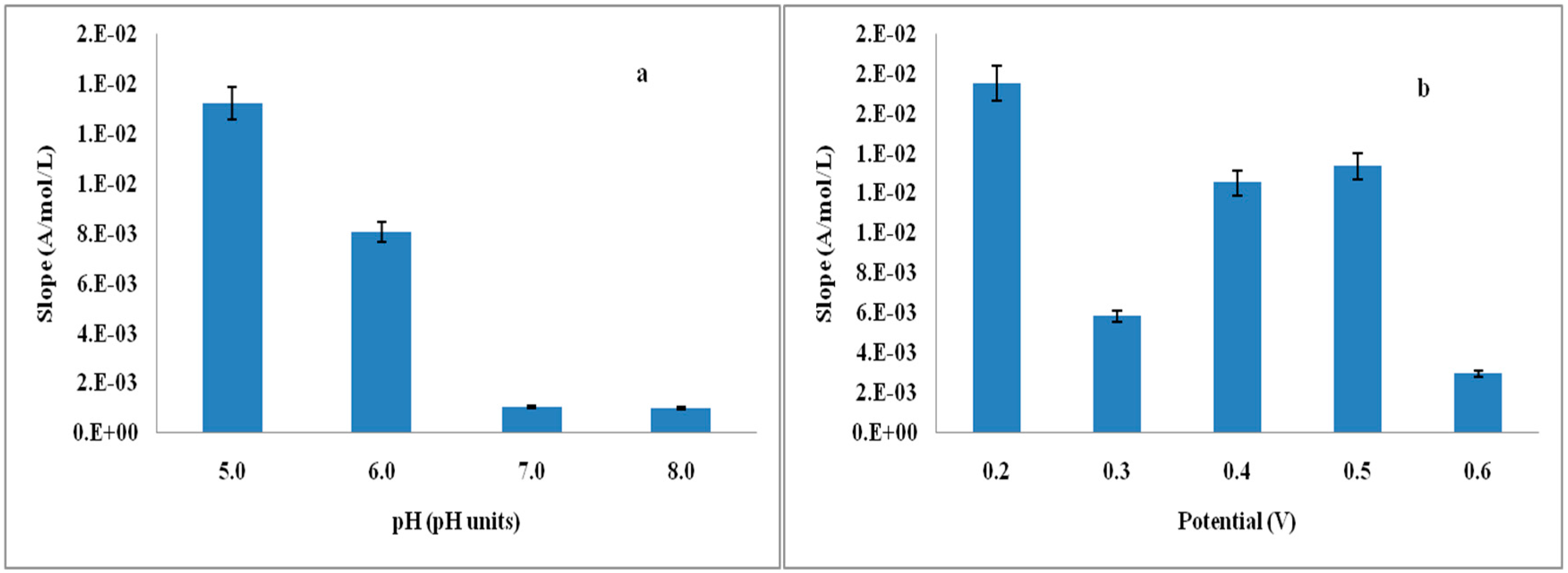
3.1. Optimization of Experimental Parameters
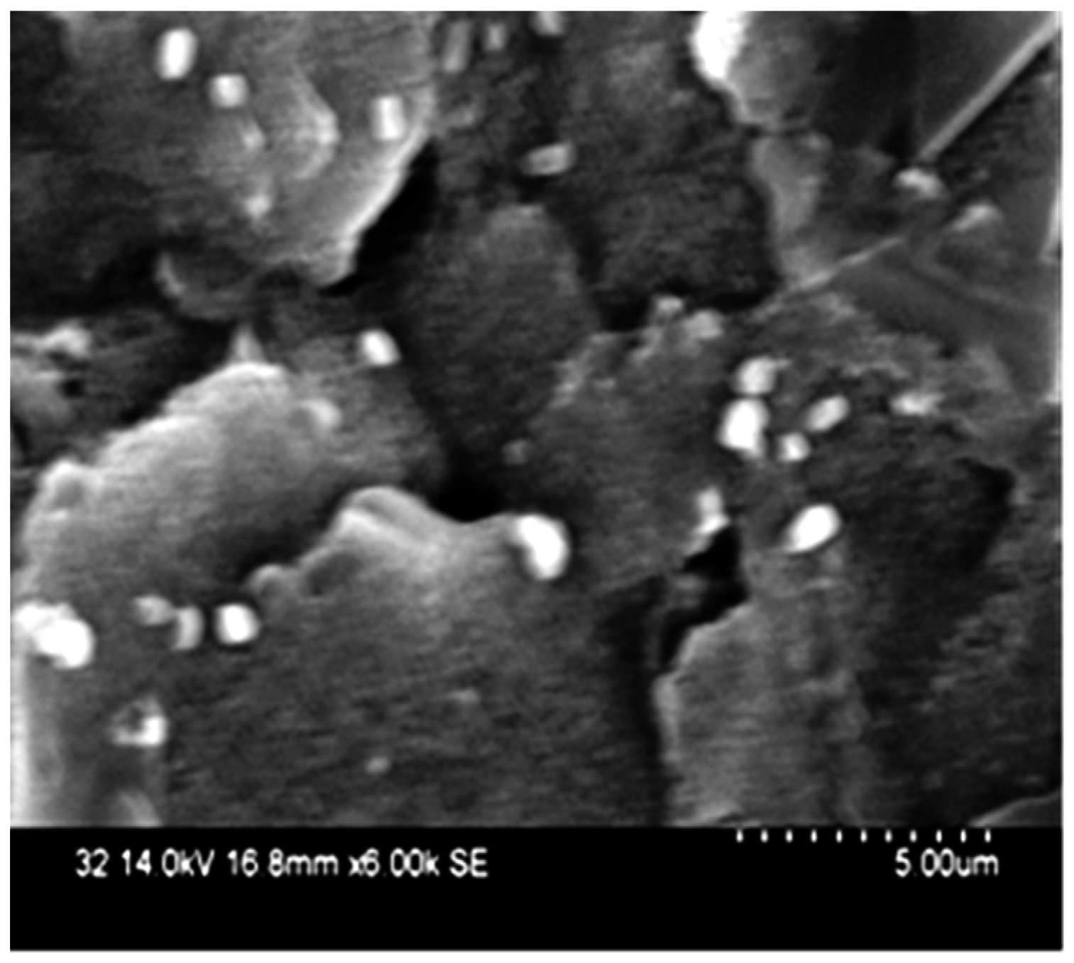
3.2. XRF and SEM for NPs/SPCTTFE Study METHOD A
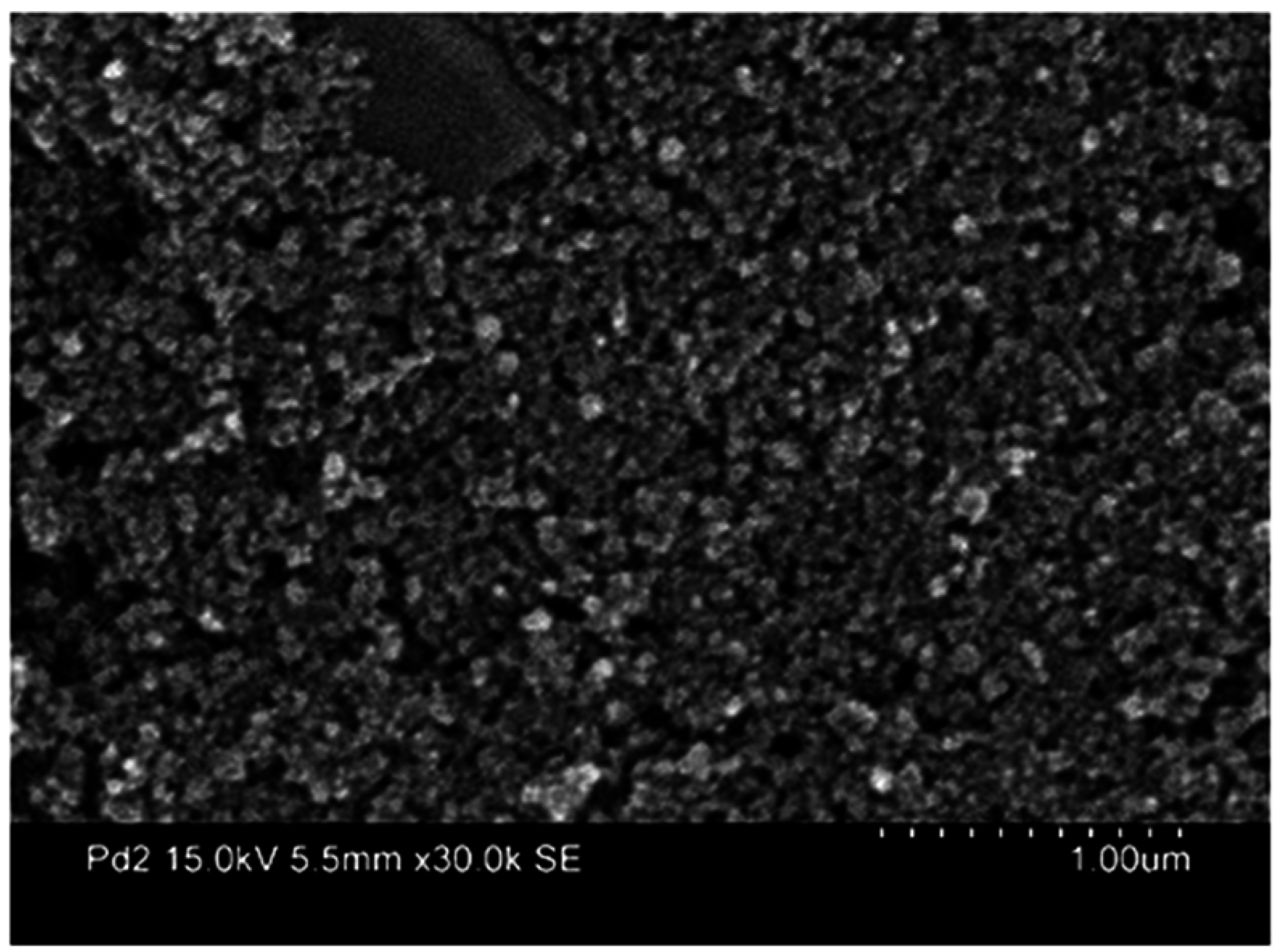
3.3. XRF and SEM for NPs/SPC TTFEs Study METHOD B
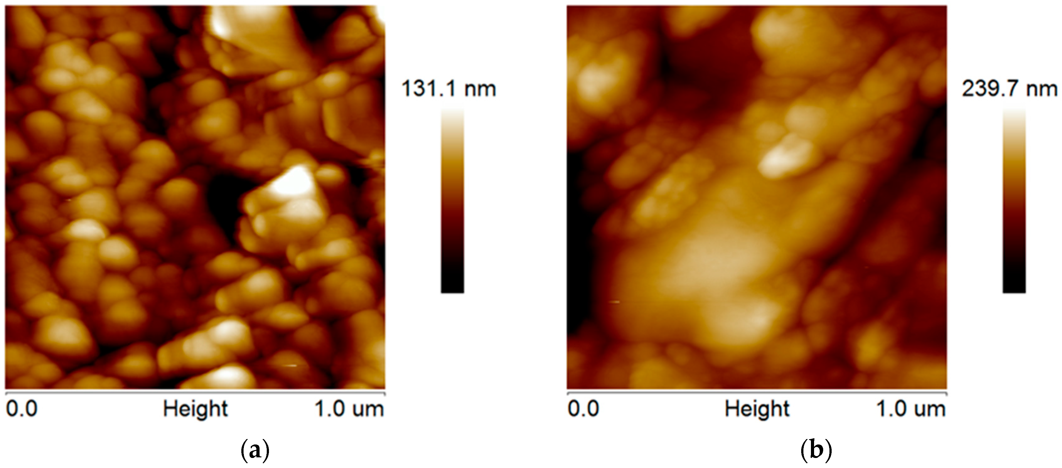
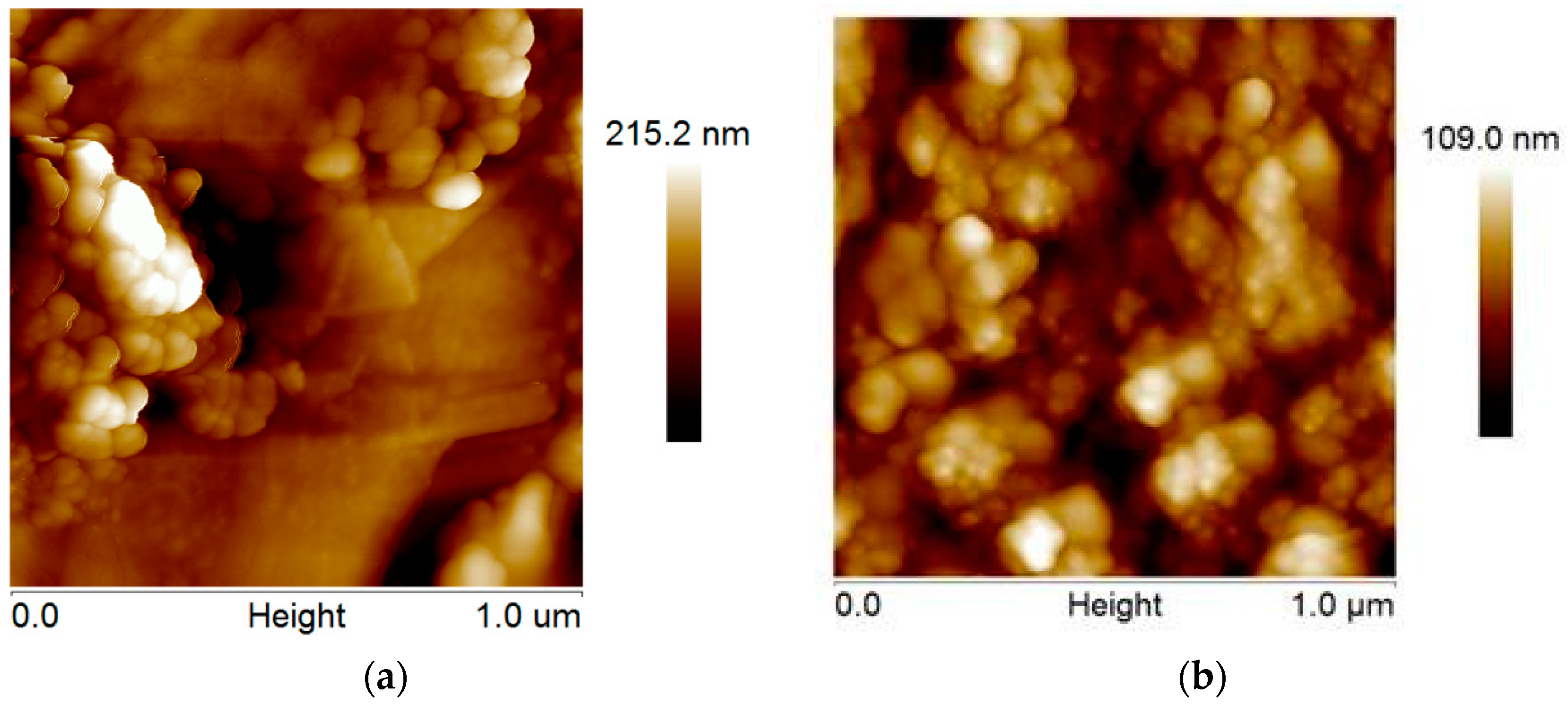
3.4. AFM Analysis of SPCTTFEs Prepared by Methods A and B
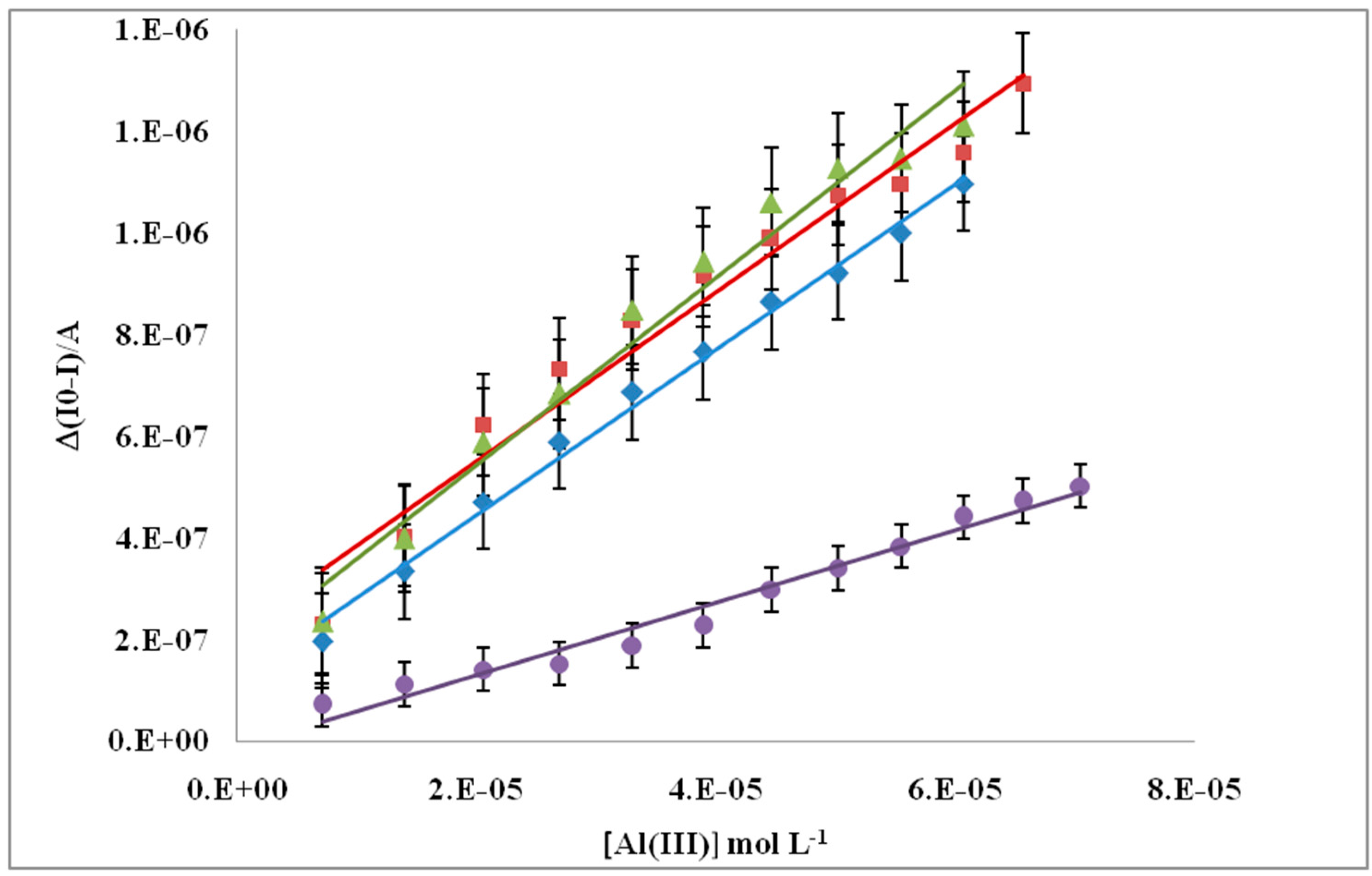
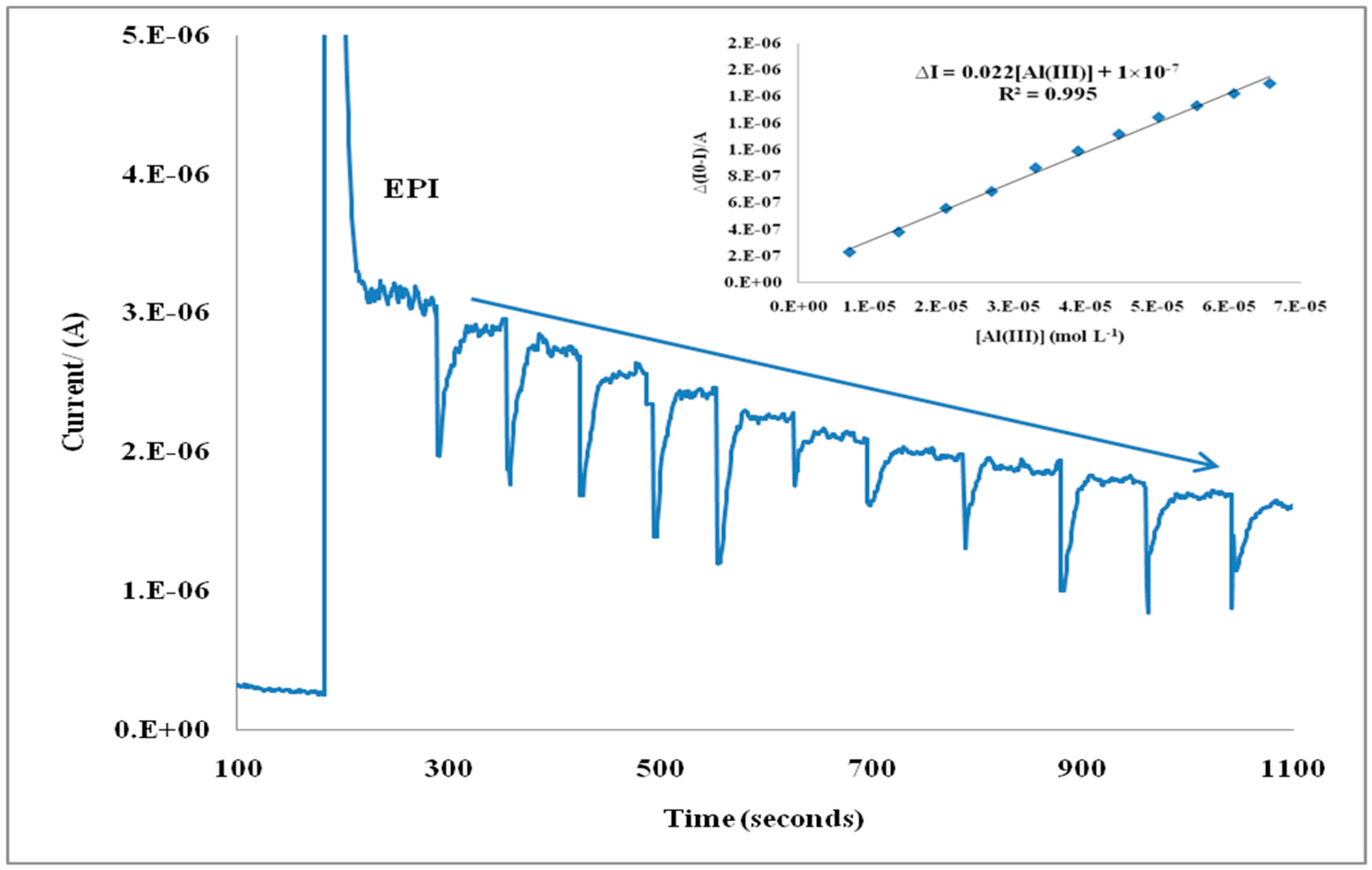
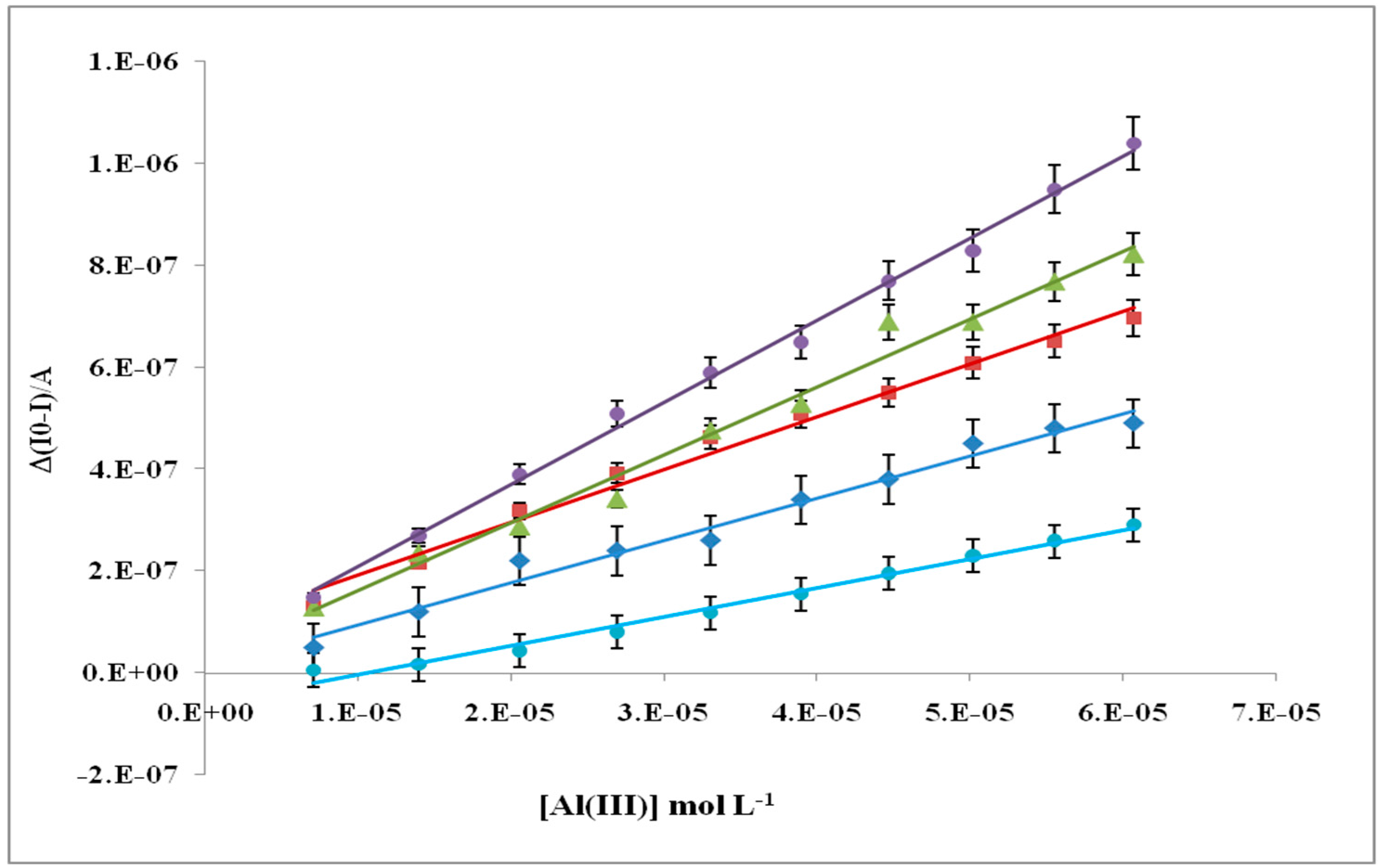
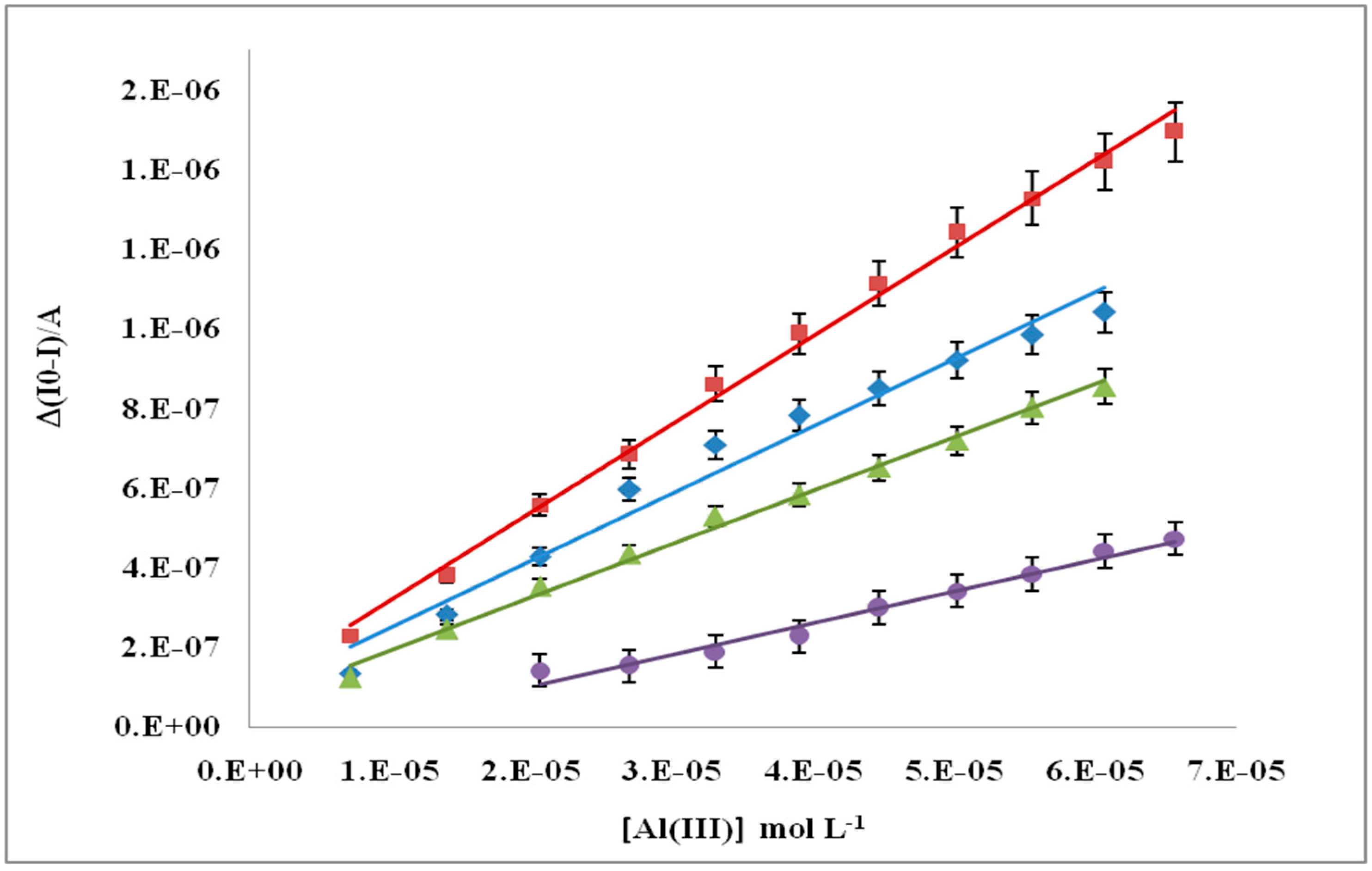
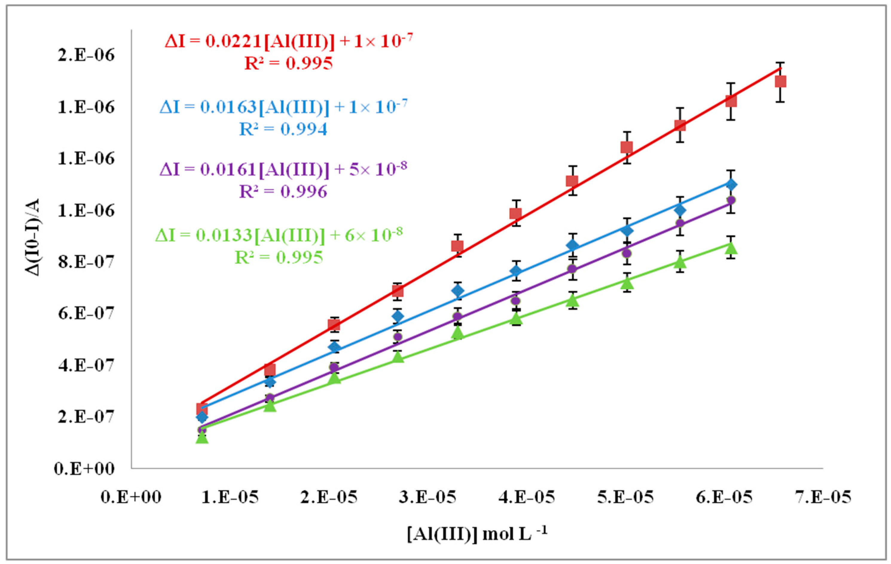
3.5. Inhibition Behavior of Al(III) on SOD Enzyme
3.6. Validation of SOD/PdNPs/TTF/SPCE Based Biosensor
3.6.1. Limit of Detection
3.6.2. Precision
3.6.3. Accuracy
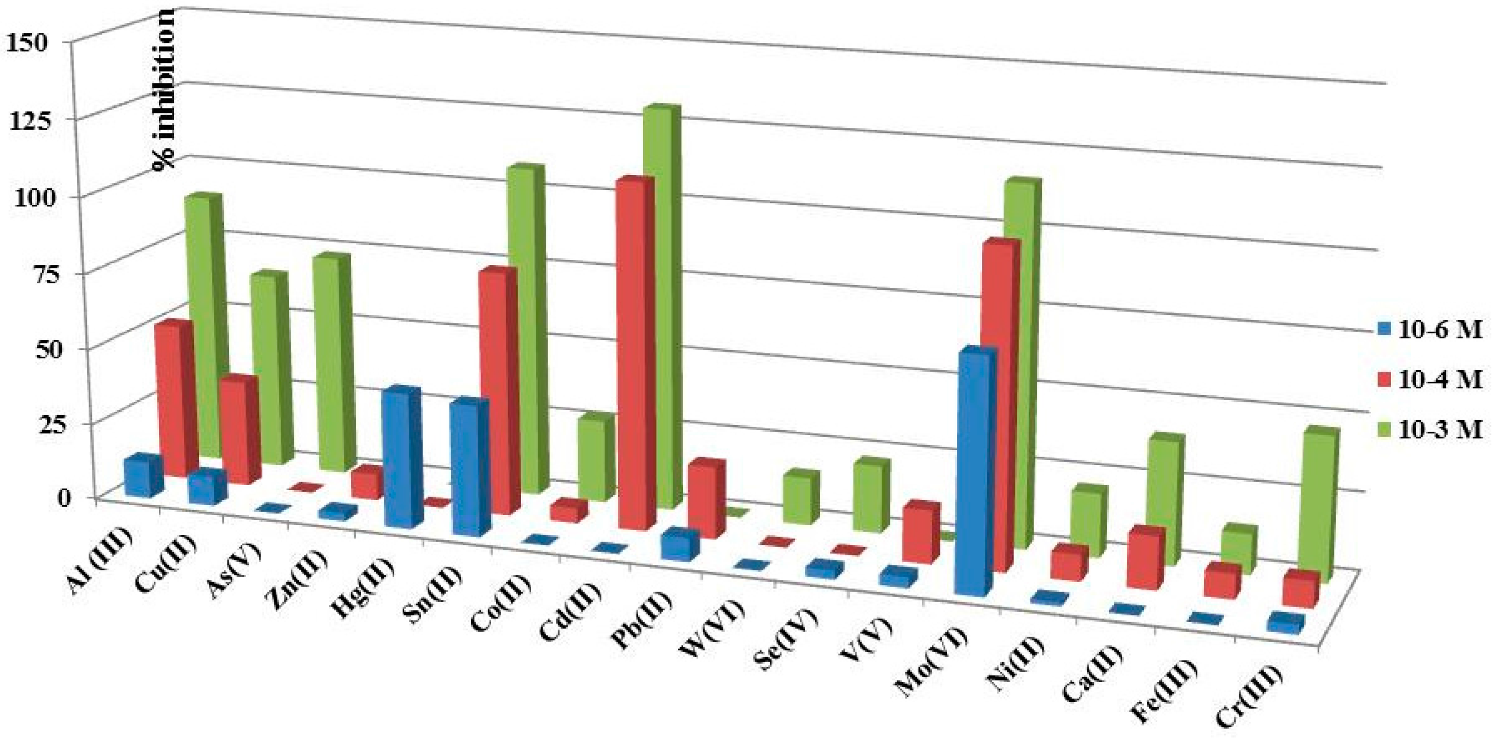
3.7. Study of Interferences on SOD/PdNPs/SPCTTFE Biosensors
4. Discussion
5. Conclusions
Acknowledgments
Author Contributions
Conflicts of Interest
References
- Exley, C. The coordination chemistry of aluminum in neurodegenerative disease. Coord. Chem. Rev. 2012, 256, 2142–2146. [Google Scholar] [CrossRef]
- Shin, R.W. Aluminum modifies the properties of Alzheimer’s disease PHF tau proteins in vivo and in vitro. J. Neurosci. 1994, 14, 7221–7233. [Google Scholar] [PubMed]
- Zatta, P.; Drago, D.; Bolognin, S.; Sensi, S.L. Alzheimer’s disease, metal ions and metal homeostatic therapy. Trends Pharmacol. Sci. 2009, 30, 346–353. [Google Scholar] [CrossRef] [PubMed]
- Wu, Z.; Du, Y.; Xue, H.; Wu, Y.; Zhou, B. Aluminum induces neurodegeneration and its toxicity arises from increased iron accumulation and reactive oxygen species (ROS) production. Neurobiol. Aging 2012, 33, 199.e1–199.e12. [Google Scholar] [CrossRef] [PubMed]
- Tian, Y.; Mao, L.; Okajima, T.; Ohsaka, T. Superoxide Dismutase-Based Third-Generation Biosensor for Superoxide Anion. Anal. Chem. 2002, 74, 2428–2434. [Google Scholar] [CrossRef] [PubMed]
- Rahman, M.A.; Kothalam, A.; Choe, E.S.; Won, M.S.; Shim, Y.B. Stability and Sensitivity Enhanced Electrochemical In Vivo Superoxide Microbiosensor Based on Covalently Co-immobilized Lipid and Cytochrome c. Anal. Chem. 2012, 84, 6654–6660. [Google Scholar] [CrossRef] [PubMed]
- Wilson, G.S.; Johnson, M.A. In-Vivo Electrochemistry: What Can We Learn about Living Systems? Chem. Rev. 2008, 108, 2462–2481. [Google Scholar] [CrossRef] [PubMed]
- Luo, X.; Jia, S.; Ma, Q.; Zhong, M.; Gao, P.; Yu, Z.; Zhang, Y. Suppressive Effects of Subchronic Aluminum Overload on the Splenic Immune Function May Be Related to Oxidative Stress in Mice. Biol. Trace Elem. Res. 2014, 157, 249–255. [Google Scholar] [CrossRef] [PubMed]
- Joshi, D.K.; Tripathi, S.; Kulshrestha, S.; Mahdi, A.A. Docosahexaenoic acid ameliorates aluminum induced biochemical and morphological alteration in rat cerebellum. Ann. Neurosci. 2014, 21, 5–9. [Google Scholar] [CrossRef] [PubMed]
- Razo-Estrada, A.C.; García-Medina, S.; Madrigal-Bujaidar, E.; Gómez-Oliván, L.M.; Galar-Martínez, M. Aluminum-Induced Oxidative Stress and Apoptosis in Liver of the Common Carp, Cyprinus Carpio. Water Air Soil Poll. 2013, 224, 1510–1518. [Google Scholar] [CrossRef]
- Bhasin, P.; Singla, N.; Dhawan, D.K. Protective role of zinc during aluminum-induced hepatotoxicity. Environ. Toxicol. 2014, 29, 320–327. [Google Scholar] [CrossRef] [PubMed]
- Viezeliene, D.; Beekhof, P.; Gremmer, E.; Rodovicius, H.; Sadauskiene, I.; Jansen, E.; Ivanov, L. Selective induction of IL-6 by aluminum-induced oxidative stress can be prevented by selenium. J. Trace Elem. Med. Biol. 2013, 27, 226–229. [Google Scholar] [CrossRef] [PubMed]
- Sivakumar, S.; Khatiwada, C.P.; Sivasubramanian, J.; Raja, B. Protective Effects of Deferiprone and Desferrioxamine in Brain Tissue of Aluminum Intoxicated Mice: An FTIR Study. Biomed. Prev. Nutr. 2014, 4, 53–61. [Google Scholar] [CrossRef]
- Yu, L.; Jiang, R.; Su, Q.; Yu, H.; Yang, J. Hippocampal neuronal metal ion imbalance related oxidative stress in a rat model of chronic aluminum exposure and neuroprotection of meloxicam. Behav. Brain Funct. 2014, 10, 6–10. [Google Scholar] [CrossRef] [PubMed]
- Jelenković, A.; Jovanović, M.D.; Stevanović, I.; Petronijević, N.; Bokonjić, D.; Živković, J.; Igić, R. Influence of the Green Tea Leaf Extract on Neurotoxicity of Aluminium Chloride in Rats: Green Tea and Aluminium Neurotoxicity. Phytother. Res. 2014, 28, 82–87. [Google Scholar] [CrossRef] [PubMed]
- Li, X.; Han, Y.; Guan, Y.; Zhang, L.; Bai, C.; Li, Y. Aluminum Induces Osteoblast Apoptosis through the Oxidative Stress-Mediated JNK Signaling Pathway. Biol. Trace Elem. Res. 2012, 150, 502–508. [Google Scholar] [CrossRef] [PubMed]
- Celik, H.; Celik, N.; Kocyigit, A.; Dikilitas, M. The relationship between plasma aluminum content, lymphocyte DNA damage, and oxidative status in persons using aluminum containers and utensils daily. Clin. Biochem. 2012, 45, 1629–1633. [Google Scholar] [CrossRef] [PubMed]
- Alonso-Lomillo, M.A.; Domínguez-Renedo, O.; Arcos-Martínez, M.J. Screen-printed biosensors in microbiology; a review. Talanta 2010, 82, 1629–1636. [Google Scholar] [CrossRef] [PubMed]
- Andreescu, S.; Njagi, J.; Ispas, C. The New Frontiers of Organic and Composite Nanotechnology; Elsevier: Oxford, UK, 2008; pp. 355–394. [Google Scholar]
- Siangproh, W.; Dungchai, W.; Rattanarat, P.; Chailapakul, O. Nanoparticle-based electrochemical detection in conventional and miniaturized systems and their bioanalytical applications: A review. Anal. Chim. Acta 2011, 690, 10–25. [Google Scholar] [CrossRef] [PubMed]
- Xia, T.; Kovochich, M.; Brant, J.; Hotze, M.; Sempf, J.; Oberley, T.; Sioutas, C.; Yeh, J.I.; Wiesner, M.R.; Nel, A.E. Comparison of the Abilities of Ambient and Manufactured Nanoparticles to Induce Cellular Toxicity According to an Oxidative Stress Paradigm. Nano Lett. 2006, 6, 1794–1807. [Google Scholar] [CrossRef] [PubMed]
- Domínguez-Renedo, O.; Alonso-Lomillo, M.A.; Arcos-Martínez, M.J. Recent developments in the field of screen-printed electrodes and their related applications. Talanta 2007, 73, 202–219. [Google Scholar] [CrossRef] [PubMed]
- Domínguez-Renedo, O.; Alonso-Lomillo, M.A.; Recio-Cebrián, P.; Arcos-Martínez, M.J. Screen-printed acetylcholinesterase-based biosensors for inhibitive determination of permethrin. Sci. Total Environ. 2012, 426, 346–350. [Google Scholar] [CrossRef] [PubMed]
- Dai, X.; Nekrassova, O.; Hyde, M.E.; Compton, R.G. Anodic Stripping Voltammetry of Arsenic(III) Using Gold Nanoparticle-Modified Electrodes. Anal. Chem. 2004, 76, 5924–5929. [Google Scholar] [CrossRef] [PubMed]
- Alonso-Lomillo, M.A.; Yardimci, C.; Domínguez-Renedo, O.; Arcos-Martínez, M.J. CYP450 2B4 covalently attached to carbon and gold screen printed electrodes by diazonium salt and thiols monolayers. Anal. Chim. Acta 2009, 633, 51–56. [Google Scholar] [CrossRef] [PubMed]
- Domínguez Renedo, O.; Arcos-Martínez, M.J. Anodic stripping voltammetry of antimony using gold nanoparticle-modified carbon screen-printed electrodes. Anal. Chim. Acta 2007, 589, 255–260. [Google Scholar] [CrossRef] [PubMed]
- Dua, D.; Ding, J.; Cai, J.; Zhang, J.; Liu, L. In situ electrodeposited nanoparticles for facilitating electron transfer across self-assembled monolayers in biosensor design. Talanta 2008, 74, 1337–1343. [Google Scholar] [CrossRef] [PubMed]
- Del Torno-de Román, L.; Alonso-Lomillo, M.A.; Domínguez-Renedo, O.; Merino-Sánchez, C.; Merino-Amayuelas, M.P.; Arcos-Martínez, M.J. Fabrication and characterization of disposable sensors and biosensors for detection of formaldehyde. Talanta 2011, 86, 324–328. [Google Scholar] [CrossRef] [PubMed]
- Rand, E.; Periyakaruppan, A.; Tanaka, Z.; Zhang, D.A.; Marsh, M.P.; Andrews, R.J.; Lee, K.H.; Chen, B.; Meyyappan, M.; Koehne, J.E. A carbon nanofiber based biosensor for simultaneous detection of dopamine and serotonin in the presence of ascorbic acid. Biosens. Bioelectron. 2008, 24, 632–637. [Google Scholar]
- Araque, E.; Arenas, C.B.; Gamella, M.; Reviejo, J.; Villalonga, R.; Pingarrón, J.M. Graphene–polyamidoamine dendrimer–Pt nanoparticles hybrid nanomaterial for the preparation of mediatorless enzyme biosensor. J. Electroanal. Chem. 2014, 717–718, 96–102. [Google Scholar] [CrossRef]
- Wang, S.Q.; Lu, L.P.; Lin, X.Q. A selective voltammetric method for uric acid detection at a glassy carbon electrode modified with electrodeposited film containing DNA and Pt-Fe2O3 nanocomposites. Electroanalysis 2004, 16, 1734–1738. [Google Scholar] [CrossRef]
- Sanllorente-Méndez, S.; Domínguez-Renedo, O.; Arcos-Martínez, M.J. Determination of Arsenic(III) Using Platinum Nanoparticle-Modified Screen-Printed Carbon-Based Electrodes. Electroanalysis 2009, 21, 635–639. [Google Scholar] [CrossRef]
- Thiagarajan, S.; Yang, R.F.; Chen, S.M. Palladium nanoparticles modified electrode for the selective detection of catecholamine neurotransmitters in presence of ascorbic acid. Bioelectrochemistry 2009, 75, 163–169. [Google Scholar] [CrossRef] [PubMed]
- Atta, N.F.; El-Kady, M.F. Novel poly (3-methylthiophene)/Pd, Pt nanoparticle sensor: Synthesis, characterization and its application to the simultaneous analysis of dopamine and ascorbic acid in biological fluids. Sens. Actuat. B 2010, 145, 299–310. [Google Scholar] [CrossRef]
- Vitulli, G.; Evangelisti, C.; Pertici, P.; Caporusso, A.M.; Panziera, N.; Salvadori, P.; Faga, M.G.; Manfredotti, C.; Martra, G.; Coluccia, S.; et al. Supported rhodium nanoparticles in catalysis: The role of stabilizers on catalytic activity and structural features. J. Organomet. Chem. 2003, 681, 37–50. [Google Scholar] [CrossRef]
- Zapf, R.; Thiele, R.; Wichert, M.; O’Connell, M.; Ziogas, A.; Kolb, G. Application of rhodium nanoparticles for steam reforming of propane in microchannels. Catal. Commun. 2013, 41, 140–145. [Google Scholar] [CrossRef]
- Lokesh, K.S.; Shivaraj, Y.; Dayananda, B.P.; Chandra, S. Synthesis of phthalocyanine stabilized rhodium nanoparticles and their application in biosensing of cytochrome c. Bioelectrochemistry 2009, 75, 104–109. [Google Scholar] [CrossRef] [PubMed]
- Chandra, S.; Lokesh, K.S.; Nicolai, A.; Lang, H. Dendrimer-rhodium nanoparticle modified glassy carbon electrode for amperometric detection of hydrogen peroxide. Anal. Chim. Acta 2009, 632, 63–68. [Google Scholar] [CrossRef] [PubMed]
- Poorahong, S.; Santhosh, P.; Valdés, G.; Tseng, T.F.; Wong, J.I.; Kanatharan, P.; Thavarungkul, P.; Wang, J. Development of amperometric α-ketoglutarate biosensor based on ruthenium–rhodium modified carbon fiber enzyme microelectrode. Biosens. Bioelectron. 2011, 26, 3670–3673. [Google Scholar] [CrossRef] [PubMed]
- Campanella, L.; Favero, G.; Persi, L.; Tomassetti, M. New biosensor for superoxide radical used to evidence molecules of biomedical and pharmaceutical interest having radical scavenging properties. J. Pharm. Biomed. Anal. 2000, 23, 69–76. [Google Scholar] [CrossRef]
- Campanella, L.; Bonanni, A.; Finotti, E.; Tomassetti, M. Biosensors for determination of total and natural antioxidant capacity of red and white wines: Comparison with other spectrophotometric and fluorimetric methods. Biosens. Bioelectron. 2004, 19, 641–651. [Google Scholar] [CrossRef]
- Campanella, L.; Favero, G.; Persi, L.; Tomassetti, M. Evaluation of radicals scavenging properties of several plants, fresh or from a herbalist’s, using a superoxide dismutase biosensor. J. Pharm. Biomed. Anal. 2001, 24, 1055–1064. [Google Scholar] [CrossRef]
- Campanella, L.; Bonanni, A.; Tomassetti, M. Determination of the antioxidant capacity of samples of different types of tea, or of beverages based on tea or other herbal products, using a superoxide dismutase biosensor. J. Pharm. Biomed. Anal. 2003, 32, 725–736. [Google Scholar] [CrossRef]
- Campanella, L.; Bonanni, A.; Favero, G.; Tomassetti, M. Determination of antioxidant properties of aromatic herbs, olives and fresh fruit using an enzymatic sensor. Anal. Bioanal. Chem. 2003, 375, 1011–1016. [Google Scholar] [PubMed]
- Campanella, L.; Martini, E.; Tomassetti, M. Antioxidant capacity of the algae using a biosensor method. Talanta 2005, 66, 902–911. [Google Scholar] [CrossRef] [PubMed]
- Campanella, L.; Bonanni, A.; Bellantoni, D.; Tomassetti, M. Biosensors for determination of total antioxidant capacity of phytotherapeutic integrators: Comparison with other spectrophotometric, fluorimetric and voltammetric. J. Pharm. Biomed. Anal. 2004, 35, 303–320. [Google Scholar] [CrossRef]
- Campanella, L.; Bonanni, A.; Bellantoni, D.; Favero, G.; Tomassetti, M. Comparison of fluorimetric, voltammetric and biosensor methods for the determination of total antioxidant capacity of drug products containing acetylsalicylic acid. J. Pharm. Biomed. Anal. 2004, 36, 91–99. [Google Scholar] [CrossRef] [PubMed]
- Tomassetti, M.; Serone, M.; Angeloni, R.; Campanella, L.; Mazzone, E. Amperometric Enzyme Sensor to Check the Total Antioxidant Capacity of Several Mixed Berries. Comparison with Two Other Spectrophotometric and Fluorimetric Methods. Sensors 2015, 15, 3435–3452. [Google Scholar] [CrossRef] [PubMed]
- Santharaman, P.; Das, M.; Singh, S.K.; Sethy, N.K.; Bhargava, K.; Claussen, J.C.; Karunakaran, C. Label-free electrochemical immunosensor for the rapid and sensitive detection of the oxidative stress marker superoxide dismutase 1 at the point-of-care. Sens. Actuators B 2016, 236, 546–553. [Google Scholar] [CrossRef]
- Tian, Y.; Mao, L.; Okajima, T.; Ohsaka, T. A carbon fiber microelectrode-based third-generation biosensor for superoxide. Biosens. Bioelectron. 2005, 21, 557–564. [Google Scholar] [CrossRef] [PubMed]
- Zhu, X.; Liu, T.; Zhao, H.; Shi, L.; Li, X.; Lan, M. Ultrasensitive detection of superoxide anion released from living cells using a porous Pt–Pd decorated enzymatic sensor. Biosens. Bioelectron 2016, 79, 449–456. [Google Scholar] [CrossRef] [PubMed]
- Salem, F.; Tavakoli, H.; Sadeghi, M.; Riazi, A. Developing a high performance superoxide dismutase based electrochemical biosensor for radiation dosimetry of thallium 201. Radiat. Phys. Chem. 2014, 102, 128–134. [Google Scholar] [CrossRef]
- Wang, Z.; Liu, D.; Gu, H.; Zhu, A.; Tian, Y.; Shi, G. NTA-modified carbon electrode as a general relaying substrate to facilitate electron transfer of SOD: Application to in vivo monitoring of O 2− in a rat brain. Biosens. Bioelectron. 2013, 43, 101–107. [Google Scholar] [CrossRef] [PubMed]
- Campanella, L.; Favero, G.; Tomassetti, M. A modified amperometric electrode for the determination of free radicals. Sens. Actuators B 1997, 44, 559–565. [Google Scholar] [CrossRef]
- Tang, J.; Zhu, X.; Niu, X.; Liu, T.; Zhao, H.; Lan, M. Anamperometric superoxide anion radical biosensor based on SOD/PtPd-PDARGO modified electrode. Talanta 2015, 137, 18–24. [Google Scholar] [CrossRef] [PubMed]
- Braik, M.; Barsan, M.M.; Dridi, C.; Ali, M.B.; Brett, C.M.A. Highly sensitive amperometric enzyme biosensor for detection of superoxide based on conducting polymer/CNT modified electrodes and superoxide dismutase. Sens. Actuators B 2016, 236, 574–582. [Google Scholar] [CrossRef]
- Ghica, M.E.; Brett, C.M.A. Simple and efficient epinephrine sensor based on carbon nanotube modified carbon film electrodes. Anal. Lett. 2013, 46, 1379–1393. [Google Scholar] [CrossRef]
- Ruipérez, F.; Mujika, J.I.; Ugalde, J.M.; Exley, C.; Lopez, X. Pro-oxidant activity of aluminum: Promoting the Fenton reaction by reducing Fe(III) to Fe(II). J. Inorg. Biochem. 2012, 117, 118–123. [Google Scholar]
- Thiagarajan, S.; Yang, R.F.; Chen, S.M. Palladium Nanoparticles Modified Electrode for the Selective Detection of Catecholamine Neurotransmitters in Presence of Ascorbic Acid. Bioelectrochemistry 2009, 75, 163–169. [Google Scholar] [CrossRef] [PubMed]
- Nunes, G.S.; Jeanty, G.; Marty, J.L. Enzyme immobilization procedures on screen-printed electrodes used for the detection of anticholinesterase pesticides: Comparative study. Anal. Chim. Acta 2004, 523, 107–115. [Google Scholar] [CrossRef]
- Alonso-Lomillo, M.A.; Domínguez-Renedo, O.; Arcos-Martínez, M.J. Enzyme Modified Screen Printed Electrodes in Biosensors: Properties, Materials and Applications; Nova Science Publishers: New York, NY, USA, 2009. [Google Scholar]
- Raposo, M.; Ferreira, Q.; Ribeiro, P.A. A Guide for Atomic Force Microscopy Analysis of Soft Condensed Matter. Mod. Res. Educ. Top. Microsc. 2007, 1, 758–769. [Google Scholar]
- Miller, J.C.; Miller, J.N. Estadística y Quimiometría para Química Analítica; Prentice Hall: Madrid, Spain, 2002. [Google Scholar]


| Technique | Electrode | Potential | Modification | Range | LOD | Sample/Analite | Reference |
|---|---|---|---|---|---|---|---|
| CV 1 | SPCE | −0.8–+0.8 V | Pyrrole/SAMs | 0.5 × 10⁻⁹–5 M | 0.5 × 10⁻⁹ M | cultured human keratinocytes NO2- | [49] |
| Amperometry | CFME 2 | +0.25 V | cysteine/AuNPs | (13–104) × 10−9 M | - | O2•− | [50] |
| Amperometry | SPCE | −0.1 V | porous Pt-Pd/nafion | (16–1536) × 10−6 M | 0.13 × 10⁻⁶ M | Cell culture medium/O2•− | [51] |
| CV 1 Chronoamperometry | Gold electrode | −0.2–+0.5 V | Au/Cys/SOD 9, Au/GNP/Cys/SOD 10 and Au/GNP/Cys/SOD/Chit 11 | 0.5–4 Gy | 0.03 × 10⁻⁶ M | thallium 201/water | [52] |
| Amperometry | GC CFME 2 | 0.2 V | NTA/HT 7 | 10−7–10−4 M | 21 × 10−9 M | brain tissue/O2•− | [53] |
| CV 1 | Carbon paste electrode Electrochem | 0–0.3 V | cytochrome c in solution and Fe(III)-protoporphyrin immobilized | (1–6) × 10−3 M | 0.3 × 10⁻³ M | Xantine | [54] |
| System Carbon paste electrode | Protoporphyrin and cytochrome both immobilized | (1–8) × 10−3 M | 0.2 × 10⁻³ M | ||||
| Cronoamperometry | Composite electrode | −0.3 V | PtPd-PDARGO 6 | (0.016–0.24) × 10−3 M | 2 × 10⁻⁶ M | DMEM 5/O2•− | [55] |
| Amperometry | GC 8 | −0.3 V | MWCNT 4 | (0.01–0.3) × 10−3 M | 1 × 10⁻⁶ M | Wines, berry juice/O2•− | [56] |
| PEDOT 3 | |||||||
| Amperometry | SPCTTFE 12 | 0.2 V | SOD/PdNP 13 | (1.0–60) × 10−5 M | 2 × 10⁻⁶ M | Al(III) | This article |
| water | |||||||
| samples |
| Method A | Method B | |||
|---|---|---|---|---|
| Element | XRF% | XRF% | XRF% | XRF% |
| (+0.18 V, 15 s) | (+0.30 V, 15 s) | CV1 | CV2 | |
| Pd | 0.136 | 0.00 | 0.557 | 0.632 |
| Pt | 0.223 | 1.48 | 2.74 | 2.71 |
| Rh | 0.693 | 0.380 | 4.49 | 2.95 |
| Au | 1.42 | - | 1.87 | 2.23 |
| NPs/SPCTTFEs | Method/Conditions | RA (nm) | RMS (nm) | Rmax (nm) | RKu | RSk |
|---|---|---|---|---|---|---|
| SPCTTFE | - | 16.8 | 21.5 | 131 | 3.53 | −0.183 |
| AuNPs/SPCTTFE | A/0.18 V | 31.3 | 39.2 | 218 | 3.34 | −0.537 |
| B/CV1 | 33.8 | 41.4 | 234 | 2.62 | 0.126 | |
| B/CV2 | 34.6 | 44.5 | 303 | 3.19 | −00292 | |
| PdNPs/SPCTTFE | B/CV1 | 24.8 | 34.6 | 216 | 4.72 | 0.0604 |
| B/CV2 | 14.7 | 18.2 | 106 | 2.69 | 0.0939 | |
| PtNPs/SPCTTFE | B/CV1 | 106 | 140 | 864 | 3.63 | 0.157 |
| B/CV2 | 106 | 140 | 864 | 3.60 | 0.141 | |
| RhNPs/SPCTTFE | B/CV1 | 22.0 | 28.1 | 173 | 3.30 | −0.0733 |
| B/CV2 | 25.6 | 33.9 | 204 | 3.65 | −0.189 |
| Km Apparent (M) | SOD/AuNPs/SPCTTFEs | SOD/PtNPs/SPCTTFEs | SOD/PdNPs/SPCTTFEs | SOD/RhNPs/SPCTTFEs |
|---|---|---|---|---|
| Method A 0.18 V | ||||
| Without Al | (7.8 ± 0.3) × 10−4 | (1.2 ± 0.3) × 10−4 | (1.5 ± 0.4) × 10−3 | (3.3 ± 0.2) × 10−3 |
| Al(III) 7.25 × 10⁻⁶ M | (1.3 ± 0.1) × 10−3 | (1.4 ± 0.3) × 10−2 | (1.0 ± 0.1) × 10−2 | (1.4 ± 0.1) × 10−2 |
| Al(III) 2.18 × 10⁻⁵ M | (3.7 ± 0.3) × 10−3 | (1.8 ± 0.6) × 10−2 | (1.3 ± 0.1) × 10−2 | - |
| Method B and CV2 conditions | ||||
| Without Al | (1.5 ± 0.2) × 10−3 | (5.8 ± 0.3) × 10−4 | (1.5 ±0.3) × 10−3 | (1.2 ± 0.1) × 10−4 |
| Al(III) 7.25 × 10⁻⁶ M | (2.7 ± 0.4) × 10−3 | (1.3 ± 0.4) × 10−3 | (5.0 ±0.4) × 10−3 | (6.4 ± 0.5) × 10−3 |
| Al(III) 2.18 × 10⁻⁵ M | (3.05 ± 0.5) × 10−3 | (3.0 ± 0.8) × 10−3 | (1.2 ±0.3) × 10−2 | (5.4 ± 0.2) × 10−3 |
| Added SRM | Found SRM | Found SRM | SRM | Recovery |
|---|---|---|---|---|
| (M) | (M) | (mg/L) | (mg/L) | - |
| 1.30 × 10⁻⁵ | 1.235 × 10⁻⁵ | 9.50 | 950 | 95.0 |
| - | 1.366 × 10⁻⁵ | 10.51 | 1051 | 105.1 |
| - | 1.370 × 10⁻⁵ | 10.51 | 1051 | 105.3 |
| - | 1.374 × 10⁻⁵ | 10.58 | 1058 | 105.8 |
| - | 1.402 × 10⁻⁵ | 10.79 | 1079 | 107.9 |
| - | - | Mean | 1038 | 103.8 |
| - | - | SD | 50.3 | 5.0 |
| - | - | RSD | 4.8 | 4.8 |
| Added SRM (mg/L) | Found SRM (mg/L) | SRM (mg/L) | SRM (mg/L) | Recovery (%) |
|---|---|---|---|---|
| 0.170 | 0.180 | 10.44 | 1044 | 104.0 |
| 0.171 | 9.90 | 990 | 99.0 | |
| 0.169 | 9.82 | 982 | 98.2 | |
| - | - | Mean | 1005 | 100.5 |
| - | - | SD | 34 | 3.4 |
| - | - | RSD | 3.4 | 3.4 |
© 2016 by the authors; licensee MDPI, Basel, Switzerland. This article is an open access article distributed under the terms and conditions of the Creative Commons Attribution (CC-BY) license (http://creativecommons.org/licenses/by/4.0/).
Share and Cite
Barquero-Quirós, M.; Arcos-Martínez, M.J. Effect of Nanoparticles on Modified Screen Printed Inhibition Superoxide Dismutase Electrodes for Aluminum. Sensors 2016, 16, 1588. https://doi.org/10.3390/s16101588
Barquero-Quirós M, Arcos-Martínez MJ. Effect of Nanoparticles on Modified Screen Printed Inhibition Superoxide Dismutase Electrodes for Aluminum. Sensors. 2016; 16(10):1588. https://doi.org/10.3390/s16101588
Chicago/Turabian StyleBarquero-Quirós, Miriam, and María Julia Arcos-Martínez. 2016. "Effect of Nanoparticles on Modified Screen Printed Inhibition Superoxide Dismutase Electrodes for Aluminum" Sensors 16, no. 10: 1588. https://doi.org/10.3390/s16101588
APA StyleBarquero-Quirós, M., & Arcos-Martínez, M. J. (2016). Effect of Nanoparticles on Modified Screen Printed Inhibition Superoxide Dismutase Electrodes for Aluminum. Sensors, 16(10), 1588. https://doi.org/10.3390/s16101588