Article

Phylogenomic Insights of Mesentotoma Salmon, 1942 (Collembola: Entomobryidae: Entomobryinae): First Mitogenome and Phylogenetic Hypothesis, Taxonomic Notes, and Description of Two New Brazilian Species †

by
Stéphanie dos Santos Viana
1,*,
Nerivania Nunes Godeiro
2,
José Wellington de Morais
1 and
Nikolas Gioia Cipola
3,*
1
National Institute of Amazonian Research-INPA-CPEN, Manaus 69067-375, Brazil
2
Natural History Research Center, Shanghai Natural History Museum, Shanghai Science & Technology Museum, Shanghai 200041, China
3
Department of Botany and Zoology, Biosciences Center, Federal University of Rio Grande do Norte-UFRN, Natal 59078-900, Brazil
*
Authors to whom correspondence should be addressed.
urn:lsid:zoobank.org:pub:15CC71-7CB6-4A1E-91C1-9450D8E14914.
Diversity 2026, 18(2), 89; https://doi.org/10.3390/d18020089
Submission received: 31 December 2025 / Revised: 23 January 2026 / Accepted: 23 January 2026 / Published: 31 January 2026
(This article belongs to the Special Issue Integrative Systematics and Evolution of Collembola)

Abstract

We used whole-genome sequencing to assemble the first complete mitogenome from a Mesentotoma Salmon, 1942 species. Mesentotoma was included in phylogenetic analyses along with 21 mitogenomes of other Entomobryinae. Maximum likelihood and Bayesian inferences yielded similar results; both analyses indicated that Mesentotoma is related to Willowsia jacobsoni and Entomobrya proxima. However, other genera of Entomobryinae, also lacking a mucronal spine (Desertia Tshelnokov, 1979 Calx Christiansen, 1958 and Isotobrya Womersley, 1934) need to be included in further analysis to verify their relationships, as well as other species of Mesentotoma. Furthermore, five species previously assigned to Entomobrya were transferred to Mesentotoma due to the absence of a mucronal spine. E. coeruleopicta Marlier, 1945 is a junior homonym of E. coeruleopicta Schött, 1917 and a replacement name is proposed: M. tranvercyana nom. nov., comb. nov. In addition, two new species of Mesentotoma from a Brazilian island are described and characterized by reduced macrochaetotaxy. This represents the first record of Mesentotoma in the Neotropical region, increasing its total number of valid species from eight to fifteen. We provided an identification key for the seven Mesentotoma species recorded in the Tropical zone.

1. Introduction

Mesentotoma Salmon, 1942 [1] is an unscaled genus of Entomobryinae [2] that resembles Entomobrya Rondani, 1861 [3], and although the phylogenetic relationships between them are not established, they differ by the mucronal spine being present (Entomobrya) or absent (Mesentotoma) [1,4,5,6,7]. The specialized microchaeta (ms) may be another difference between them, since its absence has been reported in an undescribed species of Mesentotoma [2].
The genus Mesentotoma was proposed based only on the type species, M. exalga Salmon, 1942 [1] from New Zealand, and with seven other species later included in the genus, all from the Palearctic region: M. laguna (Bacon 1913) [8] from Nearctic; M. mauka Christiansen & Bellinger, 1992 [5] and M. nani Christiansen & Bellinger, 1992 [5] from Hawaiian Islands; and M. hutchinsoni (Denis 1936) [9], M. subdollfusi Jacquemart, 1974 [10], M. dollfusi (Denis, 1924) [11], and M. hispanica Baquero, Arbea & Jordana, 2010 [7,12,13].
To date, Mesentotoma has not been recorded in the Neotropical region [13], highlighting the need for further exploration, since Entomobryoidea has great potential for new discoveries in this region [14,15,16,17,18,19,20,21,22,23,24]. Previous neotropical Entomobryoidea were sampled from the central areas of the continent, while the Entomobryoidea fauna of the coastal region is currently less known [25].
Under sampled coastal regions of South America include continental and oceanic islands, such as the Fernando de Noronha Archipelago, where the first record of Collembola for Brazil occurred in 1840 with the description of Seira musarum Ridley, 1890 [26]. Over 120 years later, Isotogastrura mucrospatulata Palacios-Vargas, Lima & Zeppelini, 2013 [27] was the second Collembola species to be recorded from the island. Subsequently, a comprehensive survey recorded 38 Collembola species (17 nominal) across 28 genera and 12 families [25,28].
The Fernando de Noronha Archipelago currently hosts 28 nominal species across 21 genera and 10 families [28,29,30,31]. This total represents only 5.5% of the known Brazilian Collembola fauna and includes just 2 endemic species out of Brazil’s 349 Collembola species: Friesea noronhaensis Lima & Zeppelini, 2021 (Neanuridae) [30] and Isotogastrura mucrospatulata Palacios-Vargas, Lima & Zeppelini, 2013 (Isotogastruridae) [25,27].
To date no species of Collembola from Fernando de Noronha Archipelago have been included in any molecular study, while Entomobryinae have been wholly sampled [32,33,34,35]. Since September 2025, a total of 186 complete Collembola mitogenomes have been available on GenBank, being only 14 taxa in 7 genera of Entomobryinae (https://www.ncbi.nlm.nih.gov/). For total molecular data, this remains low compared with the approximately 9.500 described Collembola species worldwide [13].
In this study, we performed low-coverage whole-genome sequencing and annotated a complete mitogenome for the first time of Mesentotoma, including taxa in a phylogenetic analysis. Additionally, five tropical Entomobrya species were transferred to Mesentotoma, a new name was proposed to replace the homonym E. coeruleopicta (sensu Marlier, 1945) [36], two new species were described from Brazilian coastal regions, and an identification key for the Mesentotoma species recorded in the Tropical region was created.

2. Materials and Methods

2.1. Sampling, DNA Extraction, and Sequencing

Six specimens preserved in ethanol were transported to China for DNA sequencing. One individual was used for DNA isolation by Shanghai Yaoen Biotechnology Co., Ltd., China (Shanghai, China). A TIANamp MicroDNA extraction kit (Tiangen Co., Ltd., Beijing, China) was utilized to extract DNA and a KAPA Hyper Prep Kit (Roche, Basel, Switzerland) was used to build the library. All procedures followed kit manufacturers guidelines. The library was sequenced using the Illumina NovaSeq 6000 platform (Illumina Inc., San Diego, CA, USA), producing approximately 10 Gbp of paired-end reads, with 150 bp.

2.2. Mitogenome Assembly and Annotation

Before the assembly, quality control was performed on the raw sequencing data using BBTools (https://sourceforge.net/projects/bbmap/, accessed on 10 August 2025), with the “clumpify.sh” and “bbduk.sh” pipelines. Filtered data were input to MitoZ v. 3.6 [37] and the following softwares were evoked by MitoZ: MEGAHIT [38] v. 1.2.9 for assembly; Tiara v. 1.0.1 [39] and HMMER v. 3.4 [40] for searching homologous sequences and to make the alignments; BLAST+ v. 2.16.0 [41], GeneWise v. 2.2.0 [42], Infernal v. 1.1.5 [43], and MiTFi v. 0.1 [44] for annotation; and Circos v. 0.69 [45], BWA v. 0.7.17 [46], and SAMtools v. 1.18 [47] for visualization. Accession numbers for the raw SRA data and the assembled mitogenome of Mesentotoma multicirculata sp. nov. are SRR29635445 and PX395442, respectively. Mitogenome sequences assembled from public raw data were deposited to Figshare (https://doi.org/10.6084/m9.figshare.26123944).

2.3. Phylogenetic Analyses

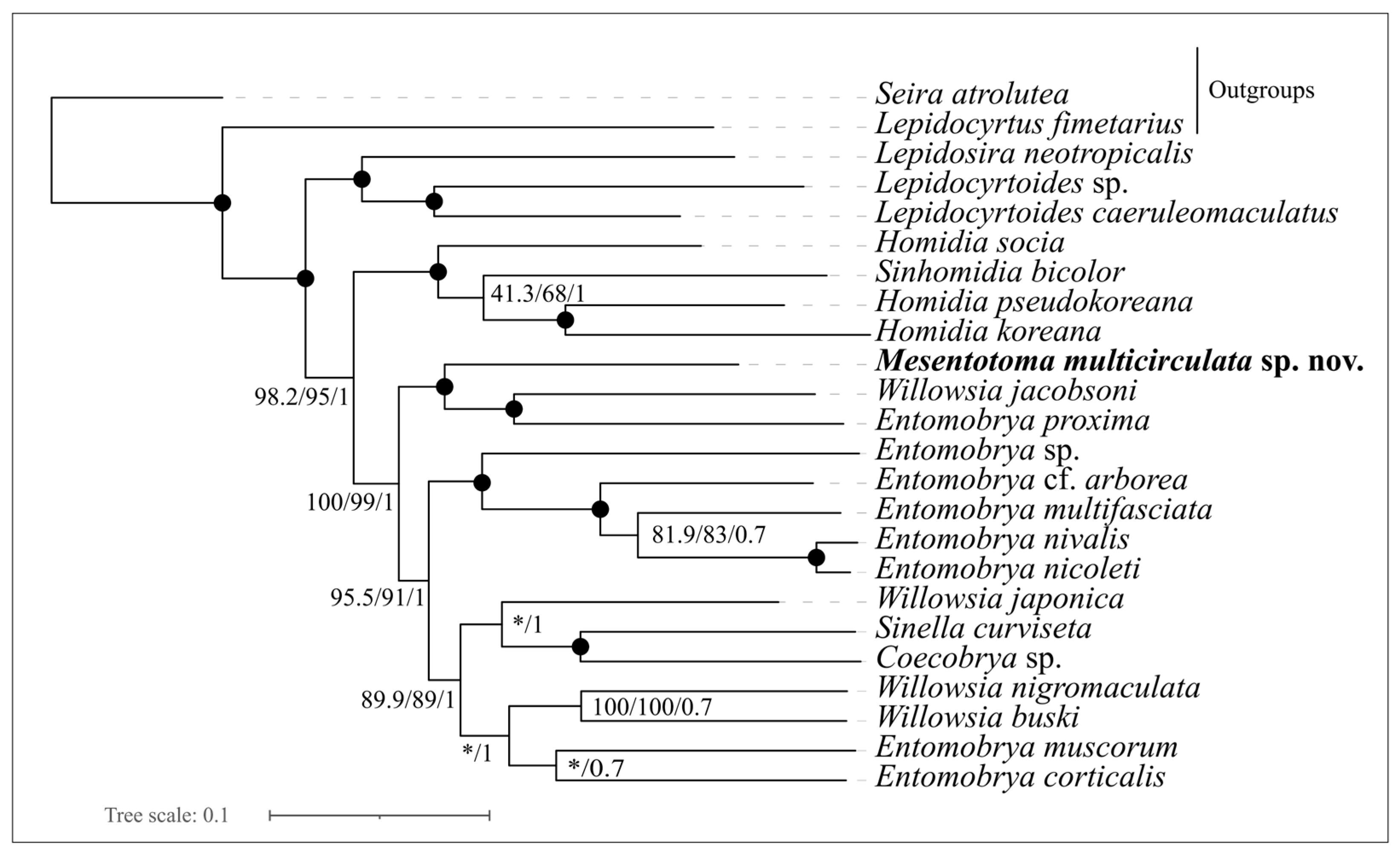
Phylogenetic analysis of Mesentotoma multicirculata sp. nov. with 21 Entomobryinae taxa (13 unscaled and 8 scaled) and 2 outgroups, Lepidocyrtus fimetarius Gisin, 1964 [48] (Lepidocyrtinae) and Seira atrolutea (Arlé, 1939) [49] (Seirinae), was performed. Both outgroups represent subfamilies currently considered to be sister groups to Entomobryinae (e.g., refs. [33,35,50]). Previous sequenced genomes were downloaded from NCBI nucleotide and SRA databases (Table 1). Nine mitogenomes were assembled using raw sequencing data SRA files in NCBI and applying the same methodology described above.
To generate the phylogenetic matrix, each of the 13 PCGs (protein coding genes) were aligned separately in MAFFT v. 7.470 with the “L-INS-I” algorithm and Trimal v. 1.4.1 [51] to trim the alignments with the “-gappyout” option. FASconCAT-G v. 1.04 [52] was used to concatenate the 13 PCGs into 1 final nucleotide matrix.
Maximum likelihood (ML) analyses were conducted in IQTree version 2.0.7 [53], employing partitions by codons with the options “-m MFP+MERGE --msub mitochondrial”. ModelFinder [54] was used to determine the best substitution model for each partition; details of the partitions scheme are presented in Supplementary Materials (Table S1).
Table 1. Taxonomical information and NCBI accession numbers (A.N.) of the species used in the phylogenetic analyses. The newly assembled mitogenomes are represented in bold.
SpeciesSubfamilyCountryA.N.Source
1Lepidocyrtus fimetarius Gisin, 1964 [48]LepidocyrtinaeChinaNC047189.1[55]
2Seira atrolutea (Arlé, 1939) [49]SeirinaeBrazilMF716602.1[32]
31 Coecobrya sp.EntomobryinaeChinaOK037064.1[33]
41 Entomobrya cf. arboreaEntomobryinaeGermanySRR22681213[35]
51 Entomobrya corticalis (Nicolet, 1842) [56]EntomobryinaeGermanySRR17308025[57]
61 Entomobrya sp.EntomobryinaeBrazilMF716608.1[32]
71 Entomobrya multifasciata (Tullberg, 1871) [58]EntomobryinaeGermanySRR17308065[35]
81 Entomobrya muscorum (Nicolet, 1842) [56]EntomobryinaeGermanySRR22586361[35]
91 Entomobrya nicoleti (Lubbock, 1870) [59]EntomobryinaeGermanySRR22681196[35]
101 Entomobrya nivalis (Linnæus, 1758) [60]EntomobryinaeGermanySRR21208386[35]
111 Entomobrya proxima Folsom, 1924 [61]EntomobryinaeChinaSRR15910091[62]
121 Homidia koreana Lee & Lee, 1981 [63]EntomobryinaeSouth KoreaMZ934725.1[64]
131 Homidia pseudokoreana Lee & Park, 2024 [65]EntomobryinaeSouth KoreaOQ852481.1[35]
141 Homidia socia Denis, 1924 [11]EntomobryinaeChinaMN480464.1[66]
152 Lepidocyrtoides caeruleomaculatus Cipola & Bellini, 2017 [16]EntomobryinaeBrazilMF716618.1[32]
162 Lepidocyrtoides sp.EntomobryinaeBrazilMF716598.1[32]
172 Lepidosira neotropicalis Nunes & Bellini, 2019 [67]EntomobryinaeBrazilMF716603.1[67]
181 Mesentotoma multicirculata sp. nov.EntomobryinaeBrazilPX395442This study
191 Sinella curviseta Brook, 1882 [68]EntomobryinaeChinaNC042755.1[69]
202 Sinhomidia bicolor (Yosii, 1965) [70]EntomobryinaeChinaOK037065.1[34]
212 Willowsia buski (Lubbock, 1870) [59]EntomobryinaeGermanySRR22681191[57]
222 Willowsia jacobsoni (Börner, 1913) [71]EntomobryinaeChinaOQ434880.1[20]
232 Willowsia japonica (Folsom, 1898) [72]EntomobryinaeChinaMT906654.1[33]
242 Willowsia nigromaculata (Lubbock, 1873) [73]EntomobryinaeGermanySRR22681218[57]
1 Species devoid of body scales, 2 scaled species.
Nodal support for ML analysis was calculated with 1000 SH- aLRT [74] and 10,000 UFBoot2 bootstrap replicates [75]. Bayesian inference (BI) analyses were performed using MrBayes 3.2.7 [76], applying the GTR model. Options: ngen = 1,000,000; lset nst = 6; lset rates = equal; nrun = 2; nchain = 4; checkfreq = 100,000; samplefreq = 1000; printfreq = 1000. The first 25% of the trees was discarded as burn-in, and a consensus tree was created from the remaining trees. The final phylogenetic tree was visualized and edited using FigTree v. 1.4.2 (https://tree.bio.ed.ac.uk/software/figtree/, accessed on 3 September 2025).

2.4. Morphological Description

Specimens preserved in ethanol (92%) were cleared with Nesbitt’s solution and then mounted on glass slides in Hoyer’s medium following the procedures described by Jordana [77]. Specimens in ethanol gel were photographed using a stereomicroscope (M165C) attached to a DFC420 digital camera with a dome as presented in Kawada & Buffington [78]. Photographs were digitally corrected using Application Suite V3.4.1. For scanning electron microscopy (SEM), specimens were transferred to absolute ethanol and critical point dried after sputter-coating with gold using the equipment BAL-TEC CPD 030 and BAL-TEC SPD 050. The images were made using a scanning electron microscope TESCAN Vega 3. Maps of species localities were made according to Shorthouse [79]. The examined material was deposited at the Invertebrate Collection of the National Institute of Amazonian Research (INPA), Manaus; Collembola Collection of the Biosciences Center of the Federal University of Rio Grande do Norte (CC/UFRN), Natal; Museum of Zoology of University of São Paulo (MZUSP), São Paulo; National Museum of Rio de Janeiro (MNRJ), Rio de Janeiro, Brazil; Museo Nacional de Ciencias Naturales (MNCN), Madrid, Spain; and Shanghai Natural History Museum (SNHM), Shanghai, China.
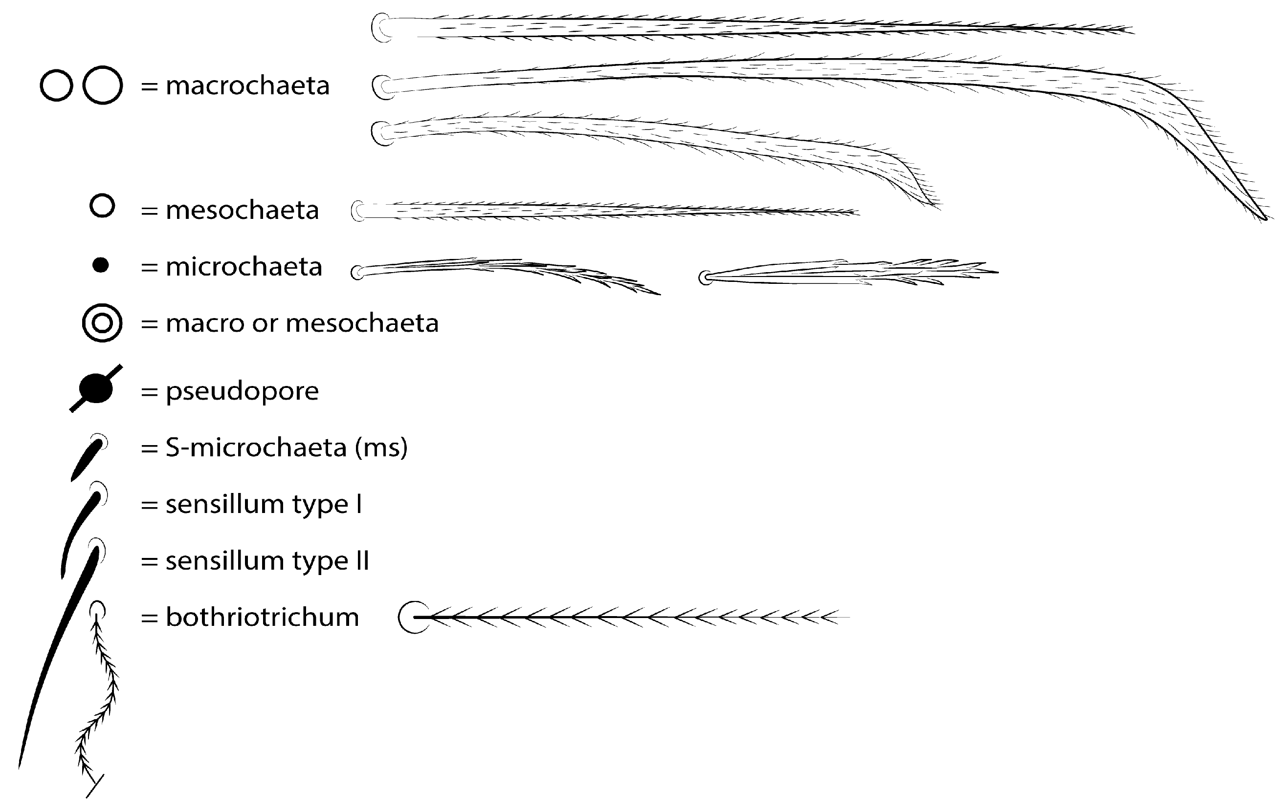
The terminology used in descriptions mainly follows for clypeal chaetotaxy [80]; labral chaetotaxy [81]; labial papillae, maxillary palp and basolateral and basomedian labial fields [80], using the Gisin system for name chaetae row [80], with additions for labeling the appendages of the sublobal plate [24,82]; postlabial chaetotaxy [83]; subcoxae outer chaetotaxy [84]; trochanteral organ [85,86]; unguiculus lamellae [87]; male’s genital plate [88]; head dorsal chaetotaxy [89], with additions [24,90,91]; trunk dorsal chaetotaxy [92], with additions [24,69,90]; and specialized chaetae (S-chaetae) [2]. Symbols used to depict the chaetotaxy are presented in Figure 4. Chaetae labels and other important taxonomic abbreviations are marked in bold on the text. Chaetae of uncertain homology are followed by a question mark (?). Chaetotaxy are all given on the left side of body only, except for the clypeal and labral regions.

3. Results

3.1. Mitochondrial Genome Features

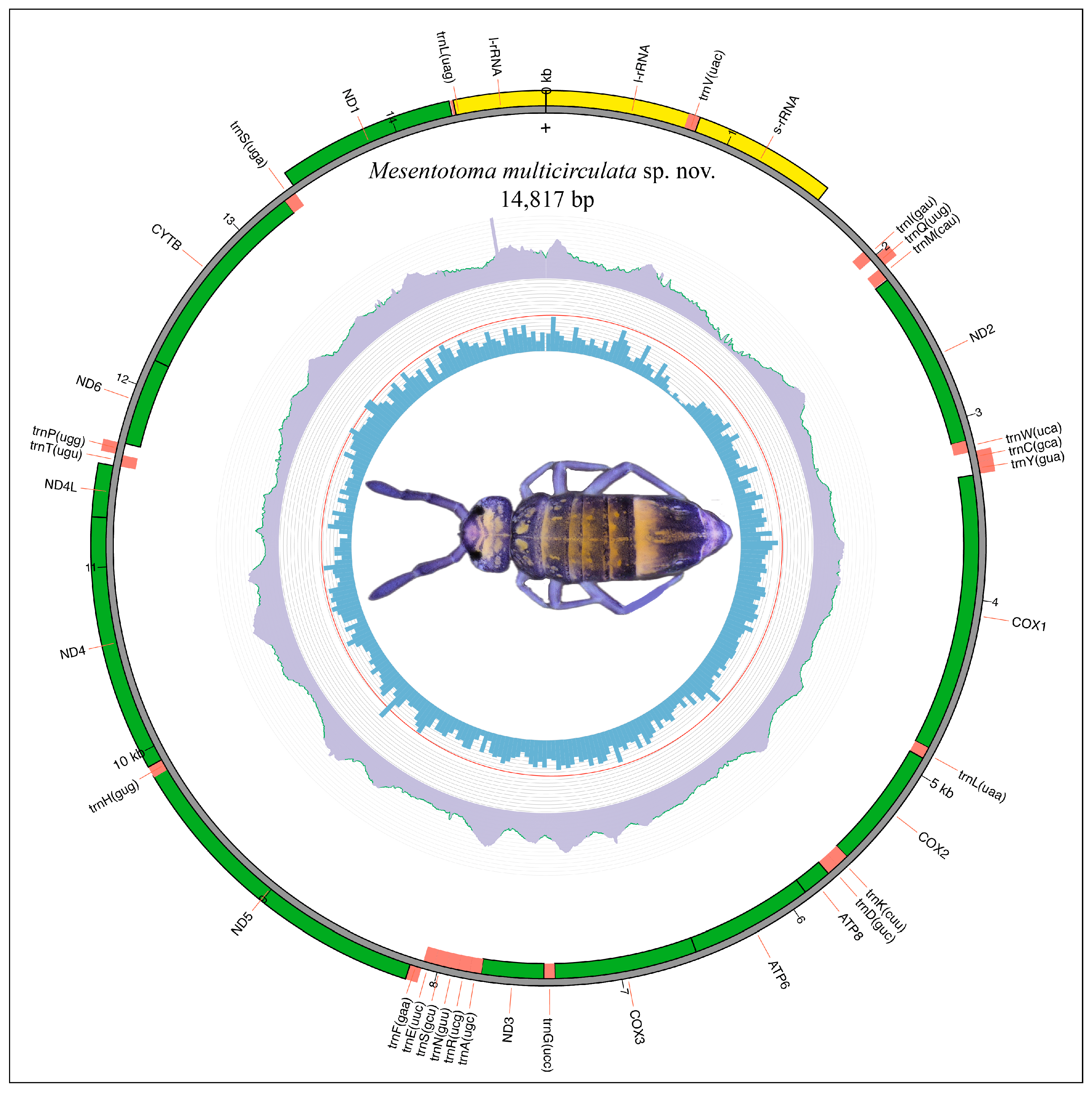
The complete mitogenome sequence of Mesentotoma multicirculata sp. nov. was 14,817 bp in length. It contained the typical set of 37 genes (13 PCGs, 22 tRNA genes, and 2 rRNA genes) usually present in Collembola mitogenomes, and a non-coding region (control region) of 367 bp (Table 2, Figure 1). The GC content was 27.43% [A (38.19%, 5659), C (16.39%, 2429), G (11.03%, 1635), T (34.38%, 5094)]. The gene order followed the presumed Pancrustacean ancestral gene arrangement (AGO), which is also the most frequent in the Collembola class. Other features such as the length and direction of each gene can be found in Table 2.

3.2. Phylogenetic Placement of Mesentotoma

Our concatenated phylogenetic matrix comprised 10,710 nucleotides and 39 partitions corresponding to 3 codons times the 13 mitochondrial PCGs (3 × 13 = 39). It was used to perform BI and ML analyses (Figure 2). Mesentotoma, Entomobrya proxima, and Willowsia jacobsoni were recovered as the basal taxa of Willowsia spp. (scaled) and the remaining Entomobryinae were unscaled and devoid of dental spines (e.g., Coecobrya, Sinella curviseta Brook, 1882, Entomobrya spp.). This result was highly supported in both phylogenetic inferences, indicating that Mesentotoma, like other Entomobryinae genera, is most likely paraphyletic. Since only one species was studied here, we emphasize the need for extended taxa sampling and a comprehensive review of the entire subfamily.

3.3. Taxonomy

Family Entomobryidae Tömösvary, 1882 [93]
Subfamily Entomobryinae Schäffer, 1896 [94] sensu Zhang & Deharveng [2]
Genus Mesentotoma Salmon, 1942 [1]

Type Species

Mesentotoma exalga Salmon, 1942 (p. 58, Pl. 19, Figures 21–30), New Zealand, Wellington municipality, Lyall Bay Island (original description) [1].

3.4. Diagnosis of Genus

Body unscaled, only with ciliate chaetae, eventually with wide tergal mic (Figures 3–5, 10, and 14); mac and mes finely ciliated, short, or elongated acuminate at the tip or foot-shaped (Figure 4); Ant IV apical bulb present (retractile or not) or absent (Figures 7A and 15A); Ant III–IV generally not annulated; Ant I–II with or without subdivisions (Figures 3A,B, 6, 7E and 15E); four labral papillae rounded or truncated, rarely with projections (Figures 8, 9A and 16A); eyes eight per side (Figures 6, 7E and 15E); dorsal macrochaetotaxy reduced or dense (Figures 11 and 17); males genital plate papillate (Figures 13D and 19B); unguiculus inner lamella excavate, truncate, or acuminate (Figures 12H and 18E); proximal dens with one–two projections on inner side (Figure 13C); dens crenulate and inner side without spines (Figure 13C); mucro with two teeth (proximal and distal) projected dorsally, proximal tooth subequal or smaller than distal tooth, proximal spine absent (Figures 13E and 19F). Adapted from Salmon [1] (p. 57); Christiansen [4] (p. 15), ref. [88] (p. 535); Christiansen & Bellinger [5] (p. 234), ref. [6] (p. 984); Jordana [7] (p. 290).

3.5. Remarks

Based on the original description, Mesentotoma differs from Entomobrya basically by Ant III–IV annulation, dens with ciliate spines, and mucro devoid of basal spines [1]. Christiansen [4], based on the type species, M. exalga, and two other species (M. dollfusi and M. laguna), redefined the genus diagnosis, removing the presence of annulations on Ant III–IV and dental spines, but including other characteristics (e.g., Ant IV apical bulb, labral papillae shape, unguiculus ai lamellae truncate/excavate, size of mucro teeth), especially the subsegmentation of Ant I–II. Later, the same author considered that Ant IV and sometimes Ant III have signs of subsegmentation or annulation, but it was not reported whether such characteristics were observed in the type species [87].
From the holotype photograph of M. exalga (Figure 3) deposited in the Museum of New Zealand Te Papa Tongarewa (number AI.000158), it is possible to note that the Ant III–IV are not annulated, as well as the absence of dental spines, therefore the absence of a mucronal spine is still the only characteristic that distinguishes Mesentotoma [4,5,6,7].

3.6. Nomenclatural Acts for Tropical Species of Mesentotoma

Here, five species inserted in Entomobrya are transferred to Mesentotoma due to the absence of the mucronal spine: M. depressa (Marlier, 1945) [36] comb. nov., M. grassei (Delamare, 1952) [94] comb. nov., M. philippinica (Gapud, 1971) [95] comb. nov., M. pseudocoeruleopicta (Delamare, 1952) [94] comb. nov., and M. tranvercyana Viana & Cipola nom. nov., comb. nov. To see the differences between species see Table 3.

3.6.1. Mesentotoma depressa (Marlier, 1945) [36] comb. nov.

Figure 20, Table 3.
Entomobrya depressa Marlier [36] (p. 254, Figure 2), Democratic Republic of Congo, Équateur Province, Mbandaka, Eala village; North Kivu province, Rutshuru municipality (original description), not examined.

3.6.2. Mesentotoma grassei (Delamare, 1952) [94] comb. nov.

Figure 20, Table 3.
Entomobrya grassei Delamare [94] in Delamare-Deboutteville & Paulian [94] (p. 71, Figures 28–30), Ivory Coast, Abidjâ [as Du Banco] municipality (original description), not examined.

3.6.3. Mesentotoma philippinica (Gapud, 1971) [95] comb. nov.

Figure 20, Table 3.
Entomobrya philippinica Gapud [95] (p. 4, Figures 1 and 2), Philippines, Laguana province (original description), not examined.

3.6.4. Mesentotoma pseudocoeruleopicta (Delamare, 1952) [94] comb. nov.

Figure 20, Table 3.
Entomobrya pseudocoeruleopicta Delamare [94] in Delamare-Deboutteville & Paulian [94] (p. 71, Figures 31–33), Ivory Coast, Abidjâ [as Du Banco] municipality (original description), not examined.

3.6.5. Mesentotoma tranvercyana Viana & Cipola nom. nov., comb. nov.

Figure 20, Table 3.
urn:lsid:zoobank.org:act:572097B7-A6B9-4980-982F-E37543963335.
Entomobrya coeruleopicta Marlier [36] in Marlier [36] (p. 254, Figure 1), Democratic Republic of Congo, North Kivu province, Rutshuru municipality (original description), not examined.
Nomenclatural Act
In Marlier [36], Entomobrya coeruleopicta Marlier [36] was described, but the specific name is preoccupied in Entomobrya coeruleopicta Schött, 1917 [96] (senior homonym), currently as Drepanura coeruleopicta (Schött) [96]. Consequently, E. coeruleopicta Marlier is a junior homonym and is hereby replaced (ICZN 2000, Chapter 6, Article 23.4, and Chapter 12, Articles 52.3, 52.4, 53.3, 57.2 and 60.3) [97] by Mesentotoma tranvercyana nom. nov., comb. nov.
Etymology
Refers to the body pigment, see Marlier [36] (p. 254, Figure 1C), in the species (from Latin: tranversus—transversal, cyaneus—blue).

3.7. New Species from Brazil

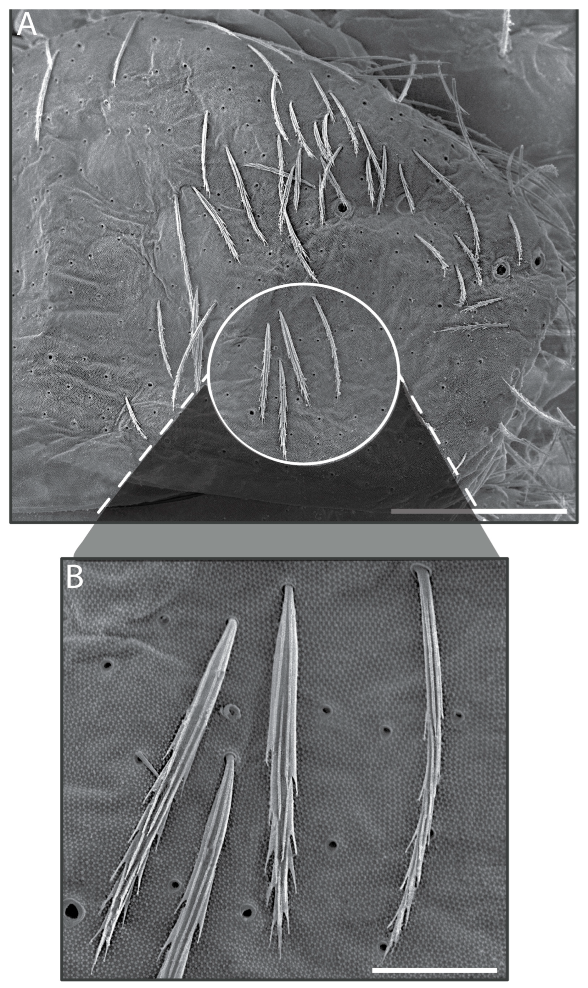
The following characters are shared by two new species of Mesentotoma herein studied and are not repeated in the descriptions: Habitus typical of Mesentotoma, head pentagon-shape, trunk oval to elliptical form (Figures 5, 6, 7E, 14 and 15E). Body covered with normal mic and wide mic (similar to scale), both ciliated (Figures 4 and 10A,B). Antennae segments with elongated sens, finger-shaped, and of different sizes and weakly to heavily ciliated chaetae (Figures 7A–C and 15A–C); Ant IV–III not annulated (Figures 7A,B and 15A,B); Ant I not subdivided (except in M. multicirculata sp. nov. from Pará State) and dorsally with three smooth chaetae at the base (Figures 6, 7E and 15E).
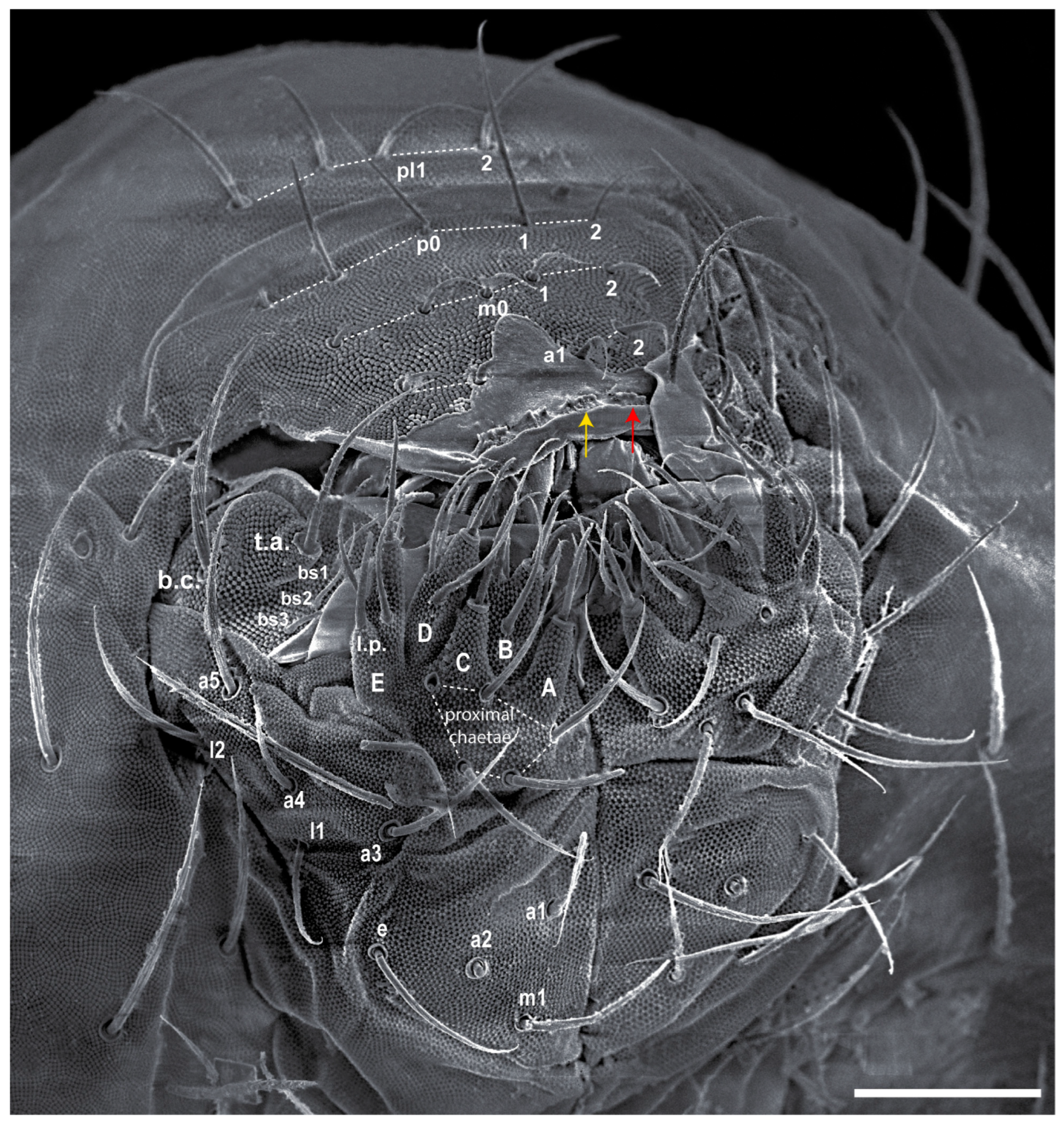
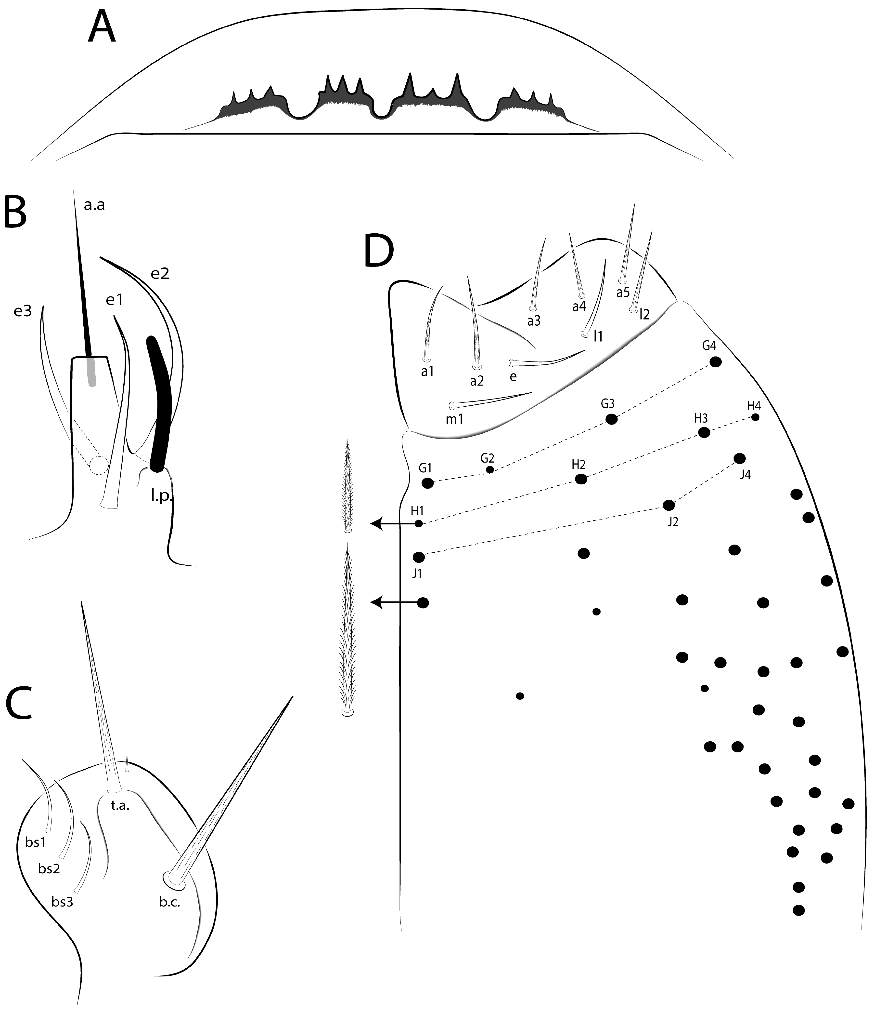
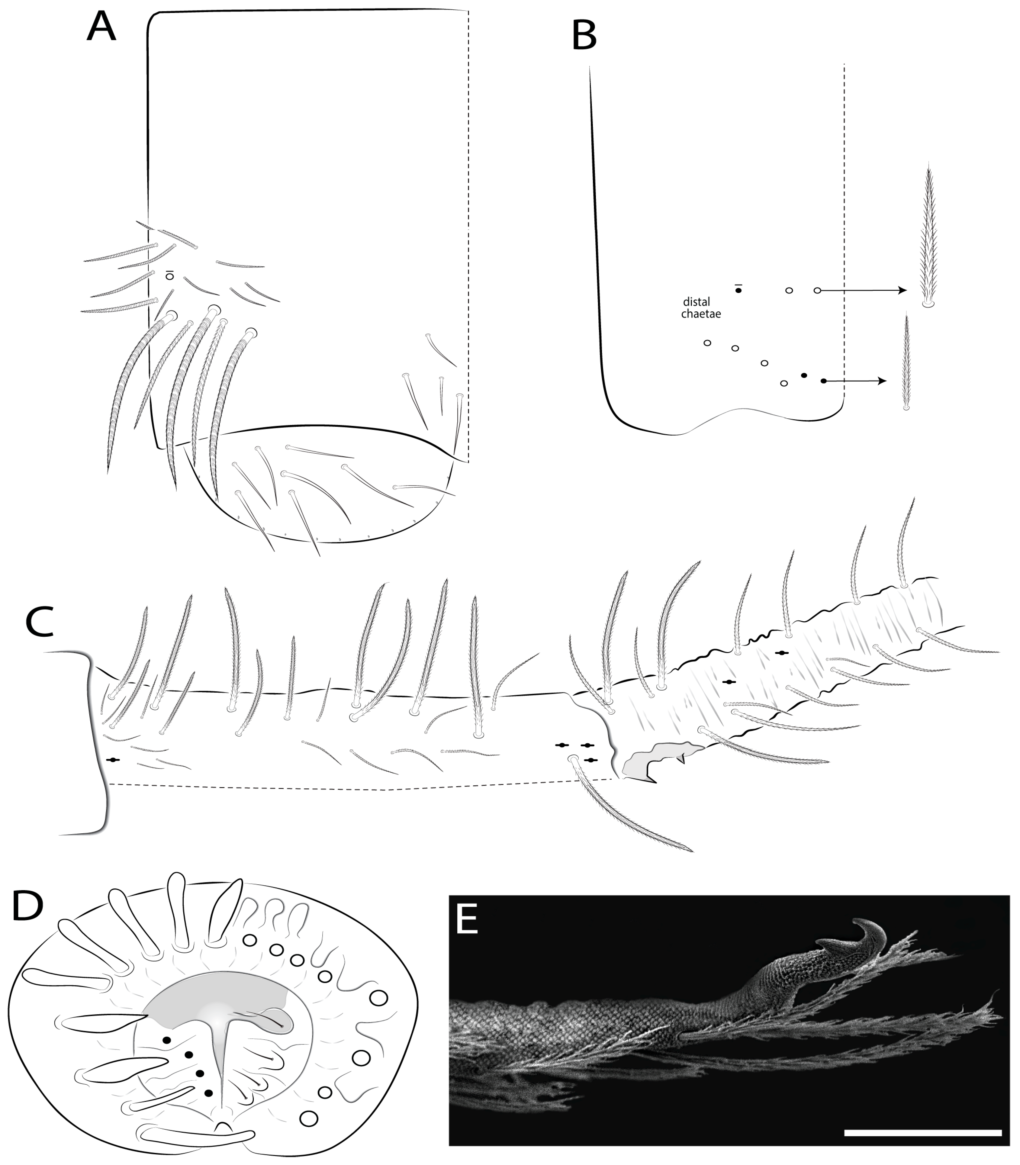
Head: With eyes 8 + 8 and a pair of post-ocellar bothriotricha (Figures 6, 7E and 15E). Four prelabral (pl1–pl2) chaetae, pl1 gently smaller than pl2 (Figures 7D, 8 and 15D); labral formula with four (a1–2), five (m0–2), five (p0–2) smooth chaetae, p0–1 largest (Figure 8). Four labral papillae (Figures 8, 9A and 16A). Labial palp with five main papillae (A–E) plus a hypostomal one (H), with zero, five, zero, four, three (e6 absent), and two guard appendages, papilla E with l.p. (Figures 8, 9B and 16B). Maxillary palp with b.c. and t.a. slightly ciliated; sublobal plate with three (bs1–3) inner appendages, plus one small distal appendix, all smooth (Figures 8 and 9C). Labium with five proximal chaetae, four slightly ciliated and one smooth (Figure 9).
Trunk: Th II–Abd V with ms and sens formula 1, 0| 1, 0, 1, 0, 0 and 2, 2 | 1, 2, 2, +, 3, respectively (Figures 11 and 17); Abd II–IV bothriotricha formula as 2 (a5, m2), 3 (a5, m2, m5), and 2 (T2, T4), respectively (Figures 11B,C and 17B,C).
Legs: Tibiotarsus outer side distally with one tenent hair discretely ciliate and apically capitate, inner side of tibiotarsus III with one smooth chaeta (Figures 12H and 18E). Unguis outer side with a pair of lateral teeth and one unpaired median tooth; unguiculus with four lamellae (ai, ae, pi, pe) (Figures 12H and 18E). Males with papillate genital plate, internally with four (atypical) small eugenital smooth chaetae (Figures 13D and 19B). Mucro bidentate, without basal spine (Figures 13E and 19F).

3.7.1. Mesentotoma multicirculata sp. nov. Viana & Cipola

urn:lsid:zoobank.org:act:FFC23A0F-0059-4721-8243-CC2F3B5384F4.
Diagnosis: Bluish body with irregular depigmented spots (circular and/or elongated) on head to Abd V (Figure 5). Ant IV apical bulb apically bilobed (Figure 7A); prelabral pl1 2 chaetae smooth (Figure 7D); head macrochaetotaxy with 6–8 ‘An’, 3 ‘A’, 1 ‘M’, 1 ‘S’, 2 ‘Pa’, 1 ‘Pm’, 2 ‘Pp’ and 3 ‘Pe’ mac, (Figure 7E); labral papillae with 3 projections (Figure 8 and Figure 9A); labial papilla E l.p. finger-shape and surpass the base of a.a. (Figure 9B); basolateral and basomedian labial fields with m1, e, l1 smooth, l2 slightly ciliate (Figure 9D); Th II a, m and p series respectively with 4, 0 and 0–1 inner mac; Th III–Abd III with 1, 0, 1 and 1 inner mac, respectively (Figure 11A,B); Abd IV with 2 inner and 13 outer mac (Figure 11C); unguis a.t. present; unguiculus III ai lamella acuminate and pe serrated (Figure 12H); collophore anteriorly with 3–4 mac (Figure 13A); mucro proximal tooth subequal than the distal one (Figure 13E).
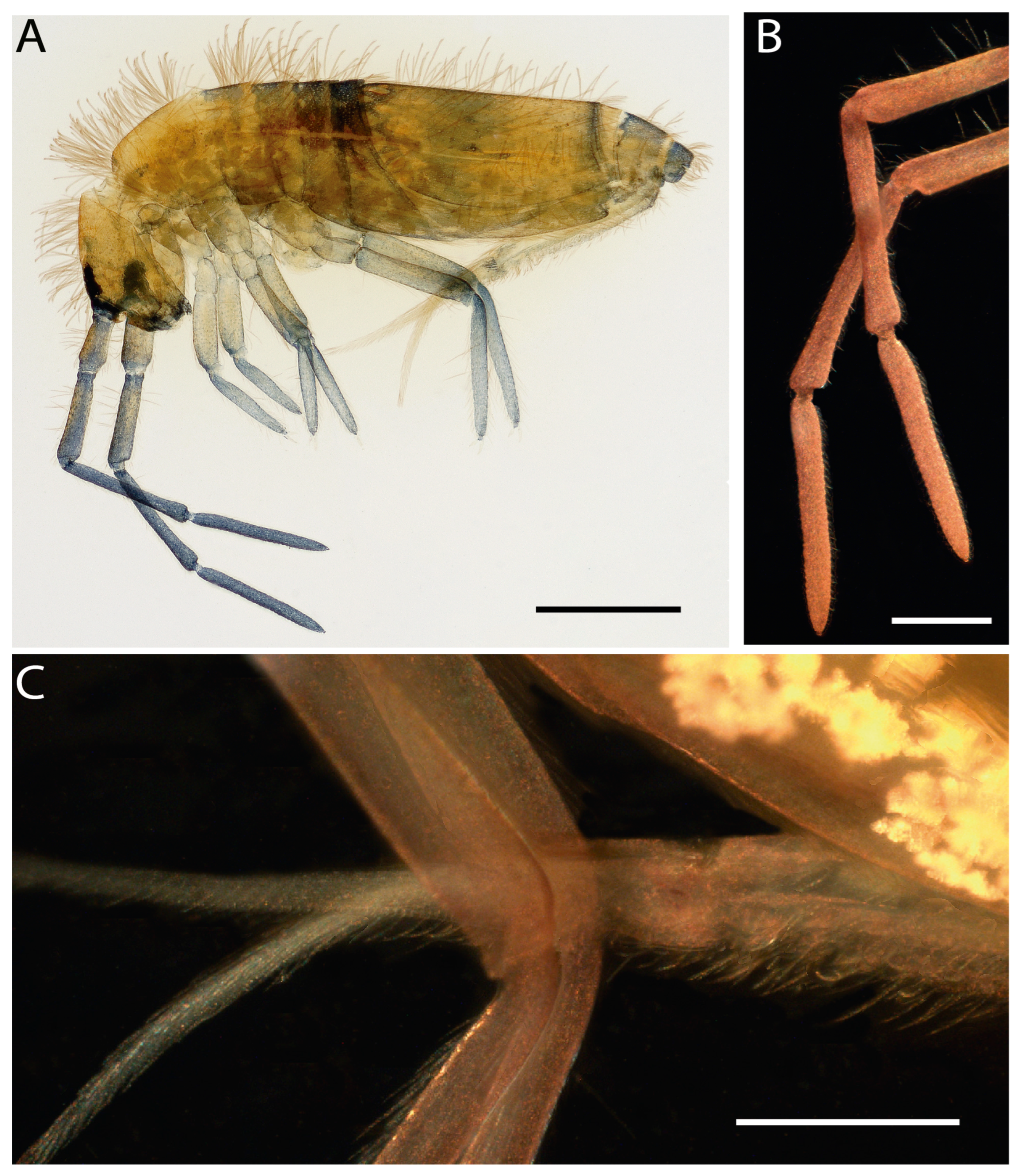
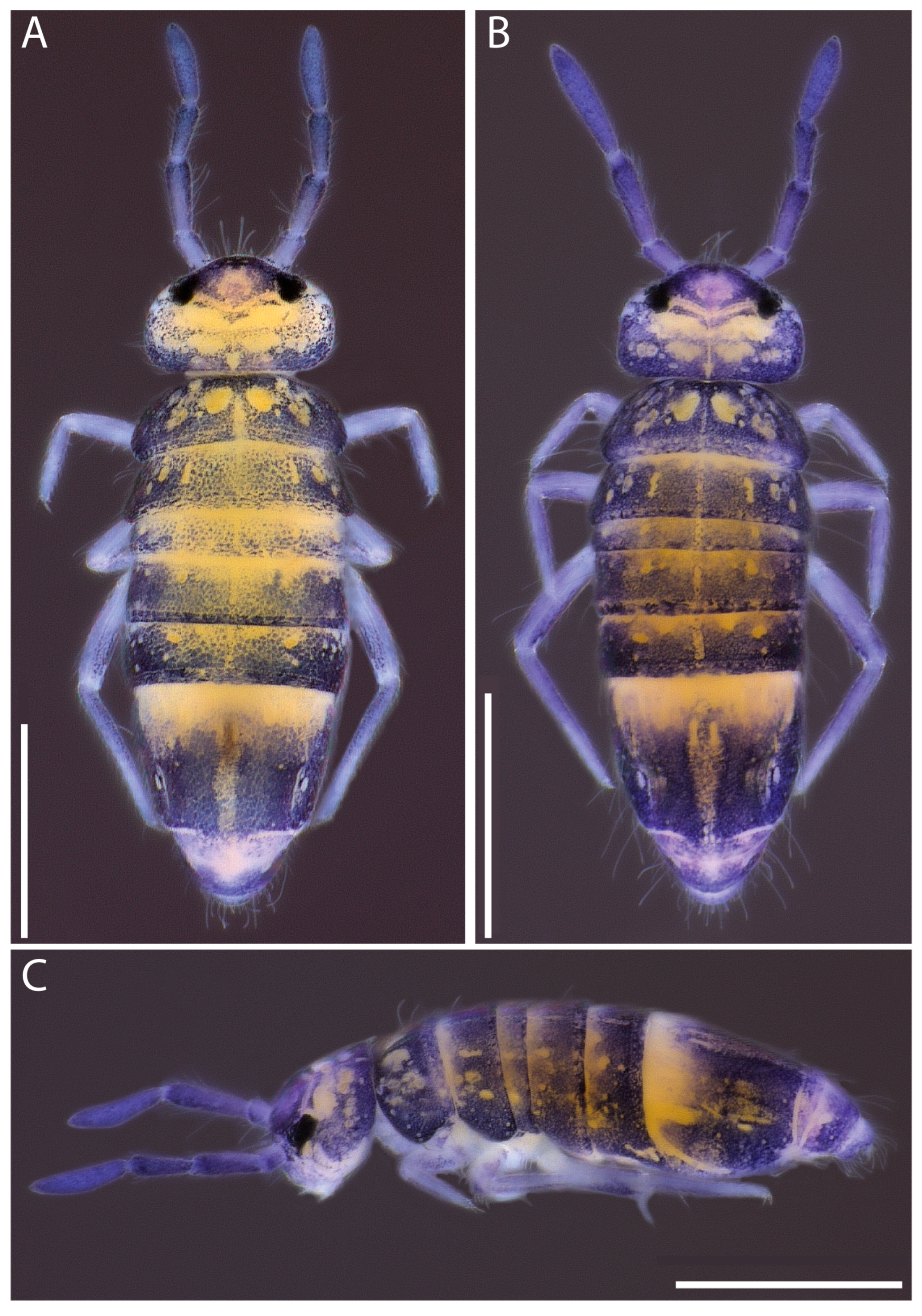
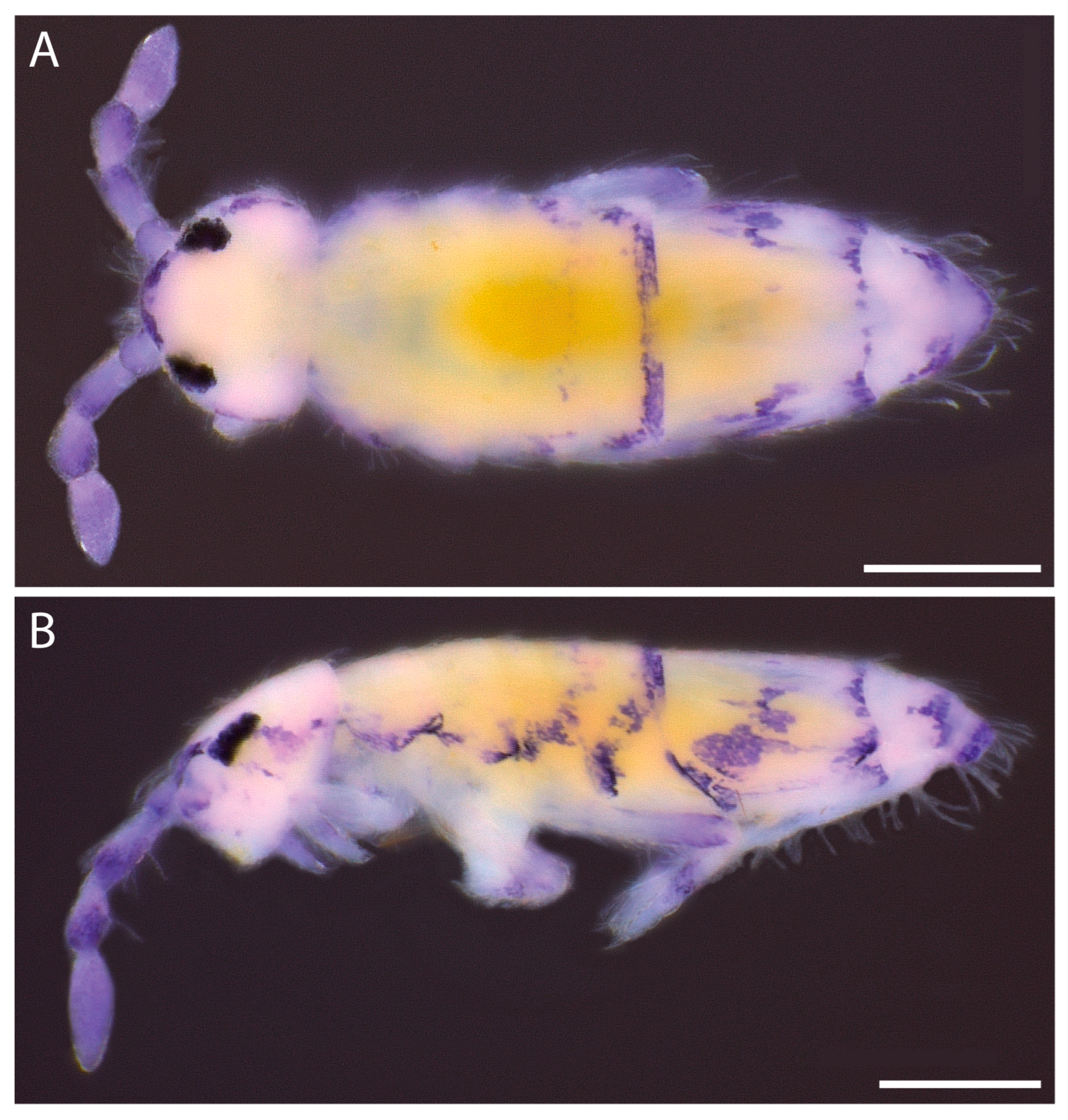
Description: Total length (head + trunk) of specimens 1.02–1.40 mm (n = 4), holotype 1.35 mm. Specimens completely black bluish, except Abd IV anteriorly and some irregular spots (circular and/or elongated) depigmented dorsally on head to Abd V, often with the dorsal medial part of the head and trunk, as well as the appendages, less pigmented; eyepatches black (Figure 5).
Figure 4. Symbols used in chaetotaxy descriptions of Mesentotoma species.
Diversity 18 00089 g004
Figure 5. Mesentotoma multicirculata sp. nov.; habitus of fixed specimens in ethanol: (A,B) dorsal view; (C) lateral view. Scale bars: 0.5 mm.
Diversity 18 00089 g005
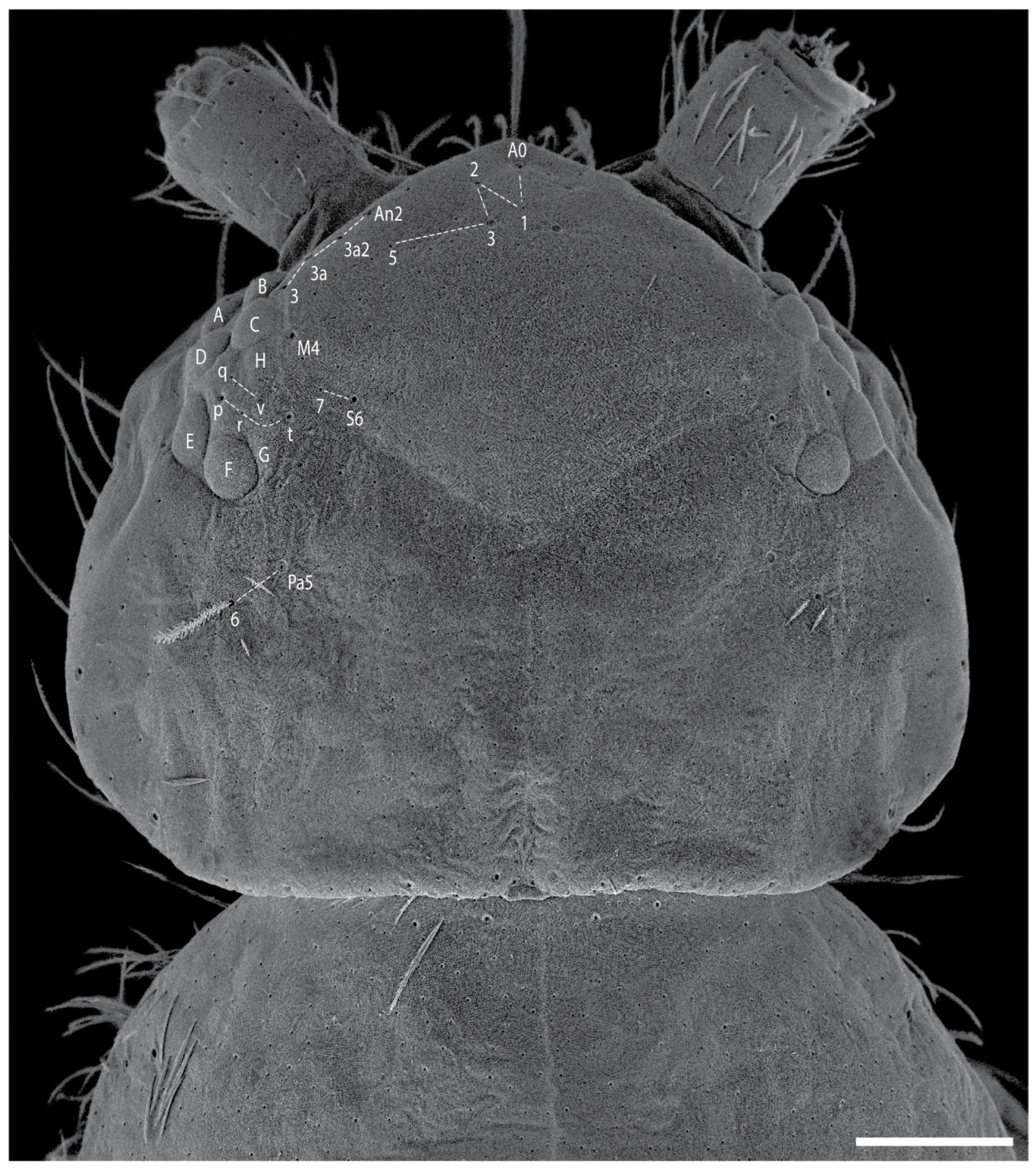
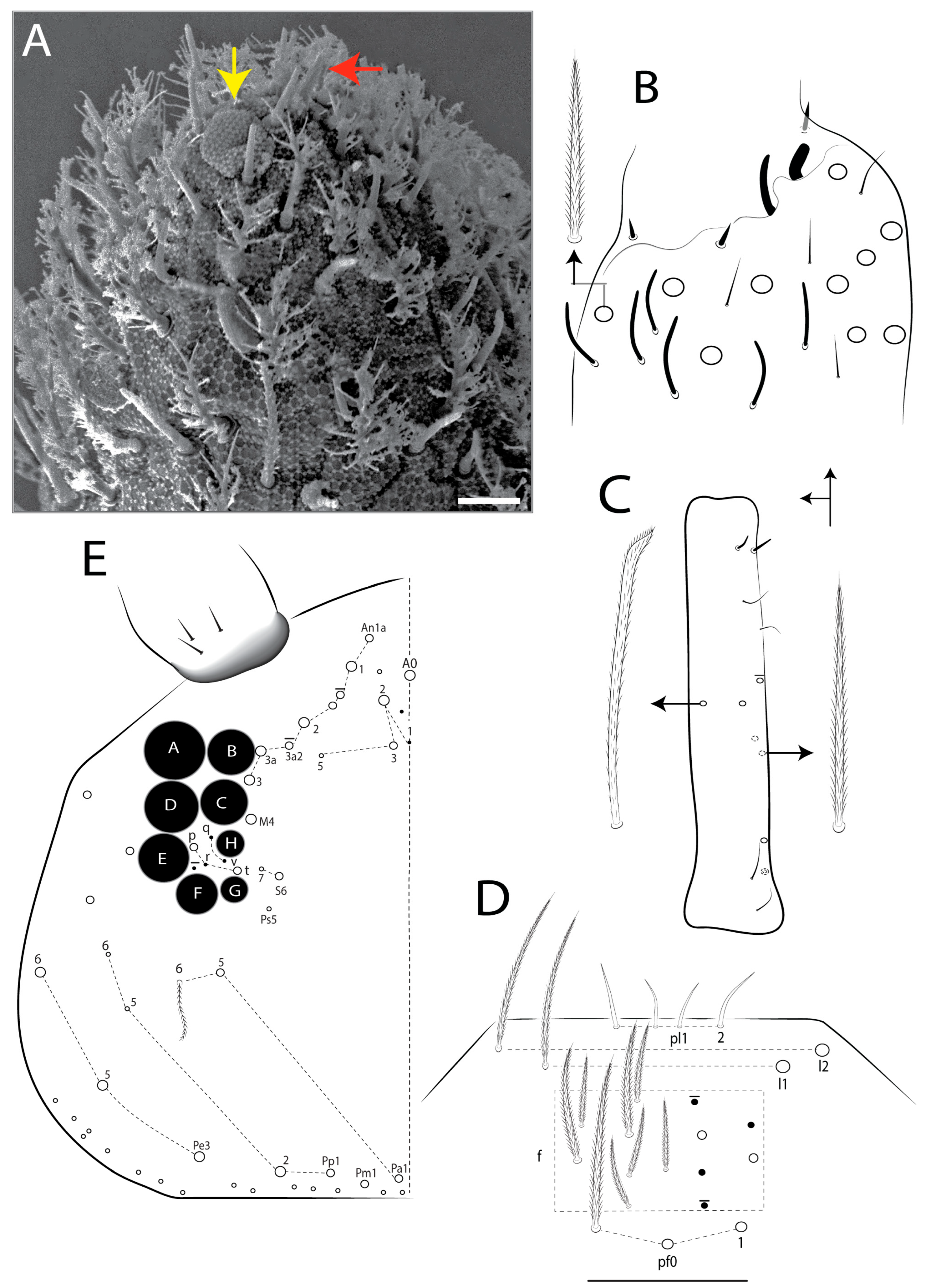
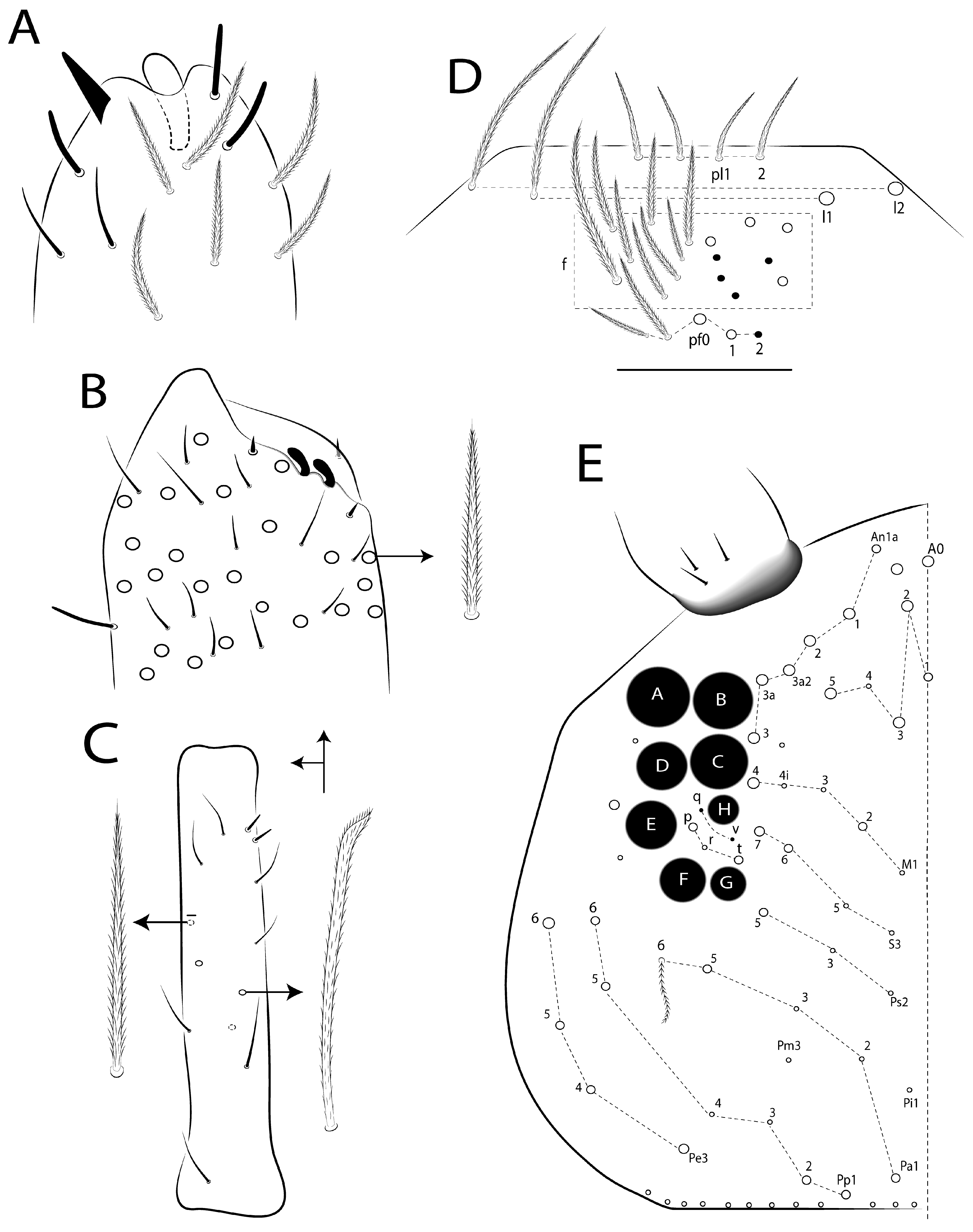
Head: Antenna shorter than the trunk length (Figure 5), ratio antennae: trunk = 1: 1.77–2.01 (n = 4), holotype 1:1.77; antennal ratio as I: II: III: IV = 1:2.07–2.71:1.81–2.26:2.76–3.47 (n = 4), holotype 1:2.71:2.26:3.47. Ant IV with an apical bulb apically bilobed (often retractable), one conical pin projection, and numerous sens of different sizes (Figure 7A). Ant III organ with one blunt-shape and one finger-shape sens, three guard sens, six finger-shape sens, and at least four thin sens (Figure 7B). Ant II with three–four dorsal and two–three ventral mac, dorsally with about four slim and elongated and two distal finger-shaped sens (Figure 7C). Eyes A larger than the others, G and H smaller, with five–six interocular chaetae (q, v, p, r, t plus one extra mic present or absent); head dorsal chaetotaxy (Figure 6 and Figure 7E) with six–eight ‘An’ (An1a–3), three ‘A’ (A0, A2–3), one ‘M’ (M4), one ‘S’ (S6), two ‘Pa’ (Pa1, Pa5), one ‘Pm’ (Pm1), two ‘Pp’ (Pp1–2), and three ‘Pe’ (Pe3, Pe5–6) mac. Clypeal formula with 4 (l1–2), 9–13 (1 unpaired), 3 (pf0–1) ciliate chaetae, l1–2 largest, pf0–1 gently larger, others subequal (Figure 7E). Prelabral pl1–2 smooth chaetae (Figure 7E and Figure 8). Labral inner papillae with three projections, median projection sometimes smaller, outer papillae with three projections subequal (Figure 8 and Figure 9A). Labial papilla E l.p. finger-shape and surpass the base of a.a. (Figure 9B). Maxillary palp b.c. 1.3 longer than t.a. (Figure 9C). Basolateral and basomedian labial fields with a1–5 and l2 slightly ciliate, m1, e, l1–2 smooth (Figure 9D). Ventral head with at least 43 ciliate chaetae; cephalic groove with 4 ciliate chaetae, b.c. absent; postlabial formula with 4 (G1–4), 4 (H1–4), 3 (J1–4) chaetae, G2, H1, and H4 smaller, others subequal (Figure 9D).
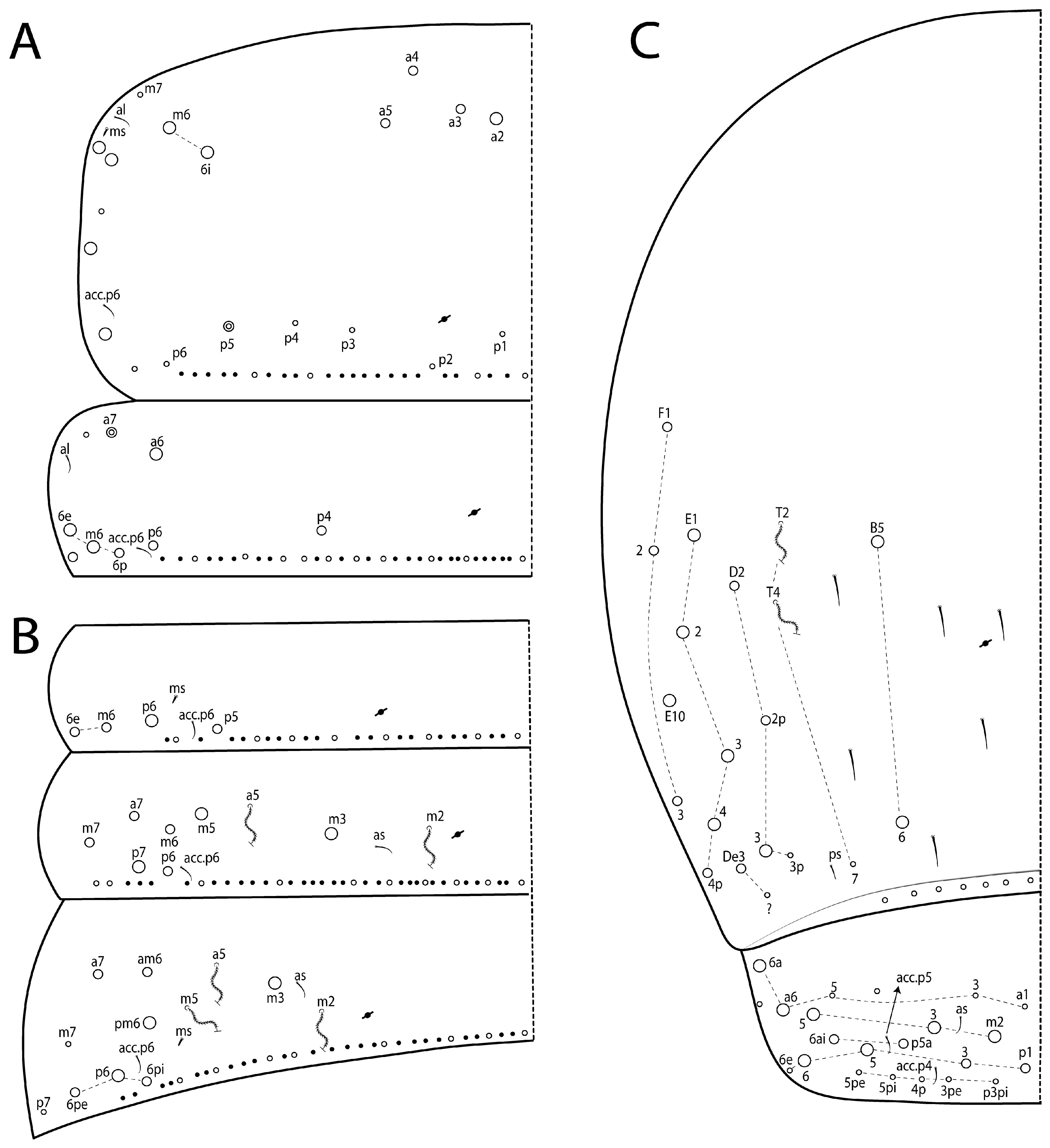
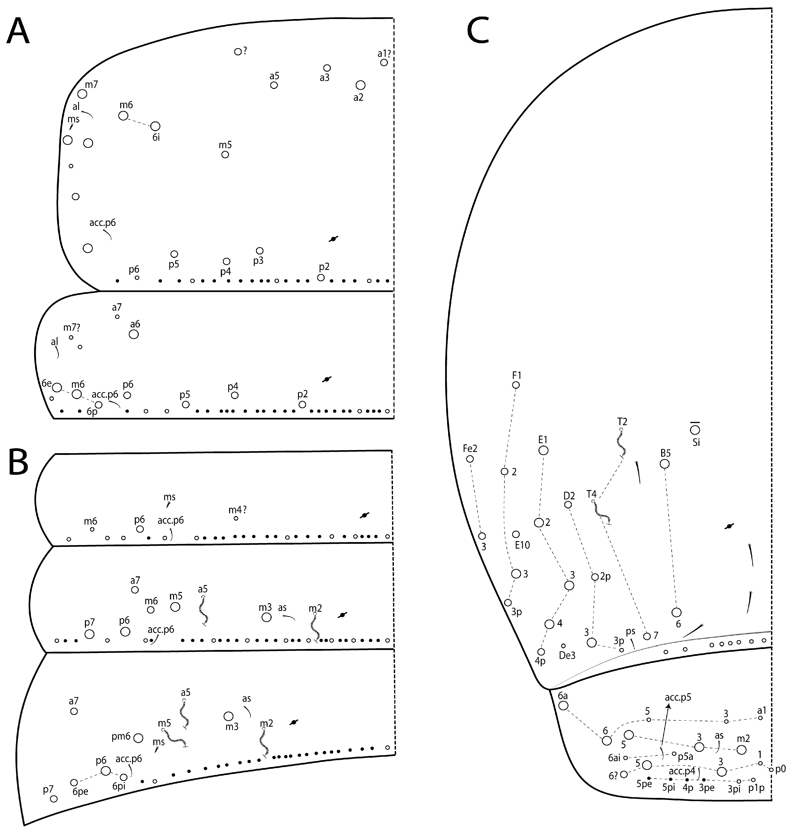
Thorax chaetotaxy (Figure 10 and Figure 11A): Th II a, m, and p series (excluding the anterior collar) with four (a2–5), two (m6–6i), and zero–one (p5) mac, respectively. Th III a, m, and p series with one–two (a6–7), three (m6p–6e), and two (p4, p6) mac, respectively. Ratio Th II:III = 1.38–1.04:1 (n = 4), holotype 1.32:1.
Abdomen chaetotaxy (Figure 11B,C): Abd I a, m, and p series with zero, two (m6–6e), and two (p5–6) mac, respectively. Abd II a, m, and p series with one (a7), four (m3, m5–7), and two mac (p6–7), respectively. Abd III a, m, and p series with one (a7), three (m3, am6, pm6), and three (p6pi–6pe) mac, respectively. Abd IV with 2 (B5–6) inner mac of A–C series and 13 (D2–3, De3, E1–4p, E10, F1–3) outer mac of T–Fe series; about 7 sens (ps type I and others type II) and 7 posterior mes. Abd V a, m, pa, and p series with two (a6–6a), three (m2–3, m5), two (p5a–6ai), and four (p1, p3, p5–6) mac, respectively. Ratio Abd III:IV = 1:3.34–3.61 (n = 4), holotype 1:3.57.
Figure 6. Mesentotoma multicirculata sp. nov.: Ant I, head and anterior region of Th II (dorsal view), A–H are eyes, dashed lines indicate dorsal chaetotaxy ‘An’, ‘A’, ‘M’, ‘S’ and ‘Pa’ series of the head. Scale bars: 0.05 mm.
Diversity 18 00089 g006
Figure 7. Mesentotoma multicirculata sp. nov.; antennae and dorsal head: (A) apex of Ant IV (left side), the red arrow indicates the pin projection and yellow the apical bulb (scale bars: 0.005 mm); (B) Ant III distally (lateral view); (C) left Ant II (dorsal view); (D) clypeal and prelabral chaetotaxy; (E) head dorsal chaetotaxy (left side).
Diversity 18 00089 g007
Figure 8. Mesentotoma multicirculata sp. nov.; head anterior region and mouthparts: in labral regions pl1–2 represent the prelabral chaetae; a1–2, m0–2, and p0–2 the labral chaetae; yellow arrows indicate inner labral papillae and red arrows the outer labral papilla; b.c. and t.a. are of the maxillary palp, bs1–3 the appendages of sublobal plate (minute distal appendix omitted); A, B, C, D, and E are the labial papillae (left side) with l.p. (lateral process) on papilla E; and a1–5, m1, e and l1–2 the labial chaetae of the basomedian and basolateral fields. Scale bars: 0.02 mm.
Diversity 18 00089 g008
Figure 9. Mesentotoma multicirculata sp. nov.; ventral head: (A) labral papillae; (B) labial papillae E (right side); (C) maxillary palp and sublobal plate (right side); (D) basomedian and basolateral labial fields and complete postlabial chaetotaxy (right side).
Diversity 18 00089 g009
Figure 10. Mesentotoma multicirculata sp. nov.; Th II left side (dorsal view): (A) chaetotaxy part; (B) different ciliated macrochaetae, wide scale-shaped, and normal. Scale bars: (A) (0.05 mm), (B) (0.01 mm).
Diversity 18 00089 g010
Figure 11. Mesentotoma multicirculata sp. nov.; dorsal chaetotaxy (left side): (A) Th II–III; (B) Abd I–III; (C) Abd IV–V.
Diversity 18 00089 g011
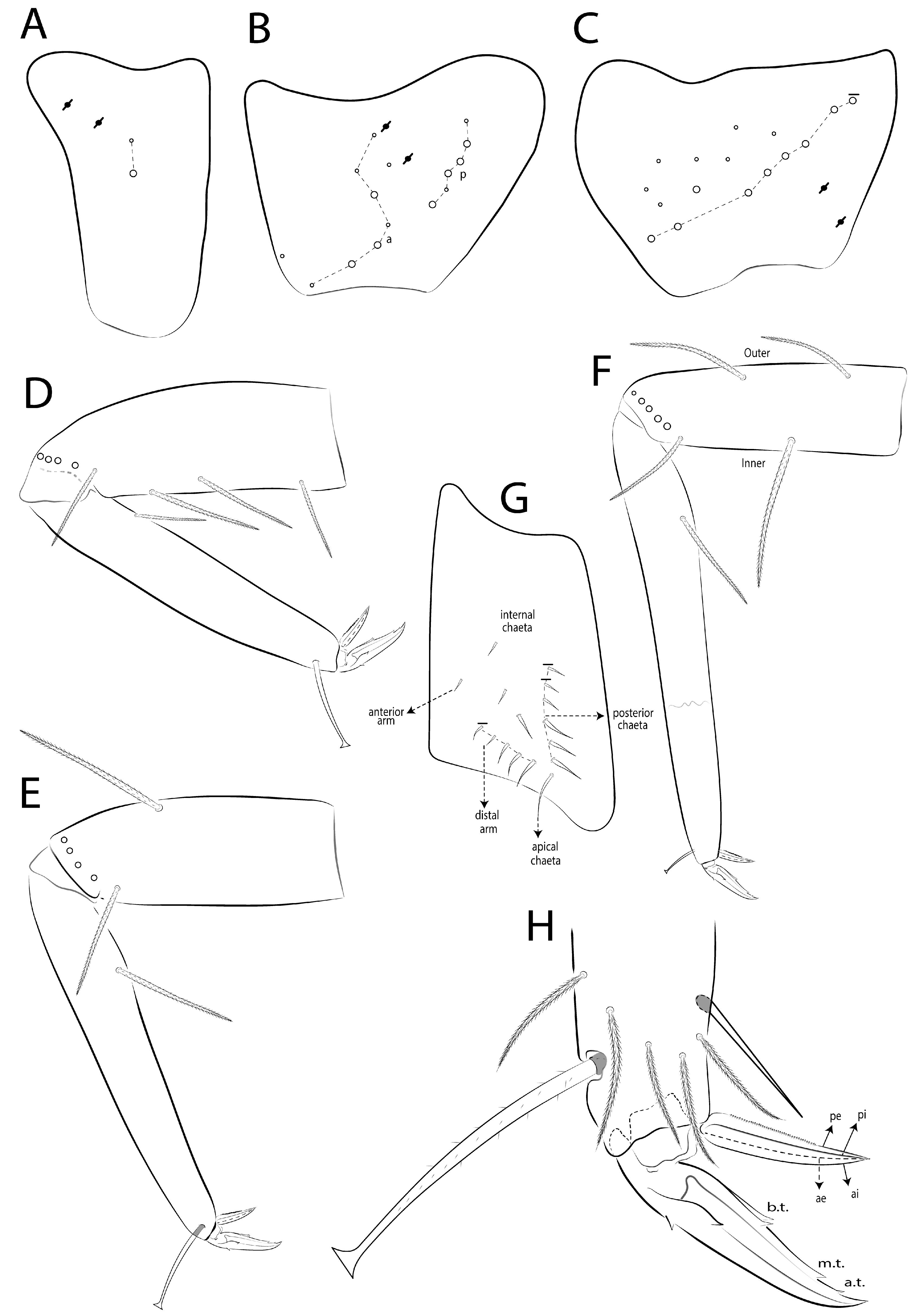
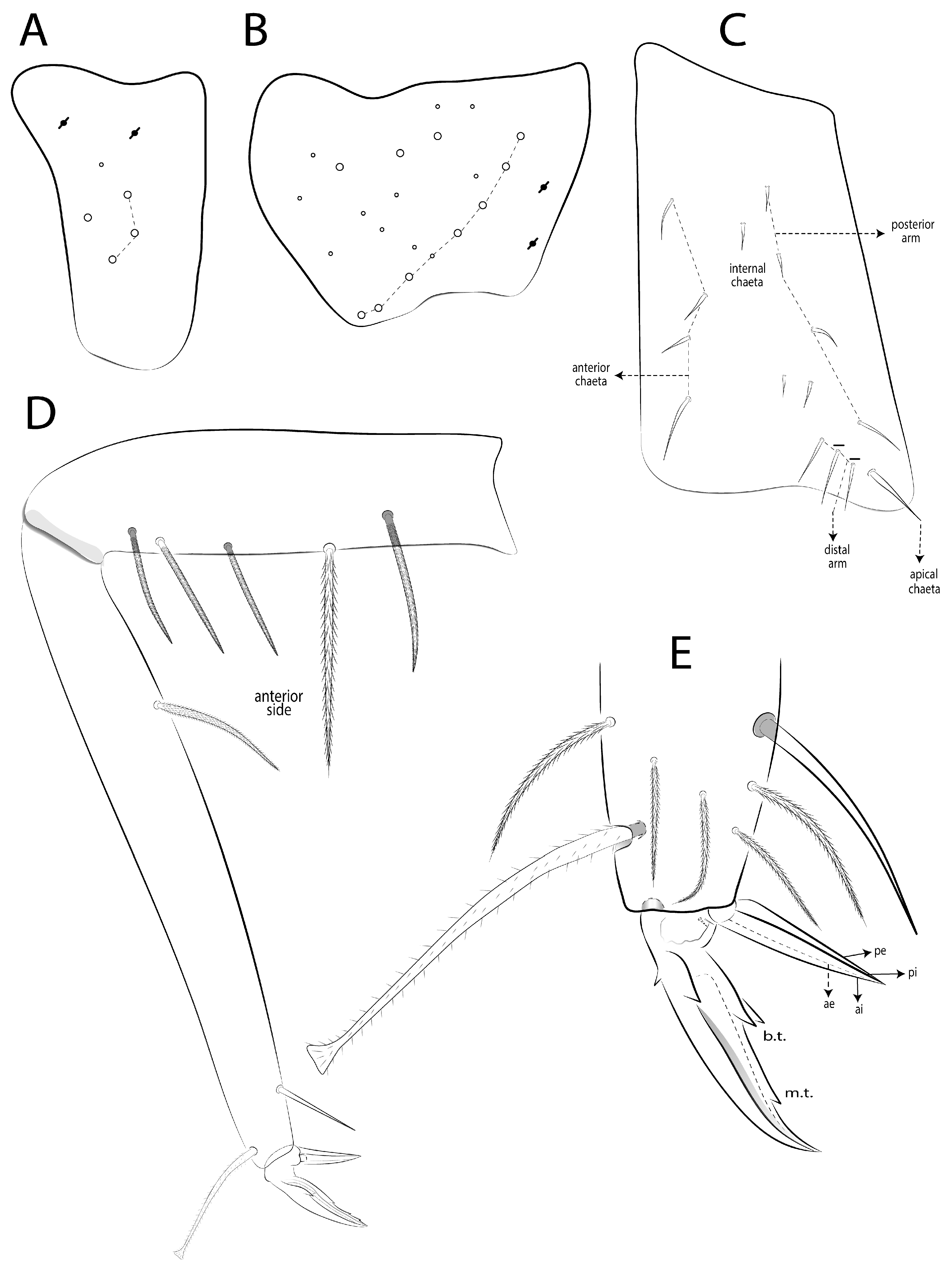
Legs (Figure 12): Subcoxa I with two chaetae and two psp; subcoxa II with an anterior row of seven chaetae, one anterior and one posterior chaeta, and two psp, posterior row of six chaetae; subcoxa III with one row of seven–eight chaetae, about eight anterior chaetae, and two psp posteriorly (Figure 12A–C). Trochanteral organ with 13–16 spine-like chaetae, 4–6 anterior, 1 posterior, 3 internal, 1 apical, and 4–5 on the distal arm (Figure 12G). Femur I–III with zero, one, two outer mac and four, zero, one inner mac. Tibiotarsus I–III with one inner mac on proximal region (Figure 12D–F). Unguis with four inner teeth, b.t. on proximal half, m.t. on distal one-fourth and subequal to b.t., a.t. on distal one-eighth and smaller than others. Unguiculus I–III lamellae (ae, ai, pe, pi) smooth and acuminate, except III pe serrated on proximal two-thirds; ratio unguis/unguiculus III = 1: 0.70. Tibiotarsus III smooth chaeta 0.79 smaller to the unguiculus; tenent hair finely ciliate and 1.10 larger that the unguis (Figure 12H).
Figure 12. Mesentotoma multicirculata sp. nov.; legs: (AC) chaetotaxy of subcoxa I–III, respectively (outer side); (DF) chaetotaxy of femur and tibiotarsus I–III, respectively (anterior side); (G) trochanteral organ (posterior view); (H) distal tibiotarsus and empodial complex III (posterior view).
Diversity 18 00089 g012
Collophore (Figure 13A): anterior side with about 15–16 ciliate chaetae, 3–4 mac (1 median present or absent), and about 12 chaetae of different sizes widely distributed; posterior side with 4 smooth chaetae distally; lateral flap with 8 smooth chaetae.
Genital plate (Figure 13D): Males with seven papillae per side and 8 + 1 (unpaired) circumgenital chaetae, three lanceolate (one unpaired), four club-shape, one finger-shape, and one pin-shape. Female plate with four anterior and three posterior (one unpaired) small smooth chaetae (as Figure 19C).
Figure 13. Mesentotoma multicirculata sp. nov.; abdominal appendages: (A) collophore chaetotaxy (lateral view); (B) ventrodistal manubrium (left side); (C) manubrium and proximal dens dorsally (right side); (D) male genital plate; (E) dens distally and mucro in outer view (Scale bars: 0.02 mm).
Diversity 18 00089 g013
Furcula (Figure 13B,C,E): Manubrium ventrally with two–three subapical and about six distal ciliate chaetae, two inner gently smaller, others subequal (Figure 13B). Manubrium dorsally with one lateral row of seven elongated and four short ciliate mac abruptly acuminate at the tip; manubrial plate with three psp and one mac (Figure 13C). Dens proximal region with two inner pointed projections, dorsally with two proximal psp, and proximal three-fourths of dens crenulate (Figure 13C). Mucro teeth subequal in size (Figure 13E).
Etymology: Refers to the body depigmented spots (Figure 5) in the new species (from Latin: multi—various, circulus—circle).
Type material: Holotype female on slide (INPA-CLL 0000279): Brazil, Pernambuco, Fernando de Noronha Island, “Sancho” Beach, 03°51′19″ S, 32°26′35″ W (Figure 20), 45 m., 27.xii–10.i.2020, malaise trap, JA Rafael, F Limeira-de-Oliveira & LC Castro coll. In total, 164 paratypes: 5 females on slides and 19 specimens in alcohol (INPA-CLL 0000280–285) same date as holotype; 5 females on slides (INPA-CLL 0000286–290): idem, except 08–23.vii.2019; 3 males and 6 females on slides and 56 specimens in alcohol (INPA-CLL 0000291–300), plus 5 in alcohol (SNHM) and 2 juveniles on slides (CC/UFRN): idem, except 23.i–12.ii.2020; 1 juvenile on slide (INPA-CLL 0000301): 4 females and 15 specimens in alcohol (MNRJ): idem, except 25.ix–08.x.2019; 1 male and 1 female on slides (MNCN) plus 1 male and 3 females on slides and 1 specimen in alcohol (INPA-CLL 0000302): idem, except trail for “Sancho” Beach, manual collect, 01–09.vi.2019, J.A. Rafael, F. Limeira-de-Oliveira & D.M.M. Mendes coll; 2 males and 2 females on slides and 6 specimens in alcohol (MZUSP): same date as holotype, except 11–27.xi.2019; 2 males and 2 females on slides and 14 specimens in alcohol (CC/UFRN): idem, except 07–21.viii.2019.
Other examined material: one male and one juvenile on slides and four specimens in alcohol (INPA): Brazil, Pará state, Belém municipality, forest near gate 4 of the Federal University of Pará (UFPA), 01°28′02″ S, 48°26′40″ W (Figure 20), 6 m., 05–07.i.2022, pitfall trap, GC Tavares coll. One male on slide (INPA): Bahia state, Salvador municipality, “Patamares” neighborhood, “Colina C” condominium, 12°57′01″ S, 38°24′30″ W, 24 m., 19–23.x.2017, malaise trap, T Mahlmann coll.

3.7.2. Mesentotoma noronhaensis sp. nov. Viana & Cipola

urn:lsid:zoobank.org:act:CEDF773D-C42F-4E36-BA1C-8AE44473DF2D.
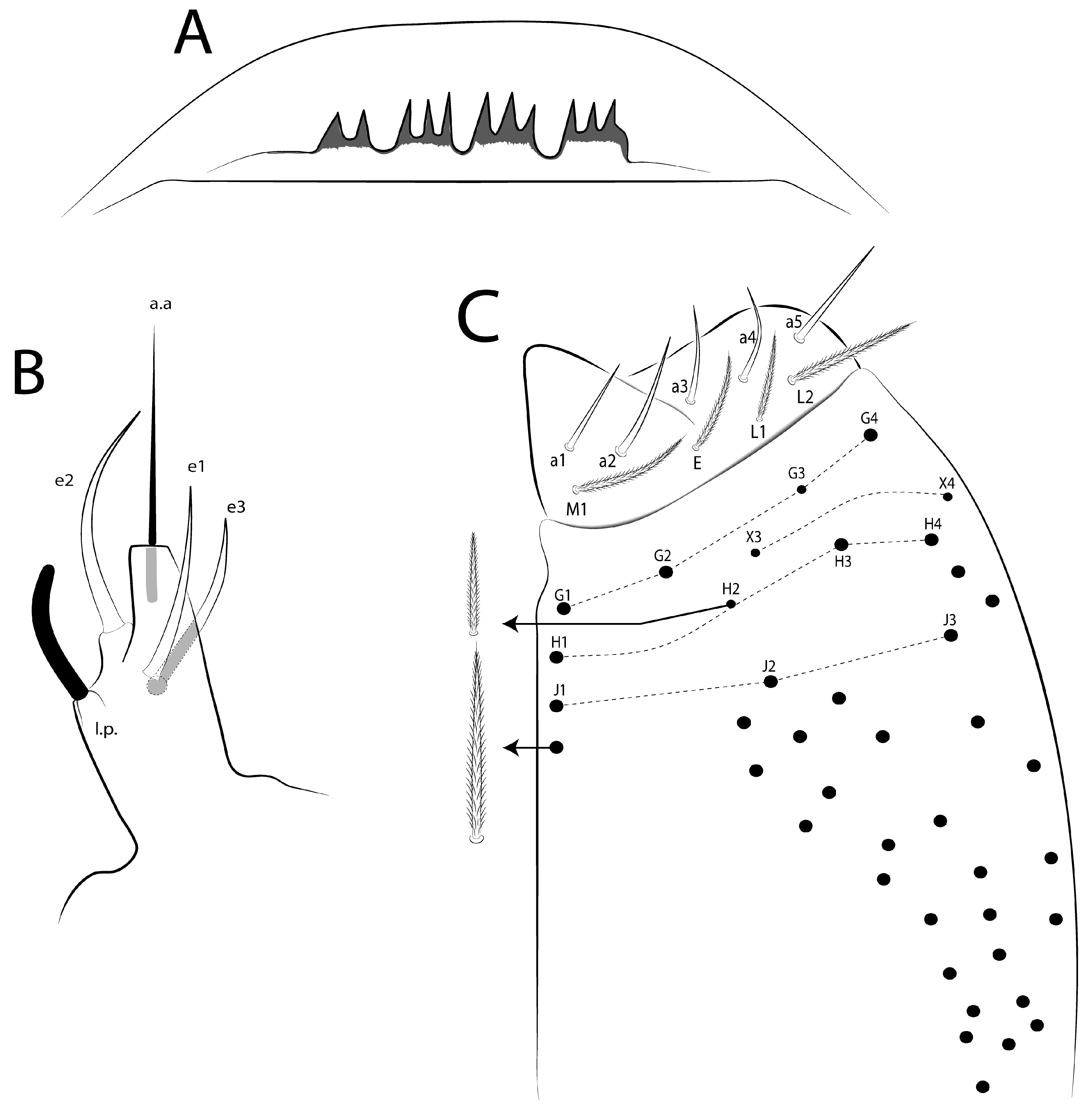
Diagnosis: Body with bluish pigments on Ant I–IV, head laterally to Abd II, where it forms a transverse band, Abd III–VI posteriorly with one, one, one (incomplete), and one transversal band, plus other incomplete band on median Abd IV; femur II–III distally and tibiotarsus I–III median region pigmented (Figure 14). Ant IV with the apical bulb unilobed (Figure 15A); prelabral pl1–2 chaetae gently ciliate (Figure 15D); head macrochaetotaxy with 6 ‘An’, 5 ‘A’, 2 ‘M’, 2 ‘S’, 1 ‘Ps’, 2 ‘Pa’, 0 ‘Pm’, 4 ‘Pp’, and 4 ‘Pe’ mac (Figure 15E); labral inner papilla with 3 projections and outer papillae with 2–3 projections (Figure 16A); labial papilla E l.p. finger-shape and reached or almost reached the base of a.a. (Figure 16B); basolateral and basomedian labial fields with M1, E, L1–2 ciliate (Figure 16C); Th II a, m, and p series with 5, 1, and 4 inner mac, respectively; Th III–Abd III with 3, 0, 1, and 1 inner mac, respectively (Figure 17A,B); Abd IV with 2–3 inner and 16 outer mac (Figure 17C); unguis a.t. absent; unguiculus III ai lamella acuminate and pe smooth (Figure 18E); collophore anteriorly with 2 mac (Figure 18A); mucro proximal tooth smaller than distal one (Figure 19F).
Description: Total length (head  +  trunk) of specimens 1.08–1.26 mm (n  =  3), holotype 1.22 mm. Specimens pale yellowish or white with irregular black bluish pigments on Ant I–IV, head anteriorly and laterally forming a band up to Abd III, which joins in a transverse band over this last segment (eventually depigmented in the dorsal region), Abd III–VI posteriorly with one, one, one (incomplete), and one transversal band, plus other incomplete band on median Abd IV; femur II–III with a distal spot and tibiotarsus I–III with a median spot; eyepatches black (Figure 14).
Figure 14. Mesentotoma noronhaensis sp. nov.; habitus of a fixed specimen in ethanol: (A) dorsal view; (B) lateral view. Scale bars: 0.2 mm.
Diversity 18 00089 g014
Head: Antenna shorter than the trunk length (Figure 14), ratio antennae/trunk = 1:2.48–2.54 (n = 2), holotype 1:2.48; antennal ratio as I:II:III:IV = 1:1.74–2.21:1.64–2.19:2.45–3.47 (n = 2), holotype 1:1.74:1.64:2.45. Ant IV with larger apical bulb apically unilobed, one conical pin projection, and numerous sens of different sizes (Figure 15A). Ant III organ with 2 club-shaped sens, 3 guard sens, at least 1 finger-shaped sens, and 12 thin sens (Figure 15B). Ant II with two dorsal and one–two ventral mac, dorsally with about seven slim and elongated sens and two distal finger-shaped sens (Figure 15C). Eyes A–C larger than the others, G and H smaller, with 5 interocular chaetae (q, v, p, r, t); head dorsal chaetotaxy (Figure 15E) with 6 ‘An’ (An1a–3), 5 ‘A’ (A0–3, A5), 2 ‘M’ (M2, M4), 2 ‘S’ (S6–7), 1 ‘Ps’ (Ps5), 2 ‘Pa’ (Pa1, Pa5), 0 ‘Pm’, 4 ‘Pp’ (Pp1–2, Pp5–6), and 4 ‘Pe’ (Pe3–6) mac, respectively; clypeal formula with 4 (l1–2), 16 (f), 5 (pf0–2) ciliate chaetae, l1–2, 1 f, and pf0 larger, others subequal (Figure 15D). Prelabral pl1–2 chaetae gently ciliate (Figure 15D). Prelabral pl1–2 chaetae gently ciliate (Figure 15D). Labral inner papilla with three projections and outer papillae with two–three projections (Figure 16A). Labial papilla E with l.p. finger-shape almost reached the base of a.a. (Figure 16B). Maxillary palp b.c. 1.05 longer than t.a. Basolateral and basomedian labial fields with a1–5 smooth, M1, E, L1–2 ciliate (Figure 16C). Ventral head with at least 40 ciliate chaetae; cephalic groove with 4 ciliate subequal chaetae, b.c. absent; postlabial formula with 4 (G1–4), 2 (X3–4), 4 (H1–4), 3 (J1–3) chaetae, G3, X2, X3, and H2 smaller, others subequal (Figure 16C).
Figure 15. Mesentotoma noronhaensis sp. nov.: antennae and dorsal head: (A) apex of right Ant IV (ventral view); (B) Ant III distally (lateral view); (C) left Ant II (dorsal view); (D) clypeal and prelabral chaetotaxy; (E) head dorsal chaetotaxy (left side).
Diversity 18 00089 g015
Figure 16. Mesentotoma noronhaensis sp. nov.: ventral head: (A) labral papillae; (B) labial papillae E (left side); (C) basomedian and basolateral labial fields and complete postlabial chaetotaxy (right side).
Diversity 18 00089 g016
Thorax chaetotaxy (Figure 17A): Th II a, m, and p series (excluding the anterior collar) with five (a1?–5, a?), four (m5–7), and four (p2–5) mac, respectively. Th III a, m, and p series with one (a6), three (m6p–6e), and four (p2, p4–6) mac, respectively. Ratio Th II: III = 1.18–1.48: 1 (n = 3), holotype 1.65: 1.
Abdomen chaetotaxy (Figure 17B,C): Abd I a, m, and p series with zero, zero, and one (p6) mac, respectively. Abd II a, m, and p series with one (a7), three (m3, m5–6), and two mac (p6–7), respectively. Abd III a, m, and p series with one (a7), two (m3, pm6), and four (p6pi–7) mac, respectively. Abd IV with 2–3 (Si, B5–6) inner mac of A–C series and 16 (T7, D2–3, E1–4p, E10, F1–3p, Fe2–3); about 5 sens (ps type I and others type II) and 8 posterior mes. Abd V a, m, pa, and p series with two (a6–6a), three (m2–3, m5), zero, and three mac (p3, p5–6?), respectively. Ratio Abd III: IV = 1: 3.19–3.96 (n = 3), holotype 1: 3.96.
Figure 17. Mesentotoma noronhaensis sp. nov.: dorsal chaetotaxy (left side): (A) Th II–III; (B) Abd I–III; (C) Abd IV–V.
Diversity 18 00089 g017
Legs (Figure 18). Subcoxa I with 3 chaetae, 2 psp and about 2 anterior chaetae; subcoxa II unclear; subcoxa III with 1 row of 8 chaetae, about 13 anterior chaetae, and 2 psp (Figure 18A,B); trochanteral organ with 13–15 spine-like chaetae, 4 anterior, 4 posterior, 3 internal, 1 apical, and 1–3 on the distal arm (Figure 18C). Femur I–III inner side with one elongated median and three subdistal mac; I and II with one anterior mac. Tibiotarsus I–III with one inner mac on proximal half (Figure 18D). Unguis b.t. on one proximal third, m.t. on one distal third, and smaller than b.t., a.t. absent. Unguiculus I–III lamellae (ae, ai, pe, pi) smooth and acuminate; ratio unguis/unguiculus = 1:1.5. Tibiotarsus III smooth chaeta 1.29 longer than the unguiculus; tenent hair 1.36 larger than the unguis (Figure 18E).
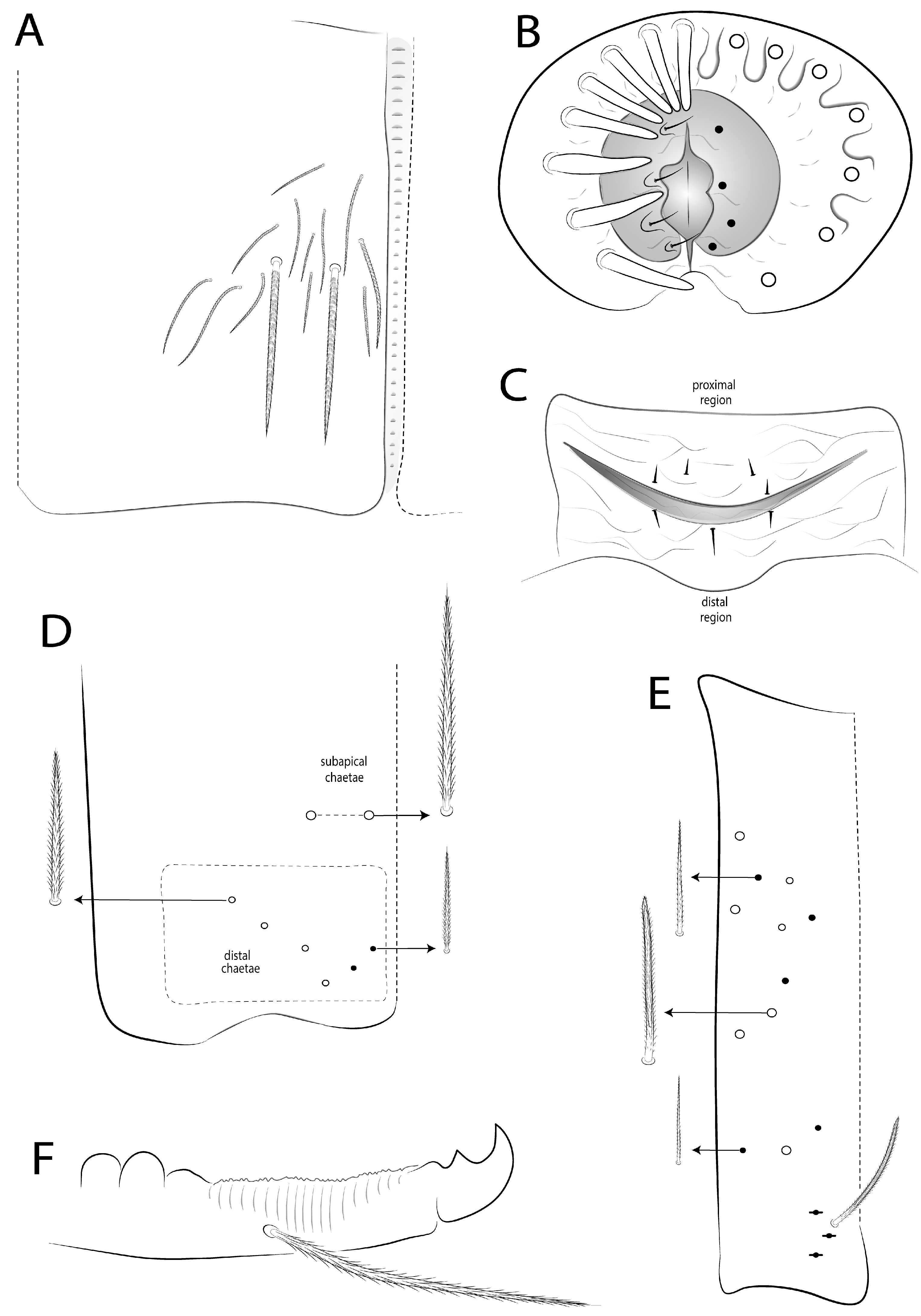
Collophore (Figure 19A): anterior side with about 14 ciliate chaetae, 2 distal mac, and about 12 chaetae of different sizes widely distributed; posterior side distally with 1 ciliate and 1 thicker smooth chaeta; lateral flap with 10 chaetae, 4 smooth chaetae, and 6 ciliate chaetae.
Genital plate (Figure 19B,C): Males with six papillae per side and 7 + 1 (unpaired) circumgenital finger-shaped chaetae, three lower blunt (Figure 19B). Female plate with four anterior and three posterior (1 unpaired) small smooth chaetae (Figure 19C).
Figure 18. Mesentotoma noronhaensis sp. nov. legs: (A,B) chaetotaxy of subcoxa I and III, respectively (outer side); (C) trochanteral organ (posterior view); (D) chaetotaxy of femur and tibiotarsus III (anterior view); (E) distal tibiotarsus and empodial complex III (posterior view).
Diversity 18 00089 g018
Furcula (Figure 19D–F): Manubrium ventrally with two subapical chaetae and six apical chaetae per side, two inner smaller, others subequal (Figure 19D). Manubrium dorsally with one lateral row of five elongated and two short ciliate mac abruptly acuminate at the tip; manubrial plate with three psp and one mac (Figure 19E). Dens proximal region with two inner projections, and proximal three-fourths of dens crenulate. Mucro proximal tooth smaller than distal tooth (Figure 19F).
Etymology. Refers to the archipelago where the species was collected, Fernando de Noronha (Figure 20).
Type material. Holotype male on slide (INPA-CLL 0000276): Brazil, Pernambuco, Fernando de Noronha Island, “Xaréu” weir, 03°51′54″ S, 32°25′41″ W (Figure 20), 26 m, 01–09.vi.2019, sweeping in vegetation, JA Rafael, F Limeira-de-Oliveira & DMM Mendes coll. Three paratypes on slides: one female and one juvenile (INPA-CLL 0000277–78): same date as holotype; one female on slide (CC/UFRN): same date as holotype, except “Sancho” Beach, 03°51′19″ S, 32°26′35″ W, 45 m, 25.ix–08.x.2019, malaise trap, JA Rafael, F Limeira-de-Oliveira & LC Castro coll.
Figure 19. Mesentotoma noronhaensis sp. nov.: abdominal appendages: (A) collophore anterior chaetotaxy; (B) male genital plate; (C) female genital plate; (D) ventrodistal manubrium (left side); (E) manubrium dorsally (left side); (F) dens distally and mucro (outer view).
Diversity 18 00089 g019

3.7.3. Remarks

The two new species described here from Brazil are similar to the other five tropical species transferred to Mesentotoma, although few characteristics are known for these species (Table 3). Mesentotoma multicirculata sp. nov. closely resembles M. grassei comb. nov., M. philippinica comb. nov., and M. pseudocoeruleopicta comb. nov. with a bluish body with depigmented spots and unguis a.t. present (Figures 5 and 12H). However, M. multicirculata sp. nov. differs from these with a dorso-central head with circular and irregular spots depigmented (only laterally in M. philippinica), an absence of foliate-type chaetae on Th II–Abd VI of males (present in M. philippinica), unguis b.t. and m.t. subequal in length (b.t. larger than m.t. in M. philippinica and M. pseudocoeruleopicta), and being the b.t. on the length half of the unguis (in proximal 1/3 in M. grassei). It also differs in unguiculus ai lamella acuminate (slightly truncated in M. philippinica) and pe lamella serrated (smooth in M. philippinica and M. pseudocoeruleopicta), and mucro tooth subequal (distal teeth larger in M. grassei).
Already M. noronhaensis sp. nov. resembles M. depressa comb. nov. and M. tranvercyana nom. nov., comb. nov., both from Congo, by dorso-central head depigmented, unguis a.t. absent, unguiculus ai lamella acuminate, and pe lamella smooth (Table 3). However, M. noronhaensis sp. nov. differs from these species by Abd III–VI posteriorly with one, one, one (incomplete), and one transversal band, respectively, plus another incomplete band on median Abd IV, while in M. depressa the body is depigmented and M. tranvercyana has pigments on all Th II and posteriorly on Th III to Abd IV. Mesentotoma noronhaensis sp. nov. also differs by tenent hairs larger than unguis length (subequal in M. depressa), unguis b.t. larger than m.t. (subequal in M. depressa), and b.t. on distal one-third of the unguis (on half in both species), and mucro distal tooth larger than proximal tooth (subequal in both species). Other differences among these species are also summarized in Table 3.

3.8. Key to Tropical Species of Mesentotoma

  • Unguis ai tooth present (Figure 12H) … 2
-
Unguis ai tooth absent (Figure 18E) … 5
2.
Mucro proximal tooth subequal (Figure 13E) … 3
-
Mucro proximal tooth shorter than the distal one (Figure 19F); Ivory Coast (Figure 20) … M. grassei (Delamare, 1952) [94] comb. nov.
3.
Th II to Abd VI only with normal mac or with some slightly wider mic (Figures 4, 10, 11, and 17); unguiculus III ai lamella acuminate (Figures 12H and 18E) … 4
-
Th II to Abd VI with foliate-type chaeta; unguiculus III ai lamella slightly truncated (see Gapud [95] p. 4–5); Philippines (Figure 20) … M. philippinica (Gapud, 1971) [95] comb. nov.
4.
Unguiculus III pe lamella serrated (Figure 12H); Brazil (Figure 20) … M. multicirculata sp. nov.
-
Unguiculus III pe lamella smooth (Figure 18E); Ivory Coast (Figure 20) … M. pseudocoeruleopicta comb. nov.
5.
Head dorsally without central pigments; body depigmented or only bd III–IV with thin transversal bands (Figure 14); unguiculus slim as “knife” (Figure 18E) … 6.
-
Head with diffuse pigments; Th II all pigmented, distal half of Th II to Abd IV pigmented; unguiculus normal (see Marlier [36] p. 254); Congo (Figure 20) … M. tranvercyana nom. nov., comb. nov.
6.
Body depigmented; unguis b.t. subequal to m.t. in length; mucro proximal tooth subequal (see Marlier [36] p. 254); Congo (Figure 20) … M. depressa (Marlier, 1945) [36] comb. nov.
-
Abd II–VI, respectively, with one, one, two, one, and one transversal band (Figure 14); unguis b.t. larger than m.t. in length (Figure 18E); mucro proximal tooth smaller than the distal one (Figure 19F); Fernando de Noronha Island (Figure 20) … M. noronhaensis sp. nov.

3.9. Distribution and Habitat of Tropical Species of Mesentotoma

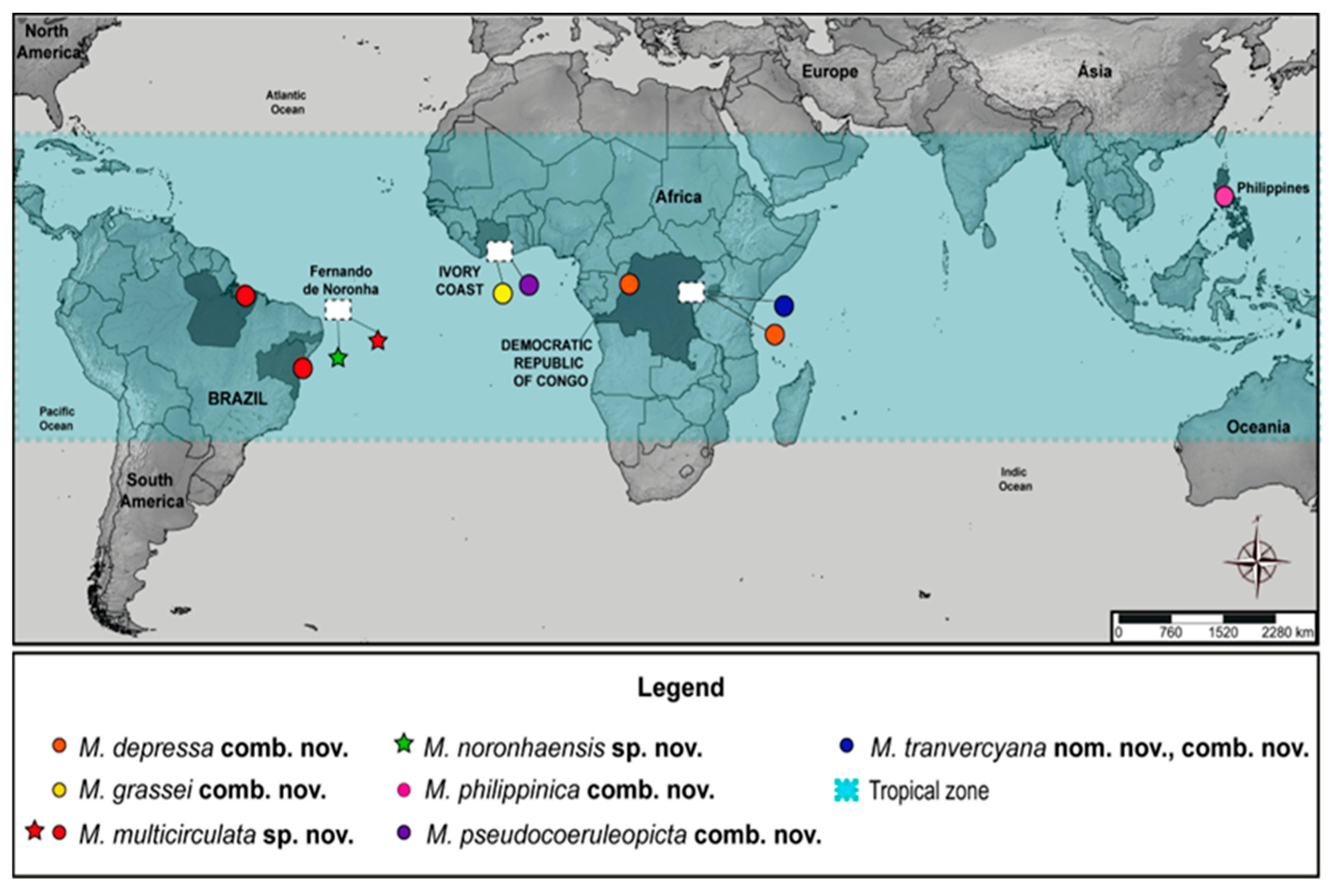
To date, seven species of Mesentotoma are present in the Tropical areas of Africa, Brazil, and the Philippines, five of which are found in coastal environments (Figure 20), which corroborates with most of the genera since they appear to be associated with this habitat [4,5,94], although there are reports of species in high altitude regions, such as the mountains of Hawaii (e.g., Christiansen & Bellinger) [5]. Even though M. depressa comb. nov. and M. tranvercyana nom. nov., comb. nov. are not from coastal areas, they were still recorded in humid areas of the Equatorial climate (Af), characterized by high monthly precipitation [98], as were the other tropical species reported here.
Figure 20. Record map of Mesentotoma tropical species, with the type locality (star) and additional material (circle) of two new species from Brazil.
Diversity 18 00089 g020
Mesentotoma multicirculata sp. nov. was found in coastal areas of the Atlantic and Amazonia Forest (Bahia and Pará States, respectively) plus in Fernando de Noronha Island, while M. noronhaensis sp. nov. was only found in this last locality (Figure 20). These three locations in Brazil are in Good’s biogeographic zones 25 and 26 of the Neotropical region [99]. Even though M. multicirculata sp. nov. was also recorded on the continent (few specimens), both new species were described from the Fernando de Noronha Archipelago. Since its discovery in 1503, the Archipelago has drawn considerable interest due to its unique biological resources and strategic biogeographical location, earning it the title of the “lost paradise of the Atlantic” [100].
Research on these small oceanic islands has sparked scientific interest in the origins and evolution of dominant oceanic crust species, as observed by Darwin, who visited the archipelago in 1832 during the historic “Beagle” expedition [101,102]. This shows that even after decades of scientific explorations, including different studies with Collembola [27,28,30,31], the archipelago can still harbor a preserved fauna, and for this reason it is still a potential place for new discoveries of hexapods [29].
Table 3. Comparison among tropical species of Mesentotoma.
Species
M. depressaM. grasseiM. multicirculataM. noronhaensisM. philippinicaM. pseudocoeruleopictaM. tranvercyana
comb. nov.comb. nov.sp. nov.sp. nov.comb. nov.comb. nov.nom. nov., comb. nov.
[36][94] [95][94][36]
CharacteristicsCongoIvory CoastBrazilBrazilPhilippinesIvory CoastCongo
Body color patterndorsal headirregular spotsirregular spotlateral bandlateral bandirregular spotsdiffuse pigments
Th II–Abd Iirregular spotsdepigmented areaslateral bandlateral bandcompletelydiffuse pigments
Abd IIirregular spotsdepigmented areasone transversal band (+/–)one transversal bandcompletelydiffuse pigments
Abd IIIirregular spotsdepigmented areasone transversal bandone transversal bandcompletelydiffuse pigments
Abd IVirregular spotsanteriorly depigmentedtwo transversal bandsanteriorly depigmentedanteriorly depigmentedanteriorly depigmented
Ant IV apical bulb ??bilobedunilobedbilobed??
Ant I subdivide ??+ (–)???
Prelabral chaetae ??SC gentlyS??
Head dorsal mac‘M’??12???
‘S’??12???
‘Ps–Pe’??811???
Labral papillae projectionsinner??333–4??
outer??32–33–4??
Head postlabial chaetotaxyX, X2??+???
Th II maca1?, m5??+???
p2–4??+???
Th III macp2, p5??+???
Abd I macp5??+???
Abd III macam6??+???
Abd IV outer macouter??1316???
Th II-Abd VI foliate-type chaetae in males??+??
Ratio unguis: tenent hairsu = t? u = tt > ut >/= ut > ut > u
Unguis III inner toothratiob.t. = m.t.b.t. = m.t. > a.t.b.t. = m.t. > a.t.b.t. > m.t.b.t. > m.t. > a.t.b.t. > m.t. > a.t.b.t. > m.t.
b.t.1/21/31/21/31/21/2<1/2
m.t.3/43/43/42/3¾3/44/5
a.t.6/77/89/107/8
Unguiculus III lamellaeinner (ai)acuminateacuminateacuminateacuminateslightly truncateacuminateacuminate
outer (pe)smooth?serratedsmoothsmoothsmoothsmooth
Collophore macanterior??3–42???
Number of male genital papillae??768??
Circumgenital chaetaenumber??8 + 1 (3 blunt)7 + 1 (3 blunt)8 + 1 (2 blunt)??
shaped??3 lanceolate, 1 pin,3 conical and7 + 1 truncate and??
??4 club, 1 finger4 + 1 finger-shape1 finger-shape??
Mucro teeth size subequaldistal tooth largersubequaldistal tooth largersubequalsubequalsubequal
Notes: (C) ciliate chaeta; (S) smooth chaeta; (+) present; (–) absent; (=) subequal; (>) larger; (<) smaller; (?) unknown.

4. Discussion

4.1. Diversity and Morphological Variations Among Tropical Species

Mesentotoma as of now has 15 species [13], of which 7 of them are in the tropical zone (Figure 20). Regarding species from New Zealand and Holarctic [1,4,5,6,10,12,77,85], these tropical species are similar in their body shape, reduced size, pigmentation pattern, unguiculus ai lamella acuminate, reduced macrochaetotaxy, and labral papillae with projections; at least these last two characteristics are in M. multicirculata sp. nov., M. noronhaensis sp. nov., and M. philippinica (Table 3). However, until a new phylogeny is proposed, it is not possible to know whether there is a relationship between these tropical species, although they have atypical differences between them.
Only M. multicirculata sp. nov. and M. noronhaensis sp. nov. have the head pentagon-shaped and wide tergal mic (similar to scale), although this last characteristic may not have been reported in the descriptions of the other species [36,94,95]. On the other hand, the presence of foliate-shaped chaetae is apparently present only in M. philippinica (see Gapud) [95].

4.2. Morphological Patterns and Atypical Characters

Another generalist structure in Entomobryioidea is the eugenital setae of the genital plate, which in males are three chaetae on each side, while in females are two anterior and two posterior chaetae [18,19,23,24,103,104,105,106,107,108,109,110,111], except for males of Pseudosinella Schäffer and Amazhomidia ducke, with two and one eugenital chaetae, respectively [14,109].
In contrast, in M. multicirculata sp. nov. and M. noronhaensis sp. nov., the males have four eugenital chaetae on each side (omitted in the description of M. philippinica), while females have an extra pair of upper chaetae and an extra unpaired lower chaeta (Figures 13D and 19B,C). Thus, these and other characteristics need to be investigated in Mesentotoma in general to see if they reinforce the current classification.

4.3. Phylogenetic Evidence, Homoplasies, and Perspectives

On the other hand, even though monophyly, as well as the relationships of Mesentotoma, is still uncertain, according to the recovered hypotheses, it is suggested that the genus has a basal relationship with species devoid of dental spines, with (Willowsia spp.) or without scales on the body (Coecobrya, Entomobrya, Sinella), which in a way was expected, since scales appeared at least three times within Entomobryinae [50].
Furthermore, according to the recovered topology, it is suggested that Mesentotoma lost the mucronal spine, but such an apomorphic condition may have arisen more than once, as it is shared in other Entomobryidae (all of Entomobryinae), whether scaled or Desertia (in Martynova et al.) [112] or unscaled as Calx Christiansen [83] and Isotobrya Womersley [113], although these genera have only one mucronal tooth, while Mesentotoma has two teeth. This reinforces the need for specimens from these genera to be included in future phylogenetic analyses, as this scenario points to two characteristics (number of teeth versus mucronal spine) whose evolutionary significance is unknown, and consequently, whether they are a significant factor in phylogenetically relating these taxa. This suggests that Mesentotoma is likely an artificial group, since many characteristics are not shared among its congeners (e.g., Ant IV apical bulb, annulations of Ant III–IV, Ant. I subsegmented, unguiculus ai lamella excavate, mucro teeth side), except for the absence of the mucronal spine, which may be a homoplastic condition within the Entomobryinae (as is the number of mucronal teeth).
This study reinforces that, among the Entomobryioidea subfamilies, Entomobryinae is the most complex in phylogenetic terms due to the great difficulty in recovering natural groups, whether from morphological and/or molecular data [2,32,33,35,50,69,114,115,116]. In this sense, even though Mesentotoma is a small genus, its origin, monophyly, as well as inter and intrageneric relationships remain largely unknown, until new representatives of the genus are included in future phylogenetic studies.

5. Conclusions

This study documents its first record of Mesentotoma in the Neotropical region and even expands the distribution of the genus to the Tropical zone. Furthermore, it provides the first mitogenomic data for genus and represents a significant step to try to understand the phylogenetic relationships with other Entomobryinae genera, as well as to trace the transformation series between these taxa. Although the relationship with other taxa is still uncertain and far from being resolved, these data suggest that Mesentotoma has a basal relationship with most scaled and unscaled taxa, and that possibly the mucronal spine may be an evolutionary marker among the Entomobryinae; if not, then it is a homoplastic characteristic with the loss of the spine in different lineages. Thus, integrative analysis including broader taxon sampling with additional Mesentotoma species and other genera devoid of mucronal spines will be essential to clarify the monophyly and evolution, as well as tracking possible synapomorphies that may support the diagnosis of different Entomobryinae taxa.

Supplementary Materials

The following supporting information can be downloaded at: https://www.mdpi.com/article/10.3390/d18020089/s1, Table S1: Best partitition model suggested by ModelFinder for partitioned analyses in IQTree.

Author Contributions

Conceptualization, N.G.C. and S.d.S.V.; methodology, N.G.C., N.N.G. and S.d.S.V.; software, N.G.C., N.N.G. and S.d.S.V.; validation, N.G.C., N.N.G., S.d.S.V. and J.W.d.M.; formal analysis, N.G.C. and N.N.G.; investigation, N.G.C. and S.d.S.V.; resources, N.G.C., N.N.G., and S.d.S.V.; data curation, N.G.C., N.N.G. and S.d.S.V.; writing—original draft preparation, N.G.C. and S.d.S.V.; writing—review and editing, N.G.C., N.N.G. and S.d.S.V.; visualization, N.G.C., N.N.G. and J.W.d.M.; supervision, N.G.C., N.N.G. and J.W.d.M.; project administration, N.G.C.; funding acquisition, N.N.G. and J.W.d.M. All authors have read and agreed to the published version of the manuscript.

Funding

This research was originally funded by INPA, project “Biology and Ecology of Amazonian Insects”, grant number 12.311. S.S.V was granted by CAPES, grant number 88887.893290/2023-00. N.N.G was granted by the National Natural Science Foundation of China—research fund for international young scientists, grant number 32350410418. And N.G.C was granted by CNPq/PDJ, grant number 174716/2023-0.

Institutional Review Board Statement

Ethical review and approval were waived for this study, due to Brazilian laws which do not require permission from an institutional ethics committee on the use of animals for taxonomical with microarthropods.

Informed Consent Statement

Not applicable.

Data Availability Statement

All data are contained within the article; to access SRA data: SRR29635445; to access new mitogenome sequence: PX395442; to access new mitogenomes assembled from SRA data: https://doi.org/10.6084/m9.figshare.26123944 (accessed on 22 January 2026). All biological material is deposited at INPA, CC/UFRN, MZUSP, MNRJ, MNCN, and SNHM as previously stated.

Acknowledgments

We would like to thank all specimens collectors, especially José Albertino Rafael (INPA) and his project associated with the Chico Mendes Institute for Biodiversity Conservation (ICMBio) for Collection Permit (62.821); to the staff of Fernando de Noronha Marine National Park, Ricardo Araújo, Viviane Vilella, and Carolina Fonseca for their administrative support; to the Territorial Authority of the State District of Fernando de Noronha (ATDEFN) for providing administrative facilities, Layane Carvalho de Castro for field support, and Phil J. Sirvid and Jean-Claude Stahl from Museum of New Zealand Te Papa Tongarewa for providing the holotype images of M. exalga. The project was supported by the Amazonas State Research Support Foundation (FAPEAM) and the National Council for Scientific and Technological Development (CNPq) (300997/2016-7 and 306661/2021-7). Also, we would like to thank the CAPES Pro-Equipamentos and Programa de Capacitação em Taxonomia—PROTAX (project 562188/2010-0 of Dra. Neusa Hamada/INPA) and Laboratório Temático de Microscopia Óptica e Eletrônica (LTMOE/NPA) for logistic support with the images. The work was supported by Programa de Apoio à Pós-Graduação (PROAP/CAPES) and Programa Institucional de Apoio à Pós-Graduação Stricto Sensu (POSGRAD/FAPEAM). We sincerely thank Reviewers 1 and 2 for their careful evaluations and insightful comments, which substantially improved the quality and clarity of this manuscript.

Conflicts of Interest

The authors declare no conflicts of interest.

Abbreviations

The following abbreviations are used in descriptions in this manuscript:
Abdabdominal segment(s)
Antantennal segment(s)
macmacrochaeta(e)
psppseudopore(s)
sensspecialized ordinary chaeta(e)
Ththoracic segment(s)
IOinterocular chaetae
ffrontal
pfprefrontal
llateral
a.a.apical appendage of labial papillae
b.c.basal chaeta of maxillary palp
bsbasal appendages of sublobal plate
l.p.lateral process of papilla E
lpclabial proximal chaetae
t.a.terminal appendage of the maxillary palp
b.t.paired basal teeth
m.t.unpaired median tooth
a.t.unpaired apical tooth
aiantero-internal lamella
aeantero-external lamella
pipostero-internal lamella
pepostero-external lamella

References

  1. Salmon, J.T. New genera and species of New Zealand Collembola. Rec. Dominion Mus. Wellingt. 1942, 1, 55–60. [Google Scholar]
  2. Zhang, F.; Deharveng, L. Systematic revision of Entomobryidae (Collembola) by integrating molecular and new morphological evidence. Zool. Scr. 2015, 44, 298–311. [Google Scholar] [CrossRef] [Scilit]
  3. Rondani, C. Entomobrya pro Degeeria Nic. In Dipterologiae Italicae Prodromus; Stocchi, A., Ed.; Parma, Italy, 1861; p. 40. [Google Scholar]
  4. Christiansen, K. The genus Mesentotoma (Collembola: Entomobryidae). Psyche 1956, 63, 14–20. [Google Scholar] [CrossRef] [Scilit]
  5. Christiansen, K.A.; Bellinger, P.F. Insects of Hawaii. Vol. 15. Collembola; University of Hawaii Press: Honolulu, HI, USA, 1992; p. 445. [Google Scholar]
  6. Christiansen, K.; Bellinger, P. The Collembola of North America North of the Rio Grande. Part 3: Entomobryidae; Grinnell College: Grinnell, IA, USA, 1998. [Google Scholar] [CrossRef] [Scilit]
  7. Jordana, R. Synopses of Palaearctic Collembola: Capbryinae & Entomobryini. Soil Org. 2012, 84, 1–390. [Google Scholar]
  8. Bacon, G.A. A new species of Collembola from Laguna Beach. J. Entomol. Zool. 1913, 5, 202–204. [Google Scholar]
  9. Denis, J.R. Report on Collembola. Mem. Conn. Acad. Arts Sci. 1936, 10, 261–282. [Google Scholar]
  10. Jacquemart, S. Collemboles nouveaux du Sahara. Bull. Inst. R. Sci. Nat. Belg. 1974, 49, 1–16. [Google Scholar]
  11. Denis, J.R. Sur les Collemboles du Muséum de Paris. Ann. Soc. Entomol. Fr. 1924, 93, 211–260. [Google Scholar] [CrossRef] [Scilit]
  12. Baquero, E.; Arbea, J.; Jordana, R. New species of Entomobryini from the Mediterranean Palaearctic. Soil Org. 2010, 82, 285–300. [Google Scholar]
  13. Checklist of the Collembola of the World. Available online: http://www.collembola.org (accessed on 30 September 2025).
  14. Cipola, N.G.; Oliveira, F.G.L. A new species of Campylothorax from the Brazilian Amazon. Zootaxa 2016, 4109, 487–495. [Google Scholar] [CrossRef] [Scilit] [PubMed]
  15. Cipola, N.G.; Morais, J.W.; Bellini, B.C. A new genus of Entomobryinae from the Brazilian Amazon. Zootaxa 2016, 4105, 261–273. [Google Scholar] [CrossRef] [Scilit]
  16. Cipola, N.G.; Morais, J.W.; Bellini, B.C. Discovery of Lepidocyrtoides from the New World. Zootaxa 2017, 4324, 201–248. [Google Scholar] [CrossRef] [Scilit]
  17. Cipola, N.G.; Morais, J.W.; Bellini, B.C. New subgenus and species of Lepidocyrtus from Amazon. Insect Syst. Evol. 2018, 50, 189–234. [Google Scholar] [CrossRef] [Scilit]
  18. Cipola, N.G.; Morais, J.W.; Bellini, B.C. Survey of Trogolaphysa of the Brazilian Amazon. Zootaxa 2022, 5093, 251–295. [Google Scholar] [CrossRef] [Scilit]
  19. Cipola, N.G.; Bellini, B.C.; Godeiro, N.N. Two new species of Seira from South America. Zootaxa 2025, 5620, 175–195. [Google Scholar] [CrossRef] [Scilit]
  20. Cipola, N.G.; Katz, A.D.; Bu, Y.; Godeiro, N.N. Systematics of Willowsia jacobsoni. Zootaxa 2025, 5604, 201–233. [Google Scholar] [CrossRef] [Scilit]
  21. Oliveira, F.G.L.; Cipola, N.G.; Almeida, E.A.B. Systematics and biogeography of Salina. Insect Syst. Evol. 2018, 51, 81–138. [Google Scholar] [CrossRef] [Scilit]
  22. Cipola, N.G.; Viana, S.S. Two new species of Lepidocyrtus (Fractocyrtus). Zootaxa 2023, 5369, 369–399. [Google Scholar] [CrossRef] [Scilit] [PubMed]
  23. Viana, S.S.; Morais, J.W.; Cipola, N.G. A new species of Entomobrya from Brazil. Zootaxa 2022, 5155, 261–279. [Google Scholar] [CrossRef] [Scilit] [PubMed]
  24. Viana, S.S.; Morais, J.W.; Cipola, N.G. Taxonomic revision of Entomobrya from the Brazilian Amazon. Zootaxa 2024, 5452, 1–110. [Google Scholar] [CrossRef] [Scilit] [PubMed]
  25. Zeppelini, D.; Queiroz, G.C.; Bellini, B.C.; Brito, R.A.; Oliveira, J.V.L.C.; Lopes, B.C.H.; Lima, E.C.A.; Ferreira, A.S.; Brito, N.P. Entomobryidae in Catálogo Taxonômico da Fauna do Brasil. Available online: http://fauna.jbrj.gov.br (accessed on 30 September 2025).
  26. Ridley, H.N. Thysanura and Collembola. Notes on the Zoology of Fernando de Noronha. Zool. J. Linn. Soc. 1890, 20, 473–570. [Google Scholar] [CrossRef] [Scilit]
  27. Palácios-Vargas, J.G.; Lima, E.C.A.; Zeppelini, D. A new species of Isotogastrura. Fla. Entomol. 2013, 96, 1579–1587. [Google Scholar] [CrossRef] [Scilit]
  28. Lima, E.C.A.; Zeppelini, D. First survey of Collembola in Fernando de Noronha. Fla. Entomol. 2015, 98, 368–369. [Google Scholar] [CrossRef] [Scilit]
  29. Rafael, J.A.; Limeira-De-Oliveira, F.; Hutchings, R.W.; Miranda, G.F.G.; Neto, A.M.d.S.; Somavilla, A.; Camargo, A.; Asenjo, A.; Pinto, Â.P.; Bello, A.d.M.; et al. Insect diversity in Fernando de Noronha. Rev. Bras. Entomol. 2020, 64, 2–26. [Google Scholar] [CrossRef] [Scilit]
  30. De Lima, E.C.A.; Lopes, B.C.H.; Oliveira-Neto, M.A.; de Mendonça, M.C.; Zeppelini, D. Synthesis of Brazilian Poduromorpha. Insects 2021, 12, 268. [Google Scholar] [CrossRef] [Scilit] [PubMed]
  31. De Lima, E.C.A.; Lopes, B.C.H.; Oliveira-Neto, M.A.; de Mendonça, M.C.; Zeppelini, D. Synthesis of Brazilian Entomobryomorpha. Diversity 2022, 14, 553. [Google Scholar] [CrossRef] [Scilit]
  32. Godeiro, N.N.; Pacheco, G.; Liu, S.; Cipola, N.G.; Berbel-Filho, W.M.; Zhang, F.; Gilbert, M.T.P.; Bellini, B.C. Phylogeny of Neotropical Seirinae (Collembola, Entomobryidae) based on mitochondrial genomes. Zool. Scr. 2020, 49, 329–339. [Google Scholar] [CrossRef] [Scilit]
  33. Godeiro, N.N.; Bellini, B.C.; Ding, N.; Xu, C.; Ding, Y.; Zhang, F. A mitogenomic phylogeny of the Entomobryoidea (Collembola): A comparative perspective. Zool. Scr. 2021, 50, 658–666. [Google Scholar] [CrossRef] [Scilit]
  34. Godeiro, N.N.; Xu, C.; Zhang, G.; Zhang, F. Mitogenomic features of three species of Entomobryoidea (Collembola) from China. Zootaxa 2022, 5120, 283–288. [Google Scholar] [CrossRef] [Scilit]
  35. Godeiro, N.N.; Ding, Y.; Cipola, N.G.; Jantarit, S.; Bellini, B.C.; Zhang, F. Phylogenomics and systematics of Entomobryoidea (Collembola): Marker design, phylogeny and classification. Cladistics 2023, 38, 101–115. [Google Scholar] [CrossRef] [Scilit]
  36. Marlier, G. Collemboles du Congo belge. Rev. Zool. Bot. Afr. 1945, 38, 252–264. [Google Scholar]
  37. Meng, G.; Li, Y.; Yang, C.; Liu, S. MitoZ: A toolkit for animal mitochondrial genome assembly, annotation and visualization. Nucleic Acids Res. 2019, 47, e63. [Google Scholar] [CrossRef] [Scilit] [PubMed]
  38. Li, D.; Liu, C.M.; Luo, R.; Sadakane, K.; Lam, T.W. MEGAHIT: An ultra-fast single-node solution for large and complex metagenomics assembly. Bioinformatics 2015, 31, 1674–1676. [Google Scholar] [CrossRef] [Scilit] [PubMed]
  39. Karlicki, M.; Antonowicz, S.; Karnkowska, A. Tiara: Deep learning-based classification system for eukaryotic sequences. Bioinformatics 2022, 38, 344–350. [Google Scholar] [CrossRef] [Scilit]
  40. Wheeler, T.J.; Eddy, S.R. nhmmer: DNA homology search with profile HMMs. Bioinformatics 2013, 29, 2487–2489. [Google Scholar] [CrossRef] [Scilit]
  41. Gertz, E.M.; Yu, Y.K.; Agarwala, R.; Schäffer, A.A.; Altschul, S.F. Composition-based statistics and translated nucleotide searches: Improving the TBLASTN module of BLAST. BMC Biol. 2006, 4, 41. [Google Scholar] [CrossRef] [Scilit] [PubMed]
  42. Birney, E.; Clamp, M.; Durbin, R. GeneWise and Genomewise. Genome Res. 2004, 14, 988–995. [Google Scholar] [CrossRef] [Scilit]
  43. Nawrocki, E.P.; Eddy, S.R. Infernal 1.1: 100-fold faster RNA homology searches. Bioinformatics 2013, 29, 2933–2935. [Google Scholar] [CrossRef] [Scilit]
  44. Jühling, F.; Putz, J.; Bernt, M.; Donath, A.; Middendorf, M.; Florentz, C.; Stadler, P.F. Improved systematic tRNA gene annotation allows new insights into mitochondrial genome evolution. Nucleic Acids Res. 2012, 40, 2833–2845. [Google Scholar] [CrossRef] [Scilit]
  45. Krzywinski, M.; Schein, J.; Birol, I.; Connors, J.; Gascoyne, R.; Horsman, D.; Jones, S.J.; Marra, M.A. Circos: An information aesthetic for comparative genomics. Genome Res. 2009, 19, 1639–1645. [Google Scholar] [CrossRef] [Scilit]
  46. Li, H.; Durbin, R. Fast and accurate short read alignment with Burrows–Wheeler transform. Bioinformatics 2009, 25, 1754–1760. [Google Scholar] [CrossRef] [Scilit] [PubMed]
  47. Li, H.; Handsaker, B.; Wysoker, A.; Fennell, T.; Ruan, J.; Homer, N.; Marth, G.; Abecasis, G.; Durbin, R. The sequence alignment/map format and SAMtools. Bioinformatics 2009, 25, 2078–2079. [Google Scholar] [CrossRef] [Scilit] [PubMed]
  48. Gisin, H. Collemboles d’Europe VI. Rev. Suisse Zool. 1964, 71, 383–400. [Google Scholar] [CrossRef] [Scilit]
  49. Arlé, R. Relatório da excursão científica do Instituto Osvaldo Cruz realizada na zona da E.F. N.O.B. em outubro de 1938. Bol. Biol. 1939, 4, 295–300. [Google Scholar]
  50. Zhang, F.; Chen, Z.; Dong, R.-R.; Deharveng, L.; Stevens, M.I.; Huang, Y.-H.; Zhu, C.-D. Molecular phylogeny reveals independent origins of body scales in Entomobryidae. Mol. Phylogenet. Evol. 2014, 70, 231–239. [Google Scholar] [CrossRef] [Scilit]
  51. Capella-Gutiérrez, S.; Silla-Martínez, J.M.; Gabaldón, T. trimAl: A tool for automated alignment trimming in phylogenetic analyses. Bioinformatics 2009, 25, 1972–1973. [Google Scholar] [CrossRef] [Scilit]
  52. Kück, P.; Longo, G.C. FASconCAT-G: Extensive functions for multiple sequence alignment preparations. Front. Zool. 2014, 11, 81. [Google Scholar] [CrossRef] [Scilit]
  53. Minh, B.Q.; Schmidt, H.A.; Chernomor, O.; Schrempf, D.; Woodhams, M.D.; von Haeseler, A.; Lanfear, R. IQ-TREE 2: New models and efficient methods for phylogenetic inference. Mol. Biol. Evol. 2020, 37, 1530–1534. [Google Scholar] [CrossRef] [Scilit] [PubMed]
  54. Kalyaanamoorthy, S.; Minh, B.Q.; Wong, T.K.; von Haeseler, A.; Jermiin, L.S. ModelFinder: Fast model selection for accurate phylogenetic estimates. Nat. Methods 2017, 14, 587–589. [Google Scholar] [CrossRef] [Scilit]
  55. Sun, X.; Yu, D.; Xie, Z.; Dong, J.; Ding, Y.; Yao, H. Phylomitogenomic analyses on collembolan higher taxa. PLoS ONE 2020, 15, e0230827. [Google Scholar] [CrossRef] [Scilit]
  56. Nicolet, H. Recherches pour servir à l’histoire des Podurelles. Nouv. Mém. Soc. Helv. Sci. Nat. 1842, 6, 1–88. [Google Scholar]
  57. Collins, G.; Schneider, C.; Boštjančić, L.L.; Burkhardt, U.; Christian, A.; Decker, P.; Ebersberger, I.; Hohberg, K.; Lecompte, O.; Merges, D.; et al. The MetaInvert soil invertebrate genome resource. Commun. Biol. 2023, 6, 1241. [Google Scholar] [CrossRef] [Scilit]
  58. Tullberg, T. Förteckning över svenska Podurider. Öfvers. K. Vetensk.-Akad. Förh. 1871, 28, 143–145. [Google Scholar]
  59. Lubbock, J. Notes on the Thysanura. Part III. Trans. Linn. Soc. Lond. 1870, 26, 295–304. [Google Scholar] [CrossRef] [Scilit]
  60. Linnæus, C. Systema Naturæ, 10th ed.; Laurentii Salvii: Holmiæ, Sweden, 1758; pp. 608–609. [Google Scholar] [CrossRef] [Scilit]
  61. Folsom, J.M. East Indian Collembola. Bull. Mus. Comp. Zool. Harv. 1924, 65, 505–517. [Google Scholar]
  62. Jin, J.; Zhao, Y.; Zhang, G.; Pan, Z.; Zhang, F. Chromosome-level genome assembly of Entomobrya proxima. Sci. Data 2023, 10, 541. [Google Scholar] [CrossRef] [Scilit]
  63. Lee, B.H.; Lee, W.K. A taxonomic study of soil microarthropods. Annu. Rep. Biol. Res. 1981, 2, 129–140. [Google Scholar]
  64. Lee, I.; Koo, J.; Park, K.H. Complete mitochondrial genome of Homidia koreana. Mitochondrial DNA B 2022, 7, 135–137. [Google Scholar] [CrossRef] [Scilit]
  65. Lee, I.; Park, K.H. A new species of Homidia from Korea. Zootaxa 2024, 5463, 262–272. [Google Scholar] [CrossRef] [Scilit]
  66. Wu, J.; Chen, K. Complete mitochondrial genome of Homidia socia. Mitochondrial DNA B 2019, 4, 3941–3942. [Google Scholar] [CrossRef] [Scilit]
  67. Nunes, R.C.; Godeiro, N.N.; Pacheco, G.; Liu, S.; Gilbert, M.T.P.; Alvarez-Valin, F.; Zhang, F.; Bellini, B.C. The discovery of Neotropical Lepidosira (Collembola, Entomobryidae) and its systematic position. Zool. Scr. 2019, 48, 783–800. [Google Scholar] [CrossRef] [Scilit]
  68. Brook, G. On a new genus of Collembola (Sinella). J. Linn. Soc. Zool. 1882, 16, 541–545. [Google Scholar] [CrossRef] [Scilit]
  69. Zhang, F.; Bellini, B.C.; Soto-Adames, F.N. New insights into the systematics of Entomobryoidea (Collembola: Entomobryomorpha): First instar chaetotaxy, homology and classification. Zool. Syst. 2019, 44, 249–278. [Google Scholar] [CrossRef]
  70. Yosii, R. On some Collembola of Japan and adjacent countries. Contrib. Biol. Lab. Kyoto Univ. 1965, 19, 1–71. [Google Scholar]
  71. Börner, C. Zur Collembolenfauna Javas. Tijdschr. Entomol. 1913, 56, 44–61. [Google Scholar]
  72. Folsom, J.W. Japanese Collembola. Part I. Bull. Essex Inst. 1898, 29, 51–58. [Google Scholar] [CrossRef] [Scilit]
  73. Lubbock, J. Monograph of the Collembola and Thysanura; Ray Society: London, UK, 1873; p. 276. [Google Scholar] [CrossRef] [Scilit]
  74. Guindon, S.; Dufayard, J.F.; Lefort, V.; Anisimova, M.; Hordijk, W.; Gascuel, O. New algorithms to estimate maximum-likelihood phylogenies: PhyML 3.0. Syst. Biol. 2010, 59, 307–321. [Google Scholar] [CrossRef] [Scilit] [PubMed]
  75. Hoang, D.T.; Chernomor, O.; von Haeseler, A.; Minh, B.Q.; Vinh, L.S. UFBoot2: Improving the ultrafast bootstrap approximation. Mol. Biol. Evol. 2018, 35, 518–522. [Google Scholar] [CrossRef] [Scilit]
  76. Huelsenbeck, J.P.; Ronquist, F. MRBAYES: Bayesian inference of phylogenetic trees. Bioinformatics 2001, 17, 754–755. [Google Scholar] [CrossRef] [Scilit]
  77. Jordana, R.; Arbea, J.I.; Simón, C.; Luciáñez, M.J. Fauna Iberica; Collembola Poduromorpha; Museo Nacional de Ciencias Naturales: Madrid, Spain, 1997; Volume 8, p. 807. [Google Scholar]
  78. Kawada, R.; Buffington, M.L. A scalable and modular dome illumination system for scientific microphotography. PLoS ONE 2016, 11, e0153426. [Google Scholar] [CrossRef] [Scilit]
  79. Shorthouse, D. SimpleMappr. Available online: https://www.simplemappr.net/ (accessed on 15 May 2025).
  80. Fjellberg, A. The labial palp in Collembola. Zool. Anz. 1999, 237, 309–330. [Google Scholar]
  81. Cipola, N.G.; Morais, J.W.; Bellini, B.C. A new species of Seira from northern Brazil. Zoologia 2014, 31, 489–495. [Google Scholar] [CrossRef] [Scilit]
  82. Cipola, N.G.; Katz, A.D. Morphological and molecular analysis of Willowsia nigromaculata. Eur. J. Taxon. 2021, 739, 92–116. [Google Scholar] [CrossRef] [Scilit]
  83. Chen, J.-X.; Christiansen, K.A. The genus Sinella of China. Orient. Insects 1993, 27, 1–54. [Google Scholar] [CrossRef] [Scilit]
  84. Yosii, R. Studies on the collembolan fauna of Malay and Singapore. Contrib. Biol. Lab. Kyoto Univ. 1959, 10, 1–65. [Google Scholar]
  85. Christiansen, K. The Nearctic members of Entomobrya. Bull. Mus. Comp. Zool. 1958, 118, 440–545. [Google Scholar]
  86. South, A. The taxonomy of British species of Entomobrya. Trans. R. Entomol. Soc. Lond. 1961, 113, 387–416. [Google Scholar] [CrossRef] [Scilit]
  87. Hüther, W. New aspects in taxonomy of Lepidocyrtus. In Proceedings of the 2nd International Seminar on Apterygota; Dallai, R., Ed.; University of Siena: Siena, Italy, 1986; pp. 61–65. [Google Scholar]
  88. Christiansen, K. The Entomobryiform male genital plate. Proc. Iowa Acad. Sci. 1958, 65, 474–476. [Google Scholar]
  89. Mari Mutt, J.A. A revision of the genus Dicranocentrus. Agric. Exp. Stn. Bull. 1979, 259, 1–79. [Google Scholar]
  90. Soto-Adames, F.N. Postembryonic development of dorsal chaetotaxy in Seira dowlingi. Zootaxa 2008, 1683, 1–31. [Google Scholar] [CrossRef] [Scilit]
  91. Pan, Z.-X.; Shi, S.; Zhang, F. New species of Homidia from eastern China. ZooKeys 2011, 152, 21–42. [Google Scholar] [CrossRef] [Scilit]
  92. Szeptycki, A. Chaetotaxy of the Entomobryidae and Its Phylogenetical Significance; Polish Academy of Sciences: Warsaw, Poland, 1979; p. 219. [Google Scholar]
  93. Tömösvary, Ö. Adatok hazánk Thysanura-faunájához. Math. Természettud. Közlem. 1882, 18, 119–131. [Google Scholar]
  94. Delamare-Deboutteville, C. Étude systématique. I. Collemboles. In Faune de Nids et des Terriers en Basse Côte-d’Ivoire; Delamare-Deboutteville, C., Paulian, R., Eds.; Lechevalier: Paris, France, 1952; pp. 62–77. [Google Scholar]
  95. Gapud, V.G. Studies on the Philippine Collembola, III. Suborder Arthropleona: Entomobryidae. Philipp. Entomol. 1971, 2, 1–50. [Google Scholar]
  96. Schött, H. Results of Dr. E. Mjöberg’s Swedish Scientific Expeditions to Australia, 1910–1913. No. 15, Collembola. Ark. Zool. 1917, 11, 1–60. [Google Scholar]
  97. International Commission on Zoological Nomenclature (ICZN). International Code of Zoological Nomenclature, 4th ed.; International Trust for Zoological Nomenclature: London, UK, 2000; p. 306. [Google Scholar]
  98. Kottek, M.; Grieser, J.; Beck, C.; Rudolf, B.; Rubel, F. World map of the Köppen–Geiger climate classification updated. Meteorol. Z. 2006, 15, 259–263. [Google Scholar] [CrossRef] [Scilit] [PubMed]
  99. Good, R. The Geography of Flowering Plants, 4th ed.; Longman Group: Harlow, UK, 1974; p. 574. [Google Scholar]
  100. Batistella, M. Espécies vegetais dominantes do Arquipélago de Fernando de Noronha. Acta Bot. Bras. 1996, 10, 223–235. [Google Scholar] [CrossRef] [Scilit]
  101. Darwin, C. Journal of Researches into the Natural History and Geology of the Countries Visited during the Voyage of H.M.S. Beagle; Murray: London, UK, 1845. [Google Scholar]
  102. Almeida, F.F.M. Arquipélago de Fernando de Noronha—Registro de monte vulcânico do Atlântico Sul. In Sítios Geológicos e Paleontológicos do Brasil; Schobbenhaus, C., Campos, D.A., Queiroz, E.T., Winge, M., Berbert-Born, M.L.C., Eds.; DNPM/CPRM–SIGEP: Brasília, Brazil, 2002; pp. 361–368. [Google Scholar]
  103. Mari Mutt, J.A. Redescription of Willowsia jacobsoni (Börner). J. Agric. Univ. P. R. 1981, 65, 361–373. [Google Scholar] [CrossRef] [Scilit]
  104. Mari Mutt, J.A.; Palacios-Vargas, J.G. Americabrya, a new genus of Entomobryidae. J. N. Y. Entomol. Soc. 1987, 95, 99–108. [Google Scholar]
  105. Katz, A.D.; Giordano, R.; Soto-Adames, F. Taxonomic review and phylogenetic analysis of North American Entomobrya. ZooKeys 2015, 525, 1–75. [Google Scholar] [CrossRef] [Scilit]
  106. Cipola, N.G.; Arbea, J.; Baquero, E.; Jordana, R.; Morais, J.W.; Bellini, B.C. Survey of Seira from Iberian Peninsula and Canary Islands. Zootaxa 2018, 4458, 1–66. [Google Scholar] [CrossRef] [Scilit]
  107. Cipola, N.G.; Morais, J.W.; Godeiro, N.N.; Bellini, B.C. Taxonomic revision of Tyrannoseira. Zootaxa 2019, 4586, 201–248. [Google Scholar] [CrossRef] [Scilit]
  108. Cipola, N.G.; Morais, J.W.; Bellini, B.C. Review of Lepidocyrtinus (African species). Zootaxa 2020, 4898, 1–110. [Google Scholar] [CrossRef] [Scilit]
  109. Cipola, N.G.; Oliveira, J.V.L.C.; Bellini, B.C.; Ferreira, A.S.; Lima, E.C.A.; Brito, R.A.; Stievano, L.C.; Souza, P.G.; Zeppelini, D. Review of eyeless Pseudosinella from Brazilian caves. Insects 2020, 11, 194. [Google Scholar] [CrossRef] [Scilit] [PubMed]
  110. Jantarit, S.; Satasook, C.; Deharveng, L. Coecobrya sirindhornae sp. n., the most highly troglomorphic Collembola in Southeast Asia. ZooKeys 2019, 824, 21–44. [Google Scholar] [CrossRef] [Scilit] [PubMed]
  111. Nilsai, A.; Detcharoen, M.; Godeiro, N.N.; Jantarit, S. Four new species of troglomorphic Coecobrya from Thailand. Subterr. Biol. 2021, 41, 1–42. [Google Scholar] [CrossRef] [Scilit]
  112. Martynova, E.F.; Chelnokov, V.G.; Kaplin, V.G. Fauna and ecology of Collembola of the Eastern Karakum. Izv. Akad. Nauk Kazakhskoi SSR Biol. 1979, 1, 35–44. [Google Scholar]
  113. Womersley, H. On some Collembola from South Africa and Southern Rhodesia. Ann. S. Afr. Mus. 1934, 30, 441–475. [Google Scholar]
  114. Zhang, F.; Sun, D.D.; Yu, D.Y.; Wang, B.X. Molecular phylogeny supports S-chaetae as a key character better than jumping organs and body scales in classification of Entomobryoidea (Collembola). Sci. Rep. 2015, 5, 12471. [Google Scholar] [CrossRef] [Scilit]
  115. Zhang, F.; Pan, Z.; Wu, J.; Ding, Y.; Yu, D.; Wang, B. Dental scales in Entomobryidae and new definition of Entomobryinae. Invert. Syst. 2016, 30, 598–615. [Google Scholar] [CrossRef] [Scilit]
  116. Ding, Y.-H.; Yu, D.-H.; Guo, W.-B.; Li, J.-N.; Zhang, F. Molecular phylogeny of Entomobrya from China. Insect Sci. 2018, 26, 587–597. [Google Scholar] [CrossRef] [Scilit]
Figure 1. Circular representation of the mitogenome of Mesentotoma multicirculata sp. nov. The innermost circle shows the GC content; the middle circle shows the reads coverage, and the outermost circle shows the gene features, rRNA (yellow), tRNA (salmon), and CDS (green). The photo in the center represents a specimen preserved in alcohol.
Diversity 18 00089 g001
Figure 2. Phylogenetic placement of Mesentotoma multicirculata sp. nov. (in bold) within Entomobryinae using Bayesian inference (BI) and maximum likelihood (ML). Numbers at the nodes represent the SH-aLRT support and bootstrap values from the ML inference and posterior probability from BI, respectively. Black circles at the nodes represent absolute support (100/100/1). (*) Clade with different topology on the ML inference.
Diversity 18 00089 g002
Figure 3. Holotype images of M. exalga on slide (number AI.000158): (A) habitus (lateral view) in stereomicroscope image; (B) Ant II distally and Ant III–IV (lateral view) in optical microscope; (C) Abd IV laterally, manubrium and proximal dens, distal femur and proximal tibiotarsus III (lateral view) in optical microscope. Scale bars: (A) (0.5 mm), (B,C) (0.2 mm). Photography provided/authorized by Jean-Claude Stahl and Museum of New Zealand Te Papa Tongarewa, New Zealand.
Diversity 18 00089 g003
Table 2. Features of the mitochondrial genome of Mesentotoma multicirculata sp. nov. (Length 14,817 bp).
Gene NameGene NameTypeDirectionLengthEndStart
16S ribosomal RNAl-rRNArRNA-9219211
tRNA-ValtrnV(uac)tRNA-68820753
12S ribosomal RNAs-rRNArRNA-7601574815
Control region36719411575
tRNA-IletrnI(gau)tRNA+6420051942
tRNA-GlntrnQ(uug)tRNA-6920732005
tRNA-MettrnM(cau)tRNA+6921382070
NADH dehydrogenase subunit 2ND2CDS+98531282144
tRNA-TrptrnW(uca)tRNA+6831933126
tRNA-CystrnC(gca)tRNA-6232533192
tRNA-TyrtrnY(gua)tRNA-6533173253
cytochrome c oxidase subunit ICOX1CDS+153548523318
tRNA-LeutrnL(uaa)tRNA+6549164852
cytochrome c oxidase subunit IICOX2CDS+68255974916
tRNA-LystrnK(cuu)tRNA+7256675596
tRNA-AsptrnD(guc)tRNA+6857335666
ATP synthase F0 subunit 8ATP8CDS+16959015733
ATP synthase F0 subunit 6ATP6CDS+68265755894
cytochrome c oxidase subunit IIICOX3CDS+78973626574
tRNA-GlytrnG(ucc)tRNA+6274227361
NADH dehydrogenase subunit 3ND3CDS+34677677422
tRNA-AlatrnA(ugc)tRNA+6578297765
tRNA-ArgtrnR(ucg)tRNA+6678937828
tRNA-AsntrnN(guu)tRNA+6679577892
tRNA-SertrnS(gcu)tRNA+6880247957
tRNA-GlutrnE(uuc)tRNA+6480878024
tRNA-PhetrnF(gaa)tRNA-6581518087
NADH dehydrogenase subunit 5ND5CDS-171798668150
tRNA-HistrnH(gug)tRNA-6499179854
NADH dehydrogenase subunit 4ND4CDS-135011,2659916
NADH dehydrogenase subunit 4LND4LCDS-28311,54611,264
tRNA-ThrtrnT(ugu)tRNA+6411,61111,548
tRNA-ProtrnP(ugg)tRNA-6411,67411,611
NADH dehydrogenase subunit 6ND6CDS+48412,15911,676
cytochrome bCYTBCDS+113513,29212,158
tRNA-SertrnS(uga)tRNA+7213,36113,290
NADH dehydrogenase subunit 1ND1CDS-93714,31213,376
tRNA-LeutrnL(uag)tRNA-6614,38314,318
16S ribosomal RNAl-rRNArRNA-48614,81714,333
Disclaimer/Publisher’s Note: The statements, opinions and data contained in all publications are solely those of the individual author(s) and contributor(s) and not of MDPI and/or the editor(s). MDPI and/or the editor(s) disclaim responsibility for any injury to people or property resulting from any ideas, methods, instructions or products referred to in the content.