Communication

Alternaria muriformis sp. nov., a New Species in Section Chalastospora Isolated from Herbivore Dung in Spain

by
Isabel Iturrieta-González
1,2 and
Josepa Gené
3,*
1
Department of Preclinic Sciences, Medicine Faculty, Laboratory of Infectology and Clinical Immunology, Center of Excellence in Translational Medicine-Scientific and Technological Nucleus (CEMT-BIOREN), Universidad de La Frontera, Temuco 4810296, Chile
2
Jeffrey Modell Center of Diagnosis and Research in Primary Immunodeficiencies, Center of Excellence in Translational Medicine, Medicine Faculty, Universidad de La Frontera, Temuco 4810296, Chile
3
Unitat de Micologia i Microbiologia Ambiental, Facultat de Medicina i Ciències de la Salut and IISPV, Universitat Rovira i Virgili, 43201 Reus, Spain
*
Author to whom correspondence should be addressed.
Diversity 2023, 15(5), 606; https://doi.org/10.3390/d15050606
Submission received: 27 March 2023 / Revised: 24 April 2023 / Accepted: 27 April 2023 / Published: 28 April 2023
(This article belongs to the Special Issue Diversity, Taxonomy and Phylogeny of Dematiaceous Fungi)

Abstract

:
In a survey of the mycobiota from the dung of herbivorous animals collected in natural areas in Spain, an Alternaria isolate was found. Morphological data and a multi-locus phylogenetic approach carried out through Maximum Likelihood and Bayesian Inference analyses with three gene markers (i.e., the internal transcribed spacer of rDNA, glyceraldehyde-3-phosphate dehydrogenase, and plasma membrane ATPase) revealed that it represents a novel Alternaria species in Chalastospora. Alternaria muriformis sp. nov. is described and illustrated here. It is closely related to Alternaria abundans, Alternaria armoraciae, and Alternaria breviramosa, but can be easily differentiated by its production of muriform conidia. Key morphological features of the members of the Chalastospora section are provided.

Graphical Abstract

1. Introduction

Alternaria, erected in 1816 [1], is currently one of the richest-species genera in the order Pleosporales (Dothideomycetes), with a wide environmental distribution and adaptation to diverse ecological lifestyles. It includes saprophytic species mainly inhabiting decaying plant material but also species associated with living plants, such as endophytes or phytopathogens [2,3,4]. Phytopathogenic species cause disease in a wide variety of important agronomic host plants, including ornamentals, fruits, vegetables, and other crops, affecting both pre- and post-harvested stages [5,6,7,8]. Several species are also able to cause animal and human infections, such as Alternaria alternata, Alternaria infectoria, Alternaria triticina, and the recently described pathogenic species Alternaria anthropophila, Alternaria atrobrunnea, and Alternaria guarroi [9,10,11,12]. Furthermore, Alternaria spp. are notable for their ability to produce secondary metabolites with phytotoxic, cytotoxic, antifungal, and antimicrobial effects, some of which have beneficial applications in the biotechnological and chemical industries [8,13,14].
In the last decade, the genus Alternaria has been taxonomically reevaluated based on several multi-gene phylogenetic analyses using a combination of various gene markers like the internal transcribed spacer of rDNA (ITS) and protein-coding genes, such as glyceraldehyde-3-phosphate dehydrogenase (gapdh), plasma membrane ATPase (ATPase), RNA polymerase second largest subunit (rpb2), and translation elongation factor 1-alpha (tef1) [2,12,15]. As a result, the genus currently contains more than 380 species, which are distributed in 29 sections and seven monotypic lineages [4,7,15,16,17,18]. The section Chalastospora, which was erected by Woudenberg et al. [15] and typified by Alternaria cetera (E.G. Simmons), was introduced to accommodate species previously included in the hyphomycetous genera Chalastospora [19] and Embellisia [20]. The section was morphologically characterized as producing simple or branched primary conidiophores from which pale to medium brown conidia originated singly or in chains. Conidia are usually narrowly ellipsoid to ellipsoid or ovoid, beakless, with no to multiple transverse eusepta, and rarely longitudinal or oblique septa [15]. The species in the section are consistently separated in terms of their genetic differences based on multilocus sequence typing (MLST) of three concatenated loci (i.e., ITS, gapdh, and ATPase) [2]. Currently, the Chalastospora section comprises seven species, most of which have been found on plant material, i.e., Alternaria abundans (E.G. Simmons), Alternaria armoraciae (E.G. Simmons and C.F. Hill), Alternaria breviramosa (Woudenberg and Crous), and A. cetera. The Alternaria obclavata (Crous and U. Braun) was described from air, and Alternaria malorum (Rühle, U. Braun, and Crous and Dugan) and Alternaria pobletensis (Iturrieta-González and Dania García and Gené) from herbivore dung, with A. malorum having also been reported as an opportunistic pathogen in humans [2,15,21].
The aim of the present study was to characterize, using a polyphasic approach combining phenotypic and sequence data, a putative novel species of Alternaria in the section Chalastospora isolated from herbivore dung collected in a natural area of Catalonia (Spain).

2. Materials and Methods

2.1. Sampling and Isolation of Fungi

Dung samples collected from different geographical regions of Spain were incubated in moist chambers at room temperature (ca. 24 °C) in darkness and examined periodically for about two months. Interesting fungi were isolated on potato dextrose agar (PDA; Pronadisa, Madrid, Spain) and preserved at the culture collection of the Medical School of Rovira i Virgili University (FMR; Reus, Spain) for further studies. The Alternaria isolate FMR 17518 was revived for morphological and molecular analysis.
Taxonomic information and nomenclature for the new species were deposited in MycoBank (https://www.mycobank.org/). Ex-type culture and holotype (as a dry colony) were deposited at the Westerdijk Fungal Biodiversity Institute (CBS, Utrecht, The Netherlands).

2.2. DNA Extraction, PCR, Sequencing, and Phylogenetic Analysis

Genomic DNA was extracted from colonies growing on PDA for 7 to 14 days at 25 °C in darkness, according to Müller et al. [22]. For a preliminary identification and later for establishing phylogenetic relationships, we amplified and sequenced the ITS region, ATPase, and gapdh gene markers according to the loci used in previous studies [2]. Amplification of the ITS barcode was performed using the primer pairs ITS5/ITS4 [23], ATPDF1/ATPDR1 for ATPase [24], and gpd1/gpd2 for gapdh [25] (Table 1).
PCR products were purified with a Qiagen PCR Purification Kit (Qiagen, Inc., Valencia, CA, USA) and stored at −20 °C until sequencing. The same pairs of primers used for the amplification were used in sequencing the products, which were processed at Macrogen Europe (Macrogen Inc., Madrid, Spain). The sequences of each isolate were edited using SeqMan v.7.0.0 (DNAStar Lasergene, Madison, WI, USA) to obtain the consensus sequences.
The sequences obtained were compared with those in the National Center for Biotechnology Information (NCBI) database, and those of the species related to our unidentified isolate were retrieved from GenBank for phylogenetic analysis (Table 2).
Multiple sequence alignments of the individual loci and combined analysis were performed using the MEGA (Molecular Evolutionary Genetics Analysis) software v.6.0 [26], through the ClustalW algorithm [27], refined with MUSCLE [28] in the same platform, and manually adjusted as necessary. Phylogenetic reconstructions were made using Maximum Likelihood (ML) and Bayesian Inference (BI) under MEGA software v.6.0 and MrBayes v.3.2.6 [29], respectively. The combined analysis of these phylogenetic markers was tested through the incongruence length difference (ILD) implemented in the Winclada program [30]. ML bootstrap values (bs) ≥ 70% were considered significant. For the BI phylogenetic analysis, the best nucleotide substitution model was determined using jModelTest [31]. The parameter settings used were two simultaneous runs of five M generations and four Markov chains, sampled every 1000 generations. The 50% majority-rule consensus tree and posterior probability values (pp) were calculated after discarding the first 25% of the samples. A pp value of ≥0.95 was considered significant. Sequence data generated in the present study were deposited in GenBank (Table 2).
Table 2. Alternaria species included in the phylogenetic analysis and their GenBank accession number.
SpeciesSectionIsolates 1SourcesGenBank Accession Numbers 2References
ITS gapdhATPase
A. abundansChalastosporaCBS 534.83 TFragaria sp. and stolonJN383485KC584154JQ671802[15,32]
A. armoraciaeChalastosporaCBS 118702 TArmoracia rusticanaKC584182 KC584099LR134098[2,15]
A. breviramosaChalastosporaCBS 121331 TTriticum sp.FJ839608 KC584148LR134099[2,15]
A. ceteraChalastosporaCBS 121340 TElymus scabrusJN383482 AY562398LR134101[15,32]
A. malorumChalastosporaCBS 135.31Malus sylvestris and fruitJQ693638JQ646278JQ671800[33]
ChalastosporaFMR 17369Rabbit dungLR134074LR134077LR134029[2]
A. obclavataChalastosporaCBS 124120 TAirKC584225 KC584149LR134100[2,15]
A. pobletensisChalastosporaFMR 16448 THerbivore dungLR133896 LR133897LR133903[2]
A. muriformisChalastosporaFMR 17518 THerbivore dungOQ421258OQ425406OQ425407Present study
A. caricisNimbyaCBS 480.90 TCarex hoodiiAY278839AY278826JQ671780[15,32]
A. scirpicolaNimbyaCBS 481.90Scirpus sp.KC584237KC584163JQ671781[15,32]
1 CBS: culture collection of the Westerdijk Fungal Biodiversity Institute, Utrecht, The Netherlands; FMR: Facultat de Medicina, Universitat Rovira i Virgili, Reus, Spain; T indicates ex-type strains. 2 ITS: internal transcribed spacers and intervening 5.8S nrDNA; ATPase: plasma membrane ATPase gene; gapdh: glyceraldehyde-3-phosphate dehydrogenase. The novel species described in this study is indicated in bold.

2.3. Phenotypic Study

Macroscopic characterization of the colonies was performed on PDA, potato carrot agar (PCA; potato 20 g, carrot 20 g, agar 13 g, and distilled water 1 L), and oatmeal agar (OA; oatmeal 30 g, agar 13 g, and distilled water 1 L) for 7 days at 25 °C in darkness. The colors of the colonies in descriptions were based on Kornerup and Wanscher [34]. Cardinal temperatures for growth were tested in duplicates on PDA after 7 days in darkness, at 5 °C intervals from 5 °C to 40 °C, as well as at 37 °C.
The microscopic characterization was carried out after 7 days at 25 °C in darkness, following the recommendations of Simmons [19]. Measurements and descriptions of the microscopic structures were taken from the specimens mounted in Shear’s solution growing on the media described above. Photomicrographs were obtained using a Zeiss Axio-Imager M1 light microscope (Zeiss, Oberkochen, Germany) with a DeltaPix Infinity X digital camera.

3. Results

The preliminary comparison of the ITS sequence in our isolate with those in the NCBI confirmed its taxonomic position in the genus Alternaria section Chalastospora, showing 98.66% of sequence identity with two species in this section, i.e., A. abundans CBS 535.83 and A. armoraciae CBS 118702. Based on this preliminary result, the phylogenetic reconstruction for each locus was performed through ML analysis. The best nucleotide substitution model determined with the MEGA program was Kimura two-parameter (K2 + G) for ITS and gapdh, and Tamura Nei with gamma distribution (T93 + G) for ATPase (Supplementary Material).
Multi-locus reconstruction of the section Chalastospora was performed using the three recommended loci for these sections, and through ML and BI analyses. The alignment comprised a total of 2216 bp (i.e., ITS 536 bp, gapdh 485 bp, and ATPase 1195 bp), including 310 variable sites (i.e., ITS 62 bp, gapdh 88 bp, and ATPase 160 bp) and 195 being phylogenetically informative (i.e., ITS 31 bp, gapdh 43 bp, and ATPase 121 bp). The best nucleotide substitution model for the ML using the combined analysis of these three loci was Tamura-Nei with gamma distribution (T93 + G) and for BI was Kimura two-parameter with gamma distribution and invariant sites (K80 + G + I) for the ITS region, and Hasegawa-Kishino-Yano with invariant sites (HKY + I) for ATPase and gapdh. The phylogenetic tree obtained showed that the isolate FMR 17518 formed a single distant branch, which was placed in a supported clade (89% bs/0.99 pp), along with the three well-supported species of A. abundans, A. armoraciae, and A. breviramosa (Figure 1). The phylogenetic distance, high support values of the lineages, and the morphological differences with the related species allow us to propose a new species in the genus Alternaria, which is described in the taxonomy section.

Taxonomy

Alternaria muriformis (Iturrieta-González and Gené) sp. nov.—MycoBank MB847820 (Figure 2).
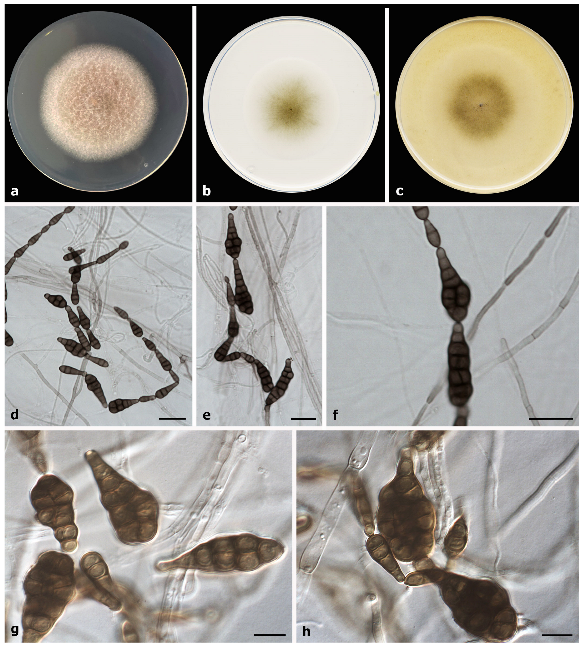
Etymology: The epithet refers to the production of muriform conidia in OA culture.
Culture characteristics (at 25 °C for 7 days): Colonies on PDA reaching 58–61 mm diam, blond-white to yellowish-white (4C4/4A2), cottony, abundant aerial mycelium, margins regular; reverse yellowish-brown (5F8/5D5), yellowish-white final edge (4A2). On PCA attaining 49–50 mm diam, flat, slightly velvety, scarce aerial mycelium, margins regular; reverse grey (1D4) to colorless towards the periphery. On OA reaching 55–56 mm diam, flat, slightly floccose, scarce aerial mycelium, margins regular; surface and reverse olive (4E3) to colorless.
Cardinal temperature for growth: minimum of 15 °C; optimum of 20 °C; and maximum of 30 °C.
Morphological description of the asexual morph (on OA at 25 °C for 7 days): Mycelium is superficial and immersed. Hyphae 1–4 μm wide, septate, branched, subhyaline to pale olivaceous, smooth-walled to verruculose. Conidiophores micronematous to semi-macronematous, arising laterally or terminally from aerial hyphae, erect to slightly flexuous, unbranched, 10.5–73 × 3–4.5 μm, pale olivaceous to yellowish brown, smooth-walled, with 1–2 terminal conidiogenous loci. Conidia forming unbranched or slightly branched chains, with up to 15 conidia in the unbranched part, commonly ellipsoidal or obclavate, 10–40 × 4–14 μm, with darkened middle transverse septa, some constricted, (1–)3–5(–7) transverse septa, and 0–1(–2) longitudinal or oblique septa per transverse segment, yellowish-brown to brown, smooth-walled; conidia with muriform conidial bodies are present, 37–45 × 16–33 μm. Secondary conidiophores can be formed as lateral conidiogenous loci from the conidial body. Furthermore, sexual morphology was not observed.
Known distribution: In Spain (as seen in this article: lifestyle-saprobic on herbivorous dung).
Specimen examined: Spain, Catalonia, Barcelona province, Pontons (N 41.40590° E 1.50918°), dung of an unidentified herbivorous animal, June 2018, J. Gené and I. Iturrieta-González (holotype FMR H-17518, culture ex-type FMR 17518).
Notes: Alternaria muriformis is placed in a supported clade in Alternaria section Chalastospora (Figure 1), and it is phylogenetically related to A. armoraciae, A. abundans, and A. breviramosa. However, the new species differs morphologically from its relatives in the production of muriform conidia [19,35,36] (Table 3). Despite following the recommendations of Simmons [19] for morphological characterization of the new fungus, we observed sporulation exclusively on OA at 25 °C.

4. Discussion

Morphological traits have long been the basis for species identification in the genus Alternaria [19]. However, due to the limited number of taxonomically informative features, especially to distinguish closely related species, the use of DNA sequence data and multi-gene analysis is now required, not only for identification purposes but also for delineating novel species. The molecular markers recommended for this purpose have been defined for various Alternaria sections over the course of several studies [2,15,33,39,40]. Therefore, based on those molecular approaches, numerous new species, mainly in the sections Alternaria, Infectoriae, Porri, and Radicina, have been described in recent years [2,12,18,41], but only a few in the section Chalastospora [2]. This section comprises a small group of eight Alternaria species, including the new species A. muriformis (Table 3), which are well delineated by using the ITS, gapdh, and ATPase gene markers. Of note is that despite the ITS barcode being considered a gene marker only able to classify Alternaria species at the section level [2,15,33,40], in section Chalastospora, each locus is able to discriminate each species (see Supplementary Material). However, significant statistical support to establish phylogenetic relationships among species can only be achieved with the combination of the three markers. In our multi-locus analysis, the isolate investigated here formed a highly supported branch with both ML and BI analysis (89/0.99) that reinforces the proposal of A. muriformis. With the exception of A. obclavata, which has been exclusively reported from air samples [35], most species in Chalastospora have been isolated from vegetal substrates, as mentioned before. However, A. malorum [21,42], A. pobletensis [2], and now A. muriformis have also been recovered from the dung of herbivorous animals, suggesting that this is a good source to find taxonomically interesting Alternaria species, not only for this section but also for other Alternaria groups, as previously reported by Marin-Felix et al. [2]. Animal dung, and specifically herbivore dung, contains hemicellulose, cellulose, lignin, high nitrogen content, minerals, and high moisture content, which constitute a good substrate for fungal growth [43]. This is how, in recent years, a significant number of new species and new records have been described from this type of substrate [2,44,45,46,47,48].
The genus Alternaria contains species with an important role as producers of metabolites [14,49]. Although some metabolites have been shown to be toxic in plants and animals, they have also been shown to have biotechnological applications with excellent antioxidative, herbicidal, antibacterial, antiparasitic, antitumor, and enzyme inhibitory properties, which reinforces the need to study potential new metabolites produced by new species in the genus [49,50,51,52].
This study introduces a new Alternaria species for the section Chalastospora based on a polyphasic approach combining morphological and molecular characterization. Future research is required in order to elucidate the ecology of A. muriformis, its role as a possible pathogenic species in toxin production, and its potential biotechnological applications.

Supplementary Materials

The following supporting information can be downloaded at: https://www.mdpi.com/article/10.3390/d15050606/s1, Figure S1: Maximum Likelihood tree of the section Chalastospora constructed with ITS region; Figure S2: Maximum Likelihood tree of the section Chalastospora constructed with gapdh; Figure S3: Maximum Likelihood tree of the section Chalastospora constructed with ATPase.

Author Contributions

Conceptualization, I.I.-G.; methodology, I.I.-G.; software, I.I.-G.; formal analysis, I.I.-G.; investigation, I.I.-G.; writing—original draft preparation, I.I.-G. and J.G.; writing—review and editing, I.I-G. and J.G.; supervision, J.G.; project administration, J.G.; funding acquisition, J.G. All authors have read and agreed to the published version of the manuscript.

Funding

This study is part of the results of the Grant PID2021-128068NB-I00 funded by MCIN/AEI/10.13039/501100011033/ and by “ERDF A way of making Europe”.

Institutional Review Board Statement

Not applicable.

Data Availability Statement

Not applicable.

Conflicts of Interest

The authors declare no conflict of interest.

References

  1. Von Esenbeck, C.G.N. Das System der Pilze und Schwämme; Stahelschen Buchhandlung: Wurzburg, Germany, 1816. [Google Scholar] [CrossRef]
  2. Marin-Felix, Y.; Hernández-Restrepo, M.; Iturrieta-González, I.; García, D.; Gené, J.; Groenewald, J.Z.; Cai, L.; Chen, Q.; Quaedvlieg, W.; Schumacher, R.K.; et al. Genera of phytopathogenic fungi: GOPHY 3. Stud. Mycol. 2019, 94, 1–124. [Google Scholar] [CrossRef]
  3. Huang, D.M.; Liu, X.; Bai, L.; Zhang, S.J.; Zhang, Z.G.; Qin, Q.P. First Report of Alternaria alternata Causing Leaf Spot Disease on Daylily in China. Plant Dis. 2022, 106, 3200. [Google Scholar] [CrossRef] [PubMed]
  4. Li, J.; Phookamsak, R.; Jiang, H.; Bhat, D.J.; Camporesi, E.; Lumyong, S.; Kumla, J.; Hongsanan, S.; Mortimer, P.E.; Xu, J.; et al. Additions to the Inventory of the Genus Alternaria Section Alternaria (Pleosporaceae, Pleosporales) in Italy. J. Fungi 2022, 8, 898. [Google Scholar] [CrossRef]
  5. Thomma, B.P.H.J. Alternaria spp.: From general saprophyte to specific parasite. Mol. Plant. Pathol. 2003, 4, 225–236. [Google Scholar] [CrossRef] [PubMed]
  6. El-Alwany, A.M. Plant Pathogenic Alternaria Species in Libya. Open Access Libr. J. 2015, 2, e1662. [Google Scholar] [CrossRef]
  7. Lawrence, D.P.; Rotondo, F.; Gannibal, P.B. Biodiversity and taxonomy of the pleomorphic genus Alternaria. Mycol. Prog. 2016, 15, 3. [Google Scholar] [CrossRef]
  8. Meena, M.; Gupta, S.K.; Swapnil, P.; Zehra, A.; Dubey, M.K.; Upadhyay, R.S. Alternaria Toxins: Potential Virulence Factors and Genes Related to Pathogenesis. Front. Microbiol. 2017, 8, 1451. [Google Scholar] [CrossRef] [PubMed] [Green Version]
  9. Pastor, F.J.; Guarro, J. Alternaria infections: Laboratory diagnosis and relevant clinical features. Clin. Microbiol. Infect. 2008, 14, 734–746. [Google Scholar] [CrossRef] [Green Version]
  10. Cardona, S.; Yusef, S.; Silva, E.; Bustos, M.G.; Torres, M.I.; Leal, A.R.; Ceballos-Garzon, A.; Josa, D.F. Cerebral phaeohyphomycosis caused by Alternaria spp.: A case report. Med. Mycol. Case Rep. 2020, 27, 11–13. [Google Scholar] [CrossRef]
  11. De Hoog, G.S.; Guarro, J.; Gené, J.; Ahmed, S.A.; Al-Hatmi, A.M.S.; Figueras, M.J.; Vitale, R.G. Atlas of Clinical Fungi, 4th ed.; Foundation Atlas of Clinical Fungi: Hilversum, The Netherlands, 2020. [Google Scholar]
  12. Iturrieta-González, I.; Pujol, I.; Iftimie, S.; García, D.; Morente, V.; Queralt, R.; Guevara-Suarez, M.; Alastruey-Izquierdo, A.; Ballester, F.; Hernández-Restrepo, M.; et al. Polyphasic identification of three new species in Alternaria section Infectoriae causing human cutaneous infection. Mycoses 2019, 63, 212–224. [Google Scholar] [CrossRef]
  13. Abbas, H.K.; Riley, R. The presence and phytotoxicity of fumonisins and aal-toxin in Alternaria alternata. Toxicon. 1996, 34, 133–136. [Google Scholar] [CrossRef]
  14. Lou, J.; Fu, L.; Peng, Y.; Zhou, L. Metabolites from Alternaria Fungi and Their Bioactivities. Molecules 2013, 18, 5891–5935. [Google Scholar] [CrossRef] [PubMed]
  15. Woudenberg, J.H.C.; Groenewald, J.Z.; Binder, M.; Crous, P.W. Alternaria redefined. Stud. Mycol. 2013, 75, 171–212. [Google Scholar] [CrossRef] [PubMed] [Green Version]
  16. Gannibal, P.B.; Orina, A.S.; Gasich, E.L. A new section for Alternaria helianthiinficiens found on sunflower and new asteraceous hosts in Russia. Mycol. Prog. 2022, 21, 34. [Google Scholar] [CrossRef]
  17. Al Ghafri, A.; Maharachchikumbura, S.S.N.; Hyde, K.D.; Al-Saady, N.A.; Al-Sadi, A.M. A new section and a new species of Alternaria encountered from Oman. Phytotaxa 2019, 405, 279–289. [Google Scholar] [CrossRef]
  18. Zhao, L.; Luo, H.; Cheng, H.; Gou, Y.N.; Yu, Z.H.; Deng, J.X. New Species of Large-Spored Alternaria in Section Porri Associated with Compositae Plants in China. J. Fungi 2022, 8, 607. [Google Scholar] [CrossRef] [PubMed]
  19. Simmons, E.G. Alternaria: An Identification Manual; CBS Biodiversity Series 6; CBS Fungal Biodiversity Centre: Utrecht, The Netherlands, 2007. [Google Scholar]
  20. Simmons, E.G. Helminthosporium allii as type of a new genus. Mycologia 1971, 63, 380–386. [Google Scholar] [CrossRef]
  21. Mirhendi, H.; Fatemi, M.J.; Bateni, H.; Hajabdolbaghi, M.; Geramishoar, M.; Ahmadi, B.; Badali, H. First case of disseminated phaeohyphomycosis in an immunocompetent individual due to Alternaria malorum. Med. Mycol. 2013, 51, 196–202. [Google Scholar] [CrossRef] [Green Version]
  22. Muller, F.M.C.; Werner, K.E.; Kasai, M.; Francesconi, A.; Chanock, S.J.; Walsh, T.J. Rapid extraction of genomic DNA from medically important yeasts and filamentous fungi by high-speed cell disruption. J. Clin. Microbiol. 1998, 36, 1625–1629. [Google Scholar] [CrossRef] [Green Version]
  23. White, T.J.; Bruns, T.; Lee, S.J.W.T.; Taylor, J. Amplification and direct sequencing 794 of fungal ribosomal RNA genes for phylogenetics. In PCR Protocols: A Guide to Methods and 796 Applications; Innis, M.A., Gelfand, D.H., Sninsky, J.J., White, T.J., Eds.; Academic Press: New York, NY, USA, 1990; pp. 315–322. [Google Scholar]
  24. Lawrence, D.P.; Gannibal, P.B.; Peever, T.L.; Pryor, B.M. The sections of Alternaria: Formalizing species-group concepts. Mycologia 2013, 105, 530–546. [Google Scholar] [CrossRef] [Green Version]
  25. Berbee, M.L.; Pirseyedi, M.; Hubbard, S. Cochliobolus Phylogenetics and the Origin of Known, Highly Virulent Pathogens, Inferred from ITS and Glyceraldehyde-3-Phosphate Dehydrogenase Gene Sequences. Mycologia 1999, 91, 964–977. [Google Scholar] [CrossRef]
  26. Tamura, K.; Stecher, G.; Peterson, D.; Filipski, A.; Kumar, S. MEGA6: Molecular evolutionary genetics analysis version 6.0. Mol. Biol. Evol. 2013, 30, 2725–2729. [Google Scholar] [CrossRef] [Green Version]
  27. Thompson, J.D.; Higgins, D.G.; Gibson, T.J. CLUSTAL W: Improving the sensitivity of progressive multiple sequence alignment through sequence weighting, position-specific gap penalties and weight matrix choice. Nucleic Acids Res. 1994, 22, 4673–4680. [Google Scholar] [CrossRef] [PubMed] [Green Version]
  28. Edgar, R.C. MUSCLE: Multiple sequence alignment with high accuracy and high throughput. Nucleic Acids Res. 2004, 32, 1792–1797. [Google Scholar] [CrossRef] [PubMed] [Green Version]
  29. Ronquist, F.; Teslenko, M.; van der Mark, P.; Ayres, D.L.; Darling, A.; Höhna, S.; Larget, B.; Liu, L.; Suchard, M.A.; Huelsenbeck, J.P. MrBayes 3.2: Efficient Bayesian Phylogenetic Inference and Model Choice across a Large Model Space. Syst. Biol. 2012, 61, 539–542. [Google Scholar] [CrossRef] [PubMed] [Green Version]
  30. Farris, J.S.; Kallersjo, M.; Kluge, A.G.; Bult, C. Testing significance of incongruence. Cladistics 1994, 10, 315–319. [Google Scholar] [CrossRef]
  31. Posada, D. jModelTest: Phylogenetic Model Averaging. Mol. Biol. Evol. 2008, 25, 1253–1256. [Google Scholar] [CrossRef] [PubMed]
  32. Deng, J.X.; Li, M.J.; Paul, N.C.; Oo, M.M.; Lee, H.B.; Oh, S.K.; Yu, S.H. Alternaria brassicifolii sp. nov. isolated from Brassica rapa subsp. pekinensis in Korea. Mycobiology 2018, 46, 172–176. [Google Scholar] [CrossRef] [Green Version]
  33. Poursafar, A.; Ghosta, Y.; Orina, A.S.; Gannibal, P.B.; Javan-Nikkhah, M.; Lawrence, D.P. Taxonomic study on Alternaria sections Infectoriae and Pseudoalternaria associated with black (sooty) head mold of wheat and barley in Iran. Mycol. Prog. 2018, 17, 343–356. [Google Scholar] [CrossRef]
  34. Kornerup, A.; Wanscher, J.H. Methuen Handbook of Colour, 3rd ed.; Methuen: London, UK, 1978; pp. 1–256. [Google Scholar]
  35. Crous, P.W.; Braun, U.; Wingfield, M.J.; Wood, A.; Shin, H.D.; Summerell, B.A.; Alfenas, A.C.; Cumagun, C.J.; Groenewald, J.Z. Phylogeny and taxonomy of obscure genera of microfungi. Persoonia 2009, 22, 139–161. [Google Scholar] [CrossRef] [Green Version]
  36. Simmons, E.G. An aggregation of Embellisia species. Mycotaxon 1983, 17, 216–241. [Google Scholar]
  37. Simmons, E.G. Alternaria themes and variations (145–149). Mycotaxon 1996, 57, 391–409. [Google Scholar]
  38. Braun, U.; Crous, P.W.; Dugan, F.; Groenewald, J.Z.; De Hoog, G.S. Phylogeny and taxonomy of Cladosporium-like hyphomycetes, including Davidiella gen. nov., the teleomorph of Cladosporium s. str. Mycol. Prog. 2003, 2, 3–18. [Google Scholar] [CrossRef]
  39. Woudenberg, J.H.C.; Truter, M.; Groenewald, J.Z.; Crous, P.W. Large-spored Alternaria pathogens in section Porri disentangled. Stud. Mycol. 2014, 79, 1–47. [Google Scholar] [CrossRef] [PubMed] [Green Version]
  40. Woudenberg, J.H.C.; Seidl, M.F.; Groenewald, J.Z.; De Vries, M.; Stielow, J.B.; Thomma, B.P.H.J.; Crous, P.W. Alternaria section Alternaria: Species, formae speciales or pathotypes? Stud. Mycol. 2015, 82, 1–21. [Google Scholar] [CrossRef] [PubMed] [Green Version]
  41. Romain, B.B.N.D.; Hassan, O.; Kim, J.S.; Chang, T. Alternaria koreana sp. nov., a new pathogen isolated from leaf spot of ovate-leaf Atractylodes in South Korea. Mol. Biol. Rep. 2022, 49, 413–420. [Google Scholar] [CrossRef]
  42. Ruehle, G.D. New apple-rot fungi from Washington. Phytopathology 1931, 21, 1141–1152. [Google Scholar]
  43. Holter, P. Herbivore dung as food for dung beetles: Elementary coprology for entomologists. Ecol. Entomol. 2016, 41, 367–377. [Google Scholar] [CrossRef] [Green Version]
  44. Richardson, M. New and interesting records of coprophilous fungi. Bot. J. Scotl. 1998, 50, 161–175. [Google Scholar] [CrossRef]
  45. Ghosta, Y.; Poursafar, A.; Qarachal, J.F. Study on coprophilous fungi: New records for Iran mycobiota. Rostaniha 2016, 17, 115–126. [Google Scholar]
  46. Melo, R.F.R.; Monte, D.B.P.D.; Gondim, N.H.B.; Maia, L.C.; Miller, A.N. Coprophilous fungi from Brazil: New records for the Neotropics. Mycotaxon 2019, 134, 335–352. [Google Scholar] [CrossRef]
  47. Guevara-Suarez, M.; García, D.; Cano-Lira, J.F.; Guarro, J.; Gené, J. Species diversity in Penicillium and Talaromyces from herbivore dung, and the proposal of two new genera of penicillium-like fungi in Aspergillaceae. Fungal Syst. Evol. 2020, 5, 39–75. [Google Scholar] [CrossRef]
  48. Iturrieta-González, I.; García, D.; Gené, J. Novel species of Cladosporium from environmental sources in Spain. Mycokeys 2021, 77, 1–25. [Google Scholar] [CrossRef] [PubMed]
  49. Zhao, S.; Li, J.; Liu, J.; Xiao, S.; Yang, S.; Mei, J.; Ren, M.; Wu, S.; Zhang, H.; Yang, X. Secondary metabolites of Alternaria: A comprehensive review of chemical diversity and pharmacological properties. Front. Microbiol. 2023, 13, 1085666. [Google Scholar] [CrossRef] [PubMed]
  50. Horiuchi, M.; Tokuda, H.; Ohnishi, K.; Yamashita, M.; Nishino, H.; Maoka, T. Porritoxins, metabolites of Alternaria porri, as anti-tumor-promoting active compounds. Nat. Prod. Res. 2006, 20, 161–166. [Google Scholar] [CrossRef]
  51. Shi, Y.N.; Pusch, S.; Shi, Y.M.; Richter, C.; Maciá-Vicente, J.G.; Schwalbe, H.; Kaiser, M.; Opatz, T.; Bode, H.B. (±)-Alternarlactones A and B, two antiparasitic alternariol-like dimers from the fungus Alternaria alternata P1210 isolated from the halophyte Salicornia sp. J. Org. Chem. 2019, 84, 11203–11209. [Google Scholar] [CrossRef] [PubMed]
  52. Kong, F.D.; Yi, T.F.; Ma, Q.Y.; Xie, Q.Y.; Zhou, L.M.; Chen, J.P.; Dai, H.F.; Wu, Y.G.; Zhao, Y.X. Biphenyl metabolites from the patchouli endophytic fungus Alternaria sp. PfuH1. Fitoterapia 2020, 146, 104708. [Google Scholar] [CrossRef]
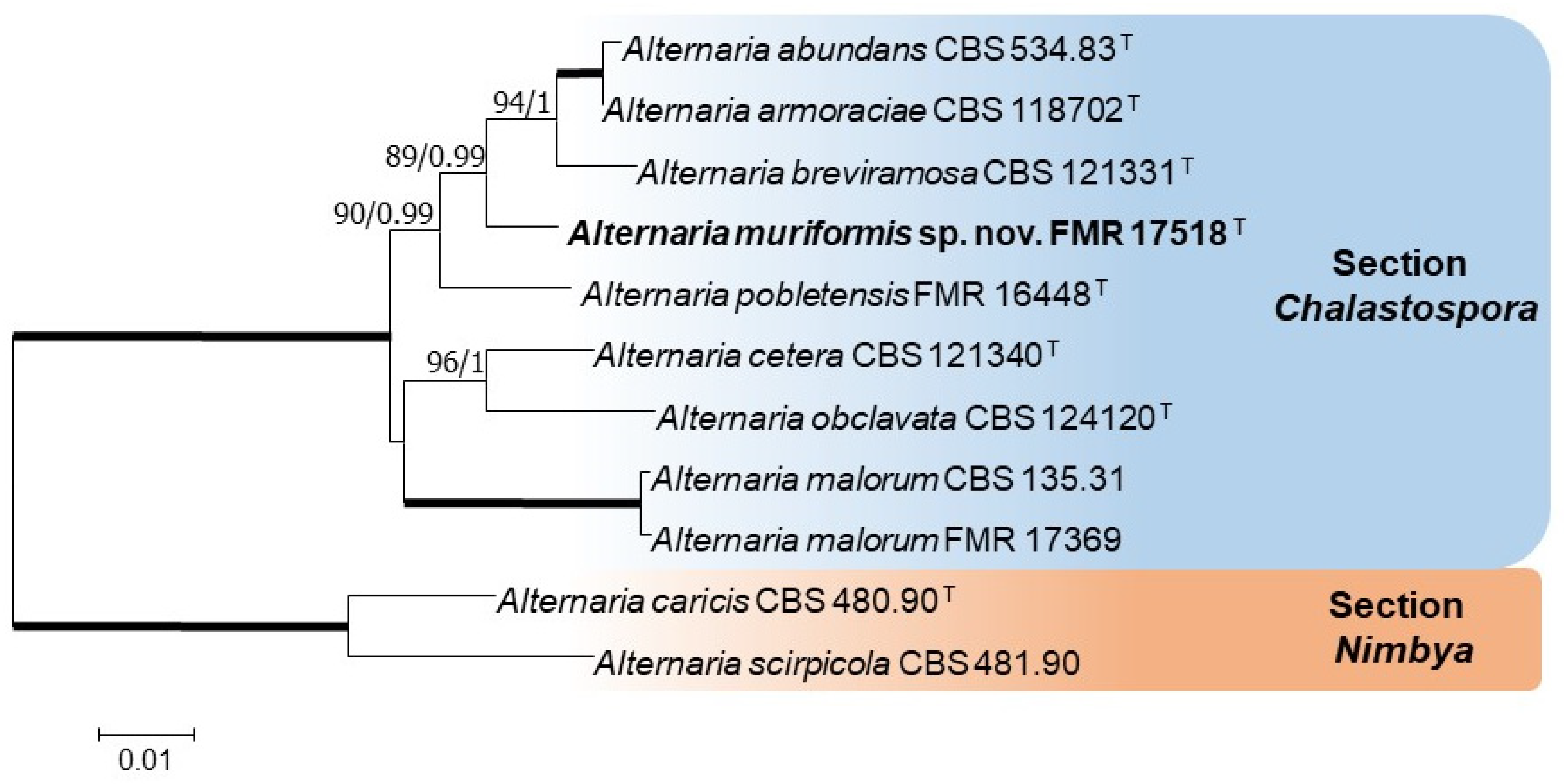
Figure 1. Phylogenetic tree constructed with ITS (536 bp), gapdh (485 bp), and ATPase (1195 bp) and sequences of ex-type strains of Alternaria species in the section Chalastospora and rooted with Alternaria caricis CBS 480.90 and Alternaria scirpicola CBS 481.90 (section Nimbya). Bootstrap support (bs) values greater than 70% and Bayesian posterior probabilities (pp) greater than 0.95 are given at the nodes (bs/pp). Bold branches indicate a bs/pp of 100/1. The novel species described in this study is indicated in bold. T indicates an ex-type of strain.
Diversity 15 00606 g001
Figure 2. Alternaria muriformis (ex-type FMR 17518). (a) Colonies on PDA; (b) colonies on PCA; (c) colonies on OA; (dh) Conidia. Scale bars (df) = 20 μm and (g,h) = 10 μm.
Diversity 15 00606 g002
Table 1. List of primer pair sets used for PCR and sequencing.
LocusPrimerSequence (5′–3′)References
Internal transcribed spacer (ITS)ITS5GGAAGTAAAAGTCGTAACAAGG[23]
ITS4TCCTCCGCTTATTGATATGC
Glyceraldehyde-3-phosphate dehydrogenase (gapdh)gpd1CAACGGCTTCGGTCGCATTG[25]
gpd2GCCAAGCAGTTGGTTGTGC
Plasma membrane ATPase (ATPase)ATPDF1ATCGTCTCCATGACCGAGTTCG[24]
ATPDR1TCCGATGGAGTTCATGATAGCC
Table 3. Comparison of the conidial morphology among Alternaria species in section Chalastospora.
SpeciesConidiaReferences
ShapeSize (µm)Transverse Septa NumbersLongitudinal or Oblique Septa Numbers (*)Ornamentation
A. abundansOvoidal
Obclavate
20–30 × 10–12
40–50 × 8–12
3–6(–8)0–1Usually smooth[36]
A. armoraciaeOvoidal to ellipsoidal15–35 × 8–123–50–1Smooth[19]
A. breviramosaEllipsoidal to fusiform(8–)10–15(–17) × 3(–3.5)0–1(–2)AbsentSmooth[35]
A. ceteraEllipsoidal to narrow-ovoid18–22 × 3–4(–5)1–3AbsentSmooth[19,37]
A. malorumEllipsoidal-ovoidal, cylindrical, or fusiform6–14(17) × 2–4AbsentAbsentSmooth[38]
A. obclavataObclavate(23–)26–30(–35) × (3.5–)40–3AbsentSmooth[35]
A. pobletensisObpyriform or obclavate, and some ellipsoidal or subcylindrical8–50 × 5–20(1–)3–7(–9)0–1(–2)Smooth or verruculose[2]
A. muriformisEllipsoidal or obclavate10–40 × 4–14(1–)3–5(–7)0–1(–2)SmoothPresent study
Muriform37–45 × 16–33
* per transverse segment. The novel species described in this study is indicated in bold.
Disclaimer/Publisher’s Note: The statements, opinions and data contained in all publications are solely those of the individual author(s) and contributor(s) and not of MDPI and/or the editor(s). MDPI and/or the editor(s) disclaim responsibility for any injury to people or property resulting from any ideas, methods, instructions or products referred to in the content.