Article

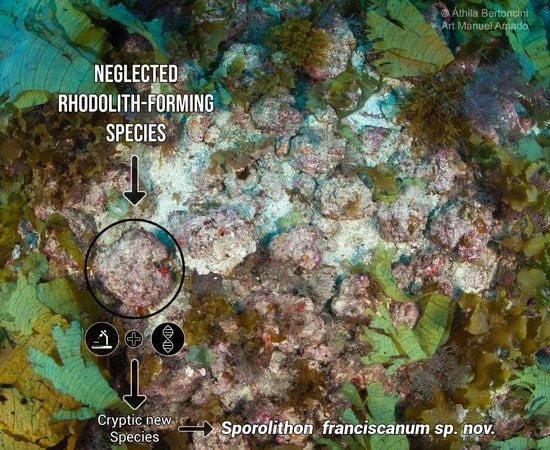
Sporolithon franciscanum sp. nov. (Sporolithales, Rhodophyta), a New Rhodolith-Forming Species from Northeast Brazil

by
Luis Alfredo S. Leão
1,
Ricardo G. Bahia
1,
Michel B. Jesionek
1,
Walter H. Adey
2,
Gabriel Johnson
3,
Leonardo T. Salgado
1 and
Renato C. Pereira
1,4,*
1
Instituto de Pesquisas Jardim Botânico do Rio de Janeiro, Diretoria de Pesquisa Científica, Rua Pacheco Leão 915, Rio de Janeiro 22460-030, RJ, Brazil
2
Botany Department, National Museum of Natural History, Smithsonian Institution, Washington, DC 20560, USA
3
Laboratory of Analytical Biology, National Museum of Natural History, Smithsonian Institution, Washington, DC 20560, USA
4
Marine Biology Department, Federal Fluminense University, Morro do Valonguinho, s/n, Niterói 24000-000, RJ, Brazil
*
Author to whom correspondence should be addressed.
Diversity 2020, 12(5), 199; https://doi.org/10.3390/d12050199
Submission received: 23 March 2020 / Revised: 1 May 2020 / Accepted: 7 May 2020 / Published: 15 May 2020
(This article belongs to the Special Issue Structure and Biodiversity of Rhodolith Seabeds)

Abstract

:
This paper describes Sporolithon franciscanum, a new rhodolith-forming species of non-geniculate coralline algae found at depths between 47–52 m near the São Francisco river mouth, the second largest and the most extensive drainage basin in Brazil, and also at the Abrolhos Bank, in the world´s largest rhodolith beds. DNA sequences from plastidial psbA and rbcL markers indicate that the species is unique compared to all other Sporolithon species that have thus far been sequenced. Since morpho-anatomical features of the new species are shared with some other Sporolithon species, its identification was only confirmed by DNA sequences.

Graphical Abstract

1. Introduction

Coralline red algae (Corallinophycidae, Rhodophyta) have as their main feature the deposition of calcium carbonate polymorphs (calcite, dolomite, magnesite, aragonite, calcite with low magnesium content) on their cell walls [1,2,3,4]. Non-geniculate coralline algae are considered one of the most difficult groups within the Rhodophyta in terms of taxonomic identification [5,6,7]. This perception can be attributed largely to the fact that, unlike most seaweeds, they have a calcified thallus that requires specific and time-consuming laboratory methods for taxonomic analyses [8]. Most identification from order (Corallinales, Hapalidiales, Sporolithales) to species level requires analyses of characteristics related to the reproductive structures (conceptacles or calcified compartments) from the tetra/bisporophytic phases [9]. However, although tetra/bisporophytic phases are the most abundant in nature, many specimens studied only have gametophytic conceptacles or, even more often, have been unfertile (and so excluded from the morpho-anatomical analysis). At the genus and sometimes at species level, the exclusive use of vegetative characteristics may also be useful in identification, such as the shape of epithallial cells or type of cell connections. However, in most cases morpho-anatomical identification in coralline algae is based on a combination of both vegetative and reproductive characteristics [9]. The main problem concerns cryptic species, which cannot be distinguished by morpho-anatomy, but are genetically different species. One example has been seen in the genus Lithophyllum Philippi (Corallinales), in which 13 different species were passed under the name Lithophyllum stictaeforme due to their similar morpho-anatomy [10]. On the other hand, a single species/genotype is often represented by different morphologies/phenotypes (e.g., [11]). The incorporation of molecular data into the coralline algae taxonomy has enabled previous classifications to be revised, confirmed or modified, in addition to enabling the identification of cryptic and/or new species established by genetic disparities or not revealed in the phenotype [12,13,14,15,16,17,18,19].
Non-geniculate coralline algae can grow attached to continuous substrates or loosen, forming free-living calcareous nodules named rhodoliths, which, by definition, are composed mainly (>50%) by non-geniculate coralline algae [20,21]. Non-geniculate coralline algae are represented worldwide by at least 75 species known to form rhodoliths [9,15,22,23,24,25,26], and the highest richness of this group (33 taxa) is found in Brazil where the genus Sporolithon Heydrich (Sporolithales) holds the largest number of species [26,27].
Sporolithon is characterized by a combination of features, such as: epithallial cells with a trapezoidal (‘flared’) lumen, contiguous filament cells connected by both cell fusions and secondary pit-connections, and cruciate divided tetrasporangia produced singly within uniporate, calcified compartments, each bearing an apical plug that are borne on a single stalk cell [28,29]. The members of this algal genus are among the main rhodolith-forming algal genera in the southwestern (SW) Atlantic where they are mainly found forming rhodoliths (and then contributing to the formation of rhodolith beds) in mesophotic regions (20–133 m deep) [26,27,30,31,32,33,34], but some species can also be found in shallow (2–7 m) reefs [26,35].
Currently, there are 29 recognized species of Sporolithon worldwide [36] from which 12 are recorded in Brazil, namely, S. amadoi [26], S. australasicum [37], S. durum [38], S. elevatum [35], S. episoredion [35], S. episporum [39,40], S. erythraeum [37], S. howei [37], S. molle [31], S. pacificum [37], S. tenue [32], and S. yoneshigueae [33]. With the exception of S. howei, which has been recorded to form carbonate concretions, all species were reported to occur in Brazilian rhodolith beds [31,32,33,34,35,37,38,39,40]. Among them, only S. amadoi, S. tenue and S. yoneshigueae were assigned based on the analysis of DNA sequences of the type material. The other nine Sporolithon species were determined based on specimens identified using only morpho-anatomical analyses. Thus, some of these names may have been incorrectly applied. In addition, and unfortunately, most specimen collections used for this identification were formalin-preserved and DNA sequencing analysis is likely to be unfruitful. Therefore, investments in new samplings of fresh rhodoliths with suitable storage methods for DNA analysis are required to assess the true diversity of these coralline algae in the SW Atlantic. Recently, several molecular studies have reported the existence of cryptic speciation in Sporolithon, which led to an increase in the number of species of this genus [15,23,24,25,26]. These studies reinforced that comparative analyses of DNA sequences based on sequences of type specimens is the best way to unequivocally identify non-geniculate coralline algae since the morpho-anatomical characteristics alone can be misleading. However, obtaining diagnostic DNA sequences from type material has been a hard task and the main challenge in the current coralline algae taxonomy, since many attempts in doing it have failed [41,42].
There is an ongoing scientific interest in rhodolith beds worldwide due, among other reasons, to the following: (1) The beds are bioengineers and provide a complex three-dimensional matrix (structural complexity) and hard structure that serves as a relatively stable microhabitat for several invertebrates, other macroalgae and fishes, some of which are endemic, rare or commercially important species [10,11,27,43,44,45,46,47,48]; (2) they are sensitive to the ongoing global climate change (e.g., ocean warming and acidification) that is predicted for the current century [49]; (3) their structures serve as paleoenvironmental indicators of past oceanic conditions [50,51]; (4) rhodolith beds are, alongside coral reefs, one of the main contributors to the global calcium carbonate budget [52,53,54]. In the tropical southwestern (SW) Atlantic Ocean (Brazil), the rhodolith-forming non-geniculate coralline algae are also known to be the main carbonate builders forming the world’s largest rhodolith beds [27] in addition to other extensive carbonate reefs [55,56].
Coralline algae exert species-specific interactions with other key organisms for community structuring. For instance, it has been demonstrated that larval settlement and metamorphosis of some species of coral and other invertebrates occurs preferably or restrictedly in the presence of some specific coralline algae [57,58,59]. Therefore, knowledge of the rhodolith-forming species is essential for understanding their specific role within the communities. In this context, the aim of this study was to report a new rhodolith-forming Sporolithon species that was found near the São Francisco River mouth (the second largest and the most extensive drainage basin within the Brazilian territory [60,61]) and in the Abrolhos Bank (the world’s largest rhodolith bed [53]). It is important to note that these two areas are characterized by presenting a high biodiversity associated with rhodoliths [27,62].

2. Materials and Methods

2.1. Sampling

Two specimens were analyzed in this study. The first (RB 797665) was sampled within the scope of the Marseal Project (Universidade Federal de Sergipe and Petrobras) that aims to generate physical, geological, chemical and biological data related to the Sergipe-Alagoas basin (Northeast Brazil). This specimen was collected offshore Aracaju (11°9’49.580’’ S; 36°53’0.814’’ W, Sergipe, Brazil, at 47 m deep), during the third campaign of the Marseal Project, in 2011, using a Van Veen grab sediment sampler. The sample was initially preserved in a 70% ethanol solution, and then preserved in silica gel. The second (Voucher GM AF5) was a small, unfertile, rhodolith fragment sampled at the Abrolhos continental shelf (17°54’01’’ S; 37°54’56’’ W, Bahia, Brazil, at 52 m deep, G.M. Amado Filho, 11.iii.2012), preserved in silica gel, and with published sequences in GenBank (KP142752), which was used to call Sporolithon sp. (voucher GM AF5) in Adey et al. [63] and Richards et al. [15,26]. The first is housed at the Rio de Janeiro Botanical Garden Herbarium (RB) under the voucher RB 797665, while the second is housed at the National Museum of Natural History (NMNH) (US, the United States National Herbarium at NMNH) under the voucher GM AF5. Herbarium abbreviations follow [64], continuously updated.

2.2. Morpho-Anatomical Analyses

Fractures from fertile areas of the holotype were decalcified according to Jesionek et al. [35] and prepared for light microscopy examination using the histological methods described by Maneveldt and Van der Merwe [65]. Other fractures were examined with scanning electron microscopy (SEM) following the protocol used by Bahia et al. [66]. The thallus anatomical terminology followed Chamberlain [67]. The morphological (growth forms) terminology followed Woelkerling et al. [68]. Cell dimensions were measured from all available images as described in Maneveldt et al. [33]. Tetrasporangial compartments and tetrasporangia were measured separately according to Kaewsuralikhit et al. [69]. Since the specimen from Abrolhos (GM AF5) corresponds to an unfertile small fragment, no anatomical analyses were performed for this specimen.

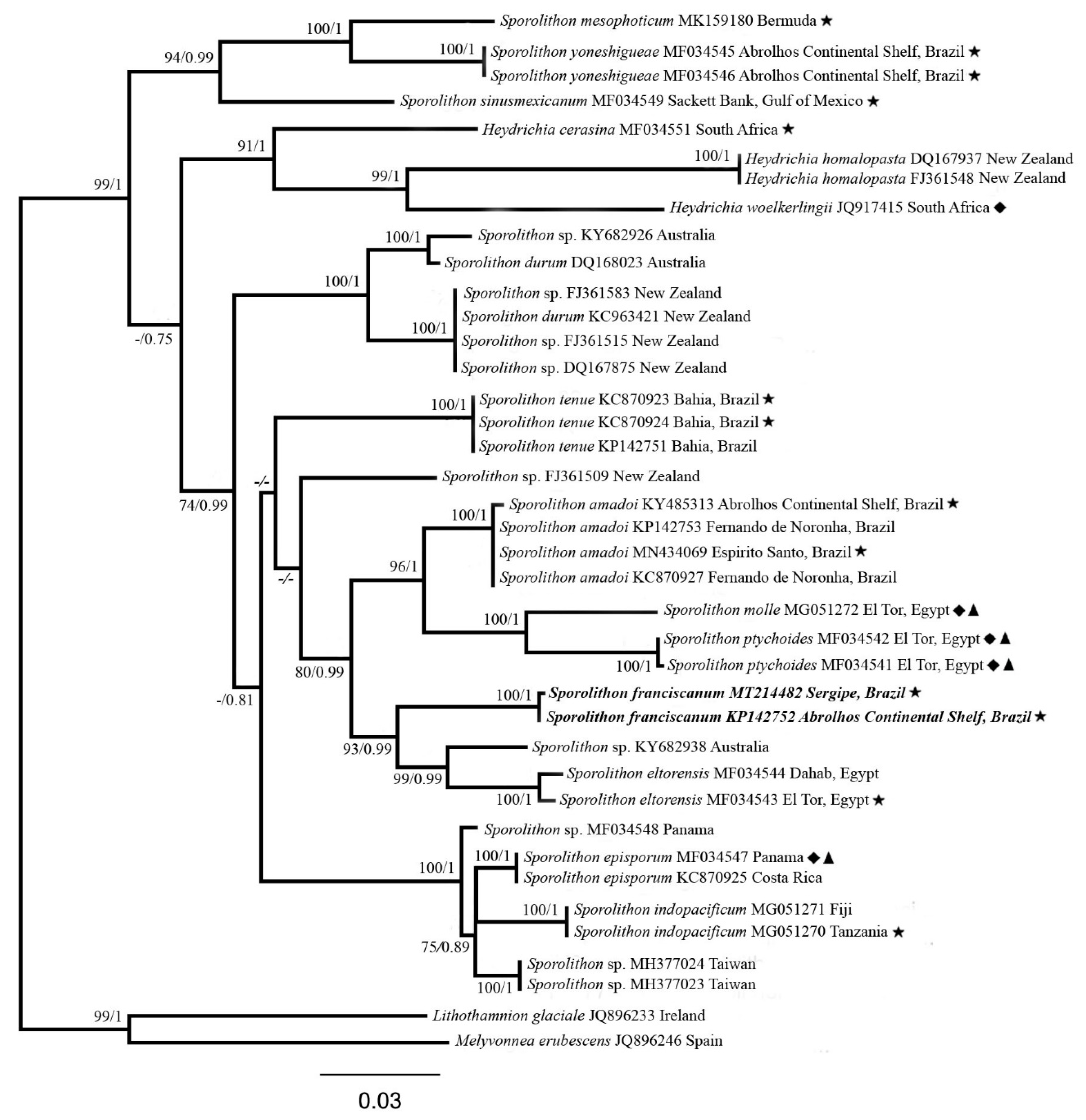
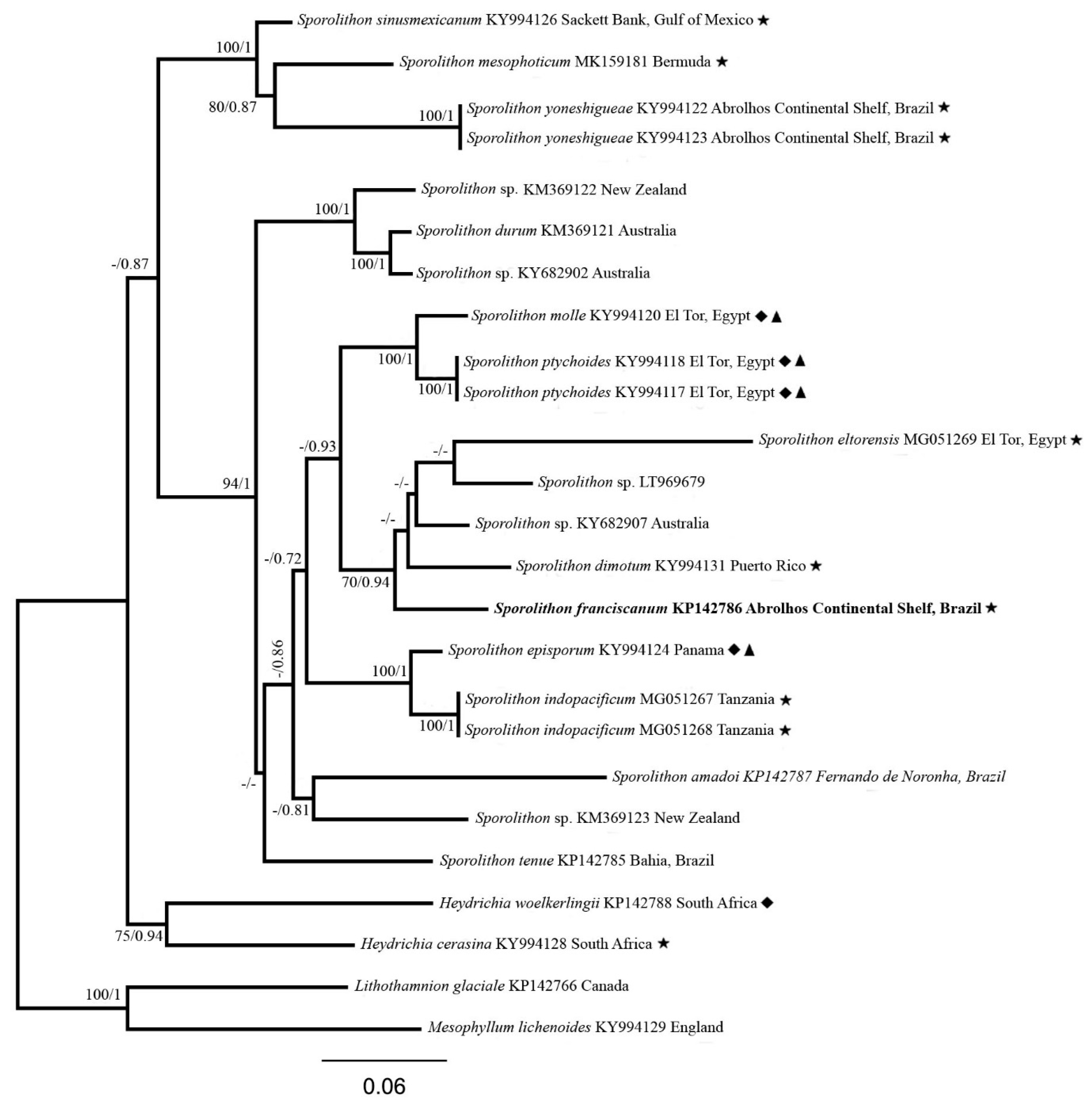
2.3. Molecular Analysis

DNA extraction, amplification and sequencing of the psbA genetic marker followed the protocol used by Jesionek et al. [70] and were performed for the specimen from Sergipe (RB 797665). DNA was extracted using the Qiagen DNeasy Blood and Tissue Kit® (Qiagen, Crawley, UK) following the modified protocol of Broom et al. [13]. The psbA gene was amplified using the primers psbAF and psbAR2 [71]. For the Abrolhos continental shelf specimen (GM AF5), the analysis for psbA and rbcL followed Adey et al. [63]. The extraction was made following the protocol of Huguey et al. [72], in which after a series of steps, it resulted in a working solution of 10:1 (water:DNA) for PCR and a negative control for each set of extraction. For psbA gene amplification, were used the same primers as for the Sergipe specimen (psbAF and psbAR2) [71], and for rbcL two pairs were used (F57—R1150 and F753—RrbcS) [73]. Since all attempts to amplify rbcL for the specimen from Sergipe (RB 797665) failed, it was not possible to use rbcL for molecular analysis. The sequences obtained for both markers were assembled and edited in Sequencher v.4.1.4 (Gene Codes Corporation) and edited and aligned in MEGA6 [74] within ClustalW [75,76]. The sequence dataset for the psbA analysis was built with the sequence of the holotype generated in this study in addition to the sequence of the paratype from GenBank (KP142752) and 37 others that were downloaded from GenBank from Sporolithon, Heydrichia and the outgroup (Lithothamnion glaciale and Melyvonnea erubescens). The sequences dataset for the rbcL analysis included the sequence of the paratype (since rbcL could not be amplified for the holotype) generated in Adey et al. [63] and 22 others from GenBank from Sporolithon and the outgroup (Heydrichia woelkerlingii and Heydrichia cerasina). The phylogenetic relationships between taxa were inferred in Geneious R7, using Maximum Likelihood (ML) with Randomized Axelerated Maximum Likelihood (RAxML 8) [77] with a bootstrap of 1000 replicates [78] and Bayesian Inference (BI) with Mr. Bayes [79] with four Monte Carlo–Markov chains. Bootstrap and Bayesian posterior probabilities (PP) values were considered high when they were equal to or higher than 70% and/or 0.70, respectively. In this analysis, 5 million generations were launched. The trees were sampled every 1000 generations and 1,250,000 trees were discarded as burn-in. Finally, the standard deviation of the split frequencies was verified in the summary statistics in Geneious R7.

3. Results

The results from psbA analyses indicated that the specimen from Sergipe (RB 797665) and the one from the Abrolhos continental shelf (GM AF5) correspond to the same and new species (namely Sporolithon franciscanum) (Figure 1). The phylogram shows that Sporolithon franciscanum formed a highly supported (93/0.99, ML and BI) clade sister to S. eltorensis and an Australian Sporolithon sp. (Figure 1). The divergence value between the specimens from Sergipe and Abrolhos continental shelf was 0.1% (an intraspecific value), and the new species diverge from the sister species from the clade by between 6.1–6.5% (Table 1). In rbcL analysis, the available sequences positioned two additional species (Sporolithon dimotum and a Sporolithon sp.) within the S. franciscanum clade (Figure 2). The new species diverged from all taxa within its clade by 6–12.3% (Table 1).

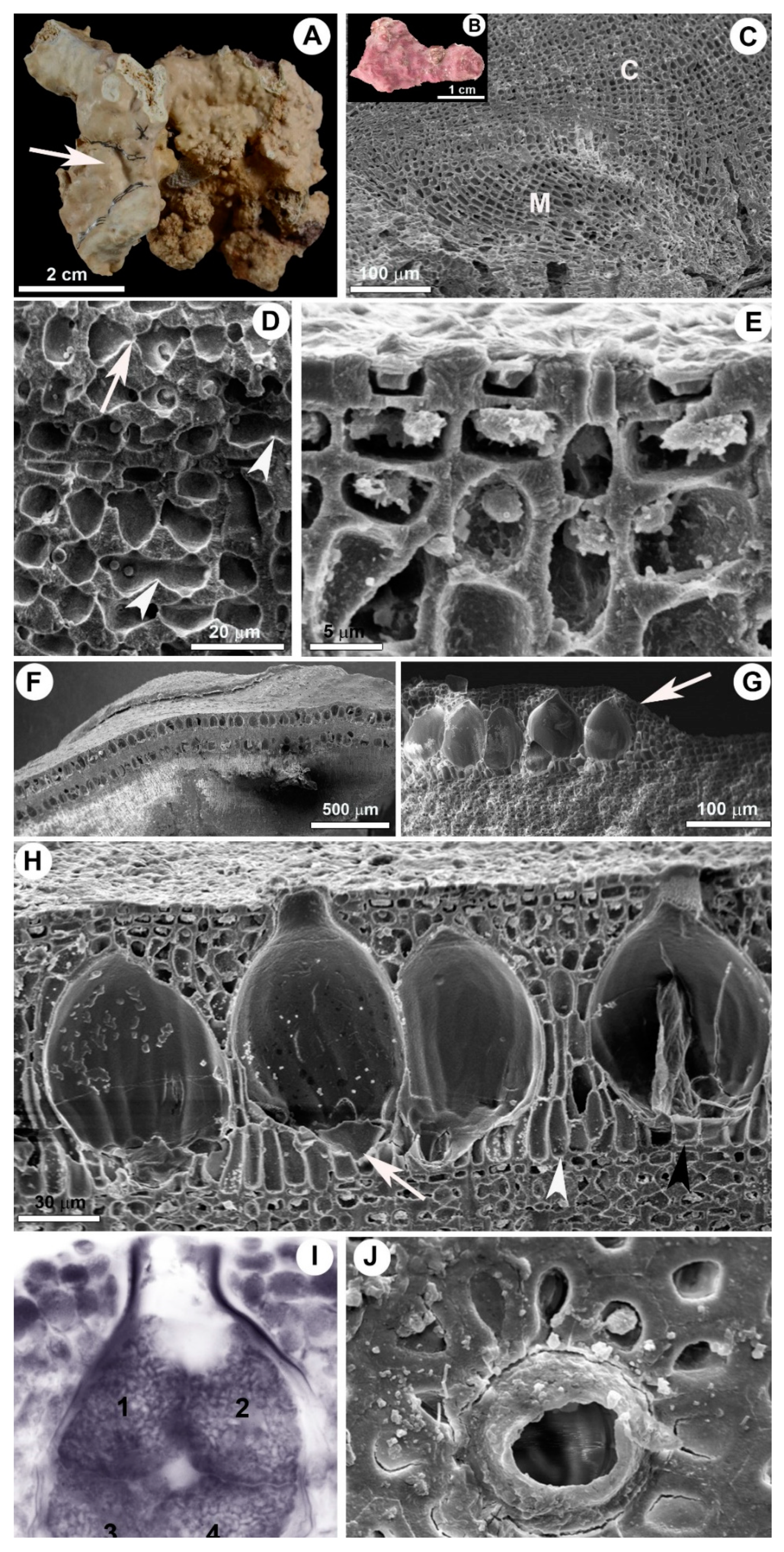
3.1. Sporolithon franciscanum L.A.S. Leão & Bahia sp. nov.

Holotype: RB 797665, collected February, 2, 2011, leg. L.C.S. Fontes, J.R. Santos, L.A. Santos, J.B.S. Mendonça, & M.S. Santos, psbA - GenBank MT214482.
Paratype: GMAF5, collected March, 11, 2012, leg. GM Amado Filho, psbA - GenBank KP142752, rbcL – GenBank KP142786 (Table S1).
Type locality: Aracaju, Sergipe State, Brazil (11°9’49.580’’ S, 36°53’0.814’’ W), subtidal region (47 m deep), free-living form as rhodolith.
Etymology: The specific epithet franciscanum is derived from the main river of Sergipe-Alagoas continental shelf, the São Francisco River.

3.2. Habit and Vegetative Anatomy

The external morphology and anatomy of two specimens were analyzed: A tetrasporophyte holotype and an unfertile paratype. The holotype was found composing the surface of a multispecific rhodolith measuring 4–7 cm in diameter (Figure 3A). The paratype constitutes a small rhodolith fragment (Figure 3B). Thalli were encrusting, with smooth surface, and pink to purple color (Figure 3A).
Thallus dorsiventrally organized, monomerous, 500–1000 μm thick, with more than 20 cell layers (Figure 3C). Medulla plumose (non-coaxial) (Figure 3C), comprise 8–17 filaments aligned in more or less parallel mode to the substratum. Medullary filaments comprise square to elongated cells, measuring 7–22 μm in length and 4–10 μm in diameter. Cortical filaments comprise square to elongated cells, measuring 6–19 μm in length and 3–8 μm in diameter. Contiguous cortical filaments are joined by both secondary pit connections and cell fusions (Figure 3D); cell fusions predominate by a ratio of 2:1. Subepithallial initials square to rectangular, with 6–11 μm in length and 4–7 μm in diameter, as long as or longer than subtending cells (Figure 3E). Epithallial cells single layered, flattened with flared upper corners (Figure 3E), and measuring 2–5 μm in length and 3–7 μm in diameter. Trichocytes absent.

3.3. Reproductive Anatomy

Tetrasporangial chambers clustered into raised sori, 3–5 cells (including epithallial cell), above the surrounding thallus surface (Figure 3G). Individual tetrasporangial chambers uniporate, elliptical, measuring 30–60 μm in diameter and 70–100 μm high, clustered in large groups, separated by 0–5 paraphyses (mostly 0–1) comprise 3–4 elongate cells each (Figure 3H). Chambers possess a basal layer of elongated cells (Figure 3H). In surface view, chamber pores possess 10–16 μm in diameter and are surrounded by 11–13 rosette cells (Figure 3J). Entire tetrasporangial sori become buried within the thallus (Figure 3F). Each chamber bears only one tetrasporangium measuring 60–80 μm in length and 30–56 μm in diameter (Figure 3I). Tetrasporangia are cruciately divided (Figure 3I). Tetrasporangia bear an apical pore plug and are borne on a single triangular stalk cell (Figure 3H).

3.4. Diagnosis

The diagnostic features of the new species are as follows: psbA DNA sequence from the holotype (MT214482) and the sequences from the paratype: psbA (KP142752) and rbcL (KP142786). No morpho-anatomical diagnostic features were found to distinguish this new species from other known Sporolithon species.

4. Discussion

The divergence values for both markers (psbA and rbcL) (Table 1) between Sporolithon franciscanum sp. nov. and the species that lie within its phylogenetic clade, are greater than those between other closely related species in Sporolithon (e.g., in psbA analysis, S. episporum and S. indopacificum diverge by 2.7% [25]; and in rbcL analysis, the divergence value between S. molle and S. ptychoides is 3.3% [15]). These results confirm the uniqueness of the new species.
Sporolithon franciscanum, the nearby clade of the species S. ptychoides and S. molle, and the sister species in rbcL phylogram, S. dimotum, cannot be distinguished morpho-anatomically, as they have tetrasporangial chambers that are overgrown and buried, tetrasporangial chambers with a basal layer of elongated cells and a similar number of rosette cells that surround each tetrasporangial chamber pore (Table 2). Sporolithon franciscanum is also morpho-anatomically similar to its sister species, S. eltorensis, in the psbA phylogram except for the abundance of secondary pit connections versus cell fusions; in the new species, cell fusions predominate at a rate of 2:1, and in S. eltorensis, this ratio is 1:1 [15]. However, a larger sample size is needed to confirm whether the ratios of secondary pit connections to cell fusions are truly a useful characteristic separating these species.
Considering Sporolithon species that occur in the SW Atlantic (S. amadoi, S. episporum, S. tenue and S. yoneshigueae), the analysis has shown that they are all positioned in distinct phylogenetic clades from that of S. franciscanum. However, it has been demonstrated that the new species and Sporolithon amadoi cannot be distinguished morpho-anatomically. Brazilian specimens with the same features present in S. franciscanum and S. amadoi (confirmed to occur in Southeast and Northeast Brazil in addition to the Gulf of Mexico) have been erroneously determined as the single species S. ptychoides [26,30,81] and considered one of the most common rhodolith-forming species in the SW Atlantic [27,81]. This finding suggests that S. franciscanum could be more common and have a wider distribution in the SW Atlantic; this result can only be confirmed through a review of all specimens assigned to S. ptychoides for Brazil, including DNA analysis and likely requiring new, freshly collected material. On the other hand, the following features were found to be useful to separate S. franciscanum from the other remaining SW Atlantic Sporolithon species: Thallus thickness, relative abundance of secondary pit connections and cell fusions, presence/absence of buried tetrasporangial chambers, dimensions of tetrasporangial chambers, number of rosette cells surrounding tetrasporangial pore and tetrasporangial chamber pore diameter (Table 2).
Richards et al. [15] concluded that the genus Sporolithon has undergone extensive speciation processes with little concomitant phenotypical/morpho-anatomical modifications. Indeed, there are records of fossil Sporolithon specimens dated from over 10 million years and attributed to present-day species due to anatomical similarity (e.g., S. ptychoides from the middle Miocene from Romania) [82]. Therefore, a global effort to review Sporolithon species including DNA sequencing is necessary to assess the real richness of this genus and understand its role within the past and present carbonate ecosystems.
Rhodolith beds are subjected to multiple threats as direct and indirect consequences of human activities, such as extraction for commercial use of calcium carbonate, bottom fishing trawling, oil and gas activities [49]. Oil and gas production can be considered the main concern to the rhodolith beds inhabiting the Sergipe-Alagoas continental shelf [62,83,84] by promoting massive sediment dislodgment, resulting mainly in burial by sediment deposition, and hampering photosynthesis of coralline algae by light attenuation as a consequence of sediment suspension [85]. In this context, taxonomic inventories are becoming even more urgent to support local conservation and management actions directed towards rhodolith beds.

5. Conclusions

This study demonstrated that the genus Sporolithon is richer than previously thought revealing a new cryptic species to science morpho-anatomically similar to other Atlantic species. This finding corroborates results from previous studies indicating that comparative analyses of DNA sequences based on sequences of type specimens is required to unequivocally identify Sporolithon species. Based on these observations, we find ourselves having to question all reports, based only on morpho-anatomy of the Sporolithon species in SW Atlantic, particularly of S. australasicum, S. durum, S. elevatum, S. episoredion, S. episporum, S. erythraeum, S. howei, and S. molle. Considering the ecological significance of Sporolithon as an important rhodolith-forming genus and that coralline algae are among the most sensitive calcified organisms to the emergent threat of ocean acidification (with negative effects that could be amplified by global warming), taxonomic studies like the present one are becoming even more urgent to support local conservation and management actions directed towards rhodolith beds.

Supplementary Materials

The following are available online at https://www.mdpi.com/1424-2818/12/5/199/s1, Table S1: List of specimens used in the phylogenetic analyses, including specimen identification number, locality, collector names, collection date and GenBank accession number.

Author Contributions

Conceptualization, L.A.S.L., R.G.B. and W.H.A.; data curation, L.A.S.L.; formal analysis, G.J., L.A.S.L., L.T.S., M.B.J., R.C.P., R.G.B. and W.H.A.; funding acquisition, L.T.S.; investigation, M.B.J.; methodology, M.B.J. and R.G.B.; project administration, L.T.S.; supervision, R.C.P.; writing — original draft, R.C.P.; writing — review and editing, G.J., L.T.S, R.C.P., R.G.B. and W.H.A. All authors have read and agreed to the published version of the manuscript.

Funding

This research received no external funding.

Conflicts of Interest

The authors declare no conflict of interest.

References

  1. Silva, P.C.; Johansen, H.W. A reappraisal of the order Corallinales (Rhodophyceae). Br. Phycol. J. 1986, 21, 245–254. [Google Scholar] [CrossRef]
  2. Borowitza, M.A. Calcification in algae: Mechanisms and the role of metabolism. Crit. Rev. Plant Sci. 1987, 6, 1–45. [Google Scholar] [CrossRef]
  3. Nash, M.C.; Troitzsch, U.; Opdyke, B.N.; Trafford, J.M.; Russel, B.D.; Kline, D.I. First discovery of dolomite and magnesite in living coralline algae and its geobiological implications. Biogeosciences 2011, 8, 3331–3340. [Google Scholar] [CrossRef] [Green Version]
  4. Krayesky-Self, S.; Richards, J.L.; Rahmatian, M.; Fredericq, S. Aragonite infill in overgrown conceptacles of coralline Lithothamnion spp. (hapalidiaceae, hapalidiales, rhodophyta): New Insights in biomineralization and phylomineralogy. J. Phycol. 2016, 52, 161–173. [Google Scholar] [CrossRef] [PubMed]
  5. Woelkerling, W.M.J. The Coralline Red Algae: An Analysis of the Genera and Sub-Families of Nongeniculate Corallinaceae; British Museum Natural History and Oxford University Press: London, UK, 1988; p. 280. [Google Scholar]
  6. Irvine, L.M.; Chamberlain, Y.M. Seaweeds of the Bristish Isles. Volume I Rhodophyta. Part 2B Corallinales, Hildenbrandiales; Her Majesty’s Stationary Office: London, UK, 1994; p. 276. [Google Scholar]
  7. Woelkerling, W.J.; Lamy, D. Non-Geniculate Coralline Red Algae and the Paris Museum: Systematics and Scientific History; Publications Scientifiques du Muséum/ADAC: Paris, France, 1998; pp. 1–767. [Google Scholar]
  8. Maneveldt, G.; Chamberlain, Y.M.; Keats, D.W. A catalogue with keys to the non-geniculate coralline algae (Corallinales, Rhodophyta) of South Africa. S. Afr. J. Bot. 2008, 555–566. [Google Scholar] [CrossRef]
  9. Harvey, A.S.; Woelkerling, W.J. A guide to nongeniculate coralline red algal (Corallinales, Rhodophyta) rhodolith identification. Cienc. Mar. 2007, 33, 411–426. [Google Scholar] [CrossRef] [Green Version]
  10. Amado-Filho, G.M.; Maneveldt, G.; Pereira-Filho, G.H.; Manso, R.C.; Bahia, R.G.; Barreto, M.B.B.; Guimaraes, S.M.P.B. Seaweed diversity associated with a Brazilian tropical rhodolith bed. Cienc. Mar. 2010, 36, 371–391. [Google Scholar]
  11. Pereira-Filho, G.H.; Amado Filho, G.M.; Guimarães, S.M.P.B.; Moura, R.L.; Sumida, P.Y.G.; Abrantes, D.P.; Bahia, R.G.; Guth, A.Z.; Jorge, R.; Francini-Filho, R.B. Reef Fish and Benthic assemblages of the Trindade and Martin Vaz island group, Southwestern Atlantic. Braz. J. Oceanogr. 2011, 59, 201–212. [Google Scholar] [CrossRef] [Green Version]
  12. Bailey, J.C.; Chapman, R.L. Evolutionary relationships among coralline red algae (Corallinaceae, Rhodophyta) determined by 18S rDNA gene sequence analysis. In Cytology, Genetics and Molecular Biology of Algae; Chaudhary, B.R., Agrawal, S.B., Eds.; SPB Academic Publishing: Amsterdam, The Netherlands, 1996; pp. 363–376. [Google Scholar]
  13. Broom, J.E.S.; Hart, D.R.; Farr, T.J.; Nelson, W.A.; Neill, K.F.; Harvey, A.S.; Woelkerling, W.J. Utility of psbA and nSSU for phylogenetics reconstruction in the Corallinales based on New Zealand taxa. Mol. Phylogenet. Evol. 2008, 46, 958–973. [Google Scholar] [CrossRef]
  14. Nelson, W.A.; Sutherland, J.E.; Farr, T.J.; Hart, D.R.; Neill, K.F.; Kim, H.J.; Yoon, H.S. Multi-gene phylogenetic analyses of New Zealand coralline algae: Corallinapetra novaezelandiae gen. et and recognition of the Hapalidiales ord. nov. J. Phycol. 2015, 51, 454–468. [Google Scholar] [CrossRef]
  15. Richards, J.L.; Sauvage, T.; Schmidt, W.E.; Fredericq, S.; Huguey, J.R.; Gabrielson, P. The coralline genera Sporolithon and Heydrichia (Sporolithales, Rhodophyta) clarified by sequencing type material of their generitypes and other species. J. Phycol. 2017, 53, 1044–1059. [Google Scholar] [CrossRef] [PubMed]
  16. Hind, K.R.; Gabrielson, P.W.; Saunders, G.W. Molecular-assisted alpha taxonomy reveals pseudocryptic diversity among species of Bossiella (Corallinales, Rhodophyta) in the eastern Pacific Ocean. Phycologia 2014, 53, 443–456. [Google Scholar] [CrossRef] [Green Version]
  17. Caragnano, A.; Foetisch, A.; Maneveldt, G.; Millet, L.; Liu, L.C.; Lin, S.M.; Rodondi, G.; Payri, C.E. Revision of Corallinaceae (Corallinales, Rhodophyta): Recognizing Dawsoniolithon, gen. nov., Parvicellularium gen. nov. and Chamberlainioideae subfam. nov. containing Chamberlainium gen. nov. and Pneophyllum. J. Phycol. 2018, 54, 391–409. [Google Scholar] [CrossRef] [PubMed]
  18. Liu, L.C.; Lin, S.M.; Caragnano, A.; Payri, C. Species diversity and molecular phylogeny of non-geniculate coralline algae (Corallinophycidae, Rhodophyta) from Taoyuan algal reefs in northern Taiwan, including Crustaphytum gen. nov. and three new species. J. Appl. Phycol. 2018, 30, 3455–3469. [Google Scholar] [CrossRef]
  19. Aki, K.; Masasuke, B. Distribution of Lithophyllum kuroshioense sp. nov., Lithophyllum subtile and L. kaiseri (Corallinales, Rhodophyta), but not L. kotschyanum, in the northwestern Pacific Ocean. Phycologia 2019. [Google Scholar] [CrossRef]
  20. Steneck, R.S. The ecology of coralline algal crusts: Convergent patterns and adaptative strategies. Annu. Rev. Ecol. Systemat. 1986, 17, 273–303. [Google Scholar] [CrossRef]
  21. Foster, M.S. Rhodoliths: Between rocks and soft places—Minireview. J. Phycol. 2001, 37, 659–667. [Google Scholar] [CrossRef]
  22. Foster, M.S.; Amado-Filho, G.M.; Kamenos, N.A.; Riosmena-Rodríguez, R.; Steller, D.L. Rhodoliths and Rhodoliths beds. In Research and Discoveries: The Revolution of Science through SCUBA; Lang, M.A., Marinelli, R.L., Roberts, S.J., Taylor, P.R., Eds.; Smithsonian Institution Scholarly Press: Washington, DC, USA, 2013; pp. 143–155. [Google Scholar]
  23. Maneveldt, G.W.; Gabrielson, P.; Kangwe, J. Sporolithon indopacificum sp. nov. (Sporolithales, Rhodophyta) from tropical western Indian and western Pacific oceans: First report, confirmed by DNA sequence data, of a widely distributed species of Sporolithon. Phytotaxa 2017, 326, 115–128. [Google Scholar] [CrossRef]
  24. Richards, J.L.; Fredericq, S. Sporolithon sinusmexicanum sp. nov. (Sporolithales, Rhodophyta): A new rhodolith-forming species from deepwater rhodolith beds in the Gulf of Mexico. Phytotaxa 2018, 350, 135–146. [Google Scholar] [CrossRef]
  25. Richards, J.L.; Gabrielson, P.; Schneider, C.W. Sporolithon mesophoticum sp. nov. (Sporolithales, Rhodophyta) from Plantagenet Bank off Bermuda at a depth of 178 m. Phytotaxa 2018, 385, 67–76. [Google Scholar] [CrossRef]
  26. Richards, J.L.; Bahia, R.G.; Jesionek, M.B.; Fredericq, S. Sporolithon amadoi sp. nov. (Sporolithales, Rhodophyta), a new rhodolith-forming non-geniculate coralline alga from offshore the northwestern Gulf of Mexico and Brazil. Phytotaxa 2019, 423, 49–67. [Google Scholar] [CrossRef] [Green Version]
  27. Amado-Filho, G.M.; Bahia, R.G.; Pereira-Filho, G.H.; Longo, L.L. South Atlantic Rhodolith Beds: Latitudinal distribution, species composition, structure and ecosystem functions, threats and conservation status. In Rhodolith/Maerl Beds: A Global Perspective; Riosmena-Rodrigues, R., Nelson, W., Aguirre, J., Eds.; Springer: New York, NY, USA, 2017; Volume 15, pp. 229–318. [Google Scholar]
  28. Verheij, E. The genus Sporolithon (Sporolithaceae fame nov., Corallinales, Rhodophyta) from the Spermonde Archipelago, Indonesia. Phycologia 1993, 32, 184–196. [Google Scholar] [CrossRef]
  29. Le Gall, L.; Payri, C.E.; Bittner, L.; Saunders, G.W. Multigene phylogenetic analyses support recognition of the Sporolithales ord. nov. Mol. Phylogenet. Evol. 2010, 54, 302–305. [Google Scholar] [CrossRef] [PubMed]
  30. Bahia, R.G.; Riosmena-Rodriguez, R.; Maneveldt, G.W.; Amado-Filho, G.M. First report of Sporolithon ptychoides (Sporolithales, Corallinophycidae, Rhodophyta) for the Atlantic Ocean. Phycol. Res. 2011, 59, 64–69. [Google Scholar] [CrossRef]
  31. Bahia, R.G.; Amado-Filho, G.M.; Maneveldt, G.W. Sporolithon molle (Heydrich) Heydrich (Sporolithales, Corallinophycidae, Rhodophyta): An addition to the Atlantic flora found on a remote oceanic island. Cryptogam. Algol. 2014, 35, 7–14. [Google Scholar] [CrossRef]
  32. Bahia, R.G.; Amado-Filho, G.M.; Maneveldt, G.W.; Adey, W.H.; Johnson, G.; Marins, B.V.; Longo, L.L. Sporolithon tenue sp. nov. (Sporolithales, Corallinophycidae, Rhodophyta): A new rhodolith-forming species from the tropical southwestern Atlantic. Phycol. Res. 2014, 62, 44–54. [Google Scholar] [CrossRef]
  33. Bahia, R.G.; Amado-Filho, G.M.; Maneveldt, G.W.; Adey, W.H.; Johnson, G.; Jesionek, M.B.; Longo, L.L. Sporolithon yoneshigueae sp. nov. (Sporolithales, Corallinophycidae, Rhodophyta), a new rhodolith-forming coralline alga from the southwest Atlantic. Phytotaxa 2015, 224, 140–158. [Google Scholar] [CrossRef]
  34. Henriques, M.C.; Coutinho, L.M.; Riosmena-Rodriguez, R.; Barros-Barreto, M.B.; Khader, S.; Figueiredo, M.A. Three deep water species of Sporolithon (Sporolithales, Rhodophyta) from the Brazilian continental shelf, with the description of Sporolithon elevatum sp. nov. Phytotaxa 2014, 190, 320–330. [Google Scholar] [CrossRef] [Green Version]
  35. Jesionek, M.B.; Bahia, R.G.; Hernández-Kantún, J.; Adey, W.H.; Yoneshigue-Valentin, Y.; Longo, L.L.; Amado-Filho, G.M. A taxonomic account of non-geniculate coralline algae (Corallinophycidae, Rhodophyta) from shallow reefs of the Abrolhos Bank, Brazil. Algae 2016, 31, 317–340. [Google Scholar] [CrossRef] [Green Version]
  36. Guiry, M.D.; Guiry, G.M. AlgaeBase. World-Wide Electronic Publication. National University of Ireland, Galway. Available online: http://www.algaebase.org (accessed on 26 April 2020).
  37. Tomita, N.Y. Contribuição ao conhecimento do gênero Sporolithon (Corallinaceae, Cryptonemiales) no Brasil. Ph.D. Thesis, Universidade de São Paulo, São Paulo, Brazil, 1976; p. 138. [Google Scholar]
  38. Wynne, M.J. A checklist of benthic marine algae of the tropical and subtropical Western Atlantic: Third revision; J. Cramer in der Gebr. Borntraeger Verlagsbuchhandlung: Stuttgart, Germany, 2011; p. 166. [Google Scholar]
  39. Costa, I.O.; Horta, P.A.; Bergstrom, E.R.; Nunes, J.M.C. Taxonomic study of crustose coralline algae off the northeastern Brazilian coast. Phytotaxa 2014, 190, 130–161. [Google Scholar] [CrossRef] [Green Version]
  40. Bahia, R.G.; Maneveldt, G.W.; Amado-Filho, G.M.; Yoneshigue-Valentin, Y. New diagnostic characters for the order Sporolithales (Corallinophycideae, Rhodophyta). J. Phycol. 2015, 51, 1137–1146. [Google Scholar] [CrossRef] [PubMed]
  41. Rosler, A.; Perfectti, F.; Peña, V.; Braga, J. Phylogenetic relationships of Corallinaceae (Corallinales, Rhodophyta): Taxonomic implications for reef-building corallines. J. Phycol. 2016, 52, 412–431. [Google Scholar] [CrossRef] [PubMed]
  42. Pezzolesi, L.; Peña, V.; Le Gall, L.; Gabrielson, P.W.; Kaleb, S.; Huguey, J.R.; Rodondi, G.; Hernandez-Kantún, J.J.; Falace, A.; Basso, D.; et al. Mediterranean lithophyllum stictiforme (corallinales, rhodophyta) is a Genetically diverse species complex: Implications for species circumscription, biogeography and conservation of coralligenous habitats. J. Phycol. 2019, 55, 473–492. [Google Scholar] [CrossRef] [PubMed]
  43. Hall-Spencer, J.M.; Grall, J.; Moore, P.G.; Atkinson, R.J.A. Bivalve fishing and maerl-bed conservation in France and the UK—Retrospect and prospect. Aquat. Conserv. 2003, 13, S33–S41. [Google Scholar] [CrossRef]
  44. Amado-Filho, G.M.; Maneveldt, G.; Manso, R.C.C.; Marins-Rosa, B.V.; Pacheco, M.R.; Guimarães, S.M.P.B. Structure of rhodolith beds from 4 to 55 meters deep along the southern coast of Espírito Santo State, Brazil. Cienc. Mar. 2007, 33, 399–410. [Google Scholar] [CrossRef] [Green Version]
  45. Nelson, W.A.; Neill, K.; Farr, T.; Barr, N.; D’Archino, R.; Miller, S.; Stewart, R. Rhodolith Beds in Northern New Zealand: Characterisation of Associated Biodiversity and Vulnerability to Environmental Stressors. N. Z. Aquat. Environ. Biodivers. Rep. 2012, 99, 102. [Google Scholar]
  46. Foster, M.S.; Riosmena-Rodríguez, R.; Steller, D.; Woelkerling, W.M.J. Living rhodolith beds in the Gulf of California and their significance for paleoenvironmental interpretation. In Pliocene Carbonates and Related Facies Flanking the Gulf of California, Baja California, Mexico; Special Paper Number 31; Johnson, M.E., Ledesma-Vazquez, J., Eds.; Geological Society of America: Boulder, CO, USA, 1997; pp. 127–139. [Google Scholar]
  47. Steller, D.L.; Riosmena-Rodríguez, R.; Foster, M.S.; Roberts, C. Rhodolith bed diversity in the Gulf of California: The importance of rhodolith structure and consequences of anthropogenic disturbances. Aquat. Conserv. Mar. Freshw. Ecosyst. 2003, 13, 5–20. [Google Scholar] [CrossRef]
  48. Littler, M.M.; Littler, D.S. Coralline algal rhodoliths form extensive benthic communities in the gulf of Chiriqui, Pacific Panama. Coral Reefs 2008, 27, 553. [Google Scholar] [CrossRef]
  49. Martin, S.; Hall-Spencer, J.M. Effects of Ocean Warming and Acidification on Rhodolith/Maërl Beds. In Rhodolith/Maerl Beds: A Global Perspective; Riosmena-Rodrigues, R., Nelson, W., Aguirre, J., Eds.; Coastal Research Library, Springer: New York, NY, USA, 2017; Volume 15, pp. 55–85. [Google Scholar]
  50. McCoy, S.J.; Kamenos, N.A. Coralline algae (Rhodophyta) in a changing world: Integrating ecological, physiological, and geochemical responses to global change. J. Phycol. 2015. [Google Scholar] [CrossRef]
  51. Kamenos, N.; Cusack, M.; Moore, P. Coralline algae are global palaeothermometers with biweekly resolution. Geochim. Cosmochim. Acta. 2008, 72, 771–779. [Google Scholar] [CrossRef]
  52. Nelson, W. Calcifi ed macroalgae–critical to coastal ecosystems and vulnerable to change: A review. Mar. Freshw. Res. 2009, 60, 787–801. [Google Scholar] [CrossRef]
  53. Amado-Filho, G.M.; Moura, R.L.; Bastos, A.C.; Salgado, L.T.; Sumida, P.Y.G.; Guth, A.Z.; Francini-Filho, R.B.; Pereira-Filho, G.H.; Abrantes, D.P.; Brasileiro, P.S.; et al. Rhodolith beds are major CaCO3 bio-factories in the Tropical South West Atlantic. PLoS ONE 2012, 7, e35171. [Google Scholar] [CrossRef] [Green Version]
  54. Amado-Filho, G.M.; Pereira-Filho, G.H.; Bahia, R.G.; Abrantes, D.P.; Veras, P.C.; Matheus, Z. Occurrence and distribution of rhodolith beds on the Fernando de Noronha Archipelago of Brazil. Aquat. Bot. 2012, 101, 41–45. [Google Scholar] [CrossRef]
  55. Amado-Filho, G.M.; Bahia, R.G.; Mariath, R.; Jesionek, M.B.; Moura, R.L.; Bastos, A.C.; Pereira-Filho, G.; Francini-Filho, R.B. Spatial and temporal dynamics of the abundance of crustose calcareous algae on the southernmost coral reefs of the western Atlantic (Abrolhos Bank, Brazil). Algae 2018, 33, 85–99. [Google Scholar] [CrossRef]
  56. Holz, V.L.; Bahia, R.G.; Karez, C.S.; Vieira, F.V.; Moraes, F.C.; Vale, N.F.; Sudatti, D.B.; Salgado, L.T.; Moura, R.L.; Amado-Filho, G.M.; et al. Structure of Rhodolith Beds and Surrounding Habitats at the Doce River Shelf (Brazil). Diversity 2020, 12, 75. [Google Scholar] [CrossRef] [Green Version]
  57. Morse, D.E.; Morse, A.N.C. Enzymatic characterization of the morphogen recognized by Agaricia humilis (scleractinian coral) larvae. Biol. Bull. 1991, 181, 104–122. [Google Scholar] [CrossRef]
  58. Harrington, L.; Fabricius, K.; De’ath, G.; Negri, A. Recognition and selection of settlement substrata determine post-settlement survival in corals. Ecology 2004, 85, 3428–3437. [Google Scholar] [CrossRef]
  59. Roberts, R.D.; Kaspar, H.F.; Barker, R.J. Settlement of abalone (Haliotis iris) larvae in response to five species of coralline algae. Shellfish. Res. 2004, 23, 975–987. [Google Scholar]
  60. Knoppers, B.; Medeiros, P.R.P.; Souza, W.F.L.; Jennerjahn, T. The São Francisco Estuary, Brazil. In The Handbook of Environmental Chemistry; Wangersky, P.J., Ed.; Springer: Berlin/Heidelberg, Germany, 2005; Volume 5, pp. 51–70. [Google Scholar]
  61. Medeiros, P.R.P.; Knoppers, B.; Souza, W.F.L.; Oliveira, E.N. Contribution of suspended matter in the lower São Francisco River (SE/AL), across diferent hydrologic conditions. Braz. J. Aquat. Sci. Technol. 2011, 15, 42–53. [Google Scholar] [CrossRef] [Green Version]
  62. Fontes, L.C.S.; Santos, J.R.; Santos, L.A.; Mendonça, J.B.S.; Santos, M.S. Sedimentos superficiais da plataforma continental de Sergipe-Alagoas. In Geologia e Geomorfologia da Bacia de Sergipe-Alagoas; Carneiro, M.E.R., Ed.; Editora UFS; Coleção Projeto Marseal: São Cristovão, Brazil, 2017; Volume 1, pp. 64–96. [Google Scholar]
  63. Adey, W.H.; Hernández-Kantún, J.; Johnson, G.; Gabrielson, P.W. Dna sequencing, anatomy, and calcification patterns support a monophyletic, subarctic, carbonate reef-forming Clathromorphum (Hapalidiaceae, Corallinales, Rhodophyta). J. Phycol. 2015, 51, 189–203. [Google Scholar] [CrossRef]
  64. Thiers, B. Index Herbariorum: A Global Directory of Public Herbaria and Associated Staff. New York Botanical Garden’s Virtual Herbarium. Available online: http://sweetgum.nybg.org/ih/ (accessed on 16 September 2019).
  65. Maneveldt, G.W.; Van der Merwe, E. Heydrichia cerasina sp. nov. (Sporolithales, Corallinophycidae, Rhodophyta) from the southernmost tip of Africa. Phycologia 2012, 51, 11–21. [Google Scholar] [CrossRef]
  66. Bahia, R.G.; Abrantes, D.P.; Brasileiro, P.S.; Pereira-Filho, G.H.; Amado-Filho, G.M. Rhodolith bed structure along a depth gradient on the northern coast of Bahia State, Brazil. Braz. J. Oceanogr. 2010, 58, 323–337. [Google Scholar] [CrossRef]
  67. Chamberlain, Y.M. The genus Leptophytum (Rhodophyta, Corallinaceae) in the British Isles with descriptions of Leptophytum bornetii, L. elatum sp. nov. and L. laeve. Br. Phycol. J. 1990, 25, 179–199. [Google Scholar] [CrossRef] [Green Version]
  68. Woelkerling, W.J.; Irvine, L.M.; Harvey, A. Growth-forms in non-geniculate coralline red algae (Corallinales, Rhodophyta). Aust. Syst. Bot. 1993, 6, 277–293. [Google Scholar] [CrossRef]
  69. Kaewsuralikhit, C.; Maneekat, S.; Noiraska, T.; Patarajinda, S.; Baba, M. First record of Sporolithon ptychoides Heydrich (Sporolithales, Corallinophycidae, Rhodophyta) from Thailand. Cryptogam. Algol. 2012, 33, 265–276. [Google Scholar] [CrossRef]
  70. Jesionek, M.B.; Bahia, R.G.; Lyra, M.B.; Leão, L.A.B.; Amado-Filho, G.M. Newly discovered coralline algae in Southeast Brazil: Tectolithon fluminense gen. et sp. nov. and Crustaphytum atlanticum sp. nov. (Hapalidiales, Rhodophyta). Phycologia 2020, 2020, 1–15. [Google Scholar] [CrossRef]
  71. Yoon, H.S.; Hackett, J.D.; Bhattacharya, D. A single origin of the peridinin and fucoxanthin containing plastids in dinoflagellates through tertiary endosymbiosis. Proc. Natl. Acad. Sci. USA 2002, 99, 11724–11729. [Google Scholar] [CrossRef] [Green Version]
  72. Hughey, J.R.; Silva, P.C.; Hommersand, M.H. Solving taxonomic and nomenclatural problems in Pacific Gigartinaceae (Rhodophyta) using DNA from type material. J. Phycol. 2001, 37, 1091–1109. [Google Scholar] [CrossRef]
  73. Freshwater, D.W.; Rueness, J. Phylogenetic relationships of some European Gelidium (Geliadiales, Rhodophyta) species, Based on rbcL nucleotide sequence analysis. Phycologia 1994, 33, 187–194. [Google Scholar] [CrossRef]
  74. Tamura, K.; Stecher, G.; Peterson, D.; Filipski, A.; Kumar, S. MEGA6: Molecular evolutionary genetics analysis version 6.0. Mol. Biol. Evol. 2013, 30, 2725–2729. [Google Scholar] [CrossRef] [Green Version]
  75. Thompson, J.D.; Higgins, D.G.; Gibson, T.J. CLUSTAL W: Improving the sensitivity of progressive multiple sequence alignment through sequence weighting, position-specific gap penalties and weight matrix choice. Nucleic Acids Res. 1994, 22, 4673–4680. [Google Scholar] [CrossRef] [PubMed] [Green Version]
  76. Larkin, M.A.; Blackshields, G.; Brown, N.P.; Chenna, R.; McGettigan, P.A.; McWilliam, H.; Valentin, F.; Wallace, I.M.; Wilm, A.; Lopez, R.; et al. Clustal W and Clustal X version 2.0. Bioinformatics 2007, 23, 2947–2948. [Google Scholar] [CrossRef] [PubMed] [Green Version]
  77. Stamatakis, A. RAxML version 8. 2014: A tool for phylogenetic analysis and post-analysis of large phylogenies. Bioinformatics 2014, 30, 1312–1313. [Google Scholar] [CrossRef] [PubMed]
  78. Felsenstein, J. Confidence limits on phylogenies: An approach using the bootstrap. Evolution 1985, 39, 783–791. [Google Scholar] [CrossRef] [PubMed]
  79. Huelsenbeck, J.P.; Ronquist, F. MRBAYES: Bayesian inference of phylogeny trees. Bioinformatics 2001, 17, 754–755. [Google Scholar] [CrossRef] [Green Version]
  80. Keats, D.W.; Chamberlain, Y.M. Sporolithon ptychoides Heydrich and S. episporum (Howe) Dawson: Two coralline red algae (Corallinales, Sporolithaceae). S. Afr. J. Bot. 1993, 59, 541–550. [Google Scholar] [CrossRef] [Green Version]
  81. Bahia, R.G. Algas Coralináceas Formadoras de Rodolitos da Plataforma Continental Tropical e Ilhas Oceânicas do Brasil: Levantamento Florístico e Taxonomia. Ph.D. Thesis, Escola Nacional de Botânica Tropical (ENBT), Rio de Janeiro, Brazil, 2014; p. 231. [Google Scholar]
  82. Chelaru, R.; Sãsãran, E.; Tãmas, T.; Bãlc, R.; Bucur, I.I.; Ples, G. Middle Miocene carbonate facies with rhodoliths from the NW Transylvanian Basin (Vălenii Șomcutei Cave, Romania). Facies 2019, 65, 1–16. [Google Scholar] [CrossRef]
  83. Agência Nacional do Petróleo. Available online: http://webmaps.anp.gov.br/mapas/Lists/DSPAppPages/MapasBrasil.aspx (accessed on 20 June 2019).
  84. EPBR. Available online: https://epbr.com.br/oferta-permanente-de-areas-de-exploracao-sobe-para-600-blocos/ (accessed on 20 June 2019).
  85. Figueiredo, M.A.O.; Coutinho, R.; Villas-Bôas, A.B.; Tâmega, F.T.S.; Mariath, R. Deep-water rhodolith productivity and growth in the southwestern Atlantic. J. Appl. Phycol. 2012, 24, 487–493. [Google Scholar] [CrossRef]
Figure 1. Phylogenetic tree inferred from RAxML (Randomized Axelerated Maximum Likelihood) and BI (Bayesian inference) analyses of the psbA dataset. Bootstrap support (1000 replicates) and Bayesian posterior probabilities (PP) are indicated at nodes. Bootstrap values lower than 70% and PP lower than 0.70 are not shown. Melyvonnea erubescens and Lithothamnion glaciale were used as outgroups. Sporolithon franciscanum sp. nov. is marked in boldface type. Stars represent holotype, isotype, neotype, or paratype specimens; diamonds represent topotype specimens (specimens from the type locality); triangles represent species whose identification was confirmed by comparison of DNA sequences with type material.
Diversity 12 00199 g001
Figure 2. Phylogenetic tree inferred from RAxML and BI analyses of the rbcL dataset. Bootstrap support (1000 replicates) and Bayesian posterior probabilities (PP) are indicated at nodes. Bootstrap values lower than 70% and PP lower than 0.70 are not shown. Lithothamnion glaciale and Mesophyllum lichenoides were used as outgroups. Sporolithon franciscanum sp. nov. is marked in boldface type. Stars represent holotype, isotype, neotype, or paratype specimens; diamonds represent topotype specimens (specimens from the type locality); triangles represent species whose identification was confirmed by comparison of DNA sequences with type material.
Diversity 12 00199 g002
Figure 3. Sporolithon franciscanum sp. nov. holotype (RB 797665) and paratype (GM AF5). (A) Habit of holotype; scale bar = 2 cm. (B) Habit of paratype; scale bar = 1 cm. (C) Scanning electron micrograph (SEM) of a vertical section of the thallus showing the monomerous, plumose (non-coaxial) construction, with medullary (M) and cortical (C) filaments; scale bar = 100 μm. (D) SEM of a vertical section showing a secondary pit connection (arrow) and a cell fusion (arrowhead) in the same thallus; scale bar = 20 μm. (E) SEM view of the upper portion of the thallus showing flared epithallial and square to rectangular subepithallial cells; scale bar = 5 μm. (F) SEM view of the thallus showing tetrasporangial compartments buried in a distinct layer; scale bar = 500 μm. (G) SEM view of the upper portion of the thallus showing tetrasporangial chambers raised, 3–5 cells (including epithallial cells), above the surrounding thallus surface; scale bar = 100 μm. (H) SEM view of the upper portion of the thallus showing the paraphyses (white arrowhead) between tetrasporangial chambers, a tetrasporangia subtended by a single stalk cell (arrow) and chambers with a basal layer of elongated cells (black arrowhead); scale bar = 30 μm. (I) Vertical section showing a cruciately divided tetrasporangia (1–4); scale bar = 15 μm. (J) SEM view of a tetrasporangial chamber pore and the surrounding rosette cells; scale bar = 10 μm.
Diversity 12 00199 g003
Table 1. Pairwise sequence divergence values (%) between S. franciscanum and other Sporolithon species from the new species phylogenetic clade.
Sporolithon SpeciespsbArbcL
S. franciscanum vs Sporolithon sp. (Australia)6.5%6%
S. franciscanum vs S. eltorensis6.1%12.3%
S. franciscanum vs S. dimotum6.8%
S. franciscanum vs Sporolithon sp. 6.8%
Table 2. Characteristics found to be informative in separating species of Sporolithon occurring in the Atlantic Ocean for which recent detailed descriptions are available. Descriptions are included only for species for where type/’topotype’ specimens and for where representative specimens were confirmed by DNA sequence data. ND = No data provided. * Measure taken from Richards et al. [15]. ** Based on images from field-collected specimens provided by Richards et al. [24]. *** Measure taken from Keats et al. [80].
References and Algal CharacteristicsSpecies
S. episporumS. franciscanumS. mesophoticumS. amadoiS. sinuxmexicanumS. tenueS. yoneshigueae
References Keats & Chamberlain [80]This studyRichards et al. [15]Richards et al. [26]Richards & Fredericq [24]Bahia et al. [32]Bahia et al. [33]
LocalityPoint Toro, near Colon, Canal Zone (Panama)Southwestern Atlantic (Aracaju, Sergipe, and Northeast region (Brazil)Plantagenet (Argus) Bank (Southwest of Bermuda)Recifes Esquecidos, (Espírito Santo, Brazil)Sackett Bank, NWGMx (USA)Southwestern Atlantic (Brazil)Abrolhos continental shelf (Bahia, Brazil)
Growth formFlat to lumpyEncrustingEncrustingEncrusting to Warty to lumpyEncrustingEncrusting to warty to fruticoseEncrusting to warty to lumpy to fruticose
HabitEpilithic or free living as rhodolithEpilithic or free living as rhodolithEpilithic, epizoic, or free living as rhodolithEpilithic, epizoic, or free living as rhodolithFree living as rhodolithFree living as rhodolithFree living as rhodolith
Relative abundance of secondary pit connections versus cell fusions (proportion)Primarily by secondary pit connections; cell fusions rareCell fusions predominate by a ratio of 2:1Cell fusions (Secondary pit-connections not observed)Secondary pit connections predominate by a ratio of 2:1; Abundant cell fusions (secondary pit connections not observed)Secondary pit connections predominate at a ratio of 2–3:1Cell fusions predominate by a ratio of 2–3:1
Vegetative thallus thicknessND>250 μm
(500–1000 μm)
120–150 μmND
(>20 cell layers)
>250 μm
(at least 800 μm) **
45–250 μm
(less than 20 cell layers)
>250 μm
(1000–2500 μm)
Tetrasporangia length up to 50 μm60–90 μmNDNDND50–75 μm90–140 μm
Tetrasporangia diameter ca. 30 μm30–60 μmNDNDND30–40 μm80–100 μm
Tetrasporangial chamber length 50–70 μm70–100 μmND65–108 μm83 μm70–95 μm140–210 μm
Tetrasporangial chamber diameter 25–40 μm30–60 μmND41–64 μm59 μm35–55 μm70–130 μm
Tetrasporangial chamber pore diameterup to 30 μm10–16 μm16–23 μm*8.5–16.5 μm16–22 μm **9–14 μm35–43 μm
Number of rosette cells surrounding tetraporangial chamber pore 12–16 ***11–1310–149–1310–128–1219–24
Layer of elongated cells at the base of tetrasporangial chamberPresentPresentNDPresentAbsentAbsentPresent
Buried tetrasporangial chambersAbsentPresentAbsentPresentAbsentAbsentPresent
Position of tetrasporangial chambers relative to the surrounding vegetative surface (number of cells) Slightly raised
(3–4 cells)
Raised, 3–5 cells (incl. epithallial cell)Flush to slightly raised (ND for number of cells layers)Raised (ND for number of cells layers)Slightly raised (ND for number of cells layers) **Raised (5–7 cells)Raised 5–8 cells

