Article

Species Diversity of Micromycetes Associated with Epipactis helleborine and Epipactis purpurata (Orchidaceae, Neottieae) in Southwestern Poland

by
Rafał Ogórek
1,*,
Klaudia Kurczaba
1,
Zbigniew Łobas
2,
Elżbieta Żołubak
2 and
Anna Jakubska-Busse
2,*
1
Department of Mycology and Genetics, Institute of Genetics and Microbiology, University of Wrocław, Przybyszewskiego Street 63-77, 51-148 Wrocław, Poland
2
Department of Botany, Institute of Environmental Biology, University of Wrocław, Kanonia Street 6/8, 50-328 Wrocław, Poland
*
Authors to whom correspondence should be addressed.
Diversity 2020, 12(5), 182; https://doi.org/10.3390/d12050182
Submission received: 30 March 2020 / Revised: 5 May 2020 / Accepted: 6 May 2020 / Published: 7 May 2020
(This article belongs to the Special Issue The Ecology and Diversity of Orchids)

Abstract

:
The Orchidaceae family is a diverse family of flowering plants that occur naturally in most parts of the world. However, fungal communities inhabiting different parts of orchids are not sufficiently described. The aim of the study was to conduct a mycological evaluation of Epipactis helleborine and E. purpurata (Orchidaceae), which grow naturally in Lower Silesia (SW Poland), by identifying the species composition of the culturable micromycetes fungi on the surfaces of the plants and from the inner layers of the tissues. Fungi were identified based on a phenotypic and genotypic analysis. To our knowledge, this is the first such analysis. This study showed that more species of micromycetes were cultured from E. helleborine compared with E. purpurata. The flowering plants of E. helleborine were inhabited by the largest number of culturable fungal species (13 species), and the fewest species were isolated from the flowering plants of E. purpurata (eight species). Some of these fungal species may be pathogens of the plants. The surface tissues of the orchids were mainly inhabited by Mucor moelleri and/or Penicillium biourgeianum. The inner layers of these plants were the most colonized by Alternaria tenuissima and/or Arthrinium arundinis and/or Fusarium sporotrichioides. The relative dominance of these fungal species depended mainly on the development phase of the plants.

Graphical Abstract

1. Introduction

Fungi, both micromycetes and macromycetes, are ubiquitous and organotrophic eukaryotes [1]. They are an important component of the biocenosis of many ecosystems because they allow their proper functioning. Fungi can function as parasites, saprotrophs or symbionts of plants and animals [2,3,4,5,6]. It seems that pathogenic and symbiotic fungi are particularly important for many plants, such as orchids, among others [7,8].
The Orchidaceae family is one of the largest and widespread family of flowering plants growing wild in most parts of the world, with the exception of polar and desert regions. Nevertheless, many species of orchids are locally distributed and generally rare. These plants are particularly associated with groups of fungi, including mycorrhizal, because of their initially mycoheterotrophic lifestyle and internal symbiotic fungi (endophytic) [7,8,9]. Orchids are plants that are obligatorily dependent on their symbiotic fungi during the protocorm stage and, in mycoheterotrophic species, throughout their life cycle [10]. For example, mycorrhizal fungal communities affect the establishment and growth of orchid seedlings, and can also indirectly participate to the process of speciation in orchids [11,12,13,14,15,16,17].
In the genus Epipactis, non-Rhizoctonia fungi were detected by Salmia [18] in small root pieces of a green and a white plant of Epipactis helleborine from Finland, i.e., Cylindrocarpon destructans, Humicola fuscoatra, Morchella sp., Mortierella nana and Sordaria fimicola. Detailed research conducted by Jacquemyn et al. [15] on the fungal communities of Epipactis palustris, E. helleborine and E. neerlandica in Belgium showed the presence of endophytic and ectomycorrhizal fungi in the roots of orchid plants. Ectomycorrhizal ascomycetes are also reported in E. helleborine, i.e., Tuberaceae [19,20,21], as well as fungi species of Pyronemataceae [22] and Herpotrichellaceae [21,23]. Researchers agree that most green orchids, including E. helleborine, associate with a Rhizoctonia-like fungi belonging to Tulasnellaceae, Ceratobasidiaceae and Serendipitaceae [8,21,24,25,26,27].
Thus, the occurrence of orchids in specific types of habitats may not only be associated with the presence of mycorrhizal partners, but also with other mycobiota naturally occurring in these habitats [15]. However, despite detailed research [15,21,28], it is still uncertain whether these species’ compositions of the natural mycobiota colonizing ecologically diverging Epipactis ssp. are rather constant and undergo only slight modifications, conditioned by habitat and geographic differences, or if the populations of the same Epipactis species can differ significantly in the composition of fungal species. The answer to this question is important in the context of the discussion on the taxonomy of the Epipactis genus, especially the definition of the separate species.
Although the research on fungi associated with orchids is carried out using molecular methods, the results obtained are not very specific, and often they will not allow the identification of a genus, and certainly not a species [8,12,13]. It is impossible to identify the species of fungi with great accuracy and even less so to determine the role that a given taxon plays in the environment using only molecular methods [2].
The mechanism of mycorrhiza of various orchid groups is already well understood, however, information on the species of fungi inhabiting different parts of these plants is still insufficient. It is widely believed that some fungi, especially endophytes, can have a positive effect on host plants by limiting the negative effects of biotic and abiotic factors on their development [29,30,31,32].
The aim of our research was (i) to precisely identify species of culturable fungi colonizing the tissue/plant bodies of two mixotrophic species of the genus Epipactis using both molecular methods and conducting fungal cultures, (ii) to identify fungal communities of Epipactis helleborine and E. purpurata and (iii) to compare the fungal communities between the internal and external parts of orchids’ tissues and the different development phases of the orchids.

2. Materials and Methods

2.1. Sample Collection

Samples were collected from two ecologically diverging species of the mixotrophic Epipactis section, i.e., Epipactis helleborine (L.) Crantz and E. purpurata Sm. Three plants from the three studied populations in Lower Silesia (SW Poland) were taken for testing in the autumn of 2018 (October—fruiting plants) and in the summer of 2019 (July—flowering/green plants). The plant material was collected from natural populations of E. purpurata Sm. from the Nieszczyce (in 2018) and Błyszcz nature reserves (in 2019), as well as Epipactis helleborine (L.) Crantz from Trestno (throughout 2018–2019). GPS coordinates are available from the authors upon request. The plants were dug up using a sterile spatula and placed into sterile sampling bags. The samples were transported the same day to the laboratory and were stored at 7 ± 0.5 °C until the mycological analysis, which was carried out within 3 days.

2.2. Study Area

Rhizomes and stems of E. purpurata were taken from two populations in SW Poland, differing in the status of habitat protection:
(I) Nieszczyce, Lubin County—a highly modified Central European oak-hornbeam forest, Galio-Carpinetum. The accompanying trees include Quercus petraea, Q. robur, Carpinus betulus and Fagus sylvatica;
(II) “Błyszcz” Nature Reserve, Legnica County—the natural plant cover here is oak-hornbeam and riparian forests. The population of E. purpurata grows linearly along the border of the reserve, a forest alley of 200-year-old oaks. In both locations, E. purpurata was accompanied by autogamous E. albensis;
(III) Rhizomes and stems of E. helleborine were collected from the population located in Trestno, (SW Poland, Wrocław County) in the regenerative forest and bush communities referring to the riparian habitat or riparian woodland classified into the Salicetea purpureae class. The accompanying trees include Quercus petraea and Q. robur, Crataegus monogyna, Crataegus laevigata, Crataegus × media, Salix sp. and Robinia pseudoacacia.

2.3. Isolation of Fungi from Host Plant

Five different fragments (roots, rhizomes, stems, leaves and inflorescences) of two orchid species (E. helleborine and E. purpurata) were used for the mycological analysis. The experiment was carried out on both disinfected (D) and non-disinfected (ND) plant fragments. In total, 90 plant fragments from each of the five plant parts tested were used for each orchid species (450 fragments were used for all examined parts of a given species). The five examined parts of the orchid species came from three independent plants, from which 15 fragments were collected for each studied part (in total, three dishes were used, each with 5 plant fragments). Thus, 45 of the 90 plant fragments tested (per plant part) of a given orchid species were surface disinfected and the other 45 were not disinfected. In the first variant of the experiment, the plants were surface disinfected in sodium hypochlorite (disinfection conditions for individual plant fragments determined experimentally: roots, flower rhizomes and stems in 1.0% NaOCl for 1 min, and leaves and inflorescences in 1.0% NaOCl for 30 s), dried on sterile filter paper and plated on a sterile potato dextrose agar (PDA) (Biocorp, Warszawa, Poland). Next, in the second variant of the experiment, the plants were plated into the PDA medium in Petri dishes without disinfection. In both cases, the incubation of the plants on the Petri dishes was carried out at 23 ± 1.0 °C for 4–28 days in darkness.

2.4. Fungal Identification

In the first step, the obtained fungi micromycetes were identified using classic methods such as a macro- and microscopic evaluation of the culture on the PDA medium. Plates with fungi were cultured at 23 ± 1.0 °C for 5–14 days. The observations were analyzed according to available monographs [33,34,35,36,37,38,39,40,41,42,43]. Microscopic slides were dyed with lactophenol cotton blue (LPCB), Sigma, Saint Louis, MO, USA), and photographs were taken with an Axio Image.M1 (Zeiss, Göttingen, Germany). Macroscopic photographs were taken with a Nikon Coolpix S3700.
To confirm the species affiliation, the fungal internal transcribed spacer region (ITS) was sequenced. DNA was extracted for a 21-day-old culture on PDA by using Bead-Beat Micro AX Gravity (A&A Biotechnology, Gdańsk, Poland) according to the manufacturer’s instructions. Fungal rDNA was amplified using the primers ITS1 (5′-TCCGTAGGTGAACCTGCGG-3′) and ITS4 (5′-TCCTCCGCTTATTGATATGC-3′) [44]. PCR was performed in a T100 Thermal Cycler (Bio-Rad, Berkeley, CA, USA), according to Ogórek et al. [2]. The PCR products were verified by electrophoretic separation on a 1.2% agarose gel, and subsequently purified using Clean-UP (A&A Biotechnology, Gdańsk, Poland) and sequenced by the sequencing service at Macrogen Europe (Amsterdam, Netherlands, http://dna.macrogen.com/eng/).

2.5. Alignment

The PCR product sequences were analyzed using the BioEdit Sequence Alignment Editor (http://www.mbio.ncsu.edu/bioedit/bioedit.html). Then, the obtained ITS sequences were compared with those deposited in the GenBank of the National Center for Biotechnology Information (NCBI, Bethesda, Rockville, MD, USA) using the BLAST algorithm (http://www.ncbi.nlm.nih.gov/), and submitted into this database (Table 1).

3. Results

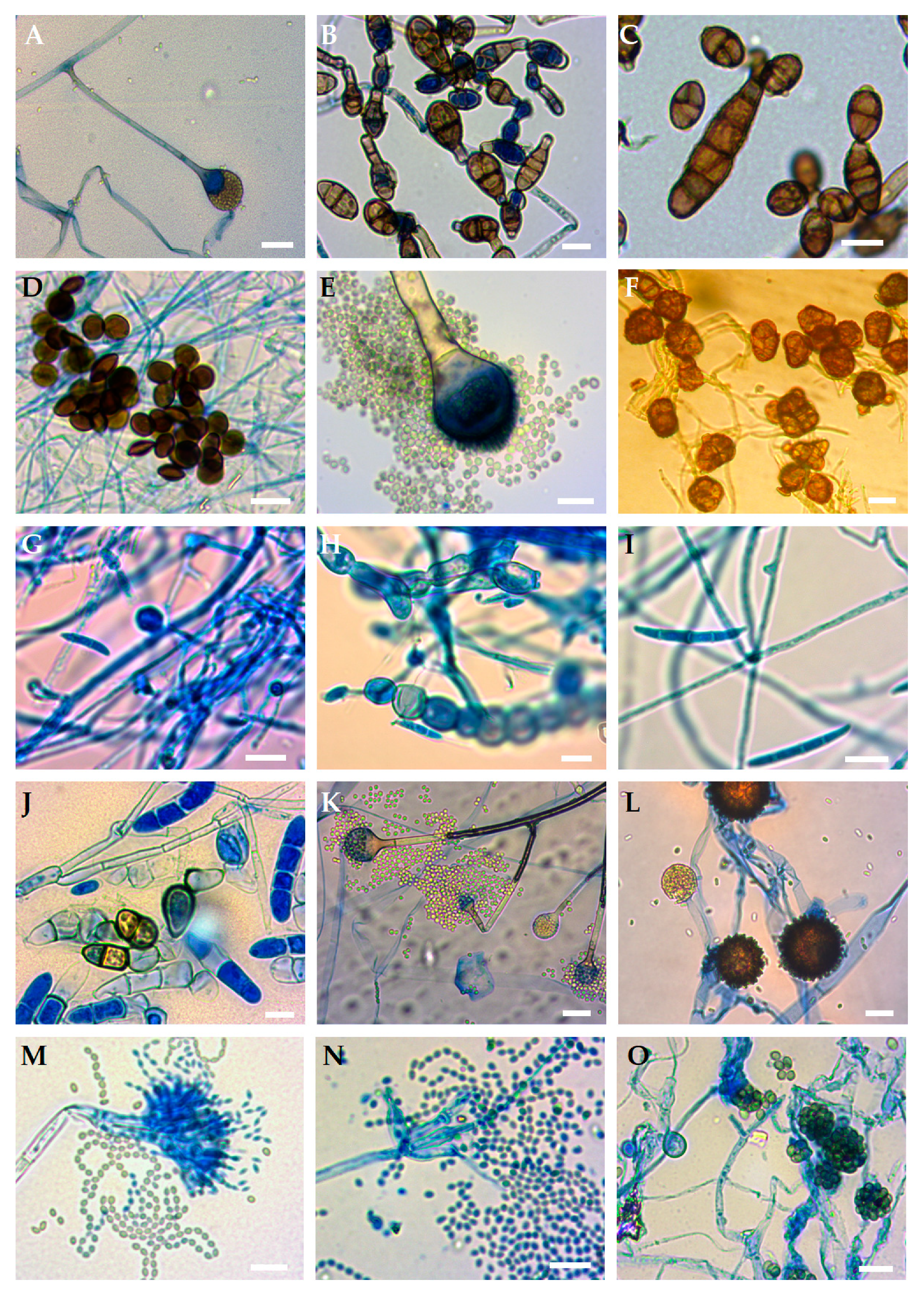
All fungi obtained from all the experimental variants were classified into 15 different fungal isolates. Phenotypic studies showed that the isolates obtained belonged to 15 species which varied phenotypically and showed different growth rates, different aerial structures as well as different mycelium pigmentation on the reverse and upper colony on the PDA medium (Figure 1). The isolated fungi also showed diversity in the production of different microscopic morphological structures (Figure 2).
The comparison of these microscopic and macroscopic observations of culture allowed for their initial identification matched to 15 species (three of them belonged to the phylum Zygomycota and the remaining to Ascomycota): Absidia cylindrospora, Alternaria alternata, Alternaria tenuissima, Arthrinium arundinis, Aspergillus fumigatus, Epicoccum nigrum, Fusarium oxysporum, Fusarium sporotrichioides, Fusarium tricinctum, Ilyonectria robusta, Mucor hiemalis, Mucor moelleri, Penicillium biourgeianum, Penicillium manginii and Trichoderma viride. Genetic analyses were carried out in order to confirm the preliminary phenotypic classification. The size of the PCR products obtained by using primers ITS1 and ITS4 were in the range of 391 to 570 bp. All the nucleotide sequences for the fungal species found in this study were submitted to GenBank under the accession numbers from MN817778 to MN817792. In the BLAST analysis done, the E values were zero, and the percentage of query cover and identity were in the range 99.0–100% and 95.34–100%, respectively (Table 1).
Overall, more species of fungi were isolated from E. helleborine compared with E. purpurata, and the flowering plants of E. helleborine were inhabited by the largest number of culturable fungal species (13 species). In turn, the fewest species were isolated from the flowering plants of E. purpurata (eight species). In the case of E. helleborine, more fungal species were isolated from the disinfected parts of the plants compared with the non-disinfected ones. On the other hand, the surface of the non-disinfected flowering plants of E. purpurata contained more fungal species (eight) than the disinfected ones (five). However, there were no differences in the number of fungal species (eight species each) for the fruiting plants of E. purpurata (Table 2 and Table 3, Figure 3).
Reverse trends were found for the disinfected and non-disinfected individual parts of both plants. Namely, the individual non-disinfected parts of both examined orchids were inhabited by a greater number of fungal species than the disinfected ones, except for the roots of the fruiting plants of E. helleborine and leaves of the fruiting plants of E. purpurata (in both cases, the same number of species was found on both experimental variants)—Table 2 and Table 3.
The inner layers of the tissues of the E. helleborine fruiting plants were most often inhabited by Fusarium sporotrichioides, taking into account the occurrence of a given species on all examined parts of these plants. This species of orchids accounted for 29.4% of all obtained fungi. For the surface tissues of these plants, M. moelleri and P. biourgeianum were the most commonly isolated species—both 17.8% each (Figure 3). On the other hand, the surface disinfected flowering plants of E. helleborine were most often colonized by A. tenuissima and A. arundinis (both 21.4% each), and the surface tissues of these plants were mainly inhibited by P. biourgeianum (23.8%)—Figure 3. In many cases, the most abundant species on the surface and internal tissues of E. purpurata were also repeated on E. helleborine. Alternaria tenuissima was the most often isolated species from the inner layers of the flowering plants’ tissues of E. helleborine, and P. biourgeianum from the surface tissues of it (41.7% and 27.8%, respectively). In turn, the surface tissues of the fruiting plants of E. helleborine were most often colonized by M. moelleri (19.0%), and the inner layers of these plants were mainly inhibited by A. tenuissima and F. sporotrichioides (both 21.4% each)—Figure 3.
The internal tissues of the roots of both orchids contained the most species of fungi among all the examined disinfected plant fragments, with the exception of rhizomes and inflorescences of the flowering E. purpurata plants (Table 2 and Table 3). The least diverse mycobiota of the disinfected plant fragments was found for the rhizomes and stems in the case of the flowering E. helleborine plants and the rhizomes and inflorescences in the case of the fruiting plants of this orchid species (Table 2). In turn, the least numbers of species of fungi were isolated from the disinfected roots, stems and leaves of the flowering E. purpurata plants, and from the disinfected stems and inflorescences of the fruiting plants of it (Table 3). Among all the examined plant fragments, the surfaces of the roots and leaves of the flowering E. helleborine plants and the roots, rhizomes and leaves of the fruiting E. helleborine plants were colonized by the largest number of fungal species. Other non-disinfected plant parts were inhabited by a similar number of species for both developmental stages of this plant (Table 2). On the other hand, the most species of fungi were isolated from the surfaces of the rhizomes, stems and inflorescences in the case of the flowering E. purpurata plants, and the number of species cultured from other non-disinfected parts of this plant was at the same level. In the case of the fruiting E. purpurata plants, among all the examined fragments, the most species of fungi were isolated from the surfaces of the roots and rhizomes, and the leaves’ surfaces were inhabited by the smallest number of fungal species (Table 3).

4. Discussion

Currently, we know that especially basidiomycetes fungi (Ceratobasidiaceae, Sebacinales and Tulasnellaceae) play a key role in the Epipactis biology [4,15,45,46,47,48]. Additionally, Epipactis helleborine can also be associated with another group of important fungi, e.g., non-Rhizoctonia fungi, endophytic fungi of Helotiales and a large number of other ectomycorrhizal taxa [8,15,18,19,20,21,22,23,24,25,26,27]. However, there are no similar reports about E. purpurata. Moreover, these results are mainly obtained by pyrosequencing, therefore, fungi are identified at most to the genus. Additionally, to our knowledge, there is no complete mycological analysis of these orchid species. Therefore, we wanted to accurately identify the species of fungi associated with the selected species of orchids (E. helleborine and E. purpurata) and also to analyze the fungal species colonizing the surfaces of the individual anatomical fragments of these plants as well as their internal tissues. Hence, we used a culture-based analysis to obtain the fungal isolates from the plants and combined classical and molecular methods to identify the species of fungi.
The results of our research showed that E. helleborine and E. purpurata, despite large differences in their biology and ecology, are not characterized by significant differences in their culturable mycobiota. Our results are in contradiction to the results of Jacquemyn et al. [15], who stated that the Epipactis species found in different habitats were inhabited by different mycorrhizal fungal communities. Further, our results do not confirm the data obtained by Salmia [18] regarding the occurrence in E. helleborine of non-Rhizoctonia fungi species such as Cylindrocarpon destructans, Humicola fuscoatra, Morchella sp., Mortierella nana and Sordaria fimicola. Discrepancies in the results obtained may be caused by the type of research methods used and/or biotic and abiotic factors prevailing at the location of the studied orchid populations. We studied orchids from SW Poland, and the mentioned researchers from Finland and Belgium. Plant species are very often associated with a characteristic fungal community. Nevertheless, other factors also determine the composition of the mycobiota inhabiting the plants, e.g., type of method used for the mycological analysis (culture-based analysis or molecular techniques), geographical location and the prevailing conditions in the habitat [5,49,50].
It should be emphasized that a culture-based analysis has several disadvantages compared with molecular techniques [51]. The former method cannot detect non-culturable fungi. In addition, some researchers suggest that this method is of little use for detecting the propagation structures of Basidiomycota fungi in the environment because these fungi grow slowly. As a consequence, they can be overtaken by faster growing colonies, e.g., by fungi belonging to the Zygomycota phylum [52,53,54,55]. This may probably be one of the reasons for not isolating Basidiomycota in our research. Although Ogórek et al. [42] report that under certain conditions (few propagation units in the environment), using a culture-based analysis, even spores of Basidiomycota can be found in the atmospheric air. It should also be noted that this method helps to explain the potential ecological roles of fungi as well as other microorganisms in environments [2].
Using both morphological culture-dependent and PCR-based methods, we identified three species of the genus Fusarium (F. oxysporum, F. sporotrichioides and F. tricinctum). These fungi are cosmopolitan species and belong to the so-called soil-borne fungi [5,56]. However, they are widely recognized as pathogenic, but they may also have other functions in ecosystems, e.g., Fusarium can inhabit the rhizospheres of plants well as act as an endophyt. Further, Tan et al. [57] confirm the information on the potential antimicrobial importance of the culturable fungal endophytes Fusarium and Ilyonectria, isolated from Dysosma versipellis (Berberidaceae).
According to the literature, Fusarium species such as F. oxysporum, F. proliferatum, F. solani, F. subglutinans and F. fractiflexum can cause foliar and root diseases in orchids. In our study, F. sporotrichioides was the most frequently isolated fungi from the inner layers of the fruiting plants’ tissues of E. purpurata, and the flowering plants’ tissues of E. helleborine (together with A. tenuissima). This is a cosmopolitan mycotoxin producer and important fungal plant pathogen, but until now, it has not been associated with orchids [58,59,60]. In turn, F. oxysporum identified by us in E. helleborine and E. purpurata was found previously, among others, in Dendrobium lindleyi [61].
It should be noted that F. oxysporum can perform a particularly diverse function in the environment, and it is a complex species composed of the phytopathogenic, saprotrophic and endosymbiotic species’ strains. A large diversity within this species is the cause of its division into many formae speciales and races [59]. Therefore, on the one hand, this species is described as one of the most common plant pathogens, including orchids, and on the other, as a species used in the biological protection of plants [61,62,63]. The extracted chemical components of this species of Fusarium, as those authors reported, may be responsible for antifungal, antioxidant and antimutagenic activities [60]. This is also confirmed by other researchers [64]. The genus Fusarium was identified also from both the stem and leaf segments of other Dendrobium species, e.g., Dendrobium nobile, D. loddigesii, D. devonianum, D. thyrsiflorum and D. moschatum [65]. Xing et al. [32] report that also endophytic fungi cultured from different Dendrobium species may be beneficial to plants due to their antibacterial and/or antifungal properties. Moreover, Fusarium cultured from germinating seeds of Cypripedium reginae may induce the germination of C. reginae seeds in vitro [66,67,68].
The surface tissues of both orchids were mainly inhibited by M. moelleri and/or P. biourgeianum. The dominance of one of these fungal species depended mainly on the development phase of the plants. Mucor moelleri, like other representatives of this genus, is a cosmopolitan filamentous fungus inhabiting various environments. Mucor spp. are mainly saprotrophs inhabiting, e.g., dead plants, but they can also contribute to food spoilage. They may also cause localized cutaneous mucormycosis in immunocompromised mammals [34]. Penicillium biourgeianum, like another species of this genus obtained by us (P. manginii), is also a cosmopolitan filamentous fungus which mainly inhabits dead matter [69]. Nevertheless, P. biourgeianum can produce biologically active compounds with an antibacterial activity, e.g., penicillenol A2 synthesized by this species exhibits activity against Staphylococcus aureus. Moreover, this compound together with beta-lactam antibiotics reduces the methicillin-resistant S. aureus’s survival [70].
The literature data include the other potential antibacterial and antifungal activities of another species identified by us, i.e., cosmopolitan species such as E. nigrum. This fungi species, previously isolated from Sobralia sp. and Dendrobium thyrsiflorum, showed a stronger antibacterial activity than ampicillin sodium [34]. Baute et al. [71] reported that the compounds such as epicorazine A and B secreted by E. nigrum show activity against S. aureus. Additionally, this fungus is described as an antagonist against many fungal pathogens of plants, among others, against selected Fusarium species [72]. It should be noted that this species is commonly known as a saprophytic and endophytic organism, but may also be a weak or opportunistic pathogen of plants [73,74].
In our study, the inner layers of orchids were the most colonized by A. tenuissima and/or A. arundinis and/or F. sporotrichioides. The dominance of one of these fungal species also depended mainly on the development phase of the plants. A study conducted by Vaz et al. [75] showed that Alternaria and F. oxysporum can also secrete compounds with an antimicrobial activity.
Alternaria tenuissima is common in the environment as a saprotrophic or opportunistic plant pathogen [76]. This species produces the allergen Alt a1 as well as mycotoxins [77,78]. In rare circumstances, A. tenuissima may be a pathogen of immunosuppressed humans and animals [79]. This species also exhibited great activity against fungal plant pathogens such as F. oxysporum, Rizoctonia solani and Sclerotinia sclerotiorum. Moreover, isolate No. CH1307 of this fungal species produced homoharringtonine, which is an effective treatment for leukaemia [80]. Arthrinium arundinis is described as an endophyte and plant and mammal pathogen, and it is a commonly occurring, widely distributed species [40,81]. On the other hand, secondary metabolites (prenylated diphenyl ethers) secreted by this species show a selective antifungal activity against Mucor hiemalis, and exhibit an inhibitory activity against A. alternata. Moreover, these compounds show in vitro cytotoxicity against the human monocytic cell line [82].
An interesting species isolated from the surface of orchids and their internal tissues is T. viride because it is used as a biofungicide [83]. This species is a free-living fungus which commonly inhabits soil and root ecosystems, and produces a variety of compounds that increase plant vigor [84]. Biocontrol mechanisms of this fungal species are mainly based on mycoparasitism [83]. Thus, the presence of this fungus on the tested orchids may contribute to their protection against pathogens.
Microscopic fungi, including entophytic fungi, are a source of various bioactive metabolites and certainly play an important role in orchid biology. It seems that understanding the importance of non-mycorrhizal fungi and their role is no less important than understanding the mycorrhizal process. We believe that this study contributes to a better understanding of the relationship between orchids and the microscopic fungi that inhabit them. Nevertheless, in the near future, we want to study the mycobiota of other orchid species and determine the biotic relationships between fungi isolated from them as well as study the secondary metabolites secreted by these fungi.

5. Conclusions

This present study reported the colonization of internal and surface tissues of various parts of Epipactis helleborine and E. purpurata orchid samples in various different developmental phases collected in SW Poland by culture methods. To our knowledge, this is the first such analysis. The two ecologically diverging Epipactis species analyzed, although growing in diverse habitats, did not differ significantly in terms of the composition of natural mycobiota. The surface tissues of these plants were mainly inhibited by Mucor moelleri and/or Penicillium biourgeianum. On the other hand, the inner layers of orchids were the most colonized by Alternaria tenuissima and/or Arthrinium arundinis and/or Fusarium sporotrichioides. The dominance of one of these fungal species depended mainly on the development phase of the plants. Overall, some of these fungal species cultured during the study may be pathogens of the plants. However, it is possible that the presence in Helleborines of some species of fungi, especially such as A. tenuissima, Epicoccum nigrum, F. oxysporum, P. biourgeianum and Trichoderma viride, could be effective against both fungal and bacterial pathogens.

Author Contributions

Conceptualization, methodology, validation, formal analysis, R.O. and A.J.-B.; investigation, R.O., K.K., E.Ż., Z.Ł. and A.J.-B.; writing—original draft preparation, R.O. and A.J.-B.; writing—review and editing, R.O. and A.J.-B.; microscopy, R.O.; All authors have read and agreed to the published version of the manuscript.

Funding

This research received no external funding.

Acknowledgments

The authors are deeply indebted to Daniel O’Connell for improving the English style. Material sampling was done with permissions from the Regional Directors for Environmental Protection (permission nos. WPN.6400.25.2018, WPN.6400.32.2018, WPN.6400.25.2019, WPN.6400.28.2019, and WPN.6205.87.2019).

Conflicts of Interest

The authors declare no conflict of interest.

References

  1. Hawksworth, D.L. The magnitude of fungal diversity: The 1.5 million species estimate revisited. Mycol. Res. 2001, 105, 1422–1432. [Google Scholar] [CrossRef] [Green Version]
  2. Ogórek, R.; Dyląg, M.; Kozak, B. Dark stains on rock surfaces in Driny Cave (Little Carpathian Mountains, Slovakia). Extremophiles 2016, 20, 641–652. [Google Scholar] [CrossRef] [Green Version]
  3. Read, D.J.; Duckett, J.G.; Francis, R.R.; Ligrone, R.; Russe, A. Symbiotic fungal associations in ‘lower’ land plants. Philos. Trans. R. Soc. B 2000, 355, 815–830. [Google Scholar] [CrossRef] [PubMed] [Green Version]
  4. Bidartondo, M.I.; Read, D.J.; Trappe, J.M.; Merckx, V.; Ligrone, R.; Duckett, J.G. The dawn of symbiosis between plants and fungi. Biol. Lett. 2011, 7, 574–577. [Google Scholar] [CrossRef] [PubMed] [Green Version]
  5. Ogórek, R.; Lejman, A.; Sobkowicz, P. Effect of the Intensity of Weed Harrowing with Spike-Tooth Harrow in Barley-Pea Mixture on Yield and Mycobiota of Harvested Grains. Agronomy 2019, 9, 103. [Google Scholar] [CrossRef] [Green Version]
  6. Ogórek, R.; Piecuch, A.; Višňovská, Z.; Cal, M.; Niedźwiecka, K. First report on the occurrence of dermatophytes of Microsporum cookei clade and close affinities to Paraphyton cookei in the Harmanecká Cave (Veľká Fatra Mts., Slovakia). Diversity 2019, 11, 191. [Google Scholar] [CrossRef] [Green Version]
  7. Leake, J.R. The biology of myco-heterotrophic (‘saprophytic’) plants. New Phytol. 1994, 127, 171–216. [Google Scholar] [CrossRef]
  8. Pecoraro, L.; Caruso, T.; Cai, L.; Gupta, V.K.; Liu, Z.J. Fungal networks and orchid distribution: New insights from above-and below-ground analyses of fungal communities. IMA Fungus 2018, 9, 1–11. [Google Scholar] [CrossRef] [Green Version]
  9. Ma, X.; Kang, J.; Nontachaiyapoom, S.; Wen, T.; Hyde, K.D. Non-mycorrhizal endophytic fungi from orchids. Curr. Sci. 2015, 109, 72–87. [Google Scholar]
  10. Rasmussen, H.N. Terrestrial Orchids: From Seed to Mycotrophic Plant; Cambridge University Press: Cambridge, UK, 1995. [Google Scholar]
  11. Herrera, H.; Valadares, R.; Contreras, D.; Bashan, Y.; Arriagada, C. Mycorrhizal compatibility and symbiotic seed germination of orchids from the Coastal Range and Andes in south central Chile. Mycorrhiza 2017, 27, 175–188. [Google Scholar] [CrossRef]
  12. McCormick, M.K.; Whigham, D.F.; O’Neill, J.P.; Becker, J.J.; Werner, S.; Rasmussen, H.N.; Bruns, T.D.; Taylor, D.L. Abundance and distribution of Corallorhiza odonthoriza reflect variations in climate and ectomycorrhizae. Ecol. Monogr. 2009, 79, 619–635. [Google Scholar] [CrossRef] [Green Version]
  13. Jacquemyn, H.; Brys, R.; Honnay, O.; Roldán-Ruiz, I.; Lievens, B.; Wiegand, T. Non-random spatial structuring of orchids in a hybrid zone of three Orchis species. New Phytol. 2012, 193, 454–464. [Google Scholar] [CrossRef] [PubMed]
  14. Jacquemyn, H.; Brys, R.; Merckx, V.S.; Waud, M.; Lievens, B.; Wiegand, T. Co-existing orchid species have distinct mycorrhizal communities and display strong spatial segregation. New Phytol. 2014, 202, 616–627. [Google Scholar] [CrossRef] [PubMed]
  15. Jacquemyn, H.; Waud, M.; Lievens, B.; Brys, R. Differences in mycorrhizal communities between Epipactis palustris, E. helleborine and its presumed sister species E. neerlandica. Ann. Bot. 2016, 118, 105–114. [Google Scholar] [CrossRef] [PubMed] [Green Version]
  16. McCormick, M.K.; Taylor, D.L.; Juhaszova, K.; Burnett, R.K.J.R.; Whigham, D.F.; O’Neill, J.P. Limitations on orchid recruitment: Not a simple picture. Mol. Ecol. 2012, 21, 1511–1523. [Google Scholar] [CrossRef] [PubMed]
  17. McCormick, M.K.; Jacquemyn, H. What constrains the distribution of orchid populations? New Phytol. 2014, 202, 392–400. [Google Scholar] [CrossRef]
  18. Salmia, A. Endomycorrhizal fungus in chlorophyll-free and green forms of the terrestrial orchid Epipactis helleborine. Karstenia 1988, 28, 3–18. [Google Scholar] [CrossRef]
  19. Schneider-Maunoury, L.; Leclercq, S.; Clément, C.; Covès, H.; Lambourdière, J.; Sauve, M.; Richard, F.; Selosse, M.A.; Taschen, E. Is Tuber melanosporum colonizing the roots of herbaceous, nonectomycorrhizal plants? Fungal Ecol. 2018, 31, 59–68. [Google Scholar] [CrossRef]
  20. Schneider-Maunoury, L.; Deveau, A.; Moreno, M.; Todesco, F.; Belmondo, S.; Murat, C.; Courty, P.E.; Jąkalski, M.; Selosse, M.A. Two ectomycorrhizal truffles, Tuber melanosporum and T. aestivum, endophytically colonise roots of non-ectomycorrhizal plants in natural environments. New Phytol. 2020, 225, 2542–2556. [Google Scholar] [CrossRef]
  21. May, M.; Jąkalski, M.; Novotná, A.; Dietel, J.; Ayasse, M.; Lallemand, F.; Figura, T.; Minasiewicz, J.; Selosse, M.A. Three-year pot culture of Epipactis helleborine reveals autotrophic survival, without mycorrhizal networks, in a mixotrophic species. Mycorrhiza 2020, 30, 51–61. [Google Scholar] [CrossRef] [Green Version]
  22. Hansen, K.; Perry, B.A.; Dranginis, A.W.; Pfister, D.H. A phylogeny of the highly diverse cup-fungus family Pyronemataceae (Pezizomycetes, Ascomycota) clarifies relationships and evolution of selected life history traits. Mol. Phylogenet. Evol. 2013, 67, 311–333. [Google Scholar] [CrossRef] [PubMed]
  23. Jumpponen, A. Dark septate endophytes—Are they mycorrhizal? Mycorrhiza 2013, 11, 207–211. [Google Scholar] [CrossRef]
  24. Dearnaley, J.D.W.; Martos, F.; Selosse, M.A. 12 orchid mycorrhizas: Molecular ecology, physiology, evolution and conservation aspects. In Fungal Associations. The Mycota (A Comprehensive Treatise on Fungi as Experimental Systems for Basic and Applied Research); Hock, B., Ed.; Springer: Berlin, Germany, 2012; pp. 207–230. [Google Scholar]
  25. Veldre, V.; Abarenkov, K.; Bahram, M.; Martos, F.; Selosse, M.A.; Tamm, H.; Kõljalg, U.; Tedersoo, L. Evolution of Nutritional Modes of Ceratobasidiaceae (Cantharellales, Basidiomycota) as Revealed from Publicly Available ITS Sequences. Fungal Ecol. 2013, 6, 256–268. [Google Scholar] [CrossRef]
  26. Weiß, M.; Waller, F.; Zuccaro, A.; Selosse, M.A. Sebacinales—One thousand and one interactions with land plants. New Phytol. 2016, 211, 20–40. [Google Scholar] [CrossRef]
  27. Lallemand, F.; Robionek, A.; Courty, P.E.; Selosse, M.A. The 13C content of the orchid Epipactis palustris (L.) Crantz responds to light as in autotrophic plants. Bot. Lett. 2018, 165, 265–273. [Google Scholar] [CrossRef]
  28. Selosse, M.A.; Faccio, A.; Scappaticci, G.; Bonfante, P. Chlorophyllous and achlorophyllous specimens of Epipactis microphylla (Neottieae, Orchidaceae) are associated with ectomycorrhizal septomycetes, including truffles. Microb. Ecol. 2004, 47, 416–426. [Google Scholar] [CrossRef]
  29. Redman, R.S.; Sheehan, K.B.; Stout, R.; Rodriguez, R.J.; Henson, J.M. Thermotolerance generated by plant/fungal symbiosis. Science 2002, 298, 1581. [Google Scholar] [CrossRef]
  30. Arnold, A.E.; Mejía, L.C.; Kyllo, D.; Rojas, E.I.; Maynard, Z.; Robbins, N.; Herre, E.A. Fungal endophytes limit pathogen damage in a tropical tree. Proc. Natl. Acad. Sci. USA 2003, 100, 15649–15654. [Google Scholar] [CrossRef] [Green Version]
  31. Khan, S.A.; Hamayun, M.; Yoon, H.; Kim, H.Y.; Suh, S.J.; Hwang, S.K.; Kim, J.M.; Lee, I.J.; Choo, Y.S.; Yoon, U.H.; et al. Plant growth promotion and Penicillium citrinum. BMC Microbiol. 2008, 8, 231. [Google Scholar] [CrossRef] [Green Version]
  32. Xing, Y.M.; Chen, J.; Cui, J.L.; Chen, X.M.; Guo, S.X. Antimicrobial activity and biodiversity of endophytic fungi in Dendrobium devonianum and Dendrobium thyrsiflorum from Vietman. Curr. Microbiol. 2011, 62, 1218–1224. [Google Scholar] [CrossRef]
  33. Rifai, M.A. A revision of the genus Trichoderma. Mycol. Pap. 1969, 116, 1–56. [Google Scholar]
  34. Schipper, M.A.A. A study on variability in Mucor hiemalis and related species. Stud. Mycol. 1973, 4, 1–40. [Google Scholar]
  35. Seifert, K. Fuskey: Fusarium Interactive Key. Agric. Agric. Food Can. 1996, 1–65. [Google Scholar]
  36. Ho, H.-M.; Chuang, S.-C.; Chen, S.-J. Notes on Zygomycetes of Taiwan (IV): Three Absidia species (Mucoraceae). Fung. Sci. 2004, 19, 125–131. [Google Scholar]
  37. Peterson, S.W. Multilocus DNA sequence analysis shows that Penicillium biourgeianum is a distinct species closely to P. brevicompactum and P. olsonii. Mycol. Res. 2004, 108, 434–440. [Google Scholar] [CrossRef] [PubMed]
  38. Houbraken, J.; Frisvad, J.C.; Samson, R.A. Taxonomy of Penicillium section Citrina. Stud. Mycol. 2011, 70, 53–138. [Google Scholar] [CrossRef] [Green Version]
  39. Cabral, A.; Groenewald, J.Z.; Rego, C.; Oliveira, H.; Crous, P.W. Cylindrocarpon root rot: Multi-gene analysis reveals novel species within the Ilyonectria radicicola species complex. Mycol. Prog. 2012, 11, 655–688. [Google Scholar] [CrossRef] [Green Version]
  40. Crous, P.W.; Groenewald, J.Z. A phylogenetic re-evaluation of Arthrinium. IMA Fungus 2013, 4, 133–154. [Google Scholar] [CrossRef] [Green Version]
  41. Woudenberg, J.H.C.; Groenewald, J.Z.; Binder, M.; Crous, P.W. Alternaria redefined. Stud. Mycol. 2013, 75, 171–212. [Google Scholar] [CrossRef] [Green Version]
  42. Ogórek, R.; Kozak, B.; Višňovská, Z.; Tancinová, D. Phenotypic and genotypic diversity of airborne fungalspores in Demänovská Ice Cave (Low Tatras, Slovakia). Aerobiologia 2018, 34, 13–28. [Google Scholar] [CrossRef] [Green Version]
  43. Dyląg, M.; Sawicki, A.; Ogórek, R. Diversity of species and susceptibility phenotypes toward commercially available fungicides of cultivable fungi colonizing bones of Ursus spelaeus on display in Niedźwiedzia Cave (Kletno, Poland). Diversity 2019, 11, 224. [Google Scholar] [CrossRef] [Green Version]
  44. White, T.J.; Bruns, T.; Lee, S.; Taylor, J.W. Amplification and direct sequencing of fungal ribosomal RNA genes for phylogenetics. In PCR Protocols: A Guide to Methods and Applications; Innis, M.A., Gelfand, D.H., Sninsky, J.J., White, T.J., Eds.; Academic Press: New York, NY, USA, 1990; pp. 315–322. [Google Scholar]
  45. Dearnaley, J.D.W. Further advances in orchid mycorrhizal research. Mycorrhiza 2007, 17, 475–486. [Google Scholar] [CrossRef] [PubMed]
  46. Bidartondo, M.I.; Burghardt, B.; Gebauer, G.; Bruns, T.D.; Read, D.J. Changing partners in the dark: Isotopic and molecular evidence of ectomycorrhizal liaisons between forest orchids and trees. Proc. R. Soc. Lond. Ser. B 2004, 271, 1799–1806. [Google Scholar] [CrossRef] [PubMed] [Green Version]
  47. Ogura-Tsujita, Y.; Yukawa, T. High mycorrhizal specificity in a widespread mycoheterotrophic plant, Eulophia zollingeri (Orchidaceae). Am. J. Bot. 2008, 95, 93–97. [Google Scholar] [CrossRef] [PubMed] [Green Version]
  48. Těšitelová, T.; Těšitel, J.; Jersáková, J.; RÍhová, G.; Selosse, M.A. Symbiotic germination capability of four Epipactis species (Orchidaceae) is broader than expected from adult ecology. Am. J. Bot. 2012, 99, 1020–1032. [Google Scholar] [CrossRef] [PubMed]
  49. Koyama, A.; Maherali, H.; Antunes, P.M. Plant geographic origin and phylogeny as potential drivers of community structure in root-inhabiting fungi. J. Ecol. 2019, 107, 1720–1736. [Google Scholar] [CrossRef]
  50. Větrovský, T.; Kohout, P.; Kopecký, M.; Machac, A.; Man, M.; Bahnmann, B.D.; Brabcová, V.; Choi, J.; Meszárošová, L.; Human, Z.R.; et al. A meta-analysis of global fungal distribution reveals climate-driven patterns. Nat. Commun. 2019, 10, 5142. [Google Scholar] [CrossRef] [Green Version]
  51. Pasanen, A.L. A review: Fungal exposure assessment in in-door environments. Indoor Air 2001, 11, 87–98. [Google Scholar] [CrossRef]
  52. MacNeil, L.; Kauri, T.; Robertson, W. Molecular techniques and their potential application in monitoring the microbiological quality of indoor air. Can. J. Microbiol. 1995, 41, 657–675. [Google Scholar] [CrossRef]
  53. Wu, P.C.; Su, H.J.J.; Ho, H.M. A comparison of sampling media for environmental viable fungi collected in a hospital environment. Environ. Res. 2000, 82, 253–257. [Google Scholar] [CrossRef]
  54. Macher, J.M. Review of methods to collect settled dust and isolate culturable microorganisms. Indoor Air 2001, 11, 99–110. [Google Scholar] [CrossRef]
  55. Ogórek, R.; Višňovská, Z.; Tančinová, D. Mycobiota of underground habitats: Case study of Harmanecká Cave in Slovakia. Microb. Ecol. 2016, 71, 87–99. [Google Scholar] [CrossRef]
  56. Roncero, M.I.G.; Hera, C.; Ruiz-Rubio, M.; García Maceira, F.I.; Madrid, M.P.; Caracuel, Z.; Calero, F.; Delgado-Jarana, J.; Roldán-Rodríguez, R.; Martínez-Rocha, A.L.; et al. Fusarium as a model for studying virulence in soil borne plant pathogens. Physiol. Mol. Plant Pathol. 2003, 62, 87–98. [Google Scholar] [CrossRef]
  57. Tan, X.M.; Zhou, Y.Q.; Zhou, X.L.; Xia, X.H.; Wei, Y.; He, L.L.; Tang, H.Z.; Yu, L.Y. Diversity and bioactive potential of culturable fungal endophytes of Dysosma versipellis; a rare medicinal plant endemic to China. Sci. Rep. 2018, 8, 1–9. [Google Scholar] [CrossRef] [PubMed] [Green Version]
  58. Ivanová, H.; Hrehová, Ľ.; Pristaš, P. First confirmed report on Fusarium sporotrichioides on Pinus ponderosa var. jeffreyi in Slovakia. Plant Protect. Sci. 2016, 52, 250–253. [Google Scholar]
  59. Srivastava, S.; Kadooka, C.; Uchida, J.Y. Fusarium species as pathogen on orchids. Microbiol. Res. 2018, 207, 188–195. [Google Scholar] [CrossRef] [PubMed]
  60. Bungtongdee, N.; Sopalun, K.; Laosripaiboon, W.; Iamtham, S. The chemical composition, antifungal, antioxidant and antimutagenicity properties of bioactive compounds from fungal endophytes associated with Thai orchids. J. Phytopathol. 2019, 167, 56–64. [Google Scholar] [CrossRef]
  61. Latiffah, Z.; Hayati, M.Z.N.; Baharuddin, S.; Maziah, Z. Identification and pathogenicity of Fusarium species associated with root rot and stem rot of Dendrobium. Asian J. Plant Pathol. 2009, 3, 14–21. [Google Scholar] [CrossRef]
  62. Swett, C.L.; Uchida, J.Y. Characterization of Fusarium diseases on commercially grown orchids in Hawaii. Plant Pathol. 2015, 64, 648–654. [Google Scholar] [CrossRef]
  63. Thongkamngam, T.; Jaenaksorn, T. Fusarium oxysporum (F221-B) as biocontrol agent against plant pathogenic fungi in vitro and in hydroponics. Plant Protect. Sci. 2017, 53, 85–95. [Google Scholar]
  64. Gong, L.J.; Guo, S.X. Endophytic fungi from Dracaena cambodiana and Aquilaria sinensis and their antimicrobial activity. Afr. J. Biotechnol. 2009, 8, 731–736. [Google Scholar]
  65. Parthibhan, S.; Rao, M.V.; Kumar, T.S. Culturable fungal endophytes in shoots of Dendrobium aqueum Lindley–An imperiled orchid. Ecol. Genet. Genom. 2017, 3, 18–24. [Google Scholar] [CrossRef]
  66. Bernard, N. L’évolution dans la symbiose. Le orchidées et leurs champignons commensaux. Ann. Sci. Nat. Bot. 1909, 9, 1–19. [Google Scholar]
  67. Bayman, P.; Otero, J.T. Microbial endophytes of orchid roots. In Microbial Root Endophytes. Soil Biology; Schulz, B.J.E., Boyle, C.J.C., Sieber, T.N., Eds.; Springer: Berlin, Germany, 2006; pp. 153–177. [Google Scholar]
  68. Vujanovic, V.; St.-Arnaud, M.; Barabé, D.; Thibeault, G. Viability testing of orchid seed and the promotion of colouration and germination. Ann. Bot. 2000, 86, 79–86. [Google Scholar] [CrossRef] [Green Version]
  69. Azam, M.; Shahid, A.A.; Majeed, R.A.; Ali, M.; Ahmad, N.; Haider, M.S. First report of Penicillium biourgeianum causing post-harvest fruit rot of apple in Pakistan. Plant Dis. 2016, 100, 1778. [Google Scholar] [CrossRef]
  70. Li, S.; Mou, Q.; Xu, X.; Qi, S.; Leung, P.H.M. Synergistic antibacterial activity between penicillenols and antibiotics against methicillin-resistant Staphylococcus aureus. R. Soc. Open Sci. 2018, 5, 172466. [Google Scholar] [CrossRef] [Green Version]
  71. Baute, M.A.; Deffieux, G.; Baute, R.; Neveu, A. New antibiotics from the fungus Epicoccum nigrum. J. Antibiot. Res. 1978, 31, 1099–1101. [Google Scholar] [CrossRef] [Green Version]
  72. Ogórek, R.; Pląskowska, E. Epicoccum nigrum for biocontrol agents in vitro of plant fungal pathogens. Commun. Agric. Appl. Biol. Sci. 2011, 76, 691–697. [Google Scholar]
  73. Kukreja, N.; Sridhara, S.; Singh, B.P.; Arora, N. Effect of proteolytic activity of Epicoccum purpurascens major allergen, Epi p 1 in allergic inflammation. Clin. Exp. Immunol. 2008, 154, 162–171. [Google Scholar] [CrossRef]
  74. Fávaro, L.C.L.; De Melo, F.L.; Aguilar-Vildoso, C.I.; Araújo, W.L. Polyphasic analysis of intraspecific diversity in Epicoccum nigrum warrants reclassification into separate species. PLoS ONE 2011, 6, e14828. [Google Scholar] [CrossRef] [Green Version]
  75. Vaz, A.B.; Mota, R.C.; Bomfim, M.R.Q.; Vieira, M.L.; Zani, C.L.; Rosa, C.A.; Rosa, L.H. Antimicrobial activity of endophytic fungi associated with Orchidaceae in Brazil. Can. J. Microbiol. 2009, 55, 1381–1391. [Google Scholar] [CrossRef] [PubMed]
  76. Simmons, E.G. Alternaria: An Identification Manual; CBS Fungal Biodiversity Centre: Utrecht, The Netherlands, 2007; pp. 500–502. [Google Scholar]
  77. Chełkowski, J.; Visconti, A. Alternaria: Biology, Plant Diseases and Metabolites; Elsevier Science Limited: Amsterdam, The Netherlands, 1992; pp. 364–365. [Google Scholar]
  78. Ibarrola, I.; Suárez-Cervera, M.; Arilla, M.C.; Martínez, A.; Monteseirín, J.; Conde, J.; Asturias, J.A. Production profile of the major allergen Alt a 1 in Alternaria alternata cultures. Ann. Allergy Asthma Immunol. 2004, 93, 589–593. [Google Scholar] [CrossRef]
  79. Pastor, F.J.; Guarro, J. Alternaria infections: Laboratory diagnosis and relevant clinical features. Clin. Microbiol. Infect. 2008, 14, 734–746. [Google Scholar] [CrossRef] [PubMed] [Green Version]
  80. Liu, Y.; Liu, S.X.; Li, Y.C.; Li, C.F. Optimization of homoharringtonine fermentation conditions for Alternaria tenuissima CH1307, an endophytic fungus of Cephalotaxus mannii Hook. f. J. Trop. Org. 2012, 3, 236–242. [Google Scholar]
  81. Greven, S.; Egberts, F.; Buchner, M.; Beck-Jendroschek, V.; Voss, K.; Brasch, J. Cutaneous chromomycosis caused by Arthrinium arundinis. J. Dtsch. Dermatol. Ges. 2018, 5, 621–623. [Google Scholar] [CrossRef] [PubMed]
  82. Zhang, P.; Li, X.; Yuan, X.L.; Du, Y.M.; Wang, B.G.; Zhang, Z.F. Antifungal prenylated diphenyl ethers from Arthrinium arundinis, an endophytic fungus isolated from the leaves of Tobacco (Nicotiana tabacum L.). Molecules 2018, 23, 3179. [Google Scholar] [CrossRef] [PubMed] [Green Version]
  83. Druzhinina, I.S.; Seidl-Seiboth, V.; Herrera-Estrella, A.; Horwitz, B.A.; Kenerley, C.M.; Monte, E.; Mukherjee, P.K.; Zeilinger, S.; Grigoriev, I.V.; Kubicek, C.P. Trichoderma: The genomics of opportunistic success. Nat. Rev. Microbiol. 2011, 9, 749–759. [Google Scholar] [CrossRef]
  84. Harman, G.E.; Howell, C.R.; Viterbo, A.; Chet, I.; Lorito, M. Trichoderma species-opportunistic, a virulent plant symbionts. Nat. Rev. Microbiol. 2004, 2, 43–56. [Google Scholar] [CrossRef]
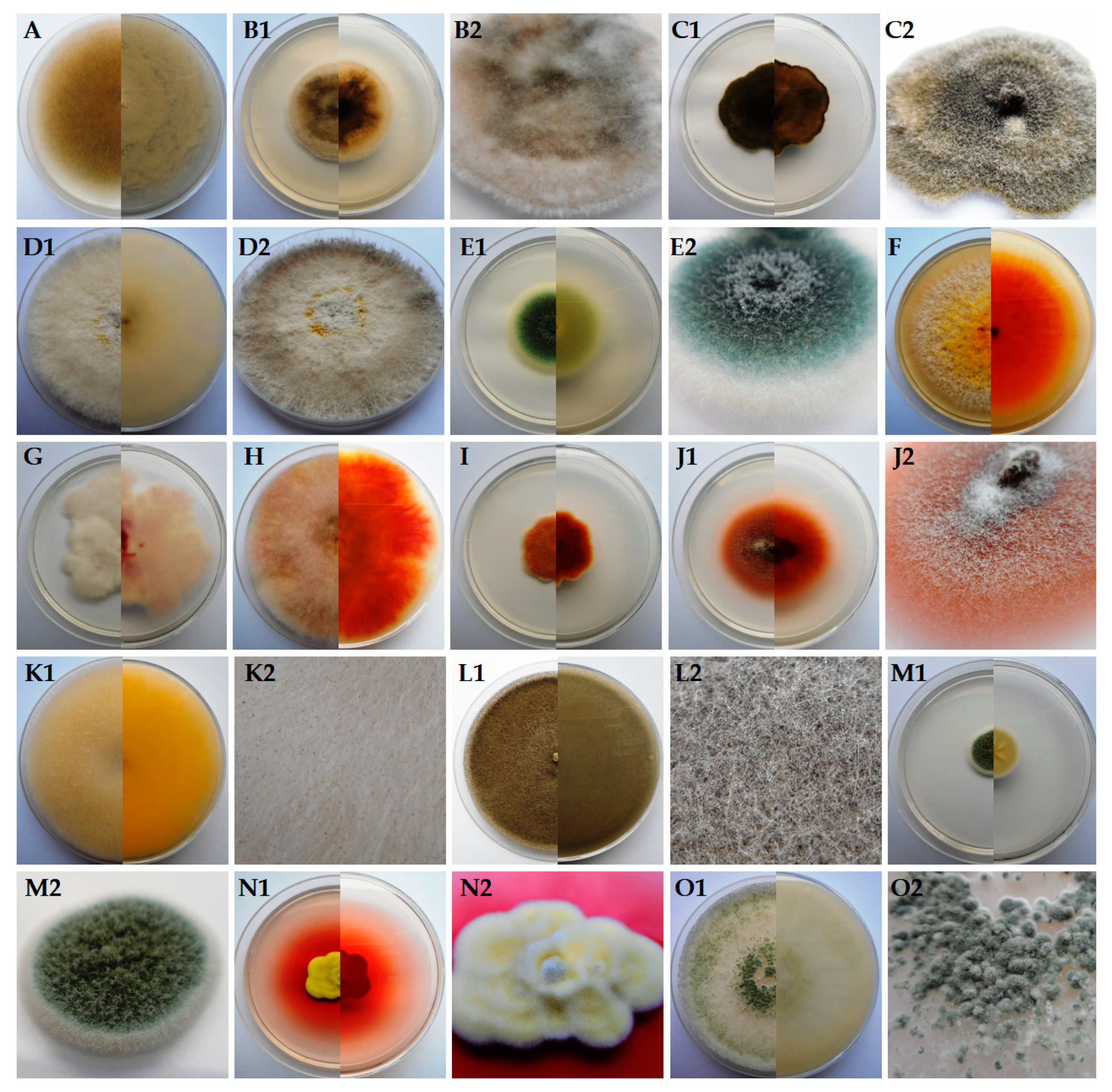
Figure 1. Macroscopic observations of 7-day-old fungal culture (exception for D2: 14-day-old) isolated from Epipactis helleborine and Epipactis purpurata occurring in Lower Silesia (SW Poland) on a PDA medium: (A) Absidia cylindrospora, (B1,B2) Alternaria alternata, (C1,C2) Alternaria tenuissima, (D1,D2) Arthrinium arundinis, (E1,E2) Aspergillus fumigatus, (F) Epicoccum nigrum, (G) Fusarium oxysporum, (H) Fusarium sporotrichioides, (I) Fusarium tricinctum, (J1,J2) Ilyonectria robusta, (K1,K2) Mucor hiemalis, (L1,L2) Mucor moelleri, (M1,M2) Penicillium biourgeianum, (N1,N2) Penicillium manginii and (O1,O2) Trichoderma viride.
Diversity 12 00182 g001
Figure 2. Microscopic observations of morphological structures of 2-week-old fungal culture isolated from Epipactis helleborine and Epipactis purpurata occurring in Lower Silesia (SW Poland) on a PDA medium: (A) Absidia cylindrospora, (B) Alternaria alternata, (C) Alternaria tenuissima, (D) Arthrinium arundinis, (E) Aspergillus fumigatus, (F) Epicoccum nigrum, (G) Fusarium oxysporum, (H) Fusarium sporotrichioides, (I) Fusarium tricinctum, (J) Ilyonectria robusta, (K) Mucor hiemalis, (L) Mucor moelleri, (M) Penicillium biourgeianum, (N) Penicillium manginii and (O) Trichoderma viride. Scale bars: 20 μm (A,F,K,L) and 10 μm (BE,GJ,N,O).
Diversity 12 00182 g002
Figure 3. Percentage of each fungal species contributing to the totals for the disinfected (D) and non-disinfected (ND) plants of Epipactis helleborine and Epipactis purpurata occurring in Lower Silesia (SW Poland).
Diversity 12 00182 g003
Table 1. Fungal communities of micromycetes cultured from Epipactis helleborine and Epipactis purpurata occurring in Lower Silesia (SW Poland), and results of the BLAST analysis (all E values were zero).
Fungi Isolated from OrchidsIdentity with Sequence from GenBank
Isolate NumberIdentified SpeciesGenBank Accession No.The Sequence Length (bp)Query Cover, %Identity, %Accession
UWR_170Absidia cylindrosporaMN817778.15149995.34JN205822.1
UWR_171Alternaria alternataMN817779.139110098.72KF996773.1
UWR_172A. tenuissimaMN817780.1487100100.00MN712241.1
UWR_173Arthrinium arundinisMN817781.1451100100.00MN593205.1
UWR_174Aspergillus fumigatusMN817782.153910099.07MN178808.1
UWR_175Epicoccum nigrumMN817783.145410095.59MG736195.1
UWR_176Fusarium oxysporumMN817784.1473100100.00MN240928.1
UWR_177F. sporotrichioidesMN817785.143510099.77MK595076.1
UWR_178F. tricinctumMN817786.1497100100.00MK102644.1
UWR_179Ilyonectria robustaMN817787.1456100100.00MK602790.1
UWR_180Mucor hiemalisMN817788.1530100100.00MH794214.1
UWR_181M. moelleriMN817789.1570100100.00MH857827.1
UWR_182Penicillium biourgeianumMN817790.1506100100.00KX067821.1
UWR_183P. manginiiMN817791.148110099.17MH858641.1
UWR_184Trichoderma virideMN817792.1552100100.00KX379164.1
Table 2. Fungal communities of micromycetes cultured from the disinfected (D) and non-disinfected (ND) individual parts of Epipactis helleborine occurring in Lower Silesia (SW Poland). A + indicates that the fungus was cultured from the plant samples.
Fungal SpeciesRootsRhizomesStemsLeavesInflorescences
DNDDNDDNDDNDDND
Fruiting plants 2018Alternaria tenuissima ++++ +
Epicoccum nigrum + ++++
Fusarium oxysporum+ +
Fusarium sporotrichioides+ +++ +++
Fusarium tricinctum + ++ + +
Ilyonectria robusta + ++
Mucor hiemalis++ + +
M. moelleri++ + + + +
Penicillium biourgeianum + + + + +
P. manginii+
Trichoderma viride++
In total6626354625
Flowering plants 2019Absidia cylindrospora++ + + +
Alternaria alternata +
A. tenuissima + + +
Arthrinium arundinis+ + +
Aspergillus fumigatus + + + +
Epicoccum nigrum ++
Fusarium tricinctum +
Ilyonectria robusta +
Mucor hiemalis++
M. moelleri +
Penicillium biourgeianum + + + + +
P. manginii+ + + + +
Trichoderma viride + +
In total4524243534
Table 3. Fungal communities of micromycetes cultured from the disinfected (D) and non-disinfected (ND) individual parts of Epipactis purpurata occurring in Lower Silesia (SW Poland). A + indicates that the fungus was cultured from the plant samples.
Fungal SpeciesRootsRhizomesStemsLeavesInflorescences
DNDDNDDNDDNDDND
Fruiting plants 2018Alternaria tenuissima + ++++
Epicoccum nigrum ++
Fusarium sporotrichioides+ + +
F. tricinctum + ++++
Ilyonectria robusta++++
Mucor hiemalis +++ + +
M. moelleri++ + + + +
Penicillium biourgeianum + + +
Trichoderma viride++
In total4535243324
Flowering plants 2019Absidia cylindrospora + ++
Alternaria alternata+ ++ + +
A. tenuissima+ + + +++
Aspergillus fumigatus + + +
Epicoccum nigrum + +
Fusarium tricinctum + +++
Penicillium biourgeianum + + + + +
P. manginii + +
In total2334242334