Article

Integrative Exploration of Paenibacillus sp. JSM-10 as a Potential Multi-Stress-Tolerant Microbial Inoculant for Sustainable Agriculture

by
Zhasmin Zhaksybek
1,2,
Adel Sattarova
1,2,
Ainur Akimbekova
1,
Aldan Shamukhan
1,
Irina Rukavitsina
3,
Sailau Abeldenov
1,* and
Anuar Rysbekovich Zhumakayev
1
1
Laboratory of Molecular Biotechnology, National Center for Biotechnology, Astana 010000, Kazakhstan
2
Department of Biotechnology and Microbiology, L.N. Gumilyov Eurasian National University, Astana 010000, Kazakhstan
3
Microbiology Laboratory, A.I. Barayev Research and Production Centre for Grain Farming, Nauchny Set., Akmola Region 021601, Kazakhstan
*
Author to whom correspondence should be addressed.
Int. J. Mol. Sci. 2026, 27(9), 4062; https://doi.org/10.3390/ijms27094062
Submission received: 28 February 2026 / Revised: 27 April 2026 / Accepted: 27 April 2026 / Published: 30 April 2026
(This article belongs to the Special Issue New Advances in Plant–Microbe Interaction)

Abstract

Abiotic stress factors, including drought and salinity, severely limit crop productivity worldwide. Furthermore, the extensive use of herbicides, such as glyphosate, disrupts beneficial soil microbiota, further impairing crop growth. Plant growth-promoting bacteria (PGPB) represent a sustainable and efficient strategy to enhance crop yields, particularly under unfavorable environmental and soil conditions. In this study, we characterized Paenibacillus sp. JSM-10, newly isolated from glyphosate-exposed agricultural soil, for its stress tolerance and plant growth-promoting potential, including its morphology examined using complementary microscopy techniques. The strain tolerated up to 0.5 g/L glyphosate, 15 g/L NaCl, and 100 g/L polyethylene glycol (PEG-6000) without significant growth inhibition (p > 0.05), demonstrating robust resilience to such multiple abiotic stresses. Beyond its tolerance, the strain exhibited several beneficial characteristics, including indole-3-acetic acid (IAA) synthesis, siderophore production, and inorganic phosphate solubilization. Furthermore, both living cells and culture filtrates of JSM-10 exhibited a positive trend toward enhancing buckwheat growth under normal and saline conditions, with effect sizes ranging from Hedges’ g = 0.56−0.92. In addition, JSM-10 exhibited antagonistic activity against a range of pathogenic microorganisms, including Nigrospora oryzae, Bipolaris sorokiniana, Alternaria spp., and Escherichia coli. Altogether, these characteristics highlight the Paenibacillus sp. JSM-10 strain and its culture filtrates as promising candidates for application in organic farming aimed at promoting plant growth and improving stress tolerance via plant–microbe interactions.

1. Introduction

Growing environmental pressures, notably pesticide pollution, water scarcity, and soil salinization, represent major limiting stress factors in modern agriculture [1]. Soil pollution by pesticide residues has emerged as a serious threat and has been recently reported as even the second major factor altering soil biodiversity [2]. This is especially concerning because pesticide pollution negatively impacts the abundance of beneficial plant-associated bacteria with plant growth-promoting characteristics [3]. A clear example is the widely utilized herbicide glyphosate N-(phosphonomethyl) glycine, which is repeatedly reported to inhibit a broad range of beneficial soil and plant microorganisms [4,5]. Such effects not only compromise microbial-mediated nutrient cycling but can also reduce crop productivity in glyphosate-treated soils. Salinity and drought also cause major abiotic stress conditions, significantly limiting crop yields. Saline conditions strongly affect plant growth by disturbing ionic and osmotic balance, altering redox status and cellular energy, as well as reducing photosynthesis [6]. Drought is another major constraint in agriculture, negatively affecting water balance, increasing the production of reactive oxygen species (ROS) [7], and causing average cereal production losses of up to 9–10% [8].
Altogether, pesticide exposure, salinity, and drought substantially constrain crop productivity, highlighting the need for advanced sustainable agricultural strategies capable of mitigating adverse abiotic conditions [9,10]. The application of environment-friendly methods, such as plant growth-promoting bacteria (PGPB), represents an effective strategy to maintain stable food production and mitigate the effects of abiotic stress factors [11,12]. PGPB enhance plant growth through multiple mechanisms, including inorganic nutrient solubilization, atmospheric nitrogen fixation, phytohormone production, and synthesis of bioactive metabolites that stimulate plant development [13]. Furthermore, stress-resilient PGPB strains represent robust biotechnological agents for sustainable agriculture, as they could enhance resistance to plant pathogens and improve crop yields under exposure to abiotic stress conditions [14].
Currently, PGPB include taxonomically diverse bacterial groups with distinct growth-promoting and protective mechanisms [15], including different strains of Pseudomonas, Bacillus, Azotobacter, and Paenibacillus. The ongoing description of new species continues to expand the diversity of promising PGPB. For example, Paenibacillus peoriae, first distinguished as a distinct species in 1993 (at that time as Bacillus peoriae) [16], has recently attracted increasing attention as a promising candidate for plant growth promotion, biocontrol, and biotechnological applications [17,18,19]. This species clearly demonstrates plant growth-promoting characteristics typical of Paenibacillus, including the production of growth-promoting substances, siderophore synthesis, and phosphorus solubilization [19], and exhibits remarkable antagonistic activity against pathogenic microorganisms, such as Fusarium oxysporum, Bipolaris sorokiniana, and Alternaria alternata [20]. These results highlight the potential of Paenibacillus-like strains to improve plant growth and productivity, suggesting their valuable potential in organic farming systems.
The Paenibacillus genus is increasingly recognized as a promising PGPB, yet its strain-specific metabolic potential, antagonistic activity, and growth stimulation across diverse crops remain insufficiently explored. In addition, tolerance of PGPB to environmentally relevant stress factors is often not comprehensively evaluated, limiting a reliable assessment of its field applicability. Therefore, the present study aimed to provide an extensive evaluation of a newly isolated glyphosate-tolerant Paenibacillus sp. JSM-10 strain and its multi-stress tolerance, plant-stimulating, and crop-protecting characteristics to assess its suitability as a robust microbial candidate for improving plant performance under adverse environmental conditions.

2. Results

2.1. Isolation, Identification, and Microscopic Evaluation of Paenibacillus sp. JSM-10

2.1.1. Isolation of JSM-10 Strain

A bacterial isolate was successfully obtained from soil collected in a soft spring wheat field (cultivar “Aqmola 2”) in Kazakhstan. Following serial dilution and plating on selective medium amended with 1 g/L glyphosate, several morphologically distinct colonies were observed. Among them, one dominant isolate with consistent growth characteristics was considered a potential glyphosate-tolerant strain and selected for further analysis. The isolate formed circular, creamy-white colonies with smooth margins, indicating a typical phenotype of Paenibacillus-like bacteria.

2.1.2. Identification of Glyphosate-Tolerant Strain JSM-10

Based on the sequence analysis of the 16S rRNA gene, the isolate was initially identified as belonging to the genus Paenibacillus; however, a precise species-level diagnosis could not be achieved through this marker alone. Consequently, sequences of the DNA gyrase subunit A (gyrA) gene and concatenated datasets of 16S rRNA and gyrA were also analyzed, indicating a close phylogenetic affiliation of the strain with Paenibacillus peoriae (Figure 1).
In addition to taxonomic markers (16S rRNA and two fragments of gyrA), subsequent amplification and sequencing of the rho gene further indicated the phylogenetic placement of the strain within the genus Paenibacillus and its close relationship to P. peoriae. This taxonomic placement is supported by high similarity scores in BLAST alignments, and the sequences were uploaded to the NCBI database (16SrRNA: PZ059921, gyrA: gyrA2: PZ241072, gyrA1: PZ287672, rho: PZ316596). Phylogenetic analysis of an amplified region of gyrA (gyrA1) and rho is given in the Supplementary Figures S1 and S2.
While the multi-marker approach indicates a close affinity to P. peoriae, the strain was conservatively designated as Paenibacillus sp. JSM-10 to reflect current genome-based (ANI/dDDH) requirements for species-level confirmation in this taxonomically complex genus.

2.1.3. Microscopic Evaluation of Paenibacillus sp. JSM-10

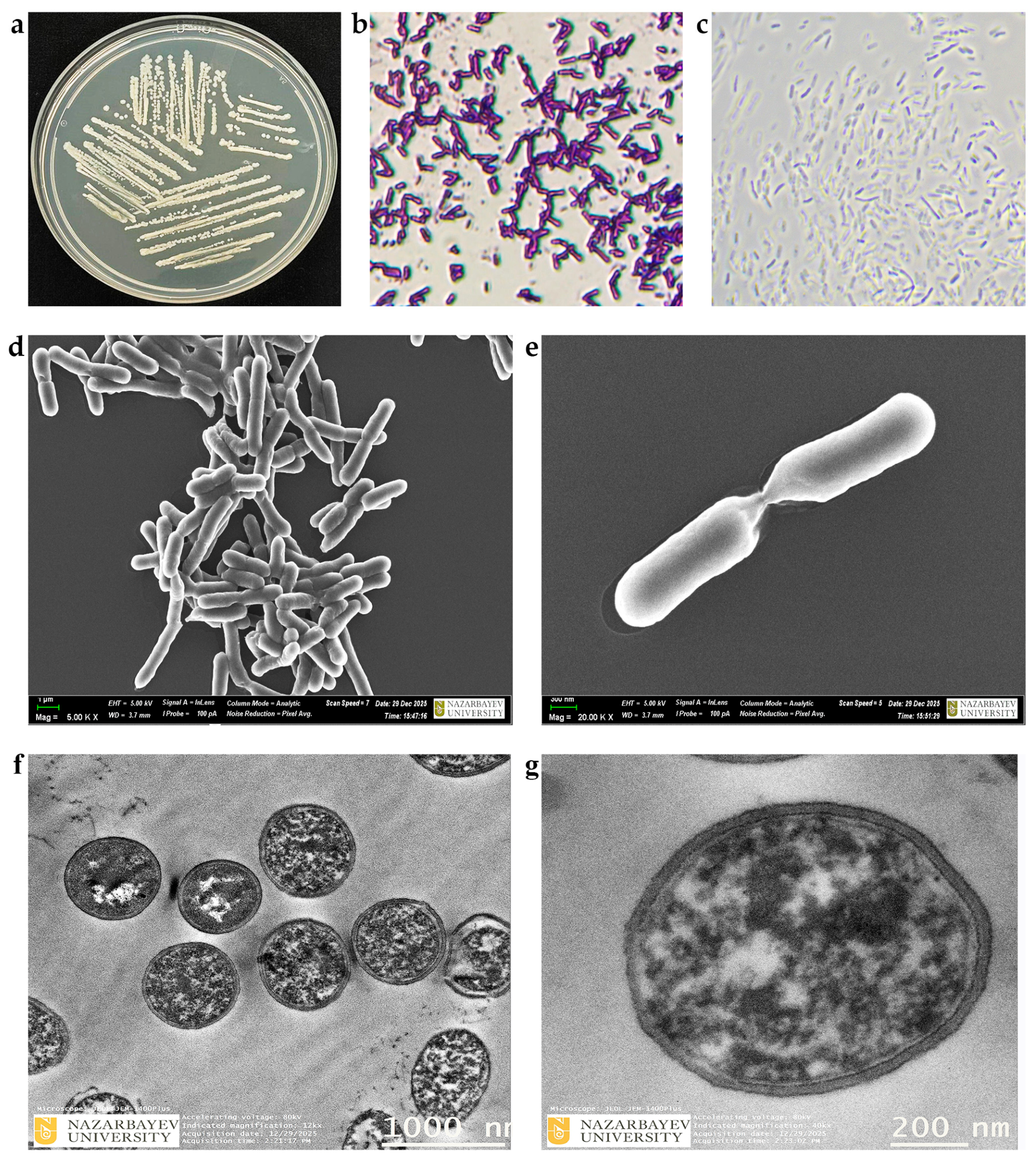
Light and phase-contrast microscopy, along with scanning and transmission electron microscopy (SEM and TEM), were employed to characterize the Gram staining, bacterial morphology, cellular morphology, and cell structure of the isolate, respectively (Figure 2).
The cellular morphology of the bacterial isolate JSM-10 grown on YEG medium (Figure 2a) represented typical Paenibacillus morphology and was further characterized using multiple microscopy techniques. Light microscopy (Figure 2b) and phase-contrast microscopy (Figure 2c) revealed Gram-positive staining and showed uniform Paenibacillus-type rod-shaped cells, respectively. Advanced microscopy, such as SEM, provided detailed images of the cell surface and overall shape, revealing smooth, rod-shaped cells with intact cell walls (Figure 2d,e). TEM analysis allowed observation of internal structures, such as cytoplasm density, and also highlight the typical ultrastructure of the isolate (Figure 2f,g). Together, these analyses demonstrated a comprehensive view of the isolate’s morphology and growth patterns of the strain at both the cellular and subcellular levels.

2.2. Abiotic Stress Tolerance of Paenibacillus sp. JSM-10

2.2.1. Evaluation of Glyphosate and NaCl Tolerance

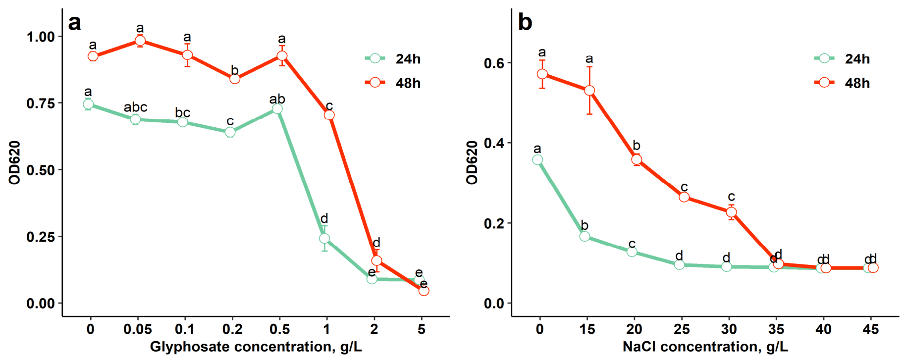
The growth of JSM-10 was not significantly reduced in the glyphosate concentration range of 0.05–0.5 g/L (Figure 3a) compared to the control (0 g/L) (p > 0.05), demonstrating its considerable tolerance to glyphosate.
The OD620 values of JSM-10 were significantly reduced only at higher glyphosate concentrations (1–5 g/L; p < 0.05). The strongest inhibition was observed at 2–5 g/L after both 24 h and 48 h (p < 0.05), indicating a concentration-dependent inhibitory effect of glyphosate (Figure 3a).
The growth of bacterial strain JSM-10 remained stable at NaCl concentrations of up to 15 g/L (Figure 3b). Although growth at this concentration was lower than the control without NaCl after 24 h (p < 0.05), no significant differences were observed at 48 h (p > 0.05), indicating growth recovery under saline stress. Cell density gradually decreased at 20–30 g/L (p < 0.05), while complete inhibition occurred at 40–45 g/L at both 24 and 48 h. PGPB capable of growth at NaCl concentrations of 1–5% (10–50 g/L) are classified as having low halotolerance [21]; accordingly, Paenibacillus sp. JSM-10 can be considered a salt-tolerant strain.
Interestingly, measurements at 24 and 48 h revealed distinct temporal patterns in the stress tolerance of strain JSM-10. Under glyphosate exposure, growth remained comparable between the two time points, whereas under NaCl stress, the strain exhibited improved growth after 48 h relative to 24 h, particularly at 15–30 g/L.

2.2.2. Examination of Drought Tolerance, Optimal Temperature Range, and Resistance to Heat-Shock Exposure

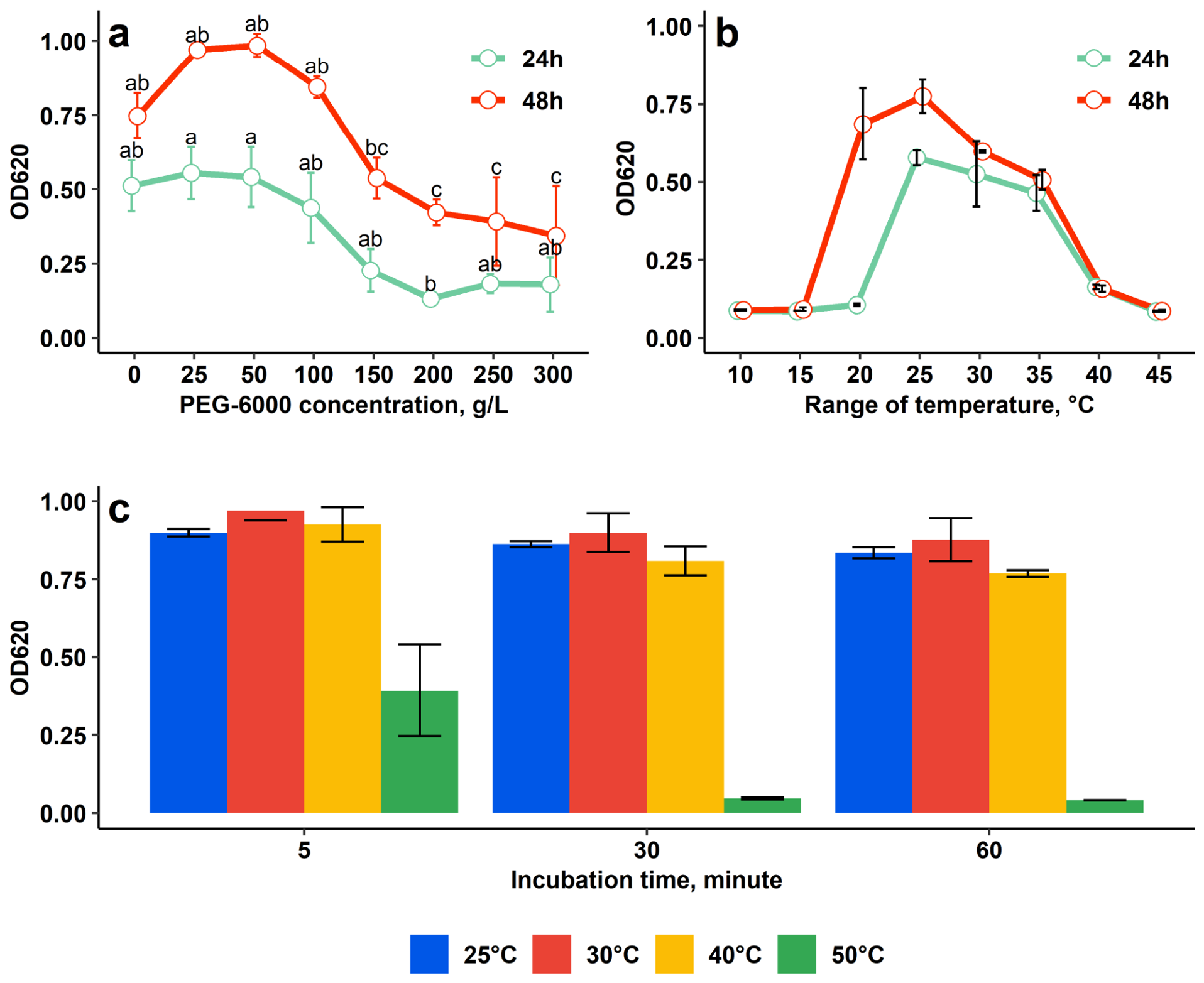
Growth of JSM-10 under PEG-6000-induced drought stress remained stable at 25–100 g/L (p > 0.05) and was significantly inhibited only at ≥150 g/L (p < 0.05) (Figure 4a). These findings indicate strong osmotic stress tolerance, as growth occurred at concentrations exceeding the 40 g/L PEG level previously associated with drought tolerance [22].
Evaluation of the optimal temperature range revealed that cell density remained low at 10–15 °C, showed the highest growth at 20–35 °C, and declined at 40 °C, with minimal growth observed at 40 and 45 °C (Figure 4b). The growth pattern was the same in both 24 and 48 h incubation, except 20 °C, at which low cell density was observed in the 24 h measurement, with a remarkable increase after a longer, 48 h incubation. Based on these findings, the optimal temperature levels for JSM-10 fall in the range of 20–35 °C.
Heat-shock testing performed to evaluate the tolerance of JSM-10 to sharp temperature changes during vegetation time in vivo showed high survival across tested temperatures, except 50 °C (Figure 4c). Bacterial recovery at 25 °C, 30 °C, and 40 °C was comparable to the control across all exposure times.

2.3. PGPB Characteristics of JSM-10

2.3.1. Indole-3-Acetic Acid (IAA) Synthesis, Phosphorus Solubilization, and Siderophore Production of Paenibacillus sp. JSM-10

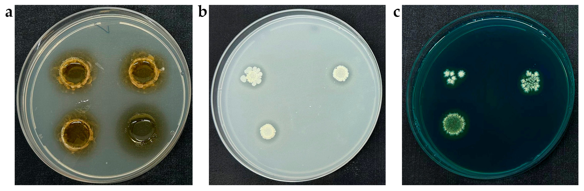
Paenibacillus sp. JSM-10 demonstrated multiple plant growth-promoting traits, including IAA synthesis, phosphate solubilization, and siderophore production (Figure 5). IAA production was confirmed by the development of a distinct pink coloration upon addition of Salkowski’s reagent to cultures grown in YEG supplemented with 0.1 g/L L-tryptophan (Figure 5a). Quantitative analysis revealed an IAA concentration of 70.3 µg/mL in the culture supernatant after three days of incubation.
Phosphate solubilization was observed on Pikovskaya agar (Figure 5b), where clear halo zones formed around the colonies, indicating the microbial dissolution of insoluble calcium phosphate (CaHPO4). On the 10th day of incubation, the solubilization index ranged from 1.30 to 1.38 and was highest at an inoculum density of 1 × 10⁶ CFU/mL. An additional PGPB characteristic, siderophore formation, was detected on Chrome Azurol S (CAS) agar by the appearance of a defined orange halo surrounding the colonies, resulting from iron chelation from the CAS-Fe3+ complex (Figure 5c).

2.3.2. The Effect of Paenibacillus sp. JSM-10 and Its Cell-Free Culture Filtrates (CCFs) on Buckwheat Growth

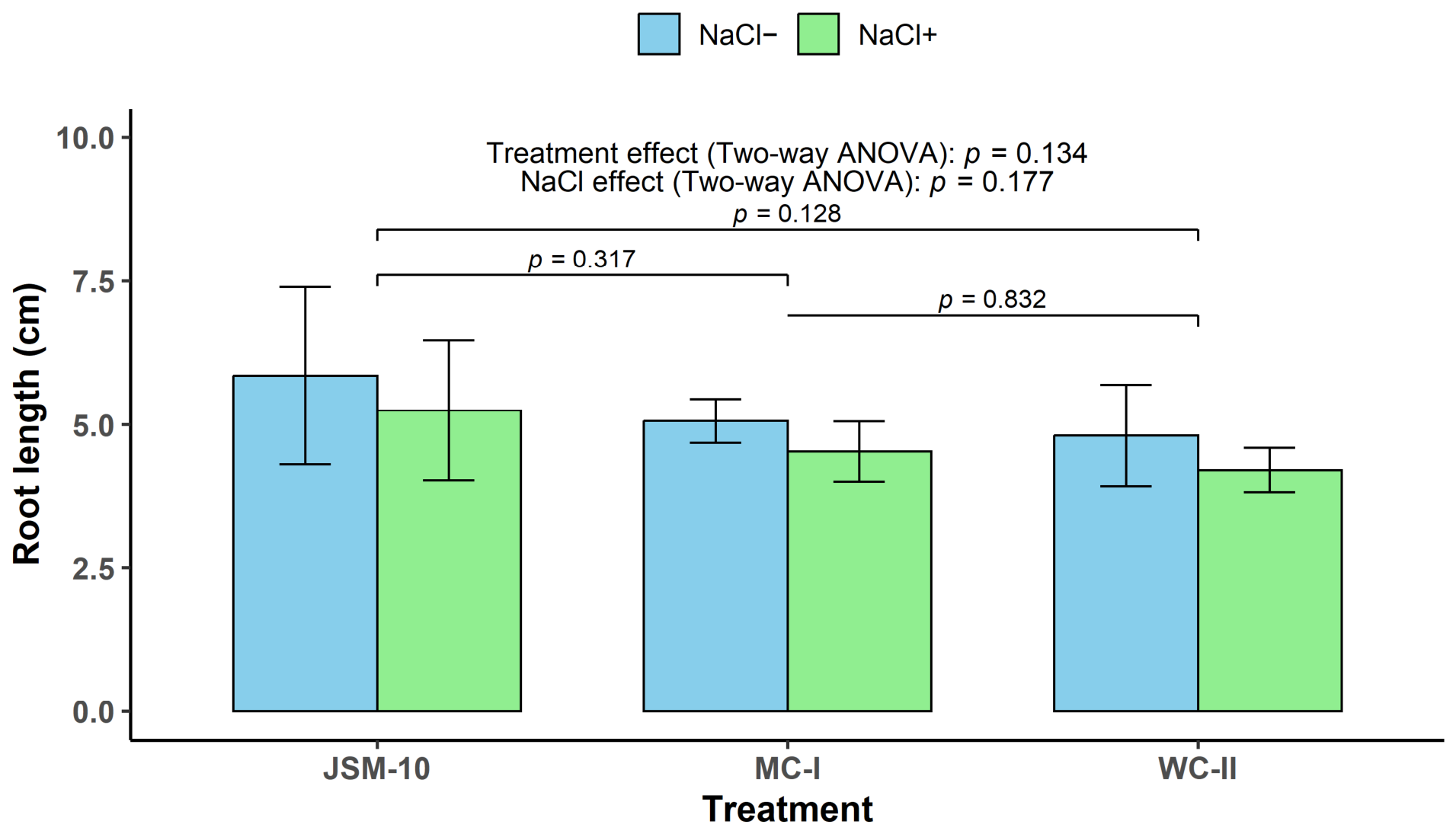
JSM-10 exhibited a positive trend toward enhancing buckwheat root length compared with both applied medium and water controls (Figure 6). Under non-saline conditions (NaCl), roots inoculated with JSM-10 reached 5.85 cm, resulting in an increase of up to 15.51 and 21.68% compared to the medium control (MC-I) and water control (WC-II), respectively.
A similar growth-promoting tendency of Paenibacillus sp. JSM-10 on the buckwheat root length was observed with the exposure to saline conditions (Figure 6). Under NaCl+, JSM-10 maintained its promoting trend on the root length, resulting in a higher root length (5.25 cm) relative to both controls: MC-I (up to 15.82%) and WC-II (up to 24.72%).
Two-way ANOVA revealed no statistically significant effects of NaCl exposure (p = 0.177) or treatment type (p = 0.134) on buckwheat root length. Post hoc Tukey’s HSD comparisons also showed no significant differences among treatments. However, the p-values indicated a decreasing tendency from the control treatments toward the bacterial inoculation. While the comparison between the two control variants (MC-I compared to WC-II) yielded a p-value of 0.83, the comparisons between JSM-10 and the MC-I and WC-II resulted in p-values of 0.32 and 0.13, respectively.
Interestingly, the growth-promoting tendency of JSM-10 on buckwheat roots was consistent across treatments, showing increases of 15.51–15.82% and 21.68–24.72% relative to the two controls (MC-I and WC-II), regardless of non-saline (NaCl) and saline (NaCl+) conditions, respectively. The effect sizes (Hedges’ g) for JSM-10 compared to treated samples with MC-I and WC-II under normal and saline conditions were 0.56 [95% CI: −0.80, 1.86] and 0.66 [95% CI: −0.73, 1.98], as well as 0.61 [95% CI: −0.76, 1.92] and 0.92 [−0.54, 2.30], respectively. Such a non-significant yet consistent positive growth trend under both normal and saline conditions suggests that JSM-10 may possess plant growth-promoting potential and may contribute to stress mitigation.

2.4. Antagonistic Potential of Glyphosate-Tolerant JSM-10 Against Different Pathogenic Species

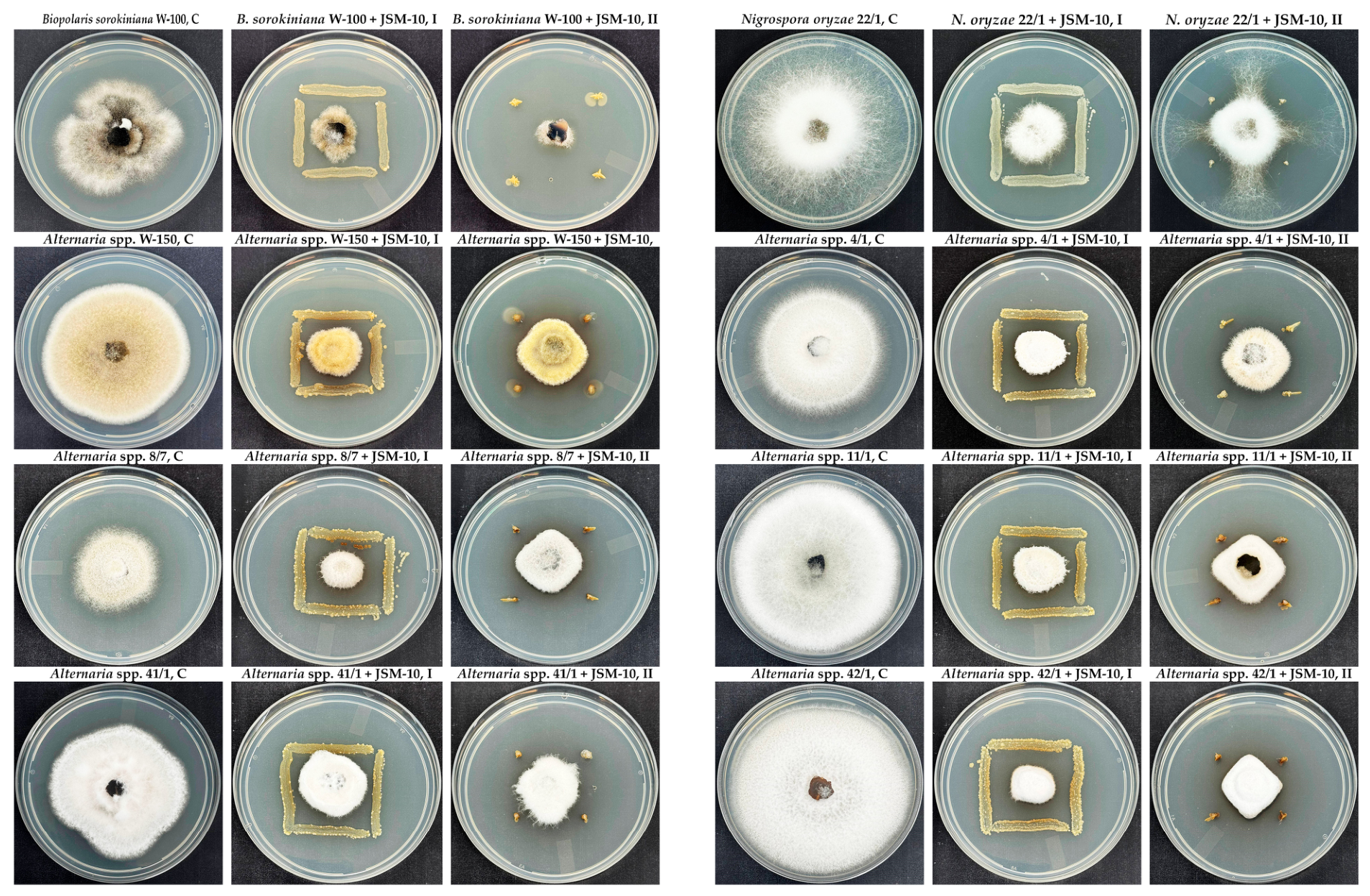
The antagonistic activity of bacterial strain JSM-10 was evaluated against the phytopathogenic fungi B. sorokiniana, N. oryzae, and Alternaria spp. on YEG medium. The strain demonstrated clear inhibitory effects on fungal pathogens, as evidenced by remarkably reduced mycelial growth compared to the control (Figure 7).
Both tested low- and high-inoculum methods inhibited fungal growth, confirming a strong antagonistic activity of JSM-10 towards all tested fungal strains. A visible color change of the medium between JSM-10 and Alternaria strains indicated that possible production of diffusible metabolites occurred during co-culture. The observed inhibition zones indicated strong antagonistic potential, suggesting the production of antifungal metabolites or competition for nutrients.
Quantitatively, the inhibition rate of JSM-10 in dual-culture assays varied between 64.2% towards B. sorokiniana, 71.2% to N. oryzae, and 72.1% against Alternaria spp. after 7 days of incubation (Table 1).
The observed inhibition, regardless of either low- or high-inoculum treatments, indicated a dose-independent mode of antagonistic activity of JSM-10. Except for Alternaria 8/7 with low-inoculum samples (36.8%), the growth of all tested fungal phytopathogens was inhibited by more than 50% on all bacterial-treated plates. The results confirm the remarkable potential of JSM-10 for application as a promising antagonistic inoculant to protect plant growth.
Additionally, the bacterial strain JSM-10 exhibited pronounced antagonistic activity against E. coli DH5α. Inhibition was observed on YEG agar plates inoculated with E. coli, where JSM-10 produced distinct, clear inhibition zones, indicating antimicrobial activity (Supplemental Figure S3). Kinetic assays performed to verify the presence of active metabolites revealed that, although 50% CCF obtained from non-inoculated medium (MM + Ala or MM + Glu) reduced cell density by only 6.80–12.56%, CCF (50%) obtained from JSM-10 grown in MM supplemented with L-alanine or glucose inhibited the growth of E. coli by up to 45.13–45.40%, respectively (Table 1). These findings suggest that extracellular metabolites produced by JSM-10 contribute substantially to its antibacterial activity.

2.5. Molecular Detection of Genes Associated with Plant Growth Promotion and Stress Tolerance

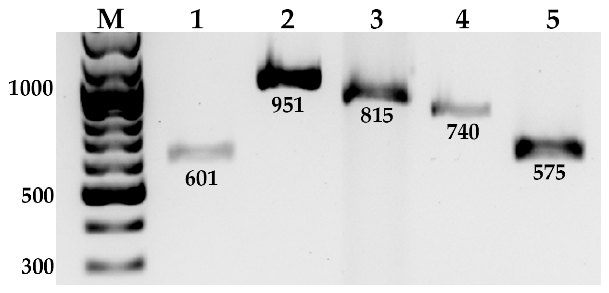
PCR amplification was performed to assess the presence of genes associated with plant growth-promoting traits and stress tolerance in strain JSM-10 (Figure 8).
PCR products of the expected sizes were obtained for gcd, ipdC, thiO, ectA, and groL, indicating the presence of genes putatively associated with IAA biosynthesis, phosphate solubilization, and stress-related functions. Bands corresponding to the expected amplicon sizes were observed for all targets, indicating the presence of genes associated with plant growth-promoting traits and stress tolerance in strain JSM-10.

3. Discussion

3.1. Plant Growth-Promoting (PGP) Potential and Mechanisms of Paenibacillus

3.1.1. Paenibacillus as a Promising PGPB Genus

A promising strategy to enhance soil quality, increase crop yields, and minimize the effects of ecological factors is the integration of organic farming relying on environmentally friendly methods, such as beneficial soil microflora or PGPB [13]. PGPB are considered cost-effective and easily accessible biological tools for mitigating both biotic and abiotic stresses. Therefore, integrating PGPB into crop production systems supports long-term sustainability and helps to preserve soil biodiversity by reducing dependence on chemical fertilizers [14]. Paenibacillus spp. represent an effective and promising PGPB genus, numerous species of which were reported to exhibit versatile plant growth–promoting and antagonistic properties (Table 2).
In addition to their PGP traits, the strains of Paenibacillus are known for their efficient ecological compatibility. For many introduced Paenibacillus strains used as PGPR/biocontrol inoculants, field and soil studies show no large, sustained disruption of resident bacterial communities [35]. As an autochthonous isolate from the Kazakh steppe, the indigenous origin of JSM-10 represents a logically compatible candidate for the local environment. These factors position the strain as a promising candidate for further biosafety and ecological impact assessments prior to large-scale field applications.

3.1.2. PGP Mechanisms of Paenibacillus sp. JSM-10 Strain

Overall, these studies demonstrate diverse PGPB traits on different crops across members of the genus Paenibacillus. One of the most important PGPB traits exhibited by Paenibacillus spp. is the production of the phytohormone IAA, a major auxin that directly influences root architecture, cell division, and nutrient acquisition [36]. In Paenibacillus species, IAA production is commonly associated with L-tryptophan-dependent pathways [37,38], which is consistent with the experimental conditions used in this study.
Siderophore production is another key trait of Paenibacillus spp., enhancing rhizosphere iron mobilization and nutrient uptake under iron limitation [39]. For example, Paenibacillus illinoisensis YZ29 increased rhizosphere iron availability by 1.8-fold and shoot biomass by 23%, demonstrating a direct link between bacterial siderophores and improved iron nutrition and growth [29]. Phosphorus solubilization is also a key trait of Paenibacillus spp., allowing plants to access otherwise unavailable phosphate pools in the rhizosphere. Inoculation of wheat with the phosphate-solubilizing Paenibacillus sp. B1 significantly increased soil available phosphorus by about 9% and shoot biomass by approximately 30% compared with non-inoculated controls, demonstrating that bacterial P solubilization can partially substitute for chemical phosphorus fertilizers [24]. Genomic analyses of Paenibacillus sonchi SBR5 revealed the presence of conserved glucose-1-dehydrogenase and gluconate dehydrogenase genes, supporting its gluconate-mediated phosphate-solubilization capacity [40].
The glyphosate-tolerant JSM-10 demonstrated key plant growth-promoting traits characteristic of the genus Paenibacillus, including IAA synthesis, siderophore production, and phosphorus solubilization (Figure 5). The co-occurrence of these traits has been widely reported in Paenibacillus strains and is considered an important factor contributing to their multifunctional role in plant growth promotion and biocontrol [26,41]. Reported specific structural genes and biosynthetic clusters—such as the ipdC gene encoding indole-3-pyruvate decarboxylase for auxin (IAA) production, and non-ribosomal peptide synthetase (NRPS) operons governing siderophore assembly—provide a concrete genetic framework. The presence of these dedicated loci elucidates the underlying molecular background driving the multiple plant growth-promoting (PGP) benefits, including enhanced root development and iron acquisition, observed in Paenibacillus sp. strain JSM-10 [42]. Altogether, these results demonstrate key plant growth-promoting traits of Paenibacillus sp. JSM-10, including phytohormone production, phosphate solubilization, and siderophore production, supporting its potential as a microbial inoculant.

3.1.3. Enhancement of Buckwheat Growth by JSM-10 Under Normal and Saline Conditions

Buckwheat is an important pseudocereal gluten-free crop widely consumed worldwide with high nutritional value and numerous health benefits [43,44]. It contains bioactive components, such as peptides, flavonoids, phenolic acids, fagopyritols, and fagopyrins, which makes it attractive both for human nutrition and for diversified cropping systems [45]. Despite its increasing global importance, research on PGPB for this crop remains limited, and reports on Paenibacillus–buckwheat interactions are particularly scarce, highlighting the relevance of evaluating new strains for this valuable crop.
The living culture of Paenibacillus sp. JSM-10 strain combined with the obtained from liquid culture CCF stimulated buckwheat root length up to 21.68-24.72% under normal and saline conditions, respectively (Figure 6). These findings indicate that the total bacterial inoculant, including both the cells and their secreted bioactive compounds, may contribute to increased plant growth. Although the differences were not statistically significant, the results suggest a tendency toward increased root length in plants treated with JSM-10. The absence of statistically significant differences is likely related to the relatively small sample size (n = 3 per treatment).
Root length is one of the most commonly used and informative indicators for evaluating PGPB effects and is suggested as a primary criterion for selecting promising strains [46]. The observed root-length increase by JSM-10 supports its further evaluation as a promising PGPB candidate. This integrated effect is consistent with reports on this genus, which is known for producing a wide range of metabolites that improve nutrient availability and stimulate root development [47].
While members of Paenibacillus genus demonstrated different plant-growth-stimulating mechanisms on a wide range of crops (Table 2), information on the effect of Paenibacillus species on buckwheat growth is limited. Seed bacterization with phosphate-solubilizing P. polymyxa KB balanced a phosphorus shortage in buckwheat [48]. The observed growth-promoting trend of strain JSM-10 broadens the portfolio of crops previously reported to be positively influenced by Paenibacillus sp.

3.2. Antagonistic Activity and Biocontrol Potential

In addition to auxin production and nutrient mobilization, Paenibacillus strains can suppress plant pathogens through diverse antimicrobial metabolites and induce plant defenses [19]. Members of this genus produce secondary metabolites, including nonribosomal peptides, lipopeptides, and volatile organic compounds (VOCs), which contribute to biocontrol activity [18]. Genomic analyses of biocontrol-active P. peoriae strains revealed gene clusters encoding nonribosomal peptide synthetases (NRPSs) and polyketide synthases (PKSs) that underpin production of these antagonistic metabolites, which can also trigger plant defense signaling [17]. Our JSM-10 strain inhibited several pathogens (Table 1), including B. sorokiniana (64.2%), N. oryzae (72%), and Alternaria spp. (72%) and E. coli (45.4%), consistent with the broad-spectrum antimicrobial activity associated with these molecular mechanisms. These findings suggest that both the living cells of JSM-10 and their bioactive metabolites contribute to pathogen suppression and may enhance systemic resistance in crops, supporting its potential as a multifunctional biocontrol agent.
Overall, JSM-10 exhibited strong antagonistic activity against E. coli and multiple fungal pathogens (B. sorokiniana, N. oryzae, and Alternaria spp.), indicating broad-spectrum biocontrol potential.

3.3. Eco-Physiological Characterization of JSM-10

3.3.1. Tolerance to Glyphosate Exposure

The global trends suggest that for successful field applications, PGPB require sufficient stress tolerance because beneficial effects can be limited by abiotic stress factors, such as soil contamination, salinity, and drought conditions. Therefore, PGPB need to be tested for systematic, multi-factor stress profiling in vitro prior to agriculturally relevant conditions [49]. For example, glyphosate is one of the most widely used herbicides worldwide and is known to exert toxic effects on non-target soil microorganisms [50], disrupting the growth, metabolism, and plant growth-promoting activities of many beneficial plant-associated fungi and bacteria [51]. Studies have shown that exposure to glyphosate can reduce microbial diversity, inhibit beneficial bacteria such as Pseudomonas and Bradyrhizobium japonicum [5], and impair their production of plant growth-promoting compounds like indole-3-acetic acid (IAA) and siderophores. The extensive utilization of this herbicide led to its dispersion and accumulation in both terrestrial and aquatic environments, heightening its potential hazards to the endemic soil microbiome and introduced PGPB [52,53].
In our studies, JSM-10 tolerated glyphosate up to 0.5 g/L without significant growth inhibition (p > 0.05), compared to the control without glyphosate (Figure 3). These findings are particularly relevant for agricultural applications since soils are frequently contaminated with glyphosate residues worldwide. Such residues can impact PGPB negatively, and, therefore, glyphosate tolerance of potential PGPB should be evaluated prior to in vivo applications.
Glyphosate showed variable levels of soil contamination but overall represented the largest contribution to total pesticide residues in soils, with a maximum concentration of 2.05 mg/kg [54]. Therefore, the glyphosate tolerance of JSM-10 examined in this study demonstrated clear tolerance and growth maintenance under herbicide stress up to 0.5 g/L, remarkably above the concentrations found in the field. This highlights its potential application as a glyphosate-tolerant PGPB to support sustainable crop production in agricultural systems, including glyphosate-polluted environments. Moreover, glyphosate exposure has been associated with endocrine disruption and genotoxic effects [55,56]. The utilization of glyphosate-tolerant PGPB is therefore a foundational step for agricultural applications in contaminated soils. Such strains can maintain their growth-promoting activities under herbicide stress and could represent potential candidates for future studies on glyphosate bioremediation to reduce environmental and health risks.
Mechanistically, the resilience of Paenibacillus to xenobiotic stress is often linked to a highly versatile genomic repertoire encoding stress-responsive enzymes, such as oxygenases and dehydrogenases, which play key roles in the degradation of complex environmental pollutants [42]. While the specific gene expression networks governing glyphosate tolerance in JSM-10 require further genetic characterization, these inherent genetic features highlight the genus’s capacity to tolerate chemical toxicity.

3.3.2. Tolerance to Salinity and Drought Stress

The Paenibacillus sp. JSM-10 was isolated from a semi-arid steppe soil environment defined by severe drought and fluctuating salinity. Such regions remain relatively under-explored for the discovery of stress-resilient PGPB capable of maintaining crop performance under adverse environmental conditions.
The JSM-10 strain demonstrated notable resilience to subsequent adverse abiotic stress factors, maintaining growth at moderate salt concentrations up to 15 g/L (Figure 3). These observations are in line with previous findings on P. polymyxa, which tolerated NaCl concentrations of 0–2%, with only gradual growth inhibition at 3–4%, demonstrating good salt tolerance in moderately saline soils [57]. In other studies, P. polymyxa strains also grew well at NaCl concentrations up to 4%, highlighting their ability to survive and remain active under saline conditions, further supporting their potential to enhance plant growth in salt-affected soils [58].
Furthermore, JSM-10 demonstrated robust osmotic stress resistance up to 150 g/L PEG 6000 and in optimal temperatures of 20–35 °C, and full recovery after prolonged heat-shock exposure up to 40 °C (Figure 4). Our findings are in accordance with previously published reports where members of the genus Paenibacillus have been increasingly recognized for their role in enhancing plant tolerance to drought stress. For example, P. polymyxa CR1 was shown to prime plants for drought by inducing dehydration-responsive genes such as RD29A and RD29B, thereby improving plant performance under water-limited conditions [59].
Importantly, drought mitigation by PGPB is often manifested not through enhanced accumulation of stress metabolites but rather through a reduction in stress-induced physiological responses. Accordingly, inoculation with Firmicutes, including Paenibacillus stellifer, was reported to significantly reduce the accumulation of osmolytes (proline and glycine betaine) and the activities of antioxidant enzymes (CAT and SOD) under moderate and severe drought stress, indicating effective stress mitigation. Similar trends observed in the present study suggest that this strain contributes to drought tolerance by stabilizing plant physiological status and reducing oxidative and osmotic stress, supporting the potential of Paenibacillus species as drought-mitigating PGPB [60].
At the molecular level, the environmental resilience of Paenibacillus is largely supported by the induction of stress-responsive genes regulating exopolysaccharide (EPS) biosynthesis—which acts as a physical biofilm barrier against osmotic shock and desiccation—and the synthesis of protective osmolytes [42]. Beyond the induction of exo-polysaccharide (EPS) biosynthesis—which forms a protective biofilm barrier against desiccation—Paenibacillus species leverages the synthesis of specific osmolytes, such as proline and trehalose, to stabilize cellular osmotic potential and protect plant enzymes under salt and water deficits [35].
Altogether, these findings, including tolerance up to 150 g/L PEG, growth under 20–35 °C, and recovery after exposure to heat-shock treatment, confirmed the drought tolerance potential of JSM-10, expanding its possible utilization under dry field conditions with sudden temperature fluctuations.

3.4. Ultrastructural Characterization of Cell Morphology and Structure

Microscopic characteristics of Paenibacillus members were reported previously, primarily based on individual techniques such as Gram staining or scanning electron microscopy [20]. However, a comprehensive morphological characterization integrating multiple microscopy approaches remains limited. In the present study, we provide a combined analysis of colony morphology, cellular structure, and ultrastructural features of Paenibacillus sp. JSM-10 using complementary microscopic techniques. This multi-scale evaluation provides an expanded understanding of its colony morphology, cellular organization, and ultrastructural features of the species, and also supports robust taxonomic and functional characterization of the isolate. The full collection of obtained microscopic images, including uncropped SEM and TEM presented in Figure 2, can be seen in Supplementary Materials (Supplementary Figures S4–S11).

3.5. Molecular Detection of Genes Putatively Involved in PGP and Stress Tolerance of JSM-10

To provide a molecular context for the observed phenotypic traits, selected genes associated with plant growth promotion and abiotic stress tolerance were examined in strain JSM-10. The targeted genes and their reported biological functions are summarized in Table 3.
The presence of ipdC and gcd correlates with the observed levels of IAA production and phosphate solubilization, respectively, while the identification of thiO supports the strain’s ability to tolerate and potentially degrade glyphosate. Furthermore, the detection of ectA and groL suggests an inherent genetic capacity for osmoprotection and proper protein folding, explaining the strain’s resilience under saline and thermal stress. While PCR-based detection of genes associated with plant growth-promoting traits and stress response supports the observed phenotypic characteristics of strain JSM-10, functional expression of these genes requires further investigation.

3.6. Potential Agricultural Applications, Current Limitations, and Future Perspectives

The successful agricultural application of promising PGPB requires a rigorous, multi-stage strategy. This roadmap consists of several subsequent steps, including the isolation and characterization of the beneficial inoculant, laboratory and greenhouse evaluations, ecological safety assessments, and finally, field trials and product certification [49].
The findings presented in this study establish the foundational stage of this strategy, providing the necessary evidence for the potential implementation of Paenibacillus sp. JSM-10. This strain was isolated from semi-arid steppe soil, which remains relatively underexplored as a source of stress-resilient plant growth-promoting bacteria. Tolerance to glyphosate exposure, salinity, and drought conditions highlights the potential of Paenibacillus sp. JSM-10 to enhance crop performance under environmental stress. Its resilience, together with the ability to induce systemic resistance and support plant growth, underscores its promise as a biological tool for sustainable and stress-resilient agriculture.
Such integrative characterization presented in the current study represents the essential foundational phase of a structured discovery-to-application pipeline. The following steps for agricultural applications include several aspects that remain to be addressed within the established evaluation pipeline for PGPB. Further genetic and molecular analyses are required to elucidate the mechanisms underlying stress tolerance and plant growth promotion. Longer-term greenhouse studies and detailed ecological safety assessments, including evaluation of potential effects on native rhizosphere microbiota, are necessary to ensure ecological compatibility. Subsequent field validation across diverse soil types and crop systems is required to confirm the strain’s efficiency under real agricultural conditions. These future studies will build upon the strong foundational dataset presented here and support the further development of strain JSM-10 for sustainable agricultural applications.

4. Materials and Methods

4.1. Isolation, Molecular Identification, and Microscopic Observation of Paenibacillus sp. JSM-10

4.1.1. Isolation of Glyphosate-Tolerant JSM-10

The previously reported strategy [64] was applied with minor modifications. The soil sample was collected from a field of soft spring wheat (Triticum aestivum L.) previously treated with glyphosate-type herbicides (“Uragan Forte” and “Faraon Gold”). Soil suspension was prepared by mixing 5 g of soil in 40 mL physiological saline (0.9% NaCl), and 50 µL of a subsequent 1:10 dilution was spread on solid PMM medium (g/L: (NH4)2SO4 4.49; K2HPO4 1.5; MgSO4·7H2O 0.2; agarose 20) [65]. PMM was supplemented with 1 g/L glyphosate formulation (“Uragan Forte”, 500 g/L; Syngenta, Switzerland) as the sole carbon source. Nystatin and fluconazole (0.1 g/L each) were added to suppress fungal growth. Plates were incubated at 25 °C for 7 days. Colonies that appeared on PMM-glyphosate medium were considered glyphosate-tolerant bacteria, and individual colonies were purified by repeated streaking on YEG agar medium (g/L: yeast extract 5; glucose 10; agar 15), which was also used for routine maintenance of the purified isolate JSM-10.

4.1.2. Molecular Identification and Detection of Functional Genes in JSM-10

Overnight bacterial culture was adjusted to 1 × 10⁷ CFU/mL in 50 μL double-distilled water (DDW) and used as a DNA template. Polymerase Chain Reaction (PCR) was performed using primer pairs and amplification programs, summarized in Table 4.
The PCR master mix for 16S rRNA (50 µL) consisted of the following components (final concentrations): 1 µL DNA template, 0.2 mM dNTPs, 1 µL Phusion Polymerase, 1 × HF buffer, 0.4 µM each primer; the final volume was adjusted with sterile MilliQ water. PCR was performed in a T100 Thermal Cycler (Bio-Rad, Singapore). The PCR products were visualized by 1% agarose gel electrophoresis and subsequently submitted for Sanger sequencing using an external service (National Scientific Shared Laboratory of Biotechnology, National Center for Biotechnology). All reagents were of molecular biology grade and purchased from New England Biolabs.
Two fragments (gyrA1 and gyrA2) of the gyrA gene were employed as a secondary molecular marker [67] to ensure accurate phylogenetic placement of strain JSM-10. The primers used in this study were designed based on multiple sequence alignment of gyrA gene sequences from representatives of the genus Paenibacillus using Vector NTI. Conserved regions were selected as primer binding sites, while the internal amplified fragment contained variable positions suitable for species-level discrimination after sequencing.
All amplification and post-PCR processing steps, including sequence assembly, were identical for both regions, except for the annealing temperature at 62 °C for the gyrA2 reaction (Table 4). The PCR master mix (25 µL) consisted of the following components (final concentrations): 1 µL DNA template, 0.2 mM dNTPs, 1 µL Taq polymerase, 1 × Taq buffer, 0.4 µM each primer; the final volume was adjusted with sterile MilliQ water. PCR was performed in a T100 Thermal Cycler.
Additionally, the rho gene (transcription termination factor) was amplified and sequenced to further support molecular identification of strain JSM-10 [18]. Primer design, PCR master mix composition, and post-PCR processing were identical to those used for the gyrA fragments.
Raw sequencing chromatograms obtained after Sanger sequencing were visually inspected and trimmed to remove low-quality regions. Forward and reverse reads were assembled into consensus sequences using Vector NTI Advance 11.0 software. The resulting contigs were checked to ensure correct base calling and absence of ambiguous nucleotides.
The obtained nucleotide sequences were compared with publicly available sequences in the NCBI GenBank database using the BLASTn algorithm in order to determine the closest homologs and confirm taxonomic affiliation. Reference sequences showing the highest similarity scores were selected for subsequent phylogenetic analysis.
Multiple sequence alignment of the obtained sequences together with the selected reference sequences was performed using the MUSCLE algorithm. For additional phylogenetic reconstruction, sequences of the 16S rRNA gene and gyrA gene fragments were concatenated, resulting in a combined alignment of 1390 and 1118 bp with gyrA1 and gyrA2, respectively. Concatenation of 16S rRNA and rho gene sequences resulted in an alignment of 1191 bp. Phylogenetic trees were constructed using the Maximum Likelihood (ML) method with the best-fit nucleotide substitution model determined automatically by the MEGA software (version 12.1.2). The robustness of the inferred phylogenetic relationships was evaluated by bootstrap analysis with 1000 replicates.
For amplification of genes associated with plant growth-promoting traits and stress tolerance (Table 4), specific primers were designed for ipdC (IAA synthesis), gcd (phosphate solubilization), thiO (glyphosate degradation), and groL and ectA (osmotic and saline stress tolerance). Primer design, PCR master mix composition, and amplification conditions were generally consistent with those used for gyrA and rho, except for groL and ectA, for which a modified reaction mixture was applied. The PCR master mix for groL and ectA contained 0.2 µM of each primer, 0.2 mM dNTPs, 1× Q5 buffer, 0.5 µL Q5 polymerase, 4% DMSO, and 1 µL DNA template in a final volume of 25 µL.
PCR amplicons were separated on 1% agarose gels (90 V, 40 min) and visualized using a GelDoc Go Imaging System (Bio-Rad, USA). The presence of target genes was assessed based on the expected amplicon size.

4.1.3. Microscopic Evaluations of Glyphosate-Tolerant Paenibacillus sp. JSM-10

Bacterial morphology was examined after cultivation of JSM-10 on YEG for 2 days. Typical morphological characteristics, such as color, form, and size, were recorded based on visual observations.
The Gram reaction was performed using a fresh bacterial culture in accordance with the manufacturer’s instructions for the Gram Stain Kit (HiMedia, Maharashtra, India). Gram-stained cells were observed under a light microscope (CX40, China) at 100× magnification; images were captured with a SOPTOP OD400UHW-P digital microscope camera.
Phase-contrast microscopy was used to observe bacterial cell morphology without staining. The bacterial strain pre-grown on YEG plates was collected in 5 mL of 0.9% NaCl using sterile cotton swabs. Ten µL of the suspension was placed on a glass slide, and observations were performed with a phase-contrast microscope (Zeiss Primostar 3) at 100× magnification; images were recorded at the selected magnification using a digital imaging system (Axiocam 212 color microscope camera).
For Scanning Electron Microscopy (SEM) and Transmission Electron Microscopy (TEM), the bacterial strain JSM-10 was grown in liquid YEG medium for 24 h at 130 rpm, with an initial density of 1 × 10⁵ CFU/mL. After incubation, 1 mL of the culture was transferred to a 2 mL Eppendorf tube containing 1 mL of 2.5% glutaraldehyde (GA), and then it was mixed gently. The prepared sample was stored at 4 °C overnight for fixation prior to submission for SEM and TEM analyses to an external service (Electron Microscopy Laboratory, Core Facilities and HPC, Nazarbayev University).
For SEM analysis (Auriga Crossbeam 540, Carl Zeiss, Oberkochen, Germany), bacterial cells were collected by centrifugation and fixed in 2.5% GA prepared in phosphate buffer (pH 7.2–7.4) at 4 °C for 1–2 h. The samples were then washed with the same buffer and post-fixed in 1% osmium tetroxide for approximately 1 h. Subsequently, the samples were dehydrated through a graded ethanol series (30–100%). The dehydrated specimens were placed onto collagen-coated coverslips. After drying, the samples were mounted on holders and coated with a thin conductive carbon layer prior to imaging.
For TEM analysis (JEM 1400 Plus, JEOL, Tokyo, Japan), cells were fixed in 2.5% GA in phosphate buffer (pH 7.2–7.4) at 4 °C for 1–2 h, followed by washing in buffer and post-fixation in 1% osmium tetroxide for approximately 1 h. The samples were then dehydrated in a graded ethanol series (30–100%), infiltrated with epoxy resin, and polymerized at 60 °C. Ultrathin sections (60–90 nm) were prepared from the polymerized blocks using an ultramicrotome (Leica UC7, Wetzlar, Germany).

4.2. Abiotic Stress Tolerance of JSM-10

4.2.1. Glyphosate Tolerance Evaluation

Glyphosate tolerance of the isolated JSM-10 strain was examined using a broth microdilution method. The YEG medium supplemented with glyphosate “Uragan Forte” (500 g/L) at final concentrations of 0.05, 0.1, 0.2, 0.5, 1, 2, and 5 g/L was dispensed (180 µL) to a 96-well Nunc microplate (ThermoFisher, Rochester, NY, USA). Inoculated YEG medium without glyphosate and non-inoculated YEG containing corresponding glyphosate concentrations served as the control and blank samples, respectively. Bacterial cell suspension was collected from overnight-grown YEG plate cultures in 5 mL 0.9% NaCl using cotton swabs, and the cell density was measured at OD620. Based on the measured cell density, the subsequent bacterial suspension for inoculation was adjusted to 1 × 10⁶ CFU/mL in 5 mL 0.9% NaCl. The bacterial suspension (20 µL) was used to inoculate each well containing 180 µL YEG with or without glyphosate, resulting in an initial density of 1 × 10⁵ CFU/mL. The plate was incubated in a temperature-controlled CAPP Rondo 4-Place Incubating Shaker, CRPI-412X (AHN Biotechnologie GmbH, Nordhausen, Germany) at 25 °C and 200 rpm. Cell density was measured at OD620 after 24 and 48 h of incubation using a Multiskan SkyHigh Microplate Spectrophotometer (Thermo Fisher Scientific, Singapore). The described protocol for bacterial suspension preparation, inoculation, and incubation was applied throughout this subsection in order to ensure quantifiability, reproducibility, and compatibility among all tests. Measurements at 24 and 48 h were consistently applied in all subsequent stress tolerance assays to evaluate the time-dependent adaptation of the strain to adverse environmental conditions. All analyses, unless specified, were performed in triplicate.

4.2.2. Examination of Salinity Tolerance

For testing salinity tolerance, the bacterial strain was incubated in a 96-well microplate containing 180 µL liquid YEG adjusted with different concentrations of NaCl (g/L: 0, 15, 20, 25, 30, 35, 40, and 45), where YEG without NaCl served as the control. Cell suspension preparation, inoculation, incubation, and growth monitoring (at 24 and 48 h) were performed according to the protocols described in Section 4.2.1.

4.2.3. Drought Tolerance Assay

Polyethylene glycol with a molecular mass of 6000 (PEG 6000) was used to simulate osmotic stress conditions [68]. The strain was cultured in YEG supplemented with PEG 6000 at final concentrations of 0 (applied as the control), 25, 50, 100, 150, 200, 250, and 300 g/L. Inoculation, incubation conditions, and growth monitoring (OD620 at 24 and 48 h) were conducted as detailed in Section 4.2.1.

4.2.4. Determination of an Optimal Temperature Range

The temperature range (10, 15, 20, 25, 30, 35, 40, and 45 °C) suitable for growth of Paenibacillus sp. JSM-10 was evaluated in YEG medium using the standardized inoculation and growth measurement protocols described in Section 4.2.1.

4.2.5. Heat-Shock Stress Examination

The heat-shock method was applied to study the resilience of JSM-10 to sharp temperature fluctuations occurring under field conditions. Heat-shock tolerance was mimicked by exposing 1 mL of cell suspension (prepared in 0.9% NaCl at 1 × 10⁷ CFU/mL in 2 mL safe-lock Eppendorf tubes) to a range of temperatures of 30, 40, and 50 °C as mild, moderate, and severe shock levels; and for three exposure times: short, medium, and long of 5, 30, and 60 min, respectively. After heat exposure, 100 µL of each treated sample was transferred into wells of a 96-well plate containing 100 µL of fresh sterile double-concentrated YEG medium. As the control, YEG was inoculated with 100 µL of cell suspension maintained at 25 °C throughout the heat-shock assay. Bacterial recovery was measured after 24 h of incubation at 25 °C with shaking at 200 rpm in a shaker-incubator using a Multiskan SkyHigh microplate spectrophotometer at OD620. The assay was run in three replicates.

4.3. Plant Growth-Promoting Traits of JSM-10 Strain

4.3.1. Indole-3-Acetic Acid (IAA) Synthesis

IAA production was evaluated using a rapid plate assay with minor modifications [69]. Bacterial cell suspensions were prepared in 5 mL 0.9% NaCl at final concentrations of 1 × 10⁵, 1 × 10⁶, and 1 × 10⁷ CFU/mL. An aliquot of 200 μL from each cell suspension was transferred into 1 cm diameter holes made in YEG agar supplemented with 0.1 g/L tryptophan as a precursor of IAA synthesis. Holes containing 200 μL sterile 0.9% NaCl served as the non-inoculated control. Following a five-day incubation at 25 °C, the holes were cleared of bacterial biomass, and 200 μL of Salkowski reagent (12 g/L FeCl3 in 37% H2SO4) [70] was added. The reaction was incubated for 20 min at 25 °C in the dark, and IAA production was indicated by the development of a pink-yellow coloration surrounding the holes. All assays were performed in triplicate.
IAA concentration in bacterial culture supernatants was determined using a calibration curve. Strain JSM-10 was cultivated in YEG supplemented with 0.1 g/L L-tryptophan, and the supernatant was collected on the third day after incubation at 25 °C and 130 rpm. For color development, 100 µL of sample or standard solution was mixed with 100 µL of Salkowski reagent and incubated for 20 min at 25 °C in the dark. Absorbance was measured at 530 nm. The exact IAA concentrations in the supernatants were determined using the constructed calibration curve in SkanIT software (version 7.0.2). For the calibration curve, a 0.5 mg/mL IAA stock solution was prepared in DMSO and used to obtain standards ranging from 0 to 500 µg/mL. Standards were treated in the same way as the bacterial supernatant samples and measured in triplicate. A calibration curve was constructed using linear regression and used to determine IAA concentrations in bacterial culture supernatants.

4.3.2. Phosphorus Solubilization Capacity

Phosphate solubilization was evaluated using Pikovskaya agar (g/L: glucose 10; (NH4)2SO4 0.5; MgSO4·7H2O 0.1; KCl 0.2; yeast extract 0.5; NaCl 0.2; MnSO4·H2O 0.002; FeSO4·7H2O 0.002; agar 20) [71] supplemented with 5 g/L CaHPO4 as the inorganic phosphate source [72]. Bacterial suspensions were adjusted to 1 × 10⁵, 1 × 10⁶, and 1 × 10⁷ CFU/mL, and 10 µL aliquots were spot-inoculated onto the agar plates in triplicate and air-dried for 10 min. After 10 days of incubation at 25 °C, well-defined halo zones surrounding the bacterial colonies were observed, indicating the phosphorus solubilization potential.
Colony diameter and halo-zone diameter were measured, and the phosphorus solubilization index was calculated according to the following formula:
Solubilization index = (Diameter of solubilization zone, mm + Diameter of colony growth, mm)/(Diameter of colony growth, mm)

4.3.3. Siderophore Production

The siderophore-producing capacity of the JSM-10 isolate was investigated using the Chrome Azurol S (CAS) agar method [73]. The CAS reagent was prepared in distilled water by mixing 10 mL of 1 mM FeCl3·6H2O in 10 mM HCl with 50 mL of 2 mM CAS solution, followed by the addition of 40 mL of 5 mM hexadecyltrimethylammonium bromide (HDTMA). Bacterial suspensions at target concentrations were prepared as described above, and 10 µL aliquots of each suspension were inoculated onto LB agar (g/L: peptone 7.5; NaCl 2.5; yeast extract 2.5; agar 15), mixed with autoclaved CAS reagent at a 1:1 ratio [74], air-dried for 10 min, and incubated at 25 °C for 7 days. Siderophore production was indicated by the formation of an orange halo zone around the colonies. All assays were tested in three replicates.

4.3.4. The Effect of JSM-10 and Its Culture Filtrates on Buckwheat (Fagopyrum esculentum L.) Growth with and Without Saline Conditions

The experiment was designed to assess both the direct effect of living cultures of Paenibacillus sp. JSM-10 and the contribution of its extracellular metabolites (CCF) on buckwheat growth with and without exposure to saline stress conditions. Plastic pots (25 mL) were filled with 10 g of soil substrate. To simulate salinity stress, 3 mL of 125 mM NaCl solution was added to the substrate and allowed to dry for 24 h. For non-saline conditions, 3 mL of distilled water (dH2O) was applied and dried under the same conditions. At the time of sowing, three treatments were applied. The JSM-10 treatment consisted of 2 mL of a 3-day culture of strain JSM-10 grown in Minimal Medium supplemented with glucose (MM + Glu), combined with 1 mL of the corresponding cell-free culture filtrate (CCF) (obtained as described below in Section 4.4.2) and 2 mL dH2O. The medium control (MC-I), included to control for medium effects, received 3 mL of CCF from non-inoculated MM + Glu and 2 mL of dH2O, while the water control (WC-II) received 5 mL of dH2O. Seven seeds were sown in each pot and covered with approximately 5 g of soil.
On the third day of incubation, a secondary treatment was applied. For the JSM-10 treatment, 2 mL of a 24 h culture grown in YEG supplemented with 0.5 g/L tryptophan was combined with 1 mL of CCF obtained from JSM-10 grown in MM + Glu and 2 mL of dH2O. The medium control received 2 mL of non-inoculated YEG supplemented with 0.5 g/L tryptophan, 1 mL of CCF from non-inoculated MM + Glu, and 2 mL of H2O. The water control received 5 mL of dH2O.
Plant growth parameters were assessed on the fifth day after sowing. Root length, shoot (seedling) length, and total seedling length (sum of root and shoot) were recorded. All treatments were performed in triplicate and incubated at 25 °C under a natural day/night cycle.

4.4. Antagonistic Activity Assays

4.4.1. Inhibition of Fungal Plant Pathogens

Initial assessment of the antagonistic potential of bacterial strain JSM-10 was evaluated against two phytopathogenic fungal isolates (Bipolaris sorokiniana W-100 and Alternaria spp. W-150) isolated in this study, following the method described by [75]. Once the antagonistic potential was confirmed, additional strains (Alternaria spp. 4/1, 8/7, 11/1, 41/1, 42/1, and Nigrospora oryzae 22/1) were included in the study. Identification of the newly isolated strains B. sorokiniana W-100 and Alternaria spp. W-150 (submitted in NCBI under accession number: PZ067503 and PZ067504) was performed as reported for the retrieved fungal strains [76]. The fungal isolates were maintained on YEG and incubated at 25 °C for 3–7 days, depending on growth rate. For the antagonism assay, a 6 mm diameter agar plug containing actively growing mycelium was transferred to the center of a fresh YEG plate. To evaluate the inhibitory capacity of JSM-10 under different bacterial inoculum loads, two co-cultivation designs were applied. In the high-inoculum treatment, a 24 h culture of strain JSM-10 was streaked using sterile cotton swabs at a distance of 2.5 mm from the fungal plug on four equidistant sides. In the low-inoculum treatment, the bacterial strain was spot-inoculated using sterile toothpicks at a distance of 3.5 mm from the fungal plug. The control plates consisted of fungal isolates grown without bacterial inoculation. The experiment was carried out in triplicate. Plates were incubated at 25 °C for 7 days. The inhibitory activity (IR) was determined using the following formula:
IR = (D1 − D2)/D1 × 100%
where IR represents the percentage of inhibitory activity, D1 is the diameter of the phytopathogen colony in the control (mm), and D2 is the diameter of the phytopathogen colony (mm) in the presence of the JSM-10 strain [77].

4.4.2. Antagonistic Activity of Living Cultures of Paenibacillus sp. JSM-10 and Its Culture Filtrates Towards Escherichia coli

The antagonistic activity of the JSM-10 strain against E. coli DH5α was evaluated using a co-culture assay. Cell suspensions of Paenibacillus sp. JSM-10 were prepared at final concentrations of 1 × 10⁵, 1 × 10⁶, and 1 × 10⁷ CFU/mL (to apply low-, moderate-, and high-concentration treatment, respectively) while E. coli was prepared at 1 × 10⁵ CFU/mL. Then, 5 mL of the prepared E. coli suspension was used to cover the surface of YEG agar plates, and the excess was removed. The inoculated plates were air-dried for 15 min, followed by spot inoculation of 10 µL of each bacterial suspension (three concentrations per plate). The plates were air-dried for an additional 10 min and then incubated at 25 °C for 5 days. Individual plates inoculated solely with E. coli or JSM-10 served as the untreated controls. Antagonistic activity was assessed based on the presence of visible inhibition zones surrounding JSM-10 spots, presented in Supplemental Figure S3.
For obtaining the cell-free culture filtrate (CCF) to evaluate potential inhibitory metabolites, JSM-10 was inoculated at 1 × 10⁵ CFU/mL as the initial cell density in 50 mL Minimal Medium (g/L: KH2PO4, 1.0; MgSO4·7H2O, 0.5; (NH4)2SO4, 5.0) in distilled water [78], supplemented individually with 2 g/L glucose or L-alanine. Three-day shaking cultures grown at 25 °C in an orbital shaker (IKA KS 260, Staufen im Breisgau, Germany) at 130 rpm were centrifuged at 10,000× g for 10 min, and the collected supernatant was filter-sterilized via 0.22 µm syringe filters (TPP, Schaffhausen, Switzerland). The sterility of the obtained CCF was confirmed by plating 10 µL on YEG agar, followed by incubation for 3 days at 25 °C with daily monitoring for growth. The CCF of non-inoculated MM prepared in the same way served as the control (C-CCF) to evaluate the medium effects on E. coli growth. The CCF was stored at 4 °C during the experimental period.
The antagonistic activity of CCF produced by strain JSM-10 against E. coli was evaluated using a microplate-based kinetic growth assay. Liquid YEG was amended with the obtained CCF at final concentrations of 25 and 50% (v/v), while YEG without CCF was set as the untreated control. Aliquots of 10 µL of E. coli suspension adjusted to 1 × 10⁶ CFU/mL were added to wells containing 90 µL of YEG medium with or without CCF, resulting in an initial cell density of 1 × 10⁵ CFU/mL per well. All treatments were performed in triplicate. The 96-well plate was incubated at 37 °C in a Multiskan SkyHigh microplate spectrophotometer operating in kinetic mode. Bacterial growth was monitored by measuring absorbance at OD620 at 20 min intervals for 24 h without shaking. The inhibition rate (%) was derived from final-cycle OD620 measurements and expressed as the relative decrease in E. coli growth in CCF-treated samples compared to the untreated control.

4.5. Statistical Analyses and Data Visualization

Statistical analyses were performed using one-way analysis of variance (ANOVA) to evaluate the effects of abiotic stress factors (glyphosate, salinity, and drought) on bacterial growth. Prior to ANOVA, all data were tested for normality using the Shapiro–Wilk test and for homogeneity of variances using Levene’s test from the car package [79]. For variants that did not meet the assumptions for parametric ANOVA, a non-parametric alternative (Kruskal–Wallis test) was applied. The significance threshold was applied as 95% (p < 0.05). When ANOVA indicated significant differences, the data were subsequently analyzed using Tukey’s honestly significant difference (HSD) post hoc comparisons from the agricolae package [80]. The significance threshold was set at 95% (p < 0.05).
Temperature range and heat-shock experiments were conducted as physiological tolerance assays to determine growth and survival ranges. Therefore, the results were evaluated descriptively (means and standard deviations) based on growth patterns and recovery trends rather than inferential statistical comparisons.
In the plant growth assessment studies, two-way ANOVA was performed to evaluate the effects of bacterial treatment (JSM-10 inoculation vs. controls) and NaCl exposure on buckwheat root length. The assumptions of ANOVA were tested prior to analysis, as described above. As no significant interaction between factors was detected in the full factorial model, the additive model was applied. Tukey’s HSD test was used for post hoc pairwise comparisons. In addition, to quantify the magnitude of the difference between the JSM-10 strain and the respective controls (MC-I and WC-II), effect sizes were calculated using Hedges’ g via the effectsize package [81]. Hedges’ g was selected over Cohen’s d to provide a more accurate estimate for n = 3 sample sizes, employing a pooled standard deviation. Hedges’ g values were calculated separately for each NaCl condition. All effect sizes are reported with their corresponding 95% confidence intervals (CI). Descriptive statistics (means and standard deviations) were calculated for each experimental group (n = 3 per group) and presented in Figure 6.
All graphs were constructed using the ggplot2 package [82] and merged using the patchwork package [83]. All statistical analyses and data visualization were conducted in the R statistical environment (R Core Team, version 4.4.1; https://www.r-project.org/contributors.html (accessed on 26 April 2026)) using RStudio, version 2023.06.1 (Posit, https://posit.co/download/rstudio-desktop (accessed on 26 April 2026)). All data are presented as mean ± standard deviation of three biological replicates, unless otherwise stated.

5. Conclusions

This study expands current knowledge by providing an integrative assessment of the Paenibacillus sp. JSM-10 strain, including its detailed microscopic characterization, tolerance to multiple abiotic stresses, resistance to glyphosate, time-dependent responses to salinity and osmotic stress, plant growth-promoting trend, and antagonistic activity. The observed functional diversity suggests the involvement of multiple yet insufficiently explored molecular mechanisms, potentially including phytohormone-mediated signaling, stress-responsive metabolic pathways, and metabolite-driven antagonistic interactions.
Importantly, the demonstrated tolerance to environmentally relevant stress factors, together with the multifunctional plant-beneficial traits, supports the potential applicability of glyphosate-tolerant Paenibacillus sp. JSM-10 as a functional, stress-resilient PGPB candidate for improving crop performance under abiotic stress in sustainable agricultural systems.
The present study represents the foundational stage in the development and evaluation of a novel plant growth-promoting bacterial inoculant. Ongoing and future research, including genomic and molecular analyses, ecological safety assessments, greenhouse experiments, and multi-site field validation, will be essential to further evaluate the environmental safety and agricultural efficiency of strain JSM-10. These subsequent studies will support the further development of Paenibacillus sp. JSM-10 as a promising stress-tolerant microbial inoculant for sustainable agriculture.

Supplementary Materials

The following supporting information can be downloaded at: https://www.mdpi.com/article/10.3390/ijms27094062/s1.

Author Contributions

Conceptualization, A.R.Z.; methodology, Z.Z., A.S. (Adel Sattarova), I.R. and A.R.Z.; software, Z.Z. and A.S. (Aldan Shamukhan); validation, Z.Z., A.S. (Adel Sattarova) and A.R.Z.; formal analysis, Z.Z., A.S. (Adel Sattarova), A.A. and A.S. (Aldan Shamukhan); investigation, Z.Z. and A.S. (Adel Sattarova); resources, I.R. and S.A.; data curation, Z.Z. and A.S. (Adel Sattarova); writing—original draft preparation, Z.Z. and A.S. (Adel Sattarova); writing—review and editing, A.A., A.S. (Aldan Shamukhan), I.R., S.A. and A.R.Z.; visualization, Z.Z., A.A. and A.S. (Aldan Shamukhan); supervision, A.R.Z.; project administration, S.A. and A.R.Z.; funding acquisition, S.A. and A.R.Z. All authors have read and approved the published version of the manuscript.

Funding

This research was funded by the Science Committee of the Ministry of Science and Higher Education of the Republic of Kazakhstan (Grant No. AP23485562).

Institutional Review Board Statement

Not applicable.

Informed Consent Statement

Not applicable.

Data Availability Statement

The sequences of 16S rRNA gyrA2, gyrA1 of gyrA and rho of Paenibacillus sp. JSM-10 and ITS of fungal isolates obtained during this data were uploaded to NCBI (accession numbers PZ059921, PZ241072, PZ287672, PZ316596, PZ067503, and PZ067504, respectively). All collected data of this study are available on a reasonable request from the corresponding author.

Acknowledgments

The authors acknowledge the Electron Microscopy Laboratory, Core Facilities and HPC, Nazarbayev University, for providing SEM and TEM analysis services, and thank Nurgul Daniyeva for her valuable guidance.

Conflicts of Interest

The authors declare no conflicts of interest.

References

  1. FAO. The State of Food and Agriculture 2023—Revealing the True Cost of Food to Transform Agrifood Systems; FAO: Rome, Italy, 2023. [Google Scholar]
  2. Köninger, J.; Labouyrie, M.; Ballabio, C.; Dulya, O.; Mikryukov, V.; Romero, F.; Franco, A.; Bahram, M.; Panagos, P.; Jones, A.; et al. Pesticide Residues Alter Taxonomic and Functional Biodiversity in Soils. Nature 2026, 650, 367–373. [Google Scholar] [CrossRef] [Scilit]
  3. Qiu, D.; Wang, Y.; Xu, N.; Chen, B.; Zhu, Y.; Zhang, Z.; Zhang, Q.; Lu, T.; Dong, H.; Shou, J.; et al. Global Variation in Plant-Beneficial Bacteria in Soil under Pesticide Stress. Nat. Commun. 2025, 16, 10685. [Google Scholar] [CrossRef] [Scilit]
  4. Asrat, A.; Sitotaw, B.; Dawoud, T.M.; Nafidi, H.-A.; Bourhia, M.; Mekuriaw, A.; Wondmie, G.F. Effect of Glyphosate on the Growth and Survival of Rhizobia Isolated from Root Nodules of Grass Pea (Lathyrus sativus L.). Sci. Rep. 2023, 13, 21535. [Google Scholar] [CrossRef] [Scilit]
  5. Sibalekile, A.; Araya, T.; Castillo Hernandez, J.; Kotzé, E. Glyphosate-Microbial Interactions: Metagenomic Insights and Future Directions. Front. Microbiol. 2025, 16, 1570235. [Google Scholar] [CrossRef] [Scilit]
  6. Hussain, Q.; Asim, M.; Zhang, R.; Khan, R.; Farooq, S.; Wu, J. Transcription Factors Interact with ABA through Gene Expression and Signaling Pathways to Mitigate Drought and Salinity Stress. Biomolecules 2021, 11, 1159. [Google Scholar] [CrossRef] [Scilit] [PubMed]
  7. Krishna, R.; Ansari, W.A.; Soumia, P.S.; Yadav, A.; Jaiswal, D.K.; Kumar, S.; Singh, A.K.; Singh, M.; Verma, J.P. Biotechnological Interventions in Tomato (Solanum lycopersicum) for Drought Stress Tolerance: Achievements and Future Prospects. BioTech 2022, 11, 48. [Google Scholar] [CrossRef] [Scilit] [PubMed]
  8. Liang, X.; Yu, S.; Ju, Y.; Wang, Y.; Yin, D. Multi-Scale Remote-Sensing Phenomics Integrated with Multi-Omics: Advances in Crop Drought–Heat Stress Tolerance Mechanisms and Perspectives for Climate-Smart Agriculture. Plants 2025, 14, 2829. [Google Scholar] [CrossRef] [Scilit] [PubMed]
  9. Pergner, I.; Lippert, C. On the Effects That Motivate Pesticide Use in Perspective of Designing a Cropping System without Pesticides but with Mineral Fertilizer—A Review. Agron. Sustain. Dev. 2023, 43, 24. [Google Scholar] [CrossRef] [Scilit]
  10. Foley, J.A.; Ramankutty, N.; Brauman, K.A.; Cassidy, E.S.; Gerber, J.S.; Johnston, M.; Mueller, N.D.; O’Connell, C.; Ray, D.K.; West, P.C.; et al. Solutions for a Cultivated Planet. Nature 2011, 478, 337–342. [Google Scholar] [CrossRef] [Scilit]
  11. Pérez-Montaño, F.; Aparicio, N.; Arenas, F.; Arjona, J.M.; Camacho, M.; Fernández-García, N.; García-Fraile, P.; Goicoechea, N.; Macías-Naranjo, S.; Matías, J.; et al. Emerging Crops and Plant Growth-Promoting Bacteria (PGPB): A Synergistic Approach to Climate-Resilient Agriculture. Microbiome 2025, 13, 228. [Google Scholar] [CrossRef] [Scilit]
  12. Zampieri, E.; Franchi, E.; Giovannini, L.; Brescia, F.; Sillo, F.; Fusini, D.; Pietrini, I.; Centritto, M.; Balestrini, R. Diverse Plant Promoting Bacterial Species Differentially Improve Tomato Plant Fitness under Water Stress. Front. Plant Sci. 2023, 14, 1297090. [Google Scholar] [CrossRef] [Scilit]
  13. Jalal, A.; da Silva Oliveira, C.E.; Galindo, F.S.; Rosa, P.A.; Gato, I.M.; de Lima, B.H.; Teixeira Filho, M.C. Regulatory Mechanisms of Plant Growth-Promoting Rhizobacteria and Plant Nutrition against Abiotic Stresses in Brassicaceae Family. Life 2023, 13, 211. [Google Scholar] [CrossRef] [Scilit]
  14. Fanai, A.; Bohia, B.; Lalremruati, F.; Lalhriatpuii, N.; Lalrokimi; Lalmuanpuii, R.; Singh, P.K.; Zothanpuia. Plant Growth Promoting Bacteria (PGPB)-Induced Plant Adaptations to Stresses: An Updated Review. PeerJ 2024, 12, e17882. [Google Scholar] [CrossRef] [Scilit]
  15. Bianco, C. Plant-Growth-Promoting Bacteria. Plants 2024, 13, 1323. [Google Scholar] [CrossRef] [Scilit]
  16. Montefusco, A.; Nakamura, L.K.; Labeda, D.P. Bacillus peoriae sp. nov. Int. J. Syst. Evol. Microbiol. 1993, 43, 388–390. [Google Scholar] [CrossRef] [Scilit]
  17. Zheng, T.; Li, M.; Kong, Z.; Ji, L.; Fu, X.; Dai, L.; Kan, J.; Men, Q.; Wang, H.; Du, B.; et al. Identification, Genome Characterization, and Growth Optimization of Paenibacillus peoriae MHJL1 for Biocontrol and Growth Promotion of Cotton Seedlings. Microorganisms 2025, 13, 261. [Google Scholar] [CrossRef] [Scilit]
  18. Yuan, L.; Jiang, H.; Jiang, X.; Li, T.; Lu, P.; Yin, X.; Wei, Y. Comparative Genomic and Functional Analyses of Paenibacillus peoriae ZBSF16 with Biocontrol Potential against Grapevine Diseases, Provide Insights into Its Genes Related to Plant Growth-Promoting and Biocontrol Mechanisms. Front. Microbiol. 2022, 13, 975344. [Google Scholar] [CrossRef] [Scilit]
  19. Dobrzyński, J.; Kulkova, I. Paenibacillus peoriae: Current Knowledge and Agricultural Biotechnology Potential of a Close Relative of P. polymyxa. Antonie Van Leeuwenhoek 2025, 118, 120. [Google Scholar] [CrossRef] [Scilit]
  20. Wang, K.; Lin, Z.; Dou, J.; Jiang, M.; Shen, N.; Feng, J. Identification and Surveys of Promoting Plant Growth VOCs from Biocontrol Bacteria Paenibacillus peoriae GXUN15128. Microbiol. Spectr. 2023, 11, e04346-22. [Google Scholar] [CrossRef] [Scilit] [PubMed]
  21. Khumairah, F.H.; Setiawati, M.R.; Fitriatin, B.N.; Simarmata, T.; Alfaraj, S.; Ansari, M.J.; El Enshasy, H.A.; Sayyed, R.Z.; Najafi, S. Halotolerant Plant Growth-Promoting Rhizobacteria Isolated From Saline Soil Improve Nitrogen Fixation and Alleviate Salt Stress in Rice Plants. Front. Microbiol. 2022, 13, 905210, Erratum in Front. Microbiol. 2022, 13, 1107282. https://doi.org/10.3389/fmicb.2022.1107282. [Google Scholar] [CrossRef] [Scilit] [PubMed]
  22. Nader, A.A.; Hauka, F.I.A.; Afify, A.H.; El-Sawah, A.M. Drought-Tolerant Bacteria and Arbuscular Mycorrhizal Fungi Mitigate the Detrimental Effects of Drought Stress Induced by Withholding Irrigation at Critical Growth Stages of Soybean (Glycine max, L.). Microorganisms 2024, 12, 1123. [Google Scholar] [CrossRef] [Scilit]
  23. Pishchik, V.N.; Filippova, P.S.; Mirskaya, G.V.; Khomyakov, Y.V.; Vertebny, V.E.; Dubovitskaya, V.I.; Ostankova, Y.V.; Semenov, A.V.; Chakrabarty, D.; Zuev, E.V.; et al. Epiphytic PGPB Bacillus megaterium AFI1 and Paenibacillus nicotianae AFI2 Improve Wheat Growth and Antioxidant Status under Ni Stress. Plants 2021, 10, 2334. [Google Scholar] [CrossRef] [Scilit]
  24. Li, Y.; Li, Y.; Zhang, H.; Wang, M.; Chen, S. Diazotrophic Paenibacillus Beijingensis BJ-18 Provides Nitrogen for Plant and Promotes Plant Growth, Nitrogen Uptake and Metabolism. Front. Microbiol. 2019, 10, 1119. [Google Scholar] [CrossRef] [Scilit]
  25. Li, X.; Ma, S.; Meng, Y.; Wei, W.; Peng, C.; Ling, C.; Fan, S.; Liu, Z. Characterization of Antagonistic Bacteria Paenibacillus polymyxa ZYPP18 and the Effects on Plant Growth. Plants 2023, 12, 2504. [Google Scholar] [CrossRef] [Scilit]
  26. Liu, X.; Li, Q.; Li, Y.; Guan, G.; Chen, S. Paenibacillus Strains with Nitrogen Fixation and Multiple Beneficial Properties for Promoting Plant Growth. PeerJ 2019, 7, e7445. [Google Scholar] [CrossRef] [Scilit]
  27. Yegorenkova, I.V.; Tregubova, K.V.; Krasov, A.I.; Evseeva, N.V.; Matora, L.Y. Effect of Exopolysaccharides of Paenibacillus polymyxa Rhizobacteria on Physiological and Morphological Variables of Wheat Seedlings. J. Microbiol. 2021, 59, 729–735. [Google Scholar] [CrossRef] [Scilit]
  28. Wang, D.; Poinsot, V.; Li, W.; Lu, Y.; Liu, C.; Li, Y.; Xie, K.; Sun, L.; Shi, C.; Peng, H.; et al. Genomic Insights and Functional Analysis Reveal Plant Growth Promotion Traits of Paenibacillus mucilaginosus G78. Genes 2023, 14, 392. [Google Scholar] [CrossRef] [Scilit]
  29. Liu, D.; Yang, Q.; Ge, K.; Hu, X.; Qi, G.; Du, B.; Liu, K.; Ding, Y. Promotion of Iron Nutrition and Growth on Peanut by Paenibacillus illinoisensis and Bacillus sp. strains in Calcareous Soil. Braz. J. Microbiol. 2017, 48, 656–670. [Google Scholar] [CrossRef] [Scilit] [PubMed]
  30. Hu, Y.; Chen, Y.; Chao, S.; Zhang, Y.; Song, L.; Wang, H.; Hu, Y.; Lv, B. Multi-Omics Analyses Reveal the Biocontrol Potential of Endophytic Paenibacillus peoriae 3-B4 against Maize Seedling Blight. Front. Microbiol. 2025, 16, 1686411. [Google Scholar] [CrossRef] [Scilit] [PubMed]
  31. Koo, Y.M.; Heo, A.Y.; Choi, H.W. Isolation and Identification Antagonistic Bacterium Paenibacillus tianmuensis YM002 against Acidovorax Citrulli. Front. Plant Sci. 2023, 14, 1173695. [Google Scholar] [CrossRef] [Scilit] [PubMed]
  32. Lin, S.; Chen, X.; Xie, L.; Zhang, Y.; Zeng, F.; Long, Y.; Ren, L.; Qi, X.; Wei, J. Biocontrol Potential of Lipopeptides Produced by Paenibacillus polymyxa AF01 against Neoscytalidium Dimidiatum in Pitaya. Front. Microbiol. 2023, 14, 1188722. [Google Scholar] [CrossRef] [Scilit]
  33. Khan, M.S.; Gao, J.; Chen, X.; Zhang, M.; Yang, F.; Du, Y.; Moe, T.S.; Munir, I.; Xue, J.; Zhang, X. Isolation and Characterization of Plant Growth-Promoting Endophytic Bacteria Paenibacillus polymyxa SK1 from Lilium Lancifolium. BioMed Res. Int. 2020, 2020, 8650957. [Google Scholar] [CrossRef] [Scilit]
  34. Wang, X.; Li, Q.; Sui, J.; Zhang, J.; Liu, Z.; Du, J.; Xu, R.; Zhou, Y.; Liu, X. Isolation and Characterization of Antagonistic Bacteria Paenibacillus jamilae HS-26 and Their Effects on Plant Growth. BioMed Res. Int. 2019, 2019, 3638926. [Google Scholar] [CrossRef] [Scilit]
  35. Khan, I.; Ullah, N.; Maqbool, H.; Hashmi, A.; Khan, B.; Khalid, A.; Naz, I.; Paker, N.P.; Munis, M.F.H.; Chaudhary, H.J. Holistic Review on Paenibacillus: The Multifunctional Genus in Heavy Metal Detoxification, Pesticide Degradation, and Abiotic Stress Mitigation for Sustainable Agriculture. Plant Soil 2026, 518, 1245–1280. [Google Scholar] [CrossRef] [Scilit]
  36. Tariq, H.; Subramanian, S.; Geitmann, A.; Smith, D.L. Bacillus and Paenibacillus as Plant Growth-Promoting Bacteria in Soybean and Cannabis. Front. Plant Sci. 2025, 16, 1529859. [Google Scholar] [CrossRef] [Scilit]
  37. Weselowski, B.; Nathoo, N.; Eastman, A.W.; MacDonald, J.; Yuan, Z.-C. Isolation, Identification and Characterization of Paenibacillus polymyxa CR1 with Potentials for Biopesticide, Biofertilization, Biomass Degradation and Biofuel Production. BMC Microbiol. 2016, 16, 244. [Google Scholar] [CrossRef] [Scilit] [PubMed]
  38. Sun, H.; Zhang, J.; Liu, W.; Wenhui, E.; Wang, X.; Li, H.; Cui, Y.; Zhao, D.; Liu, K.; Du, B.; et al. Identification and Combinatorial Engineering of Indole-3-Acetic Acid Synthetic Pathways in Paenibacillus polymyxa. Biotechnol. Biofuels Bioprod. 2022, 15, 81. [Google Scholar] [CrossRef] [Scilit] [PubMed]
  39. Zhang, Y.; Ren, J.; Wang, W.; Chen, B.; Li, E.; Chen, S. Siderophore and Indolic Acid Production by Paenibacillus triticisoli BJ-18 and Their Plant Growth-Promoting and Antimicrobe Abilities. PeerJ 2020, 8, e9403. [Google Scholar] [CrossRef] [Scilit]
  40. Brito, L.F.; López, M.G.; Straube, L.; Passaglia, L.M.P.; Wendisch, V.F. Inorganic Phosphate Solubilization by Rhizosphere Bacterium Paenibacillus sonchi: Gene Expression and Physiological Functions. Front. Microbiol. 2020, 11, 588605. [Google Scholar] [CrossRef] [Scilit]
  41. Yang, F.; Jiang, H.; Ma, K.; Hegazy, A.; Wang, X.; Liang, S.; Chang, G.; Yu, L.; Tian, B.; Shi, X. Genomic and Phenotypic Analyses Reveal Paenibacillus polymyxa PJH16 Is a Potential Biocontrol Agent against Cucumber Fusarium Wilt. Front. Microbiol. 2024, 15, 1359263. [Google Scholar] [CrossRef] [Scilit]
  42. Grady, E.N.; MacDonald, J.; Liu, L.; Richman, A.; Yuan, Z.-C. Current Knowledge and Perspectives of Paenibacillus: A Review. Microb. Cell Fact. 2016, 15, 203. [Google Scholar] [CrossRef] [Scilit]
  43. Suzuki, T.; Noda, T.; Morishita, T.; Ishiguro, K.; Otsuka, S.; Brunori, A. Present Status and Future Perspectives of Breeding for Buckwheat Quality. Breed. Sci. 2020, 70, 48–66. [Google Scholar] [CrossRef] [Scilit]
  44. Zamaratskaia, G.; Gerhardt, K.; Knicky, M.; Wendin, K. Buckwheat: An Underutilized Crop with Attractive Sensory Qualities and Health Benefits. Crit. Rev. Food Sci. Nutr. 2024, 64, 12303–12318. [Google Scholar] [CrossRef] [Scilit] [PubMed]
  45. Sofi, S.A.; Ahmed, N.; Farooq, A.; Rafiq, S.; Zargar, S.M.; Kamran, F.; Dar, T.A.; Mir, S.A.; Dar, B.N.; Mousavi Khaneghah, A. Nutritional and Bioactive Characteristics of Buckwheat, and Its Potential for Developing Gluten-Free Products: An Updated Overview. Food Sci. Nutr. 2023, 11, 2256–2276. [Google Scholar] [CrossRef] [Scilit] [PubMed]
  46. Grover, M.; Bodhankar, S.; Sharma, A.; Sharma, P.; Singh, J.; Nain, L. PGPR Mediated Alterations in Root Traits: Way Toward Sustainable Crop Production. Front. Sustain. Food Syst. 2021, 4, 618230. [Google Scholar] [CrossRef] [Scilit]
  47. Gupta, A.; Mishra, R.; Rai, S.; Bano, A.; Pathak, N.; Fujita, M.; Kumar, M.; Hasanuzzaman, M. Mechanistic Insights of Plant Growth Promoting Bacteria Mediated Drought and Salt Stress Tolerance in Plants for Sustainable Agriculture. Int. J. Mol. Sci. 2022, 23, 3741. [Google Scholar] [CrossRef] [Scilit]
  48. Svietlova, N.; Sytar, O.; Volkogon, M.; Storozhenko, V.; Kalinichenko, O.; Ganchurin, V.; Taran, N. Remodeling of the Composition of the Membrane’s Lipids of Buckwheat Plants (Fagopyrum esculentum Moench.) under Conditions of Phosphorous Deficiency and Seed Bacterization with Phosphate Solubilizing Microorganisms. J. Cent. Eur. Agric. 2017, 18, 879–888. [Google Scholar] [CrossRef] [Scilit]
  49. Basu, A.; Prasad, P.; Das, S.N.; Kalam, S.; Sayyed, R.Z.; Reddy, M.S.; El Enshasy, H. Plant Growth Promoting Rhizobacteria (PGPR) as Green Bioinoculants: Recent Developments, Constraints, and Prospects. Sustainability 2021, 13, 1140. [Google Scholar] [CrossRef] [Scilit]
  50. Li, W.; Wang, K.; Wang, P.; Yang, P.; Xu, S.; Tong, J.; Zhang, Y.; Yang, Y.; Han, L.; Ye, M.; et al. Impact of Glyphosate on Soil Bacterial Communities and Degradation Mechanisms in Large-Leaf Tea Plantations. J. Hazard. Mater. 2025, 483, 136626. [Google Scholar] [CrossRef] [Scilit]
  51. Guerrero Ramírez, J.R.; Ibarra Muñoz, L.A.; Balagurusamy, N.; Frías Ramírez, J.E.; Alfaro Hernández, L.; Carrillo Campos, J. Microbiology and Biochemistry of Pesticides Biodegradation. Int. J. Mol. Sci. 2023, 24, 15969. [Google Scholar] [CrossRef] [Scilit] [PubMed]
  52. Borggaard, O.K.; Gimsing, A.L. Fate of Glyphosate in Soil and the Possibility of Leaching to Ground and Surface Waters: A Review. Pest Manag. Sci. 2008, 64, 441–456. [Google Scholar] [CrossRef] [Scilit] [PubMed]
  53. Van Bruggen, A.H.C.; He, M.M.; Shin, K.; Mai, V.; Jeong, K.C.; Finckh, M.R.; Morris, J.G. Environmental and Health Effects of the Herbicide Glyphosate. Sci. Total Environ. 2018, 616–617, 255–268. [Google Scholar] [CrossRef] [Scilit] [PubMed]
  54. Silva, V.; Mol, H.G.J.; Zomer, P.; Tienstra, M.; Ritsema, C.J.; Geissen, V. Pesticide Residues in European Agricultural Soils—A Hidden Reality Unfolded. Sci. Total Environ. 2019, 653, 1532–1545. [Google Scholar] [CrossRef] [Scilit]
  55. Costas-Ferreira, C.; Durán, R.; Faro, L.R.F. Toxic Effects of Glyphosate on the Nervous System: A Systematic Review. Int. J. Mol. Sci. 2022, 23, 4605. [Google Scholar] [CrossRef] [Scilit]
  56. Bartholomew, S.K.; Winslow, W.; Sharma, R.; Pathak, K.V.; Tallino, S.; Judd, J.M.; Leon, H.; Turk, J.; Pirrotte, P.; Velazquez, R. Glyphosate Exposure Exacerbates Neuroinflammation and Alzheimer’s Disease-like Pathology despite a 6-Month Recovery Period in Mice. J. Neuroinflamm. 2024, 21, 316. [Google Scholar] [CrossRef] [Scilit]
  57. Wang, C.; Pei, J.; Li, H.; Zhu, X.; Zhang, Y.; Wang, Y.; Li, W.; Wang, Z.; Liu, K.; Du, B.; et al. Mechanisms on Salt Tolerant of Paenibacillus polymyxa SC2 and Its Growth-Promoting Effects on Maize Seedlings under Saline Conditions. Microbiol. Res. 2024, 282, 127639. [Google Scholar] [CrossRef] [Scilit]
  58. Ran, J.; Wu, Y.; Zhang, B.; Su, Y.; Lu, N.; Li, Y.; Liang, X.; Zhou, H.; Shi, J. Paenibacillus polymyxa Antagonism towards Fusarium: Identification and Optimisation of Antibiotic Production. Toxins 2023, 15, 138. [Google Scholar] [CrossRef] [Scilit]
  59. Liu, W.; Sikora, E.; Park, S.-W. Plant Growth-Promoting Rhizobacterium, Paenibacillus polymyxa CR1, Upregulates Dehydration-Responsive Genes, RD29A and RD29B, during Priming Drought Tolerance in Arabidopsis. Plant Physiol. Biochem. 2020, 156, 146–154. [Google Scholar] [CrossRef] [Scilit] [PubMed]
  60. Devi, N.S.A.; Kumutha, K.; Anandham, R.; Krishnamoorthy, R. Induction of Moisture Stress Tolerance by Bacillus and Paenibacillus in Pigeon Pea (Cajanus cajan. L). 3 Biotech 2021, 11, 355. [Google Scholar] [CrossRef] [Scilit]
  61. Xie, J.; Shi, H.; Du, Z.; Wang, T.; Liu, X.; Chen, S. Comparative Genomic and Functional Analysis Reveal Conservation of Plant Growth Promoting Traits in Paenibacillus polymyxa and Its Closely Related Species. Sci. Rep. 2016, 6, 21329. [Google Scholar] [CrossRef] [Scilit]
  62. Mulati, M.; Chai, L.; Xu, H.; Wu, S.; Zhang, W. Diversity of Glyphosate-Degrading Bacteria and Degradation Genes from Xinjiang Cotton Field’s Unique Soil Environment. Environ. Microbiome 2025, 20, 138. [Google Scholar] [CrossRef] [Scilit]
  63. Zamanzadeh-Nasrabadi, S.M.; Mohammadiapanah, F.; Hosseini-Mazinani, M.; Sarikhan, S. Salinity Stress Endurance of the Plants with the Aid of Bacterial Genes. Front. Genet. 2023, 14, 1049608. [Google Scholar] [CrossRef] [Scilit]
  64. Zhumakayev, A.R.; Vörös, M.; Szekeres, A.; Rakk, D.; Vágvölgyi, C.; Szűcs, A.; Kredics, L.; Škrbić, B.D.; Hatvani, L. Comprehensive Characterization of Stress Tolerant Bacteria with Plant Growth-Promoting Potential Isolated from Glyphosate-Treated Environment. World J. Microbiol. Biotechnol. 2021, 37, 94. [Google Scholar] [CrossRef] [Scilit] [PubMed]
  65. Zamule, S.M.; Dupre, C.E.; Mendola, M.L.; Widmer, J.; Shebert, J.A.; Roote, C.E.; Das, P. Bioremediation Potential of Select Bacterial Species for the Neonicotinoid Insecticides, Thiamethoxam and Imidacloprid. Ecotoxicol. Environ. Saf. 2021, 209, 111814. [Google Scholar] [CrossRef] [Scilit]
  66. Muyzer, G.; de Waal, E.C.; Uitterlinden, A.G. Profiling of Complex Microbial Populations by Denaturing Gradient Gel Electrophoresis Analysis of Polymerase Chain Reaction-Amplified Genes Coding for 16S RRNA. Appl. Environ. Microbiol. 1993, 59, 695–700. [Google Scholar] [CrossRef] [Scilit]
  67. Iiyama, K.; Otao, M.; Mori, K.; Mon, H.; Lee, J.M.; Kusakabe, T.; Tashiro, K.; Asano, S.-I.; Yasunaga-Aoki, C. Phylogenetic Relationship of Paenibacillus Species Based on Putative Replication Origin Regions and Analysis of an YheCD-like Sequence Found in This Region. Biosci. Biotechnol. Biochem. 2014, 78, 891–897. [Google Scholar] [CrossRef] [Scilit]
  68. Devi, N.S.A.; Kumutha, K.; Anandham, R.; Krishnamoorthy, R.; Babu, R.; Gnanachitra, M. Plant Growth Promoting Traits of Firmibacteria under Drought Stress. Res. J. Agric. Sci. 2018, 9, 1294–1299. [Google Scholar]
  69. Shrivastava, U.P.; Ashok, K. A Simple and Rapid Plate Assay for the Screening of Indole-3-Acetic Acid (Iaa) Producing Microorganisms. Int. J. Appl. Biol. Pharm. Technol. 2011, 2, 120–123. [Google Scholar]
  70. Bric, J.M.; Bostock, R.M.; Silverstone, S.E. Rapid in Situ Assay for Indoleacetic Acid Production by Bacteria Immobilized on a Nitrocellulose Membrane. Appl. Environ. Microbiol. 1991, 57, 535–538. [Google Scholar] [CrossRef] [Scilit]
  71. Pikovskaya, R.I. Mobilization of Phosphorus in Soil in Connection with the Vital Activity of Some Microbial Species. Microbiology 1948, 17, 362–370. [Google Scholar]
  72. Teles, E.A.P.; Xavier, J.F.; Arcênio, F.S.; Amaya, R.L.; Gonçalves, J.V.S.; Rouws, L.F.M.; Zonta, E.; Coelho, I.S. Characterization and Evaluation of Potential Halotolerant Phosphate Solubilizing Bacteria from Salicornia fruticosa Rhizosphere. Front. Plant Sci. 2024, 14, 1324056. [Google Scholar] [CrossRef] [Scilit]
  73. Schwyn, B.; Neilands, J.B. Universal Chemical Assay for the Detection and Determination of Siderophores. Anal. Biochem. 1987, 160, 47–56. [Google Scholar] [CrossRef] [Scilit]
  74. Lakshmanan, V.; Shantharaj, D.; Li, G.; Seyfferth, A.L.; Janine Sherrier, D.; Bais, H.P. A Natural Rice Rhizospheric Bacterium Abates Arsenic Accumulation in Rice (Oryza sativa L.). Planta 2015, 242, 1037–1050. [Google Scholar] [CrossRef] [Scilit]
  75. Vörös, M.; Manczinger, L.; Kredics, L.; Szekeres, A.; Shine, K.; Alharbi, N.S.; Khaled, J.M.; Vágvölgyi, C. Influence of Agro-Environmental Pollutants on a Biocontrol Strain of Bacillus velezensis. Microbiologyopen 2019, 8, e00660. [Google Scholar] [CrossRef] [Scilit]
  76. Shaizadinova, A.; Amanzholova, M.; Rukavitsina, I.; Abeldenov, S.; Zhumakayev, A.R. CRISPR/Cas12a-Based Method Coupled with Isothermal Amplification to Identify Alternaria spp. Isolated from Wheat Grain Samples. Front. Microbiol. 2025, 15, 1468336. [Google Scholar] [CrossRef] [Scilit] [PubMed]
  77. Pineda-Suazo, D.; Montero-Vargas, J.M.; Ordaz-Ortiz, J.J.; Vázquez-Marrufo, G. Growth Inhibition of Phytopathogenic Fungi and Oomycetes by Basidiomycete Irpex Lacteus and Identification of Its Antimicrobial Extracellular Metabolites. Pol. J. Microbiol. 2021, 70, 131–136. [Google Scholar] [CrossRef] [Scilit]
  78. Vágvölgyi, C.; Magyar, K.; Papp, T.; Palágyi, Z.; Ferenczy, L.; Nagy, Á. Value of Substrate Utilization Data for Characterization of Mucor Isolates. Can. J. Microbiol. 1996, 42, 613–616. [Google Scholar] [CrossRef] [Scilit]
  79. Fox, J.; Weisberg, S. An R Companion to Applied Regression, 3rd ed.; Sage: Thousand Oaks, CA, USA, 2019. [Google Scholar]
  80. de Mendiburu, F. agricolae: Statistical Procedures for Agricultural Research, R package version 1.3-6; R Core Team: Vienna, Austria, 2023. [Google Scholar]
  81. Ben-Shachar, M.; Lüdecke, D.; Makowski, D. effectsize: Estimation of Effect Size Indices and Standardized Parameters. J. Open Source Softw. 2020, 5, 2815. [Google Scholar] [CrossRef] [Scilit]
  82. Wickham, H. ggplot2: Elegant Graphics for Data Analysis; Springer-Verlag: New York, NY, USA, 2016. [Google Scholar]
  83. Pedersen, T.L. patchwork: The Composer of Plots, R package version 1.3.0; R Core Team: Vienna, Austria, 2024. [Google Scholar]
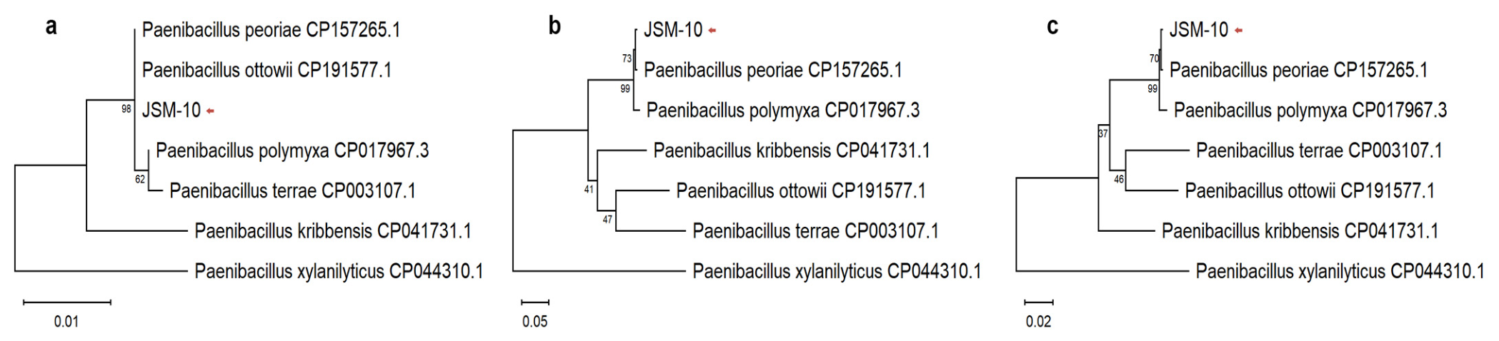
Figure 1. Phylogenetic analysis of strain JSM-10 and closely related Paenibacillus species based on different genetic markers. (a) Maximum Likelihood phylogenetic tree based on 16S rRNA gene sequences. (b) Maximum Likelihood phylogenetic tree based on gyrA gene (gyrA2 fragment) sequences. (c) Maximum Likelihood phylogenetic tree based on concatenated 16S rRNA and gyrA gene (gyrA2 fragment) sequences. Bootstrap values (1000 replicates) are shown at branch nodes. The strain JSM-10 is indicated with a red arrow.
Ijms 27 04062 g001
Figure 2. Morphological characterization of Paenibacillus sp. JSM-10. (a) Colony morphology of JSM-10 grown on YEG medium. (b) Gram-stained cells observed under light microscopy. (c) Phase-contrast microscopy image. (d) Scanning electron microscopy (SEM) image of clusters of rod-shaped cells; (e) SEM of high-magnification image of a dividing cell. (f) Transmission electron microscopy (TEM) image of intracellular ultrastructure. (g) TEM of intracellular components and cellular morphology.
Ijms 27 04062 g002
Figure 3. Growth response (OD620) of Paenibacillus sp. JSM-10 under (a) glyphosate and (b) NaCl stress factors, across concentration gradients. Different letters indicate statistically significant differences between treatments (p < 0.05, one-way ANOVA followed by Tukey’s HSD test).
Ijms 27 04062 g003
Figure 4. Drought tolerance assay (a), optimal temperature range (b), and resistance to heat-shock treatment (c) of glyphosate-tolerant Paenibacillus sp. JSM-10. Different letters indicate statistically significant differences between treatments (p < 0.05, one-way ANOVA followed by Tukey’s HSD test).
Ijms 27 04062 g004
Figure 5. Plant growth-promoting traits of Paenibacillus sp. JSM-10: (a) IAA production on YEG medium, (b) phosphate solubilization on Pikovskaya agar, (c) siderophore production on CAS blue agar medium.
Ijms 27 04062 g005
Figure 6. Root length of buckwheat under normal (NaCl) and saline (NaCl+) conditions. JSM-10 is the combined effect of living culture of Paenibacillus sp. JSM-10 strain and its CCF; MC-I represents CCF from non-inoculated medium; WC-II is distilled H2O. Brackets indicate p-values for pairwise comparisons between treatments across NaCl conditions based on two-way ANOVA followed by Tukey’s HSD test.
Ijms 27 04062 g006
Figure 7. Antagonistic activity of Paenibacillus sp. JSM-10 strain against phytopathogenic fungi B. sorokiniana, N. oryzae, and 6 strains of Alternaria spp. C is the control (growth in the absence of JSM-10), bacterial-treated plates are indicated as “+JSM-10”; I and II represent high- and low-inoculum treatments, respectively.
Ijms 27 04062 g007
Figure 8. PCR-based detection of taxonomic and functional genes in Paenibacillus sp. JSM-10. Lane M: DNA marker (SM0333, ThermoFisher Scientific); lane 1–5: gcd, ipdC, thiO, ectA, and groL, respectively.
Ijms 27 04062 g008
Table 1. Antagonistic potential of Paenibacillus sp. JSM-10 against different pathogenic species.
SampleCD, cm
/OD620
Inhibition Rate, %
Fungal Plant Pathogens, Co-Culture Plate Assay
Bipolaris sorokiniana W-100, C3.98 ± 2.14-
B. sorokiniana W-100 + JSM-10, I2.00 ± 0.1849.7
B. sorokiniana W-100 + JSM-10, II1.43 ± 0.1964.2
Nigrospora oryzae 22/1, C8.10 ± 0.17-
N. oryzae 22/1 + JSM-10, I2.43 ± 0.1270.0
N. oryzae 22/1 + JSM-10, II2.33 ± 0.1571.2
Alternaria spp. W-150, C7.03 ± 0.29-
Alternaria spp. W-150 + JSM-10, I2.33 ± 0.3066.9
Alternaria spp. W-150 + JSM-10, II3.25 ± 0.3353.7
Alternaria spp. 4/1, C5.73 ± 1.37-
Alternaria spp. 4/1 + JSM-10, I2.03 ± 0.3164.5
Alternaria spp. 4/1 + JSM-10, II2.66 ± 0.2153.5
Alternaria spp. 8/7, C3.85 ± 0.07-
Alternaria spp. 8/7 + JSM-10, I1.63 ± 0.2357.6
Alternaria spp. 8/7 + JSM-10, II2.43 ± 0.1536.8
Alternaria spp. 11/1, C6.60 ± 1.14-
Alternaria spp. 11/1 + JSM-10, I2.16 ± 0.2367.2
Alternaria spp. 11/1 + JSM-10, II2.96 ± 0.3155.1
Alternaria spp. 41/1, C5.65 ± 0.64-
Alternaria spp. 41/1 + JSM-10, I1.76 ± 0.4568.7
Alternaria spp. 41/1 + JSM-10, II2.26 ± 0.0659.9
Alternaria spp. 42/1, C6.33 ± 1.53-
Alternaria spp. 42/1 + JSM-10, I1.76 ± 0.1572.1
Alternaria spp. 42/1 + JSM-10, II2.70 ± 0.1057.4
Escherichia coli, kinetic measurements
E. coli, C0.15 ± 0.001-
JSM-10, Ala, 50%0.08 ± 0.00145.1
C-CCF, Ala, 50%0.13 ± 0.01212.6
JSM-10, Ala, 25%0.14 ± 0.0056.8
C-CCF, Ala, 25%0.14 ± 0.0025.1
JSM-10, Glu, 50%0.08 ± 0.00145.4
C-CCF, Glu, 50%0.14 ± 0.0086.8
JSM-10, Glu, 25%0.14 ± 0.0054.1
C-CCF, Glu, 25%0.14 ± 0.0137.8
For fungi, co-culture plate assay: C is the control (growth in the absence of JSM-10), highlighted in bold for convenience, while bacterial-treated plates are indicated as “+JSM-10”; I and II represent high- and low-inoculum treatments, respectively. For E. coli, kinetic assay: C is the untreated control (growth in the absence of CCF), highlighted in bold; C-CCF is the control treated with CCF (growth in the presence of filtrates from non-inoculated MM); JSM-10 denotes treatment with CCF obtained from JSM-10 (growth in the presence of CCF of JSM-10 grown in MM); Ala and Glu are filtrates from MM amended with L-alanine or glucose, respectively; 25 and 50% represent the applied CCF concentrations.
Table 2. List of reported PGPB of Paenibacillus species with beneficial effects on different crops.
Paenibacillus SpeciesTested Crops/PestReported EffectReferences
PGPB Characteristics
Paenibacillus nicotianae AFI2Wheat (Triticum aestivum L.)Increased shoot length by 21.1% under Ni stress[23]
Paenibacillus peoriae MHJL1Cotton (Gossypium hirsutum)Plant growth; increased plant height, root length, stem diameter and fresh weight by 14.12–120.47%[17]
Paenibacillus beijingensis BJ-18Wheat (T. aestivum L.)Increased shoot and root dry weight by 86.1% and 46.0% under low-nitrogen conditions[24]
Maize (Zea mays)Increased shoot and root dry weight by 46.6% and 47.5% under low-nitrogen conditions
Cucumber (Cucumis sativus)Increased shoot and root dry weight by 103.6% and 20.3% under low-nitrogen conditions
Paenibacillus polymyxa ZYPP18Wheat (T. aestivum L.)Enhanced seedling growth and reduced disease incidence by 37.4–65.6%[25]
Paenibacillus sp.Wheat (T. aestivum L.)Increased shoot length by 30.9%[26]
Cucumber
(C. sativus)
Increased shoot and root length by 50.0% and 94.4%
Tomato (Solanum lycopersicum)Increased shoot and root length by 64.6% and 55.2%
P. polymyxa 92Wheat (T. aestivum L.)Increased shoot and root length up to 22% and dry weight up to 28%[27]
Paenibacillus mucilaginosus G78Tomato
(S. lycopersicum)
Increased plant height and fresh weight by 44.1% and 90.0%[28]
Paenibacillus illinoisensis YZ29Peanut
(Arachis hypogaea)
Increased yield by 37.05%[29]
Antagonistic activity
P. peoriae GXUN15128in vitro (plant-pathogenic fungi)Inhibited growth of 10 fungal species by 48.4–86.1% (in vitro)[20]
P. peoriae 3-B4Fusarium verticillioidesInhibited growth by 59.92%[30]
P. polymyxa ZYPP18Rhizoctonia cerealisInhibited fungal growth by 92.68%[25]
Paenibacillus sp.Fusarium graminearumFormed inhibition zones larger than 25 mm[26]
Fusarium solaniFormed inhibition zones of 5–15 mm
Paneibacillus tianmuensis YM002Acidovorax citrulli (cucumber leaves)Formed inhibition zones with diameters ranging from 1.95 to 9.97 mm[31]
P. polymyxa AF01Botrytis cinereaInhibited growth by 78.29%[32]
Bipolaris cactivoraInhibited growth by 60.94%
Fusarium equisetiInhibited growth by 66.28%
P. polymyxa SK1Botryosphaeria dothideaInhibited growth by 66.67%[33]
B. cinereaInhibited growth by 61.19%
Fusarium fujikuroiInhibited growth by 60.71%
Fusarium oxysporumInhibited growth by 55.54%
Paenibacillus jamilae HS-26F. oxysporumInhibited growth by 46.30%[34]
Bipolaris sorokinianaInhibited growth by 63.86%
Rhizoctonia solaniInhibited growth by 44%
Table 3. Molecular detection of functional genes in JSM-10.
GeneAmplicon Size (bp)FunctionReference
PGP Characteristics
gcd601Glucose-1-dehydrogenase;
involved in oxidation of glucose to gluconic acid
[61]
ipdC951Indole-3-pyruvate decarboxylase;
involved in indole-3-acetic acid (IAA) biosynthesis
[42,61]
Abiotic stress tolerance
thiO815Glycine oxidase;
involved in glyphosate degradation
[62]
ectA740Diaminobutyric acid acetyltransferase;
involved in ectoine biosynthesis (osmoprotection)
[63]
groL575Chaperonin GroEL;
assists in protein folding under normal and stress conditions
[63]
Table 4. Primer design and PCR programs for differential genes of JSM-10.
Amplified Genes and Corresponding Primers
F: 5′-3′
R: 3′-5′
PCR ProgramReferences
16S rRNA
Eub 341-F:
CCTACGGGAGGCAGCAG
Eub 1060-R:
CGACACGAGCTGACGACA
95 °C, 2 min (1 cycle);
95 °C, 30 s; 57 °C, 45 s;
72 °C, 1 min (35 cycles);
72 °C, 7 min (1 cycle)
[66]
gyrA fragment gyrA1
FW_434_gyrA_1:
GCATTAACCTCTTGCTCCTTGAAGCGTAT
RV_1326_gyrA_1:
TGGAAGGTTTGGTCAAGGCGCTGAACATTC
95 °C, 30 s (1 cycle);
95 °C, 15 s; 58 °C, 20 s;
72 °C, 58 s (30 cycles),
72 °C, 5 min (1 cycle)
This
study
gyrA fragment gyrA2
FW_1775_gyrA_2:
TCCTTGATGCCTTCGCCACCCATAATGAGTC
RV_2415_gyrA_2:
GTCGGTGACCGCTTTGGCCGATATTCCA
95 °C, 30 s (1 cycle);
95 °C, 15 s; 62 °C, 20 s;
72 °C, 58 s (30 cycles),
72 °C, 5 min (1 cycle)
This
study
rho
FW_rho:
GCCAATAGCATTTCTACCAACAATCCCG
RV_rho:
GTTGTTGCCCACCAGAACTGCTTTG
95 °C, 30 s (1 cycle)
95 °C, 15 s; 56 °C, 20 s;
72 °C, 40 s (32 cycles)
72 °C, 2 min (1 cycle)
This
study
gcd
FW_gcd:
CCCAATGTAAAGAAGTTCCGATTGC
RV_gcd:
CTGACAATGGCTCCTTTGGTAGCTG
95 °C, 30 s (1 cycle)
95 °C, 15 s; 63 °C, 20 s;
72 °C, 40 s (32 cycles)
72 °C, 2 min (1 cycle)
This
study
ipdC
FW_ipdC:
CCTGAAGTTAGGTCAACCAATGAATTAC
RV_ipdC:
CAATGTGGACGATTTTAGCTTGAGGAG
95 °C, 30 s (1 cycle)
95 °C, 15 s; 54 °C, 20 s;
72 °C, 57 s (32 cycles) 72 °C, 2 min (1 cycle)
This
study
thiO
FW_thiO:
CTGAATGCTTGGTCATAGGTGGAGGTG
RV_thiO:
CTACGGTCACTTCTTCGTCATATTGATG
95 °C, 30 s (1 cycle)
95 °C, 15 s; 53 °C, 20 s;
72 °C, 57 s (32 cycles)
72 °C, 2 min (1 cycle)

This
study
ectA
FW_ectA:
CTGTTAAAGTAACACTCGGACCGAAAG
RV_ectA:
CAGCGTTGAATGTACCACGCAGTTTG
98 °C, 3 min (1 cycle)
98 °C, 15 s; 69 °C, 20 s;
72 °C, 18 s (31 cycles)
72 °C, 2 min (1 cycle)
This
study
groL
FW_groL:
CGAACGATGTAGCTGGTGATGGTAC
RV_groL:
CAGCGTTGAATGTACCACGCAGTTTG
This
study
Disclaimer/Publisher’s Note: The statements, opinions and data contained in all publications are solely those of the individual author(s) and contributor(s) and not of MDPI and/or the editor(s). MDPI and/or the editor(s) disclaim responsibility for any injury to people or property resulting from any ideas, methods, instructions or products referred to in the content.