Transcription Factor–Based Classification of Pituitary Neuroendocrine Tumors: Practical Immunohistochemical Algorithms, Molecular Correlates, and Diagnostic Challenges in the 5th WHO Era




Abstract
1. Introduction
2. Historical and Conceptual Background
2.1. Evolution of Pituitary Tumor Classification
2.2. Rationale for Lineage-Based, TF-Centered Classification
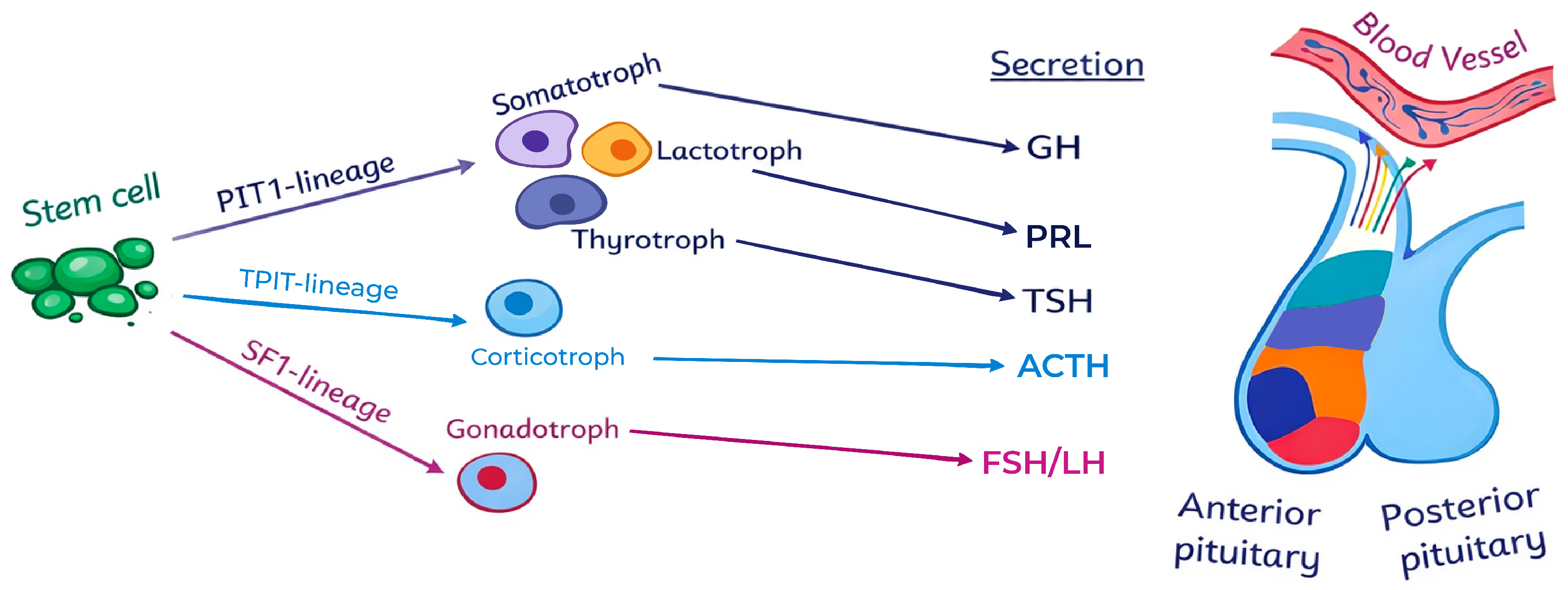
- PIT1-lineage specifies somatotrophs (GH-secreting), lactotrophs (PRL-secreting), and thyrotrophs (TSH-secreting).
- TPIT-lineage specifies corticotrophs (ACTH-secreting).
- SF1-lineage specifies gonadotrophs (FSH/LH-secreting).
- PIT1-lineage aggressive tumors include sparsely granulated somatotroph tumors (more aggressive and often resistant to somatostatin analogue (SSA) therapy than their densely granulated counterparts), acidophil stem cell tumors, and immature PIT1-lineage tumors (formerly classified as silent subtype 3 adenomas with poor differentiation and increased potential for recurrence and invasiveness).
- TPIT-lineage aggressive tumors include silent corticotroph tumors (that are more likely to exhibit enhanced proliferative and invasive capacity compared to silent gonadotroph tumors), and the rare but highly aggressive Crooke cell tumors.
3. Biology of Pituitary Lineage-Defining Transcription Factors
3.1. PIT1 (POU1F1) and PIT1-Lineage Tumors
3.2. TPIT (TBX19) and Corticotroph Tumors
3.3. SF1 (NR5A1) and Gonadotroph Tumors
3.4. Supporting and Additional Markers: GATA3, ERα, Others
- Markers supporting routine lineage assignment:
- 2.
- Developmental and contextual markers used in selected cases
3.5. Link with Germline and Somatic Driver Events
4. Practical TF-Based Immunohistochemical Classification
4.1. Recommended IHC Panels and Technical Issues
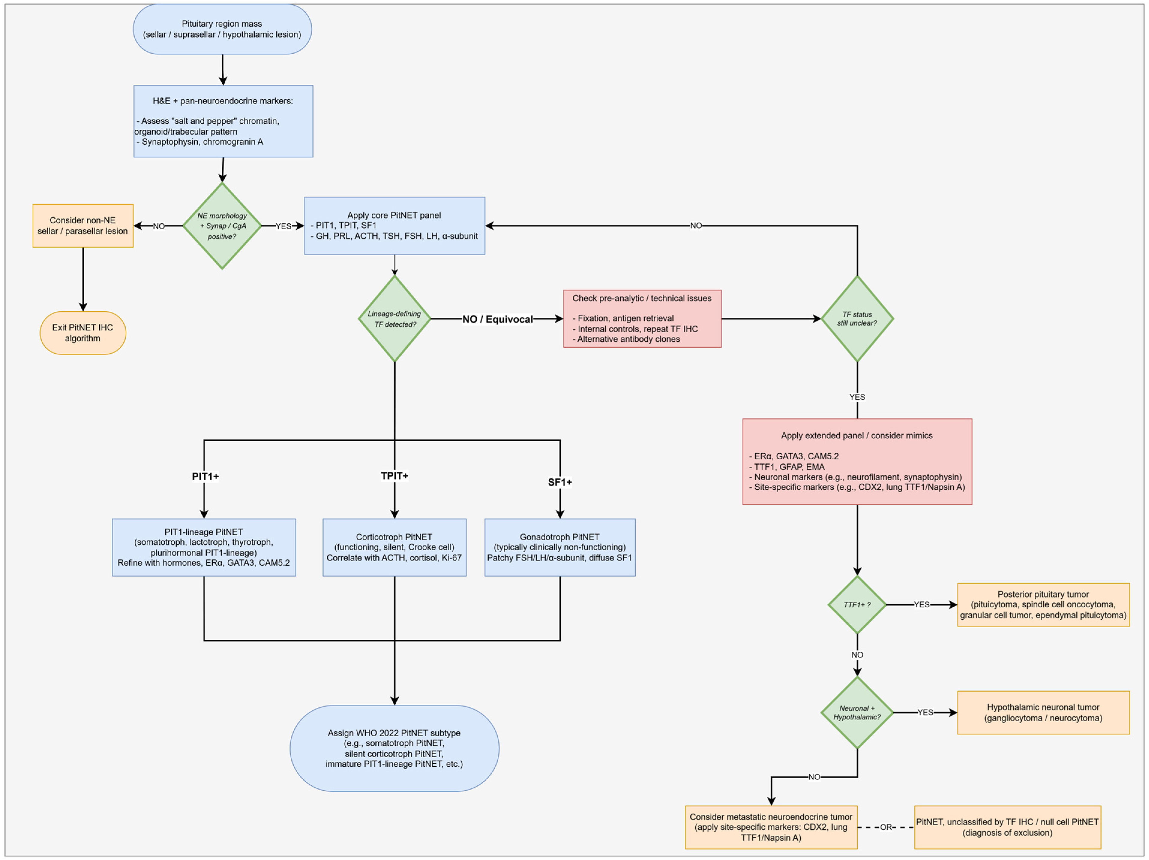
4.2. Stepwise Algorithm for Work-Up of a Pituitary Region Neoplasm
4.3. Handling Discordant or Non-Diagnostic TF Results
4.4. Mapping TF/Hormone Patterns to WHO 5th PitNET Subtypes
4.5. Distinguishing PitNETs from TTF-1–Positive Posterior Pituitary Tumors
5. Molecular and Clinicopathologic Correlates
5.1. Genetic and Epigenetic Features by Lineage
5.2. Proliferation, High-Risk Histotypes, and Aggressiveness
5.3. Treatment Implications
6. Diagnostic Challenges and Pitfalls
6.1. TF-Negative or “Triple Negative” Neoplasms
6.2. Multilineage/Mixed-TF Tumors
6.3. Discordant Hormone and TF Profiles
6.4. Differentiating Lineage-Unassigned PitNETs from Non-PitNET Sellar Tumors
7. Implementation in Reporting and Future Directions
7.1. How to Report: Minimum Dataset and Suggested Wording
- Basic clinical context (where available): patient age and sex, functional status (clinically functioning vs. non-functioning), and main presenting syndrome.
- Tumor site and extent: sellar/suprasellar location, approximate size, and, where assessable, evidence of invasion into adjacent structures (e.g., cavernous sinus, sphenoid).
- Morphology of the adenohypophyseal component: growth pattern (diffuse, papillary, trabecular, etc.), cytologic features (acidophilic, basophilic, chromophobic), granulation pattern (densely vs. sparsely granulated where applicable), and characteristic features such as fibrous bodies or Crooke hyaline change.
- Immunophenotype—core neuroendocrine and lineage markers:
- o
- Pan-neuroendocrine markers (e.g., synaptophysin, chromogranin A).
- o
- Lineage-defining transcription factors (PIT1, TPIT, and SF1), with documentation of their nuclear staining pattern and intensity.
- o
- Auxiliary markers when used (e.g., ERα and GATA3) to support lineage assignment in difficult cases.
- Immunophenotype—pituitary hormones and adjunct markers:
- o
- The six anterior pituitary hormones (GH, PRL, ACTH, TSH, FSH, and LH) and the α-subunit, indicating which are positive and whether staining is diffuse vs. focal [9].
- o
- Proliferation assessment:
- o
- Ki-67 labelling index, measured in hotspots and reported as a percentage (or number of positive cells per mm2) [26].
- o
- Mitotic activity, if counted, is expressed as mitoses per 10 high-power fields.
- Additional markers:
7.2. Impact on Multidisciplinary Management
7.3. Future Perspectives
8. Conclusions
Author Contributions
Funding
Institutional Review Board Statement
Informed Consent Statement
Data Availability Statement
Conflicts of Interest
Abbreviations
| ACTH | Adrenocorticotropic Hormone |
| AIP | Aryl Hydrocarbon Receptor–Interacting Protein |
| ASC | Acidophil Stem Cell |
| ATRX | Alpha Thalassemia/Mental Retardation Syndrome X-Linked |
| BRAF | B-Raf Proto-Oncogene |
| cAMP | Cyclic Adenosine Monophosphate |
| CDX2 | Caudal Type Homeobox 2 |
| DRD2 | Dopamine Receptor D2 |
| EGFR | Epidermal Growth Factor Receptor |
| EMA | Epithelial Membrane Antigen |
| EMT | Epithelial–Mesenchymal Transition |
| ERα | Estrogen Receptor Alpha |
| FSH | Follicle-Stimulating Hormone |
| GATA3 | GATA Binding Protein 3 |
| GCT | Granular Cell Tumor |
| GFAP | Glial Fibrillary Acidic Protein |
| GH | Growth Hormone |
| GNAS | Guanine Nucleotide-binding Protein alpha stimulating |
| H&E | Hematoxylin and Eosin |
| IHC | Immunohistochemistry |
| LH | Luteinizing Hormone |
| LMWCK | Low Molecular Weight Cytokeratin |
| MAPK | Mitogen-Activated Protein Kinase |
| MEG3 | Maternally Expressed Gene 3 |
| MEN1 | Multiple Endocrine Neoplasia Type 1 |
| MGMT | O6-Methylguanine-DNA Methyltransferase |
| MRI | Magnetic Resonance Imaging |
| NEN | Neuroendocrine Neoplasm |
| NF | Non-Functioning |
| NF-PitNET | Non-Functioning Pituitary Neuroendocrine Tumor |
| NR5A1 | Nuclear Receptor Subfamily 5 Group A Member 1 |
| PA | Pituitary Adenoma |
| PAS | Periodic Acid–Schiff |
| PIT1 | Pituitary-Specific Positive Transcription Factor 1 |
| POU1F1 | POU Class 1 Homeobox 1 |
| PitNET | Pituitary Neuroendocrine Tumor |
| PITX1 | Pituitary Homeobox 1 |
| PITX2 | Pituitary Homeobox 2 |
| POMC | Pro-opiomelanocortin |
| PROP1 | Prophet of PIT1 |
| PRL | Prolactin |
| PRLR | Prolactin Receptor |
| PPT | Posterior Pituitary Tumor |
| SCT | Silent Corticotroph Tumor |
| SCO | Spindle Cell Oncocytoma |
| SF1 | Steroidogenic Factor 1 |
| SF3B1 | Splicing Factor 3B Subunit 1 |
| SSTR | Somatostatin Receptor |
| SSTR2 | Somatostatin Receptor Type 2 |
| SSTR5 | Somatostatin Receptor Type 5 |
| SSA | Somatostatin Analogue |
| TBX19 | T-Box Transcription Factor 19 |
| TF | Transcription Factor |
| TP53 | Tumor Protein p53 |
| TPIT | T-Box Pituitary Transcription Factor |
| TSH | Thyroid-Stimulating Hormone |
| TTF-1 | Thyroid Transcription Factor 1 |
| USP8 | Ubiquitin-Specific Protease 8 |
| USP48 | Ubiquitin-Specific Protease 48 |
| V600E | Valine-to-Glutamic Acid Substitution at Codon 600 |
| WHO | World Health Organization |
References
- Asa, S.L.; Mete, O.; Cusimano, M.D.; McCutcheon, I.E.; Perry, A.; Yamada, S.; Nishioka, H.; Casar-Borota, O.; Uccella, S.; La Rosa, S.; et al. Pituitary neuroendocrine tumors: A model for neuroendocrine tumor classification. Mod. Pathol. 2021, 34, 1634–1650. [Google Scholar] [CrossRef] [Scilit]
- Villa, C.; Birtolo, M.F.; Perez-Rivas, L.G.; Righi, A.; Assie, G.; Baussart, B.; Asioli, S. Grading and staging for pituitary neuroendocrine tumors. Brain Pathol. 2024, 35, e13299. [Google Scholar] [CrossRef] [Scilit] [PubMed]
- Dai, C.; Kang, J.; Liu, X.; Yao, Y.; Wang, H.; Wang, R. How to Classify and Define Pituitary Tumors: Recent Advances and Current Controversies. Front. Endocrinol. 2021, 12, 604644. [Google Scholar] [CrossRef] [Scilit]
- Trouillas, J.; Jaffrain-Rea, M.-L.; Vasiljevic, A.; Raverot, G.; Roncaroli, F.; Villa, C. How to Classify Pituitary Neuroendocrine Tumors (PitNET)s in 2020. Cancers 2020, 12, 514. [Google Scholar] [CrossRef] [Scilit]
- Asa, S.L.; Mete, O.; Perry, A.; Osamura, R.Y. Overview of the 2022 WHO Classification of Pituitary Tumors. Endocr. Pathol. 2022, 33, 6–26. [Google Scholar] [CrossRef] [Scilit]
- Casar-Borota, O.; Burman, P.; Lopes, M.B. The 2022 WHO classification of tumors of the pituitary gland: An update on aggressive and metastatic pituitary neuroendocrine tumors. Brain Pathol. 2024, 35, e13302. [Google Scholar] [CrossRef] [Scilit]
- Ho, K.K.Y.; Melmed, S. Pituitary adenomas: Biology, nomenclature and clinical classification. Rev. Endocr. Metab. Disord. 2025, 26, 137–146. [Google Scholar] [CrossRef] [Scilit] [PubMed]
- Chiloiro, S.; De Marinis, L. From Pituitary Adenoma to Pituitary Neuroendocrine Tumors: How Molecular Pathways May Impact the Therapeutic Management? Endocr. Metab. Immune Disord. Drug Targets 2021, 21, 1744–1759. [Google Scholar] [CrossRef] [Scilit]
- Goyal-Honavar, A.; Chacko, G. Practical approaches to diagnosing PitNETs/adenomas based on cell lineage. Brain Pathol. 2024, 35, e13298. [Google Scholar] [CrossRef] [Scilit]
- Kontogeorgos, G. Update on pituitary adenomas in the 2017 World Health Organization classification: Innovations and perspectives. Hormones 2021, 20, 287–291. [Google Scholar] [CrossRef] [Scilit] [PubMed]
- Wan, X.-Y.; Chen, J.; Wang, J.-W.; Liu, Y.-C.; Shu, K.; Lei, T. Overview of the 2022 WHO Classification of Pituitary Adenomas/Pituitary Neuroendocrine Tumors: Clinical Practices, Controversies, and Perspectives. Curr. Med. Sci. 2022, 42, 1111–1118. [Google Scholar] [CrossRef] [Scilit]
- Burcea, I.F.; Năstase, V.-N.; Poiană, C. Pituitary transcription factors in the immunohistochemical and molecular diagnosis of pituitary tumours—A systematic review. Endokrynol. Pol. 2021, 72, 53–63. [Google Scholar] [CrossRef] [Scilit]
- Inomoto, C.; Tahara, S.; Oyama, K.; Kimura, M.; Matsuno, A.; Teramoto, A.; Osamura, R.Y. Molecular, functional, and histopathological classification of the pituitary neuroendocrine neoplasms. Brain Tumor Pathol. 2021, 38, 183–188. [Google Scholar] [CrossRef] [Scilit]
- Kobalka, P.J.; Huntoon, K.; Becker, A.P. Neuropathology of Pituitary Adenomas and Sellar Lesions. Neurosurgery 2021, 88, 900–918. [Google Scholar] [CrossRef] [Scilit]
- Portovedo, S.; Neto, L.V.; Soares, P.; de Carvalho, D.P.; Takiya, C.M.; Miranda-Alves, L. Aggressive nonfunctioning pituitary neuroendocrine tumors. Brain Tumor Pathol. 2022, 39, 183–199. [Google Scholar] [CrossRef] [Scilit]
- Lenders, N.F.; Earls, P.E.; Inder, W.J.; McCormack, A.I. The evolution in pituitary tumour classification: A clinical perspective. Endocr. Oncol. 2023, 3, e220079. [Google Scholar] [CrossRef] [Scilit]
- Asa, S.L.; Mete, O.; Ezzat, S. Genomics and Epigenomics of Pituitary Tumors: What Do Pathologists Need to Know? Endocr. Pathol. 2021, 32, 3–16. [Google Scholar] [CrossRef] [Scilit]
- Drouin, J. The corticotroph cells from early development to tumorigenesis. J. Neuroendocr. 2022, 34, e13147. [Google Scholar] [CrossRef] [Scilit] [PubMed]
- Hinojosa-Amaya, J.M.; Lam-Chung, C.E.; Cuevas-Ramos, D. Recent Understanding and Future Directions of Recurrent Corticotroph Tumors. Front. Endocrinol. 2021, 12, 657382. [Google Scholar] [CrossRef] [Scilit] [PubMed]
- Mouchtouris, N.; Smit, R.D.; Piper, K.; Prashant, G.; Evans, J.J.; Karsy, M. A review of multiomics platforms in pituitary adenoma pathogenesis. Front. Biosci. 2022, 27, 77. [Google Scholar] [CrossRef] [Scilit] [PubMed]
- Relav, L.; Doghman-Bouguerra, M.; Ruggiero, C.; Muzzi, J.C.D.; Figueiredo, B.C.; Lalli, E. Steroidogenic Factor 1, a Goldilocks Transcription Factor from Adrenocortical Organogenesis to Malignancy. Int. J. Mol. Sci. 2023, 24, 3585. [Google Scholar] [CrossRef] [Scilit] [PubMed]
- Zheng, A.C.; Wang, E.J.; Aghi, M.K. Recent advancements in the molecular biology of pituitary adenomas. Expert Rev. Endocrinol. Metab. 2022, 17, 293–304. [Google Scholar] [CrossRef] [Scilit] [PubMed]
- Chang, M.; Yang, C.; Bao, X.; Wang, R. Genetic and Epigenetic Causes of Pituitary Adenomas. Front. Endocrinol. 2021, 11, 596554. [Google Scholar] [CrossRef] [Scilit]
- Möller, K.; Uhlig, R.; Gorbokon, N.; Dum, D.; Menz, A.; Büscheck, F.; Luebke, A.M.; Hube-Magg, C.; Hinsch, A.; Höflmayer, D.; et al. Comparison of INSM1 immunostaining with established neuroendocrine markers synaptophysin and chromogranin A in over 14,000 neuroendocrine and non-neuroendocrine tumors. Mol. Cell. Endocrinol. 2023, 581, 112106. [Google Scholar] [CrossRef] [Scilit]
- Gheorghișan-Gălățeanu, A.-A.; Ilieșiu, A.; Lambrescu, I.M.; Țăpoi, D.A. The Complex Histopathological and Immunohistochemical Spectrum of Neuroendocrine Tumors—An Overview of the Latest Classifications. Int. J. Mol. Sci. 2023, 24, 1418. [Google Scholar] [CrossRef] [Scilit] [PubMed]
- Guaraldi, F.; Ambrosi, F.; Ricci, C.; Di Sciascio, L.; Asioli, S. Histopathology of growth hormone-secreting pituitary tumors: State of the art and new perspectives. Best Pract. Res. Clin. Endocrinol. Metab. 2024, 38, 101894. [Google Scholar] [CrossRef] [Scilit]
- Kontogeorgos, G.; Thodou, E.; Osamura, R.Y.; Lloyd, R.V. High-risk pituitary adenomas and strategies for predicting response to treatment. Hormones 2022, 21, 1–14. [Google Scholar] [CrossRef] [Scilit]
- Iglesias, P. Histopathological Types, Clinical Presentation, Imaging Studies, Treatment Strategies, and Prognosis of Posterior Pituitary Tumors: An Updated Review. J. Clin. Med. 2025, 14, 4553. [Google Scholar] [CrossRef] [Scilit]
- Faraj, C.A.; McCutcheon, I.E.; Gubbiotti, M.A. PIT-1/SF-1 co-expression in pituitary neuroendocrine tumors (PitNETs) with comprehensive review of the literature: How should we best characterize these neoplasms? Ann. Diagn. Pathol. 2024, 74, 152398. [Google Scholar] [CrossRef] [Scilit]
- Uhlig, R.; Dum, D.; Gorbokon, N.; Menz, A.; Büscheck, F.; Luebke, A.M.; Hube-Magg, C.; Hinsch, A.; Höflmayer, D.; Fraune, C.; et al. Synaptophysin and chromogranin A expression analysis in human tumors. Mol. Cell. Endocrinol. 2022, 555, 111726. [Google Scholar] [CrossRef] [Scilit]
- Neou, M.; Villa, C.; Armignacco, R.; Jouinot, A.; Raffin-Sanson, M.-L.; Septier, A.; Letourneur, F.; Diry, S.; Diedisheim, M.; Izac, B.; et al. Pangenomic Classification of Pituitary Neuroendocrine Tumors. Cancer Cell 2020, 37, 123–134.e5. [Google Scholar] [CrossRef] [Scilit] [PubMed]
- Serioli, S.; Agostini, L.; Pietrantoni, A.; Valeri, F.; Costanza, F.; Chiloiro, S.; Buffoli, B.; Piazza, A.; Poliani, P.L.; Peris-Celda, M.; et al. Aggressive PitNETs and Potential Target Therapies: A Systematic Review of Molecular and Genetic Pathways. Int. J. Mol. Sci. 2023, 24, 15719. [Google Scholar] [CrossRef] [Scilit]
- Fleseriu, M.; Varlamov, E.V.; Akirov, A.; Langlois, F.; Petersenn, S.; Melmed, S. Prolactin-secreting adenomas: Pathogenesis, diagnosis, and management. Lancet Diabetes Endocrinol. 2025, 13, 874–890. [Google Scholar] [CrossRef] [Scilit]
- Peverelli, E.; Treppiedi, D.; Mantovani, G. Molecular mechanisms involved in somatostatin receptor regulation in corticotroph tumors: The role of cytoskeleton and USP8 mutations. Endocr. Oncol. 2022, 2, R24–R30. [Google Scholar] [CrossRef] [Scilit]
- Lenders, N.F.; Inder, W.J.; McCormack, A.I. Towards precision medicine for clinically non-functioning pituitary tumours. Clin. Endocrinol. 2021, 95, 398–409. [Google Scholar] [CrossRef] [Scilit] [PubMed]
- Mete, O.; Wenig, B.M. Update from the 5th Edition of the World Health Organization Classification of Head and Neck Tumors: Overview of the 2022 WHO Classification of Head and Neck Neuroendocrine Neoplasms. Head Neck Pathol. 2022, 16, 123–142. [Google Scholar] [CrossRef] [Scilit] [PubMed]
- Rindi, G.; Mete, O.; Uccella, S.; Basturk, O.; La Rosa, S.; Brosens, L.A.A.; Ezzat, S.; de Herder, W.W.; Klimstra, D.S.; Papotti, M.; et al. Overview of the 2022 WHO Classification of Neuroendocrine Neoplasms. Endocr. Pathol. 2022, 33, 115–154. [Google Scholar] [CrossRef] [Scilit]
- Nf, L.; AI, M. Refractory Pit1 plurihormonal tumours and thyrotroph adenomas. Pituitary 2023, 26, 182–186. [Google Scholar] [CrossRef] [Scilit]



| Category | 2004 WHO | 2017 WHO | 2022 WHO (5th ed.) |
|---|---|---|---|
| Terminology | Adenoma | Adenoma vs. tumor vs. PitNET | Pituitary Neuroendocrine Tumor (PitNET) |
| Ihc Basis | Hormonal | Transcription factors (TF) and hormonal | Transcription factors (TF) & hormonal |
| Type/Lineage | ‘Typical’ | SF1 lineage Gonadotroph | SF1 lineage Gonadotroph |
| ‘Atypical’ | TPIT lineage Corticotroph PIT1 lineage Lactotroph (sparsely granulated, densely granulated, ASC) Somatotroph (sparsely granulated, densely granulated, mammosomatotroph, mixed somatotroph–lactotroph) Thyrotroph Plurihormonal (PIT1-positive plurihormonal a, unusual combinations) | TPIT lineage Corticotroph PIT1 lineage Lactotroph (sparsely granulated, densely granulated) Somatotroph (sparsely granulated, densely granulated) Mammosomatotroph b Mixed somatotroph and lactotroph b Thyrotroph Mature plurihormonal PIT1 lineage c Immature PIT1 lineage c Acidophil stem cell b | |
| No Distinct Cell Lineage | Null cell | Null cell | Null cell Plurihormonal |
| Proliferative Markers | Ki-67 > 3% Elevated mitotic index p53 ↑ | Ki-67 > 3% Elevated mitotic index — | — — — |
| Carcinoma/Metastasis | Craniospinal or distant metastases | Craniospinal or distant metastases | Term omitted; replaced with “Metastatic PitNET” c |
| Markers | Primary Function |
|---|---|
| Minimal Panel | |
| PIT1 | Lineage marker for somatotroph, lactotroph, and thyrotroph cells |
| TPIT | Lineage marker for corticotroph cells |
| SF1 | Lineage marker for gonadotroph cells |
| Synaptophysin | Pan-neuroendocrine marker to confirm neuroendocrine differentiation; highly sensitive |
| Chromogranin A | Pan-neuroendocrine marker; less sensitive than synaptophysin |
| Ki-67 (MIB1) | Proliferation marker to assess tumor growth potential |
| Extended Panel | |
| GATA3 | Marker for thyrotroph and gonadotroph |
| ERα | Marker for lactotroph and mammosomatotroph |
| CAM5.2 | Distinguishes high-risk subtypes; detects fibrous bodies of sparsely granulated somatotroph PitNETs |
| PitNET Subtype | Transcription Factor(s) | Hormone IHC Pattern | Clinical/Functional Status | Key Comments |
|---|---|---|---|---|
| Densely Granulated Somatotroph PitNET | PIT1+ | GH+, α-subunit+ | Acromegaly | Acidophilic cells; diffuse GH staining; perinuclear LMWCK; usually responsive to SSA |
| Sparsely Granulated Somatotroph PitNET | PIT1+ | GH weak/focal | Acromegaly or clinically NF | High-risk subtype; chromophobic cells; fibrous bodies (CAM5.2+); aggressive behavior and reduced SSA response |
| Lactotroph PitNET | PIT1+, ERα+ | PRL+ | Hyperprolactinemia | Most are sparsely granulated with juxtanuclear “dot-like” PRL; excellent dopamine agonist response |
| Thyrotroph PitNET | PIT1+, GATA3+ | TSH+, α-subunit+ | Hyperthyroidism (rare) | Often fibrotic or spindle-celled; rare but diagnostically challenging |
| Corticotroph PitNET | TPIT+ | ACTH+ | Cushing’s disease | Basophilic cells; strong PAS positivity; typically, microadenomas |
| Crooke Cell Tumor | TPIT+ | ACTH+ (often weak) | Cushing’s disease or NF | High-risk variant; perinuclear hyaline keratin rings; aggressive and invasive |
| Silent Corticotroph PitNET | TPIT+ | ACTH+ | Clinically NF | High-risk; larger size, invasive growth, higher recurrence rate than gonadotroph tumors |
| Gonadotroph PitNET | SF1+, GATA3+ (±ERα) | FSH/LH/α-subunit (often weak or negative) | Clinically NF | Most common PitNET subtype; diffuse nuclear SF-1 is the most reliable marker |
| Mature Plurihormonal PIT1-Lineage PitNET | PIT1+ | Multiple PIT1-lineage hormones | Variable (often functional) | Well-differentiated cells; distinct from aggressive immature subtype |
| Immature PIT1-Lineage PitNET | PIT1+ (ERα/GATA3 variable) | Multihormonal (focal/patchy) | Clinically NF or mixed | High-risk; lineage infidelity; large, invasive tumors; formerly “silent subtype 3” |
| Acidophil Stem Cell PitNET | PIT1+ | Usually GH+, PRL+ | Acromegaly, Hyperprolactinemia | High-risk; scattered fibrous bodies |
| Null Cell PitNET | PIT1−/TPIT−/SF-1− | No hormone expression | Clinically NF | Diagnosis of exclusion; very rare with modern TF IHC |
| Mixed/plurihormonal PitNET | Multiple combination | Multiple combination | Variable | Variable |
| Lineage/Histotype | Typical Clinical Context | Usual Tumor Size at MRI | MRI Signal and Enhancement Pattern * | Invasion/Recurrence Pattern † |
|---|---|---|---|---|
| PIT1-lineage—densely granulated somatotroph | Acromegaly with biochemically active GH excess | Often microadenoma or small macroadenoma | Usually iso- to mildly hypointense on T1 and iso- to mildly hyperintense on T2; relatively homogeneous, strong contrast enhancement | Less frequent cavernous sinus invasion; higher surgical remission rates compared with sparsely granulated somatotroph PitNETs [27] |
| PIT1-lineage—sparsely granulated somatotroph | Acromegaly; often younger patients; treatment-resistant disease | Typically larger macroadenoma | Macroadenoma, often heterogeneous on T2 (cystic or hyperintense foci); robust enhancement but more irregular due to size and internal heterogeneity | More frequent cavernous sinus invasion and lower rates of gross total resection and biochemical remission than densely granulated tumors [27] |
| PIT1-lineage—lactotroph | Hyperprolactinemia (amenorrhea, galactorrhea, hypogonadism) | Microadenoma or macroadenoma | Signal characteristics are usually similar to other PitNETs (iso- to hypointense on T1, iso- to hyperintense on T2) with homogeneous or mildly heterogeneous enhancement | Invasive macroprolactinomas may extend into the cavernous sinus, but many lesions are resectable or controlled medically [5,20] |
| PIT1-lineage—immature PIT1-lineage/plurihormonal | Often clinically non-functioning or mixed hormone profiles; younger patients | Large macroadenoma or giant adenoma | Bulky, lobulated masses with heterogeneous T2 signal and enhancement, reflecting cystic change, hemorrhage, or necrosis | Frequently radiologically invasive with cavernous sinus and suprasellar extension and high postoperative recurrence rates [5,12,20] |
| TPIT-lineage—functioning corticotroph (Cushing’s disease) | Overt hypercortisolism; ACTH-dependent Cushing’s disease | Often microadenoma; occasionally small macroadenoma | Small, often subtle lesions; iso- or hypointense on T1, variably hyperintense on T2; may enhance less than normal gland on dynamic sequences | Limited local invasion in many cases, but residual/recurrent disease can occur, especially when the lesion is not well visualized preoperatively [14,19] |
| TPIT-lineage—silent corticotroph | Clinically non-functioning macroadenoma | Usually macroadenoma | Large, solid sellar/suprasellar mass with fairly typical PitNET signal (iso- to hypointense T1, iso- to hyperintense T2) and strong, sometimes heterogeneous enhancement | High rate of cavernous sinus invasion and postoperative recurrence compared with other clinically non-functioning PitNETs, especially SF1-lineage tumors [15,19,20] |
| TPIT-lineage—Crooke cell PitNET | Often aggressive corticotroph tumor; may present with Cushing’s disease or as non-functioning mass | Macroadenoma, frequently large | Macroadenoma with variable signal intensity and heterogeneous enhancement; no pathognomonic signal feature but often invasive at presentation | Marked local invasiveness and therapeutic resistance; high recurrence rates even after apparently adequate resection [5,19,20,27] |
| SF1-lineage—gonadotroph PitNET | Clinically non-functioning macroadenoma; symptoms from mass effect | Typically macroadenoma | Conventional PitNET appearance: iso- to hypointense on T1, iso- to moderately hyperintense on T2, with homogeneous or mildly heterogeneous enhancement | Often sizeable at diagnosis but with more indolent clinical courses and lower recurrence rates than silent corticotroph or immature PIT1-lineage PitNETs [14,20,22] |
| Tf-Defined Group | Typical Molecular Drivers/Alterations | High-Risk Histotypes (WHO 5th) | General Therapeutic Considerations |
|---|---|---|---|
| PIT1 Lineage | Somatic (GNAS, SF3B1, PRLR); Germline (AIP, MEN1) | Sparsely granulated somatotroph; Immature PIT1-lineage PitNET; Acidophil stem cell PitNET | SSR–directed therapy (octreotide, pasireotide); GH receptor antagonists (pegvisomant) in resistant acromegaly; dopamine agonists for lactotroph tumors |
| TPit Lineage | USP8, USP48, BRAF, ATRX; additional signaling pathway alterations reported | Silent corticotroph PitNET; Crooke cell tumor | Pasireotide preferred due to SSTR5 expression; emerging molecularly targeted approaches are under investigation in selected cases |
| SF-1 Lineage | No recurrent somatic driver mutations identified; epigenetic alterations (MEG3) | Rare aggressive gonadotroph PitNETs; most clinically non-functioning | Surgery remains the primary therapy; with limited and variable responses to medical therapies |
| TF-Negative/Mixed PITNets | Copy number alterations; TP53 abnormalities reported in aggressive disease | Metastatic PitNET | Temozolomide as first-line systemic therapy; treatment response associated with low MGMT expression |
Disclaimer/Publisher’s Note: The statements, opinions and data contained in all publications are solely those of the individual author(s) and contributor(s) and not of MDPI and/or the editor(s). MDPI and/or the editor(s) disclaim responsibility for any injury to people or property resulting from any ideas, methods, instructions or products referred to in the content. |
© 2026 by the authors. Licensee MDPI, Basel, Switzerland. This article is an open access article distributed under the terms and conditions of the Creative Commons Attribution (CC BY) license.
Share and Cite
Pandit, N.; Wehbeh, Y.; Itani, O.; Kanakis, D. Transcription Factor–Based Classification of Pituitary Neuroendocrine Tumors: Practical Immunohistochemical Algorithms, Molecular Correlates, and Diagnostic Challenges in the 5th WHO Era. Int. J. Mol. Sci. 2026, 27, 2307. https://doi.org/10.3390/ijms27052307
Pandit N, Wehbeh Y, Itani O, Kanakis D. Transcription Factor–Based Classification of Pituitary Neuroendocrine Tumors: Practical Immunohistochemical Algorithms, Molecular Correlates, and Diagnostic Challenges in the 5th WHO Era. International Journal of Molecular Sciences. 2026; 27(5):2307. https://doi.org/10.3390/ijms27052307
Chicago/Turabian StylePandit, Nirmal, Yahya Wehbeh, Omar Itani, and Dimitrios Kanakis. 2026. "Transcription Factor–Based Classification of Pituitary Neuroendocrine Tumors: Practical Immunohistochemical Algorithms, Molecular Correlates, and Diagnostic Challenges in the 5th WHO Era" International Journal of Molecular Sciences 27, no. 5: 2307. https://doi.org/10.3390/ijms27052307
APA StylePandit, N., Wehbeh, Y., Itani, O., & Kanakis, D. (2026). Transcription Factor–Based Classification of Pituitary Neuroendocrine Tumors: Practical Immunohistochemical Algorithms, Molecular Correlates, and Diagnostic Challenges in the 5th WHO Era. International Journal of Molecular Sciences, 27(5), 2307. https://doi.org/10.3390/ijms27052307