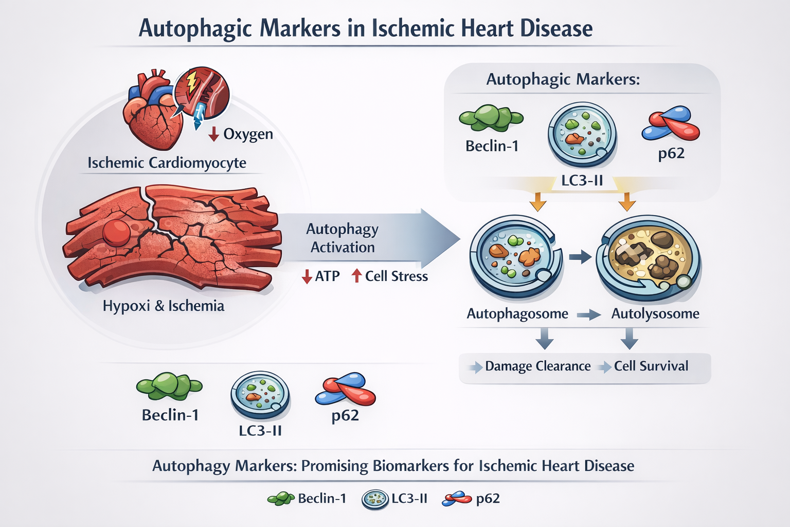
Reading the Signature of Autophagy in the Ischemic and Infarcted Heart: A Systematic Review of Circulating Biomarkers
 , , , , and
Abstract
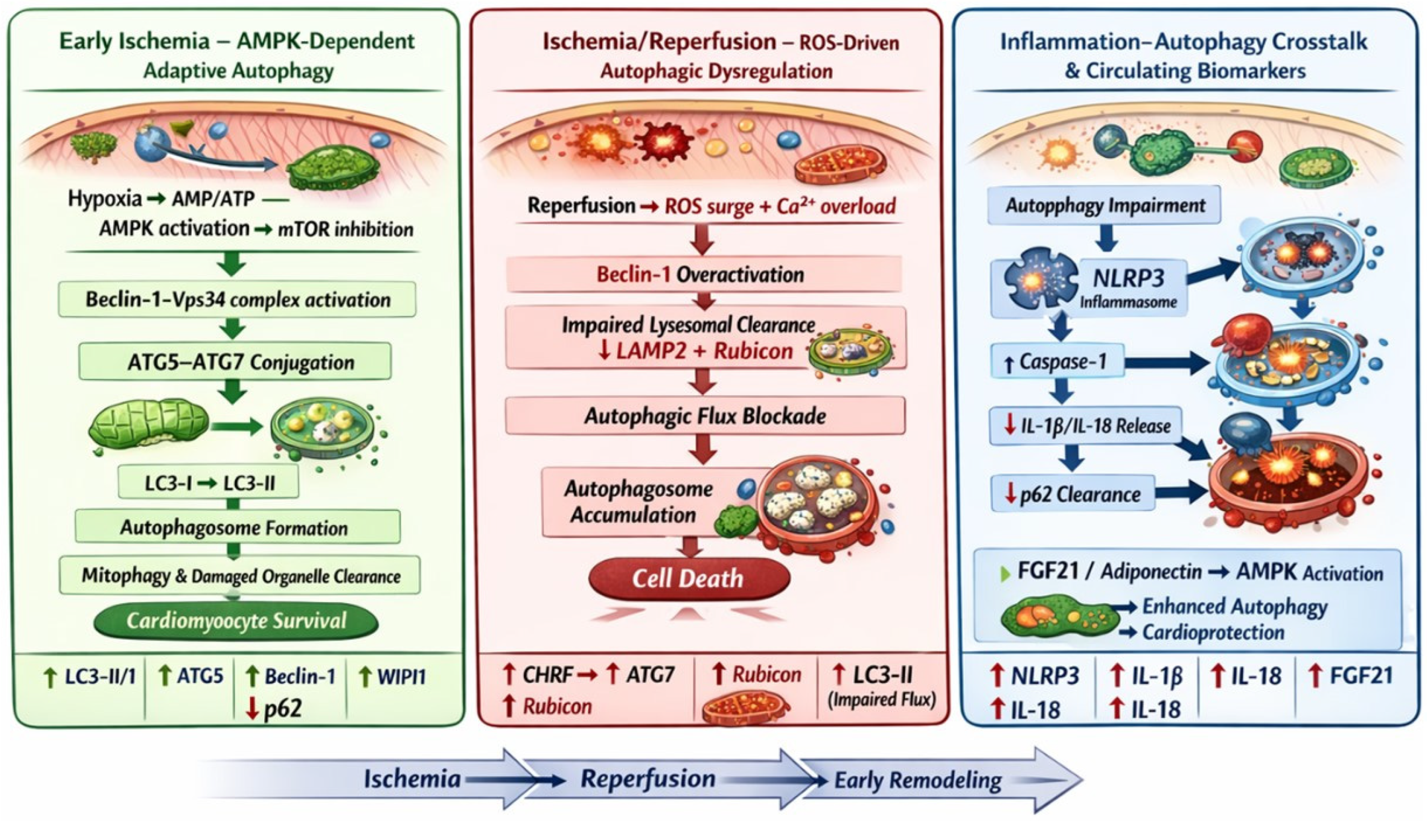
1. Introduction
2. Materials and Methods
2.1. Eligibility Criteria
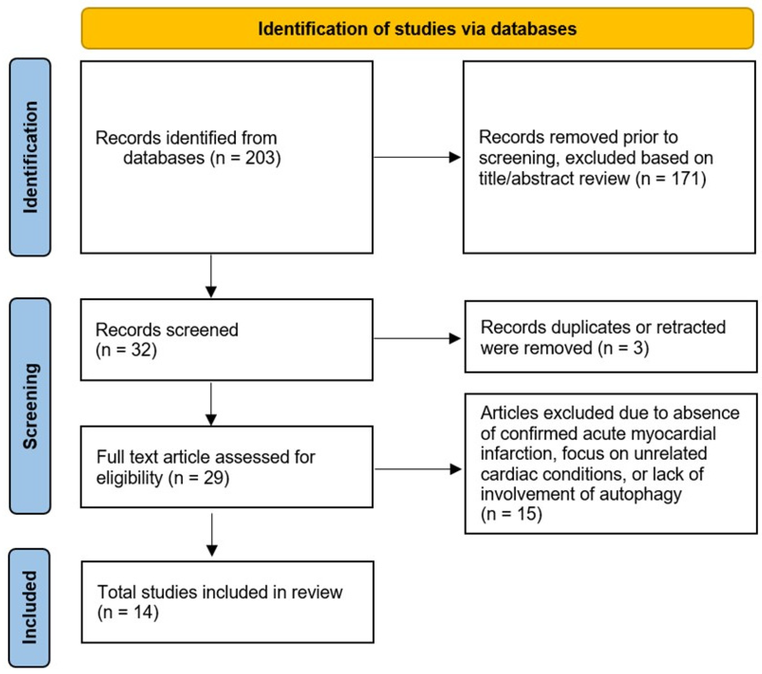
2.2. Study Selection
2.3. Data Extraction
3. Results
3.1. MAP-LC3 (Microtubule-Associated Protein Light Chain 3) (LC3II/I)
3.2. Nuclear Pore Glycoprotein P62
3.3. ATG7 (Autophagy-Related Gene 7)
3.4. CHRF
3.5. NLRP3 (NLR Family Pyrin Domain Containing 3)
3.6. IL-1β and IL-18
3.7. ATG5 (Autophagy-Related Gene 5) and Beclin 1
3.8. FGF21 (Fibroblast Growth Factor 21)
3.9. APN (Adiponectin)
3.10. WIPI1 (WD Repeat Domain Phosphoinositide-Interacting Protein 1)
3.11. Rubicon
4. Discussion
5. Conclusions
Author Contributions
Funding
Institutional Review Board Statement
Informed Consent Statement
Data Availability Statement
Acknowledgments
Conflicts of Interest
Abbreviations
| AMI | Acute Myocardial Infarction |
| APN | Adiponectin |
| ATG | Autophagy-Related Gene |
| CHRF | Cardiac Hypertrophy–Related Factor |
| ELISA | Enzyme-Linked Immunosorbent Assay |
| FGF21 | Fibroblast Growth Factor 21 |
| I/R | Ischemia/Reperfusion |
| IL-1β | Interleukin-1 beta |
| IL-18 | Interleukin 18 |
| LC3 | Light Chain 3 |
| MAP-LC3 | Microtubule-Associated Protein Light Chain 3 |
| WIPI1 | WD Repeat Domain Phosphoinositide-Interacting Protein 1 |
References
- Shahbazi, F.; Moslehi, S.; Mirzaei, Z.; Mohammadi, Y. The Effect of Addressing the Top 10 Global Causes of Death on Life Expectancy in 2019: A Global and Regional Analysis. Int. Health 2025, 17, 517–526. [Google Scholar] [CrossRef] [Scilit]
- Thygesen, K.; Alpert, J.S.; Jaffe, A.S.; Chaitman, B.R.; Bax, J.J.; Morrow, D.A.; White, H.D. Fourth Universal Definition of Myocardial Infarction. J. Am. Coll. Cardiol. 2018, 72, 2231–2264. [Google Scholar] [CrossRef] [Scilit] [PubMed]
- Roth, G.A.; Mensah, G.A.; Johnson, C.O.; Addolorato, G.; Ammirati, E.; Baddour, L.M.; Barengo, N.C.; Beaton, A.Z.; Benjamin, E.J.; Benziger, C.P.; et al. Global Burden of Cardiovascular Diseases and Risk Factors, 1990–2019: Update from the GBD 2019 Study. J. Am. Coll. Cardiol. 2020, 76, 2982–3021, Correction in J. Am. Coll. Cardiol. 2021, 77, 1958–1959. [Google Scholar] [PubMed]
- Ibanez, B.; James, S.; Agewall, S.; Antunes, M.J.; Bucciarelli-Ducci, C.; Bueno, H.; Caforio, A.L.P.; Crea, F.; Goudevenos, J.A.; Halvorsen, S.; et al. 2017 ESC Guidelines for the management of acute myocardial infarction in patients presenting with ST-segment elevation: The Task Force for the management of acute myocardial infarction in patients presenting with ST-segment elevation of the European Society of Cardiology (ESC). Eur. Heart J. 2018, 39, 119–177. [Google Scholar] [CrossRef] [Scilit] [PubMed]
- Mizushima, N.; Levine, B.; Cuervo, A.M.; Klionsky, D.J. Autophagy Fights Disease through Cellular Self-Digestion. Nature 2008, 451, 1069–1075. [Google Scholar] [CrossRef] [Scilit]
- Klionsky, D.J.; Abdel-Aziz, A.K.; Abdelfatah, S.; Abdellatif, M.; Abdoli, A.; Abel, S.; Abeliovich, H.; Abildgaard, M.H.; Abudu, Y.P.; Acevedo-Arozena, A.; et al. Guidelines for the Use and Interpretation of Assays for Monitoring Autophagy (4th Edition). Autophagy 2021, 17, 1–382. [Google Scholar] [CrossRef] [Scilit]
- Kroemer, G.; Mariño, G.; Levine, B. Autophagy and the Integrated Stress Response. Mol. Cell 2010, 40, 280–293. [Google Scholar] [CrossRef] [Scilit]
- Sciarretta, S.; Maejima, Y.; Zablocki, D.; Sadoshima, J. The Role of Autophagy in the Heart. Annu. Rev. Physiol. 2018, 80, 1–26. [Google Scholar] [CrossRef] [Scilit]
- Matsui, Y.; Takagi, H.; Qu, X.; Abdellatif, M.; Sakoda, H.; Asano, T.; Levine, B.; Sadoshima, J. Distinct Roles of Autophagy in the Heart During Ischemia and Reperfusion: Roles of AMP-Activated Protein Kinase and Beclin 1 in Mediating Autophagy. Circ. Res. 2007, 100, 914–922. [Google Scholar] [CrossRef] [Scilit]
- Ma, X.; Liu, H.; Foyil, S.R.; Godar, R.J.; Weinheimer, C.J.; Hill, J.A.; Diwan, A. Impaired Autophagosome Clearance Contributes to Cardiomyocyte Death in Ischemia/Reperfusion Injury. Circulation 2012, 125, 3170–3181. [Google Scholar] [CrossRef] [Scilit]
- Lindahl, B.; Toss, H.; Siegbahn, A.; Venge, P.; Wallentin, L. Markers of Myocardial Damage and Inflammation in Relation to Long-Term Mortality in Unstable Coronary Artery Disease. N. Engl. J. Med. 2000, 343, 1139–1147. [Google Scholar] [CrossRef] [Scilit]
- Sciarretta, S.; Yee, D.; Nagarajan, N.; Bianchi, F.; Saito, T.; Valenti, V.; Tong, M.; Del Re, D.P.; Vecchione, C.; Schirone, L.; et al. Trehalose-Induced Activation of Autophagy Improves Cardiac Remodeling After Myocardial Infarction. J. Am. Coll. Cardiol. 2018, 71, 1999–2010. [Google Scholar] [CrossRef] [Scilit] [PubMed]
- Kanamori, H.; Takemura, G.; Goto, K.; Maruyama, R.; Tsujimoto, A.; Ogino, A.; Takeyama, T.; Kawaguchi, T.; Watanabe, T.; Fujiwara, T.; et al. The Role of Autophagy Emerging in Postinfarction Cardiac Remodelling. Cardiovasc. Res. 2011, 91, 330–339. [Google Scholar] [CrossRef] [Scilit] [PubMed]
- Page, M.J.; McKenzie, J.E.; Bossuyt, P.M.; Boutron, I.; Hoffmann, T.C.; Mulrow, C.D.; Shamseer, L.; Tetzlaff, J.M.; Akl, E.A.; Brennan, S.E.; et al. The PRISMA 2020 Statement: An Updated Guideline for Reporting Systematic Reviews. BMJ Acad. Ed. 2021, 372, 1–9. [Google Scholar]
- Mo, Y.; Wu, H.; Zheng, X.; Xu, L.; Liu, L.; Liu, Z. LncRNA CHRF Aggravates Myocardial Ischemia/Reperfusion Injury by Enhancing Autophagy via Modulation of the miR-182-5p/ATG7 Pathway. J. Biochem. Mol. Toxicol. 2021, 35, e22709. [Google Scholar] [CrossRef] [Scilit]
- Bullón, P.; Cano-García, F.J.; Alcocer-Gómez, E.; Varela-López, A.; Roman-Malo, L.; Ruiz-Salmerón, R.J.; Quiles, J.L.; Navarro-Pando, J.M.; Battino, M.; Ruiz-Cabello, J.; et al. Could NLRP3–Inflammasome Be a Cardiovascular Risk Biomarker in Acute Myocardial Infarction Patients? Antioxid. Redox Signal. 2017, 27, 269–275. [Google Scholar] [CrossRef] [Scilit]
- Kong, M.G.; Suh, J.; Cho, Y.; Park, H.W.; Lee, N.H. Abstract P1050: The Activity of Autophagy Biomarkers in Acute Myocardial Infarction. Circ. Res. 2023, 133, AP1050. [Google Scholar] [CrossRef] [Scilit]
- Grazide, M.-H.; Ruidavets, J.-B.; Martinet, W.; Elbaz, M.; Vindis, C. Association of Circulating Autophagy Proteins ATG5 and Beclin 1 with Acute Myocardial Infarction in a Case-Control Study. Cardiology 2024, 149, 217–224. [Google Scholar] [CrossRef] [Scilit]
- Zhang, W.; Chu, S.; Ding, W.; Wang, F. Serum Level of Fibroblast Growth Factor 21 Is Independently Associated with Acute Myocardial Infarction. PLoS ONE 2015, 10, e0129791. [Google Scholar]
- Sunaga, H.; Koitabashi, N.; Iso, T.; Matsui, H.; Obokata, M.; Kawakami, R.; Murakami, M.; Yokoyama, T.; Kurabayashi, M. Activation of Cardiac AMPK-FGF21 Feed-Forward Loop in Acute Myocardial Infarction: Role of Adrenergic Overdrive and Lipolysis Byproducts. Sci. Rep. 2019, 9, 11841. [Google Scholar] [CrossRef] [Scilit]
- Cheng, J.; Su, X.; Qiao, L.; Zhai, C.; Chen, W. Circulating Level of Fibroblast Growth Factor 21 Is Independently Associated with the Risks of Unstable Angina Pectoris. Biosci. Rep. 2018, 38, BSR20181099. [Google Scholar] [CrossRef] [Scilit]
- Sinha, S.R.; Prakash, P.; Keshari, J.R.; Prasad, R.V. The Correlation Between Serum Fibroblast Growth Factor 21 and the Severity and Occurrence of Coronary Artery Disease. Cureus 2024, 16, e51924. [Google Scholar] [CrossRef] [Scilit]
- Shibata, R.; Sato, K.; Kumada, M.; Izumiya, Y.; Sonoda, M.; Kihara, S.; Ouchi, N.; Walsh, K. Adiponectin Accumulates in Myocardial Tissue That Has Been Damaged by Ischemia-Reperfusion Injury via Leakage from the Vascular Compartment. Cardiovasc. Res. 2007, 74, 471–479. [Google Scholar] [CrossRef] [Scilit] [PubMed]
- Kumada, M.; Kihara, S.; Sumitsuji, S.; Kawamoto, T.; Matsumoto, S.; Ouchi, N.; Arita, Y.; Okamoto, Y.; Shimomura, I.; Hiraoka, H.; et al. Association of Hypoadiponectinemia with Coronary Artery Disease in Men. Arterioscler. Thromb. Vasc. Biol. 2003, 23, 85–89. [Google Scholar] [CrossRef] [Scilit] [PubMed]
- Kojima, S.; Funahashi, T.; Sakamoto, T.; Miyamoto, S.; Soejima, H.; Hokamaki, J.; Kajiwara, I.; Sugiyama, S.; Yoshimura, M.; Fujimoto, K.; et al. The Variation of Plasma Concentrations of a Novel, Adipocyte Derived Protein, Adiponectin, in Patients with Acute Myocardial Infarction. Heart 2003, 89, 667. [Google Scholar] [CrossRef] [Scilit] [PubMed]
- Liu, W.; Shen, J.; Li, Y.; Wu, J.; Luo, X.; Yu, Y.; Zhang, Y.; Gu, L.; Zhang, X.; Jiang, C.; et al. Pyroptosis Inhibition Improves the Symptom of Acute Myocardial Infarction. Cell Death Dis. 2021, 12, 852. [Google Scholar] [CrossRef] [Scilit]
- Grazide, M.-H.; Ruidavets, J.-B.; Martinet, W.; Elbaz, M.; Vindis, C. Circulating Autophagy Regulator Rubicon Is Linked to Increased Myocardial Infarction Risk. J. Mol. Cell Cardiol. Plus 2024, 11, 100279. [Google Scholar]
- Jiang, P.; Mizushima, N. LC3- and P62-Based Biochemical Methods for the Analysis of Autophagy Progression in Mammalian Cells. Methods 2015, 75, 13–18. [Google Scholar] [CrossRef] [Scilit]
- Tanida, I.; Ueno, T.; Kominami, E. LC3 and Autophagy. In Autophagosome and Phagosome; Deretic, V., Ed.; Humana Press: Totowa, NJ, USA, 2008; pp. 77–88. [Google Scholar]
- Komatsu, M.; Kageyama, S.; Ichimura, Y. p62/SQSTM1/A170: Physiology and pathology. Pharmacol. Res. 2012, 66, 457–462. [Google Scholar] [CrossRef] [Scilit]
- Lamark, T.; Svenning, S.; Johansen, T. Regulation of Selective Autophagy: The P62/SQSTM1 Paradigm. Essays Biochem. 2017, 61, 609–624. [Google Scholar] [CrossRef] [Scilit]
- Pankiv, S.; Clausen, T.H.; Lamark, T.; Brech, A.; Bruun, J.-A.; Outzen, H.; Øvervatn, A.; Bjørkøy, G.; Johansen, T. p62/SQSTM1 Binds Directly to Atg8/LC3 to Facilitate Degradation of Ubiquitinated Protein Aggregates by Autophagy. J. Biol. Chem. 2007, 282, 24131–24145. [Google Scholar] [CrossRef] [Scilit] [PubMed]
- Collier, J.J.; Suomi, F.; Oláhová, M.; McWilliams, T.G.; Taylor, R.W. Emerging Roles of ATG7 in Human Health and Disease. EMBO Mol. Med. 2021, 13, e14824. [Google Scholar] [CrossRef] [Scilit] [PubMed]
- Mizushima, N.; Levine, B. Autophagy in Human Diseases. N. Engl. J. Med. 2020, 383, 1564–1576. [Google Scholar] [CrossRef] [Scilit] [PubMed]
- Mizushima, N. The ATG Conjugation Systems in Autophagy. Curr. Opin. Cell Biol. 2020, 63, 1–10. [Google Scholar] [CrossRef] [Scilit]
- Wang, K.; Liu, F.; Zhou, L.-Y.; Long, B.; Yuan, S.-M.; Wang, Y.; Liu, C.-Y.; Sun, T.; Zhang, X.-J.; Li, P.-F. The Long Noncoding RNA CHRF Regulates Cardiac Hypertrophy by Targeting miR-489. Circ. Res. 2014, 114, 1377–1388. [Google Scholar] [CrossRef] [Scilit]
- Xie, X.; Zhao, W.; Pang, J.; Xiong, X.; Wang, H.; Ma, L. Long Non-coding RNA, CHRF, Predicts Poor Prognosis of Lung Adenocarcinoma and Promotes Cell Proliferation and Migration. Oncol. Lett. 2018, 16, 6245–6252. [Google Scholar] [CrossRef] [Scilit]
- Swanson, K.V.; Deng, M.; Ting, J.P.-Y. The NLRP3 Inflammasome: Molecular Activation and Regulation to Therapeutics. Nat. Rev. Immunol. 2019, 19, 477–489. [Google Scholar] [CrossRef] [Scilit]
- He, Y.; Hara, H.; Núñez, G. Mechanism and Regulation of NLRP3 Inflammasome Activation. Trends Biochem. Sci. 2016, 41, 1012–1021. [Google Scholar] [CrossRef] [Scilit]
- Toldo, S.; Abbate, A. The NLRP3 Inflammasome in Acute Myocardial Infarction. Nat. Rev. Cardiol. 2018, 15, 203–214. [Google Scholar] [CrossRef] [Scilit]
- Orogo, A.M.; Gustafsson, Å.B. Therapeutic Targeting of Autophagy. Circ. Res. 2015, 116, 489–503. [Google Scholar] [CrossRef] [Scilit]
- Li, P.-L. Cardiovascular Pathobiology of Inflammasomes: Inflammatory Machinery and Beyond. Antioxid. Redox Signal. 2015, 22, 1079–1083. [Google Scholar] [CrossRef] [Scilit] [PubMed]
- Progatzky, F.; Sangha, N.J.; Yoshida, N.; McBrien, M.; Cheung, J.; Shia, A.; Scott, J.; Marchesi, J.R.; Lamb, J.R.; Bugeon, L.; et al. Dietary Cholesterol Directly Induces Acute Inflammasome-Dependent Intestinal Inflammation. Nat. Commun. 2014, 5, 5864. [Google Scholar] [CrossRef] [Scilit] [PubMed]
- Pierdominici, M.; Vomero, M.; Barbati, C.; Colasanti, T.; Maselli, A.; Vacirca, D.; Giovannetti, A.; Malorni, W.; Ortona, E. Role of Autophagy in Immunity and Autoimmunity, with a Special Focus on Systemic Lupus Erythematosus. FASEB J. 2012, 26, 1400–1412. [Google Scholar] [CrossRef] [Scilit] [PubMed]
- Kim, M.; Sandford, E.; Gatica, D.; Qiu, Y.; Liu, X.; Zheng, Y.; Schulman, B.A.; Xu, J.; Semple, I.; Ro, S.-H.; et al. Mutation in ATG5 Reduces Autophagy and Leads to Ataxia with Developmental Delay. eLife 2016, 5, e12245. [Google Scholar] [CrossRef] [Scilit]
- Ye, X.; Zhou, X.-J.; Zhang, H. Exploring the Role of Autophagy-Related Gene 5 (ATG5) Yields Important Insights Into Autophagy in Autoimmune/Autoinflammatory Diseases. Front. Immunol. 2018, 9, 2334. [Google Scholar]
- Mayes, M.D.; Bossini-Castillo, L.; Gorlova, O.; Martin, J.E.; Zhou, X.; Chen, W.V.; Assassi, S.; Ying, J.; Tan, F.K.; Arnett, F.C.; et al. Immunochip Analysis Identifies Multiple Susceptibility Loci for Systemic Sclerosis. Am. J. Hum. Genet. 2014, 94, 47–61. [Google Scholar] [CrossRef] [Scilit]
- Changotra, H.; Kaur, S.; Yadav, S.S.; Gupta, G.L.; Parkash, J.; Duseja, A. ATG5: A Central Autophagy Regulator Implicated in Various Human Diseases. Cell Biochem. Funct. 2022, 40, 650–667. [Google Scholar] [CrossRef] [Scilit]
- Maejima, Y.; Isobe, M.; Sadoshima, J. Regulation of Autophagy by Beclin 1 in the Heart. J. Mol. Cell. Cardiol. 2016, 95, 19–25. [Google Scholar] [CrossRef] [Scilit]
- Valentim, L.; Laurence, K.M.; Townsend, P.A.; Carroll, C.J.; Soond, S.; Scarabelli, T.M.; Knight, R.A.; Latchman, D.S.; Stephanou, A. Urocortin Inhibits Beclin1-Mediated Autophagic Cell Death in Cardiac Myocytes Exposed to Ischaemia/Reperfusion Injury. J. Mol. Cell. Cardiol. 2006, 40, 846–852. [Google Scholar] [CrossRef] [Scilit]
- Hariharan, N.; Zhai, P.; Sadoshima, J. Oxidative Stress Stimulates Autophagic Flux During Ischemia/Reperfusion. Antioxid. Redox Signal. 2011, 14, 2179–2190. [Google Scholar] [CrossRef] [Scilit]
- Planavila, A.; Redondo-Angulo, I.; Villarroya, F. FGF21 and Cardiac Physiopathology. Front. Endocrinol. 2015, 6, 133. [Google Scholar] [CrossRef] [Scilit]
- Flippo, K.H.; Potthoff, M.J. Metabolic Messengers: FGF21. Nat. Metab. 2021, 3, 309–317. [Google Scholar] [CrossRef] [Scilit] [PubMed]
- Fisher, F.M.; Maratos-Flier, E. Understanding the Physiology of FGF21. Annu. Rev. Physiol. 2016, 78, 223–241. [Google Scholar] [CrossRef] [Scilit] [PubMed]
- Markan, K.R.; Naber, M.C.; Ameka, M.K.; Anderegg, M.D.; Mangelsdorf, D.J.; Kliewer, S.A.; Mohammadi, M.; Potthoff, M.J. Circulating FGF21 Is Liver Derived and Enhances Glucose Uptake During Refeeding and Overfeeding. Diabetes 2014, 63, 4057–4063. [Google Scholar] [CrossRef] [Scilit] [PubMed]
- Badman, M.K.; Pissios, P.; Kennedy, A.R.; Koukos, G.; Flier, J.S.; Maratos-Flier, E. Hepatic Fibroblast Growth Factor 21 Is Regulated by PPARα and Is a Key Mediator of Hepatic Lipid Metabolism in Ketotic States. Cell Metab. 2007, 5, 426–437. [Google Scholar] [CrossRef] [Scilit]
- Uebanso, T.; Taketani, Y.; Yamamoto, H.; Amo, K.; Ominami, H.; Arai, H.; Takei, Y.; Masuda, M.; Tanimura, A.; Harada, N.; et al. Paradoxical Regulation of Human FGF21 by Both Fasting and Feeding Signals: Is FGF21 a Nutritional Adaptation Factor? PLoS ONE 2011, 6, e22976. [Google Scholar] [CrossRef] [Scilit]
- Yamamoto, T.; Sadoshima, J. Protection of the Heart Against Ischemia/Reperfusion by Silent Information Regulator 1. Trends Cardiovasc. Med. 2011, 21, 27–32. [Google Scholar] [CrossRef] [Scilit]
- Gómez-Sámano, M.Á.; Grajales-Gómez, M.; Zuarth-Vázquez, J.M.; Navarro-Flores, M.F.; Martínez-Saavedra, M.; Juárez-León, Ó.A.; Morales-García, M.G.; Enríquez-Estrada, V.M.; Gómez-Pérez, F.J.; Cuevas-Ramos, D. Fibroblast Growth Factor 21 and Its Novel Association with Oxidative Stress. Redox Biol. 2017, 11, 335–341. [Google Scholar] [CrossRef] [Scilit]
- Zhang, Y.; Liu, D.; Long, X.-X.; Fang, Q.-C.; Jia, W.-P.; Li, H.-T. The Role of FGF21 in the Pathogenesis of Cardiovascular Disease. Chin. Med. J. 2021, 134, 2931–2943. [Google Scholar]
- Ren, Z.; Xiao, W.; Zeng, Y.; Liu, M.-H.; Li, G.-H.; Tang, Z.-H.; Qu, S.-L.; Hao, Y.-M.; Yuan, H.-Q.; Jiang, Z.-S. Fibroblast Growth Factor-21 Alleviates Hypoxia/Reoxygenation Injury in H9c2 Cardiomyocytes by Promoting Autophagic Flux. Int. J. Mol. Med. 2019, 43, 1321–1330. [Google Scholar]
- Chen, F.; Zhan, J.; Yan, X.; Mamun, A.A.; Zhang, Y.; Xu, Y.; Zhang, H.; Li, X.; Zhou, K.; Xiao, J. FGF21 Alleviates Microvascular Damage Following Limb Ischemia/Reperfusion Injury by TFEB-Mediated Autophagy Enhancement and Anti-Oxidative Response. Signal Transduct. Target. Ther. 2022, 7, 349. [Google Scholar] [CrossRef] [Scilit] [PubMed]
- Choi, A.M.K.; Ryter, S.W.; Levine, B. Autophagy in Human Health and Disease. N. Engl. J. Med. 2013, 368, 651–662. [Google Scholar] [CrossRef] [Scilit] [PubMed]
- Liu, S.Q.; Tefft, B.J.; Roberts, D.T.; Zhang, L.-Q.; Ren, Y.; Li, Y.C.; Huang, Y.; Zhang, D.; Phillips, H.R.; Wu, Y.H. Cardioprotective Proteins Upregulated in the Liver in Response to Experimental Myocardial Ischemia. Am. J. Physiol. Heart Circ. Physiol. 2012, 303, H1446–H1458. [Google Scholar] [CrossRef] [Scilit] [PubMed]
- Kawano, J.; Arora, R. The Role of Adiponectin in Obesity, Diabetes, and Cardiovascular Disease. J. Cardiometab. Syndr. 2009, 4, 44–49. [Google Scholar] [CrossRef] [Scilit]
- Hopkins, T.A.; Ouchi, N.; Shibata, R.; Walsh, K. Adiponectin Actions in the Cardiovascular System. Cardiovasc. Res. 2007, 74, 11–18. [Google Scholar] [CrossRef] [Scilit]
- Ren, J.; Xu, X.; Wang, Q.; Ren, S.Y.; Dong, M.; Zhang, Y. Permissive Role of AMPK and Autophagy in Adiponectin Deficiency-Accentuated Myocardial Injury and Inflammation in Endotoxemia. J. Mol. Cell. Cardiol. 2016, 93, 18–31. [Google Scholar] [CrossRef] [Scilit]
- Lopaschuk, G.D.; Folmes, C.D.L.; Stanley, W.C. Cardiac Energy Metabolism in Obesity. Circ. Res. 2007, 101, 335–347. [Google Scholar] [CrossRef] [Scilit]
- Essick, E.E.; Wilson, R.M.; Pimentel, D.R.; Shimano, M.; Baid, S.; Ouchi, N.; Sam, F. Adiponectin Modulates Oxidative Stress-Induced Autophagy in Cardiomyocytes. PLoS ONE 2013, 8, e68697. [Google Scholar] [CrossRef] [Scilit]
- Kamareddine, L.; Ghantous, C.M.; Allouch, S.; Al-Ashmar, S.A.; Anlar, G.; Kannan, S.; Djouhri, L.; Korashy, H.M.; Agouni, A.; Zeidan, A. Between Inflammation and Autophagy: The Role of Leptin-Adiponectin Axis in Cardiac Remodeling. J. Inflamm. Res. 2021, 14, 5349–5365. [Google Scholar] [CrossRef] [Scilit]
- Sung, H.K.; Tang, J.; Jahng, J.W.S.; Song, E.; Chan, Y.K.; Lone, A.H.; Peterson, J.; Abdul-Sater, A.; Sweeney, G. Ischemia-Induced Cardiac Dysfunction Is Exacerbated in Adiponectin-Knockout Mice Due to Impaired Autophagy Flux. Clin. Transl. Sci. 2024, 17, e13758. [Google Scholar] [CrossRef] [Scilit]
- Liu, D.; Zhou, L.; Xu, B.; Tse, G.; Shao, Q.; Liu, T. WIPI1-Mediated Mitophagy Dysfunction in Ventricular Remodeling Associated with Long-Term Diabetes Mellitus. Cell. Signal. 2025, 130, 111663. [Google Scholar] [CrossRef] [Scilit] [PubMed]
- Almannai, M.; Marafi, D.; El-Hattab, A.W. WIPI Proteins: Biological Functions and Related Syndromes. Front. Mol. Neurosci. 2022, 15, 1011918. [Google Scholar] [CrossRef] [Scilit] [PubMed]
- Tzimas, C.; Rau, C.D.; Buergisser, P.E.; Jean-Louis, G.; Lee, K.; Chukwuneke, J.; Dun, W.; Wang, Y.; Tsai, E.J. WIPI1 Is a Conserved Mediator of Right Ventricular Failure. JCI Insight 2019, 5, 122929. [Google Scholar] [CrossRef] [Scilit]
- Galluzzi, L.; Green, D.R. Autophagy-Independent Functions of the Autophagy Machinery. Cell 2019, 177, 1682–1699. [Google Scholar] [CrossRef] [Scilit] [PubMed]
- Minami, S.; Nakamura, S.; Yoshimori, T. Rubicon in Metabolic Diseases and Ageing. Front. Cell Dev. Biol. 2022, 9, 816829. [Google Scholar] [CrossRef] [Scilit] [PubMed]
- Heckmann, B.L.; Teubner, B.J.W.; Tummers, B.; Boada-Romero, E.; Harris, L.; Yang, M.; Guy, C.S.; Zakharenko, S.S.; Green, D.R. LC3-Associated Endocytosis Facilitates β-Amyloid Clearance and Mitigates Neurodegeneration in Murine Alzheimer’s Disease. Cell 2019, 178, 536–551.e14. [Google Scholar] [CrossRef] [Scilit]
- Mizushima, N.; Komatsu, M. Autophagy: Renovation of Cells and Tissues. Cell 2011, 147, 728–741. [Google Scholar] [CrossRef] [Scilit]
- Deretic, V.; Saitoh, T.; Akira, S. Autophagy in Infection, Inflammation and Immunity. Nat. Rev. Immunol. 2013, 13, 722–737. [Google Scholar] [CrossRef] [Scilit]
- Wada, Y.; Sun-Wada, G.-H.; Kawamura, N.; Aoyama, M. Role of Autophagy in Embryogenesis. Curr. Opin. Genet. Dev. 2014, 27, 60–66. [Google Scholar] [CrossRef] [Scilit]
- Kobayashi, S. Choose Delicately and Reuse Adequately: The Newly Revealed Process of Autophagy. Biol. Pharm. Bull. 2015, 38, 1098–1103. [Google Scholar] [CrossRef] [Scilit]
- Levine, B.; Kroemer, G. Autophagy in the Pathogenesis of Disease. Cell 2008, 132, 27–42. [Google Scholar] [CrossRef] [Scilit] [PubMed]
- Shirakabe, A.; Ikeda, Y.; Sciarretta, S.; Zablocki, D.K.; Sadoshima, J. Aging and Autophagy in the Heart. Circ. Res. 2016, 118, 1563–1576. [Google Scholar] [CrossRef] [Scilit] [PubMed]
- Hamacher-Brady, A.; Brady, N.R.; Gottlieb, R.A. Enhancing Macroautophagy Protects against Ischemia/Reperfusion Injury in Cardiac Myocytes. J. Biol. Chem. 2006, 281, 29776–29787. [Google Scholar] [CrossRef] [Scilit] [PubMed]
- Gustafsson, Å.B.; Gottlieb, R.A. Autophagy in Ischemic Heart Disease. Circ. Res. 2009, 104, 150–158. [Google Scholar] [CrossRef] [Scilit]
- Pischon, T.; Girman, C.J.; Hotamisligil, G.S.; Rifai, N.; Hu, F.B.; Rimm, E.B. Plasma Adiponectin Levels and Risk of Myocardial Infarction in Men. JAMA 2004, 291, 1730–1737. [Google Scholar] [CrossRef] [Scilit]
- Kakavand, H.; Aghakouchakzadeh, M.; Coons, J.C.; Talasaz, A.H. Pharmacologic Prevention of Myocardial Ischemia–Reperfusion Injury in Patients with Acute Coronary Syndrome Undergoing Percutaneous Coronary Intervention. J. Cardiovasc. Pharmacol. 2021, 77, 430. [Google Scholar] [CrossRef] [Scilit]
- Ye, Y.; Perez-Polo, J.R.; Aguilar, D.; Birnbaum, Y. The Potential Effects of Anti-Diabetic Medications on Myocardial Ischemia–Reperfusion Injury. Basic Res. Cardiol. 2011, 106, 925–952. [Google Scholar] [CrossRef] [Scilit]
- Ibáñez, B.; Heusch, G.; Ovize, M.; Van de Werf, F. Evolving Therapies for Myocardial Ischemia/Reperfusion Injury. J. Am. Coll. Cardiol. 2015, 65, 1454–1471. [Google Scholar] [CrossRef] [Scilit]
- Sharma, V.; Bell, R.M.; Yellon, D.M. Targeting Reperfusion Injury in Acute Myocardial Infarction: A Review of Reperfusion Injury Pharmacotherapy. Expert Opin. Pharmacother. 2012, 13, 1153–1175. [Google Scholar] [CrossRef] [Scilit]
- Lujan, H.L.; DiCarlo, S.E. Reperfusion-induced sustained ventricular tachycardia, leading to ventricular fibrillation, in chronically instrumented, intact, conscious mice. Physiol. Rep. 2014, 2, e12057. [Google Scholar] [CrossRef] [Scilit]
- Wu, X.; Wu, M.; Huang, H.; Liu, Z.; Huang, H.; Wang, L. Incidence, predictors, and prognostic impact of reperfusion-related ventricular arrhythmias in STEMI patients undergoing primary percutaneous coronary intervention. BMC Cardiovasc. Disord. 2025, 25, 1–11. [Google Scholar] [CrossRef] [Scilit]
- Hausenloy, D.J.; Yellon, D.M. Myocardial ischemia-reperfusion injury: A neglected therapeutic target. J. Clin. Investig. 2013, 123, 92–100. [Google Scholar] [CrossRef] [Scilit] [PubMed]
- Sciarretta, S.; Hariharan, N.; Monden, Y.; Zablocki, D.; Sadoshima, J. Is autophagy in response to ischemia and reperfusion protective or detrimental for the heart? Pediatr. Cardiol. 2011, 32, 275–281. [Google Scholar] [CrossRef] [Scilit] [PubMed]
- Lin, X.L.; Xiao, W.J.; Xiao, L.L.; Liu, M.H. Molecular mechanisms of autophagy in cardiac ischemia/reperfusion injury. Mol. Med. Rep. 2018, 18, 675–683. [Google Scholar] [CrossRef] [Scilit] [PubMed]
- D’arcy, M.S. Cell death: A review of the major forms of apoptosis, necrosis and autophagy. Cell Biol. Int. 2019, 43, 582–592. [Google Scholar] [CrossRef] [Scilit]
- Jain, M.V.; Paczulla, A.M.; Klonisch, T.; Dimgba, F.N.; Rao, S.B.; Roberg, K.; Schweizer, F.; Lengerke, C.; Davoodpour, P.; Palicharla, V.R.; et al. Interconnections between Apoptotic, Autophagic and Necrotic Pathways: Implications for Cancer Therapy Development. J. Cell. Mol. Med. 2013, 17, 12–29. [Google Scholar] [CrossRef] [Scilit]
- Song, S.; Tan, J.; Miao, Y.; Li, M.; Zhang, Q. Crosstalk of Autophagy and Apoptosis: Involvement of the Dual Role of Autophagy under ER Stress. J. Cell. Physiol. 2017, 232, 2977–2984. [Google Scholar] [CrossRef] [Scilit]
- Chandrika, B.B.; Yang, C.; Ou, Y.; Feng, X.; Muhoza, D.; Holmes, A.F.; Theus, S.; Deshmukh, S.; Haun, R.S.; Kaushal, G.P. Endoplasmic Reticulum Stress-Induced Autophagy Provides Cytoprotection from Chemical Hypoxia and Oxidant Injury and Ameliorates Renal Ischemia-Reperfusion Injury. PLoS ONE 2015, 10, e0140025. [Google Scholar] [CrossRef] [Scilit]
- Liu, S.; Yao, S.; Yang, H.; Liu, S.; Wang, Y. Autophagy: Regulator of Cell Death. Cell Death Dis. 2023, 14, 648. [Google Scholar] [CrossRef] [Scilit]
- Chmurska, A.; Matczak, K.; Marczak, A. Two Faces of Autophagy in the Struggle against Cancer. Int. J. Mol. Sci. 2021, 22, 2981. [Google Scholar] [CrossRef] [Scilit]
- Li, M.; Gao, P.; Zhang, J. Crosstalk between Autophagy and Apoptosis: Potential and Emerging Therapeutic Targets for Cardiac Diseases. Int. J. Mol. Sci. 2016, 17, 332. [Google Scholar] [CrossRef] [Scilit]
- Kang, R.; Zeh, H.J.; Lotze, M.T.; Tang, D. The Beclin 1 Network Regulates Autophagy and Apoptosis. Cell Death Differ. 2011, 18, 571–580. [Google Scholar] [CrossRef] [Scilit]
- Wirawan, E.; Vande Walle, L.; Kersse, K.; Cornelis, S.; Claerhout, S.; Vanoverberghe, I.; Roelandt, R.; De Rycke, R.; Verspurten, J.; Declercq, W.; et al. Caspase-Mediated Cleavage of Beclin-1 Inactivates Beclin-1-Induced Autophagy and Enhances Apoptosis by Promoting the Release of Proapoptotic Factors from Mitochondria. Cell Death Dis. 2010, 1, e18. [Google Scholar] [CrossRef] [Scilit]
- Liu, Y.; Liu, T.; Lei, T.; Zhang, D.; Du, S.; Girani, L.; Qi, D.; Lin, C.; Tong, R.; Wang, Y. RIP1/RIP3-Regulated Necroptosis as a Target for Multifaceted Disease Therapy (Review). Int. J. Mol. Med. 2019, 44, 771–786. [Google Scholar] [CrossRef] [Scilit]


| Articles | Biomarker | Sample Source | Tissue Type | Methods | Results | |
|---|---|---|---|---|---|---|
| 1 | Mo, Yipeng et al. [15] | CHRF | Male Sprague–Dawley rats (n = 6 per group, total 5 groups) | Myocardial tissue | RT-qPCR, RNA pull-down, luciferase assay, TUNEL, TTC staining, MTT assay | Upregulation of CHRF in ischemia–reperfusion injury (I/R). Silencing CHRF leads to decrease in the size of infarct, LDH, apoptosis, and autophagy markers, which indicate a protective effect. |
| ATG7 | H9C2 rat | Cardiomyoblast cells | RT-qPCR, Western blot, luciferase assay | ATG7 is a direct target of miR-182-5p. CHRF indirectly increases ATG7. Overexpression of ATG7 restores autophagy even when CHRF is silenced. | ||
| LC3-II/I, p62 | H9C2 rat | Cardiomyoblast cells | Western blot, GFP-LC3 immunofluorescence | LC3-II/I is increased and p62 decreased under I/R (indicating enhanced autophagy). These changes are reversed by CHRF knockdown. | ||
| 2 | Bullón, Pedro, et al. [16] | NLRP3 | H9C2 rat | Cardiomyoblast cells | qPCR (SYBR Green) | Higher mRNA expression in AMI patients compared to controls. Indicates inflammation and autophagy impairment in cardiac ischemia. |
| MAP-LC3 | AMI patients 6 months after the attack (n = 150) and controls (109 healthy men) | Blood mononuclear cells | qPCR (SYBR Green) | Lower gene expression in AMI group, suggesting reduced autophagic activity. | ||
| IL-1β | AMI patients 6 months after the attack (n = 150) and controls (109 healthy men) | Blood mononuclear cells | ELISA | Elevated IL-1β in AMI patients; correlated with inflammasome activation and cardiovascular risk. | ||
| 3 | Kong, Min Gyu, et al. [17] |
| AMI patients 6 months after the attack (n = 150) and controls (109 healthy men) | Blood mononuclear cells | ELISA | LC3-II, ATG5, and Beclin-1 levels are significantly higher in AMI patients. |
| 4 | Grazide et al. [18] | ATG5 and Beclin 1 | 22 AMI patients, 19 controls | Human serum, peripheral artery and coronary artery before coronary revascularization | ELISA | The circulating concentrations of ATG5 and Beclin 1 were lower in AMI patients compared with control subject. |
| 5 | Ma et al. [10] | Beclin 1 | AMI patients (n = 100) and control subjects (n = 99) at high cardiovascular risk but without known coronary disease. | Blood | Immunoblots | Ischemia/reperfusion injury impairs autophagosome clearance mediated in part by reactive oxygen species–induced decline in lysosome-associated membrane protein-2 and upregulation of Beclin 1, contributing to increased cardiomyocyte death. |
| 6 | Zhang et al. [19] | FGF21 | Adult male cardiomyocyte-specific green fluorescent protein–light chain-3 (GFP-LC3) transgenic mice and C57BL/6 mice. | Myocardial tissue | ELISA | FGF21 levels reached the maximum within approximately 24 h after the onset of AMI and remained high on the 3rd and 7th days |
| 7 | Sunaga et al. [20] | FGF21 | Samples collected from 55 AMI patients and 45 non-AMI control patients on the 1st day. All patients were followed-up within 30 days. | Blood | Western blot analysis and ELISA | Serum FGF21 levels are rapidly elevated upon onset of AMI. AMI patients had higher serum FGF21 levels on admission than SAP. |
| 8 | Cheng et al. [21] | FGF21 | A total of 93 patients, 43 were diagnosed as SAP, and 50 were as AMI. Also, experiments using mice model of myocardial ischemia were used. | Blood | ELISA | FGF21 levels on admission were significantly higher in UAP patients than controls, and also higher than SAP patients. |
| 9 | Sinha et al. [22] | FGF21 | A total of 197 subjects were categorized into: 66 subjects with SAP, 76 subjects with UAP, 55 control subjects with normal coronary artery findings | Blood | ELISA | The case group had considerably higher levels of FGF21 compared to the control group. |
| 10 | Shibata et al. [23] | Adiponectin | The study enrolled a total of 110 subjects, of which 70 were in case groups (patients with SAP having atherosclerotic lesions on angiography) and 40 were in the control group. | Blood | Plasma adiponectin by ELISA. Heart adiponectin: by immunohistochemical, Western blot and real-time PCR analyses. | Adiponectin accumulates in the heart following ischemic damage primarily through leakage from the vascular compartment, and that adiponectin has a longer half-life in damaged heart tissue than in plasma |
| 11 | Kumada et al. [24] | Adiponectin | Wild-type (WT) and adiponectin-knockout (APN-KO) mice | Plasma, Myocardial tissue | ELISA | Male patients with hypoadiponectinemia had a significant 2-fold increase in CAD prevalence, independent of well-known CAD risk factors. |
| 12 | Kojima et al. [25] | Adiponectin | 225 male patients were enrolled from inpatients who underwent coronary angiography. Voluntary blood donors (n = 225) matched for age served as controls. | Blood | ELISA | Plasma adiponectin concentrations in the patients with AMI on admission were significantly lower than those in the control subjects. |
| 13 | Liu et al. [26] | WIPI1 | 34 patients with AMI, and the control group consisted of 35 subjects who had atypical chest pain at rest, or with some exercise, but had no significant coronary artery stenosis (<25% of luminal diameter) and no coronary spasm. | Blood | Polymerase chain reaction and Western blots | Protein WIPI1 specifically expressed in heart was significantly increased within an hour after AMI. |
| 14 | Grazide et al. [27] | Rubicon | Thirty male mice (8 weeks old) were randomly divided into different experimental groups | Myocardial tissue | ELISA | Significantly elevated plasma Rubicon levels in AMI patients compared to the control group Multivariate analysis confirmed that Rubicon levels were independently associated with an increased risk of MI. |
Disclaimer/Publisher’s Note: The statements, opinions and data contained in all publications are solely those of the individual author(s) and contributor(s) and not of MDPI and/or the editor(s). MDPI and/or the editor(s) disclaim responsibility for any injury to people or property resulting from any ideas, methods, instructions or products referred to in the content. |
© 2026 by the authors. Licensee MDPI, Basel, Switzerland. This article is an open access article distributed under the terms and conditions of the Creative Commons Attribution (CC BY) license.
Share and Cite
Radaelli, D.; Alshaeb, A.; Al-Habash, I.; Belakaposka Srpanova, V.; Jakovski, Z.; Sinagra, G.; Mihic, A.G.; D’Errico, S. Reading the Signature of Autophagy in the Ischemic and Infarcted Heart: A Systematic Review of Circulating Biomarkers. Int. J. Mol. Sci. 2026, 27, 2116. https://doi.org/10.3390/ijms27052116
Radaelli D, Alshaeb A, Al-Habash I, Belakaposka Srpanova V, Jakovski Z, Sinagra G, Mihic AG, D’Errico S. Reading the Signature of Autophagy in the Ischemic and Infarcted Heart: A Systematic Review of Circulating Biomarkers. International Journal of Molecular Sciences. 2026; 27(5):2116. https://doi.org/10.3390/ijms27052116
Chicago/Turabian StyleRadaelli, Davide, Asma Alshaeb, Ibrahim Al-Habash, Viktorija Belakaposka Srpanova, Zlatko Jakovski, Gianfranco Sinagra, Anita Galic Mihic, and Stefano D’Errico. 2026. "Reading the Signature of Autophagy in the Ischemic and Infarcted Heart: A Systematic Review of Circulating Biomarkers" International Journal of Molecular Sciences 27, no. 5: 2116. https://doi.org/10.3390/ijms27052116
APA StyleRadaelli, D., Alshaeb, A., Al-Habash, I., Belakaposka Srpanova, V., Jakovski, Z., Sinagra, G., Mihic, A. G., & D’Errico, S. (2026). Reading the Signature of Autophagy in the Ischemic and Infarcted Heart: A Systematic Review of Circulating Biomarkers. International Journal of Molecular Sciences, 27(5), 2116. https://doi.org/10.3390/ijms27052116