Embroidered Silk Fibroin Scaffolds for ACL Tissue Engineering


 , , , , and
Abstract
1. Introduction
2. Results
2.1. Characteristics of Silk and Silk-Based Scaffolds
2.1.1. Silk Fiber Dimensions
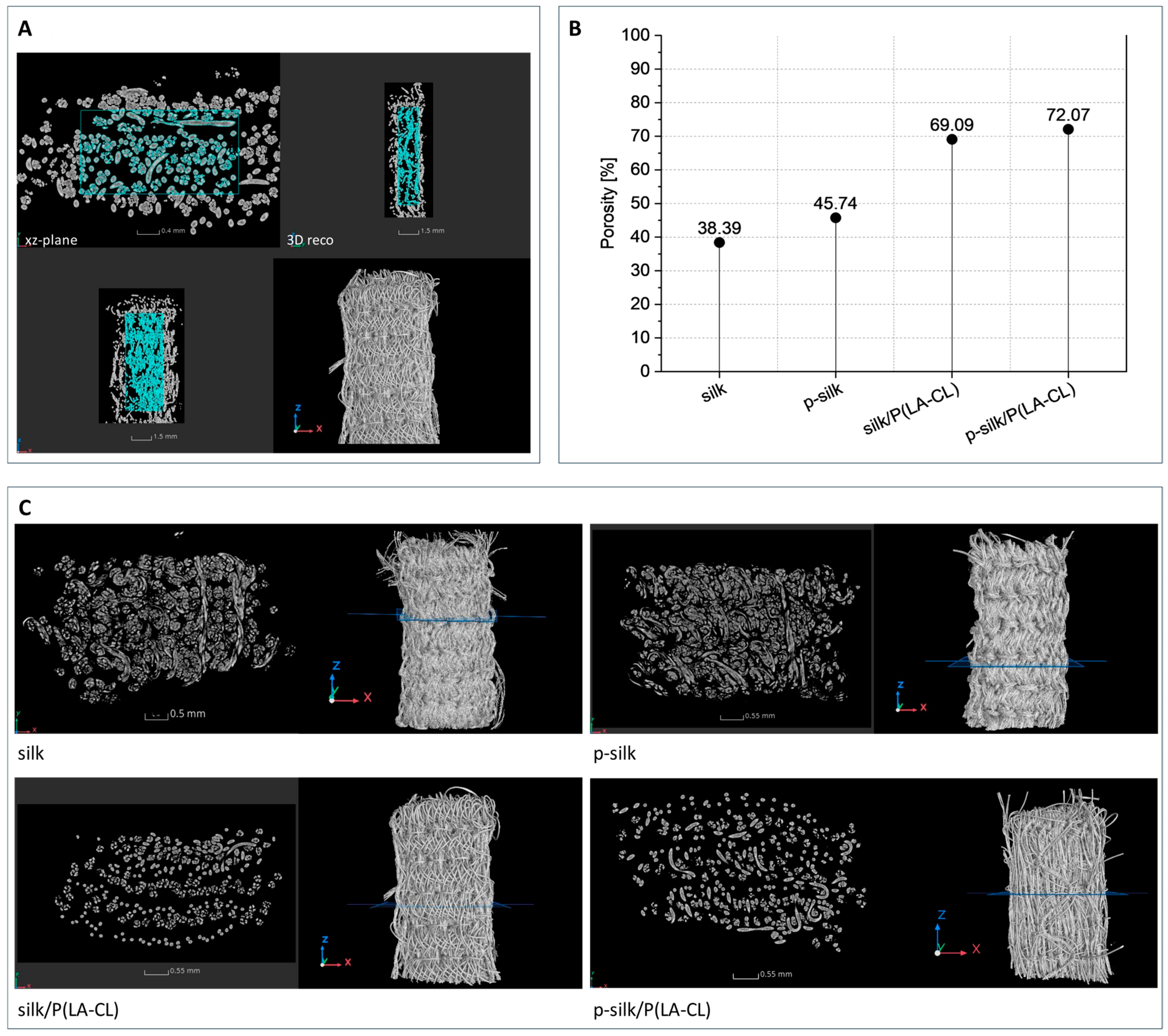
2.1.2. Characteristics of Textile Scaffold Structure
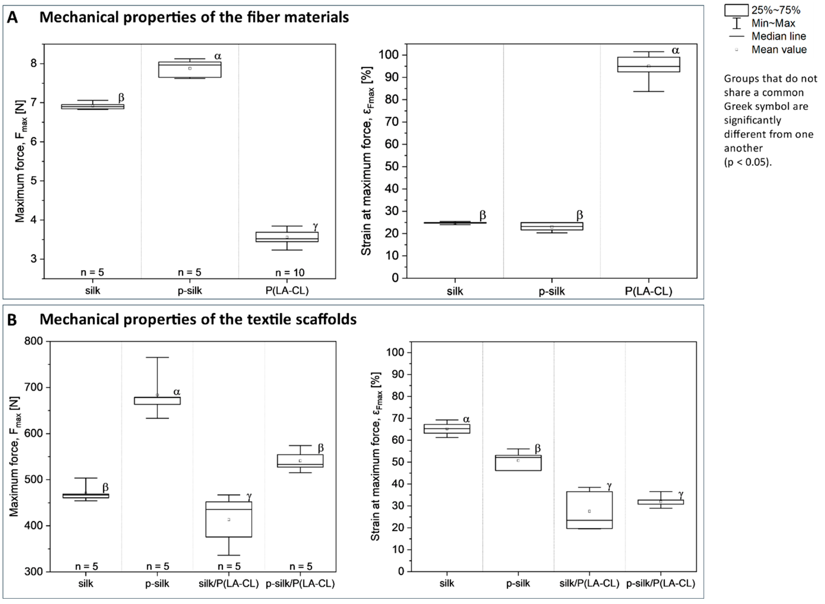
2.1.3. Biomechanical Characteristics of Fiber Materials and Scaffolds
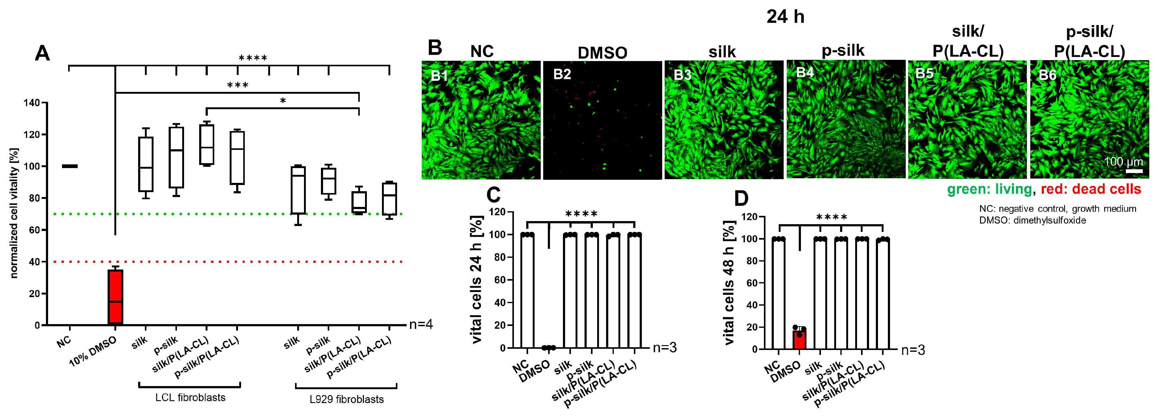
2.2. No Cytotoxic Effects of the Scaffolds on LCL Fibroblasts
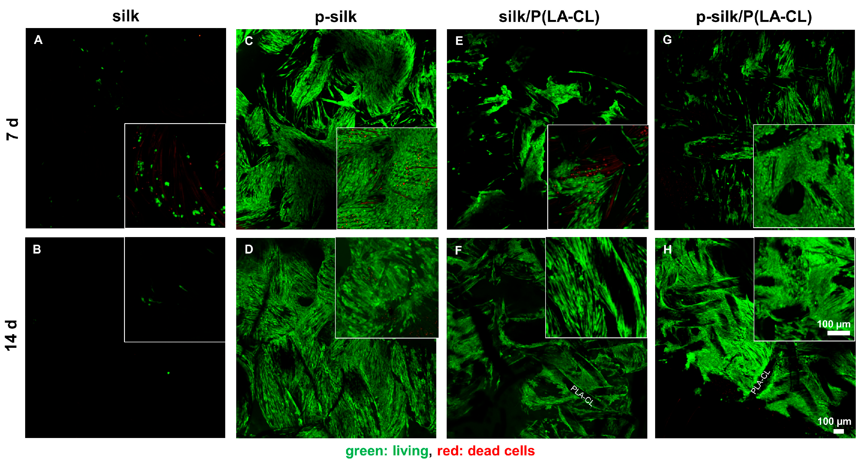
2.3. LCL Fibroblasts Survive on Silk Scaffolds
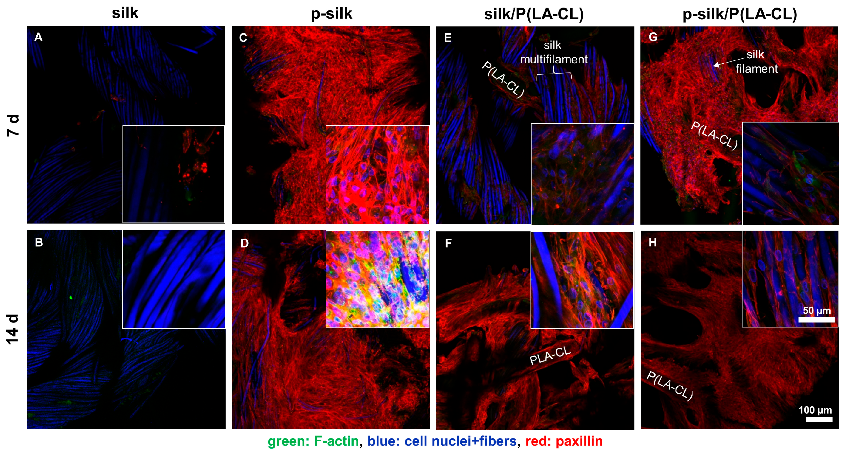
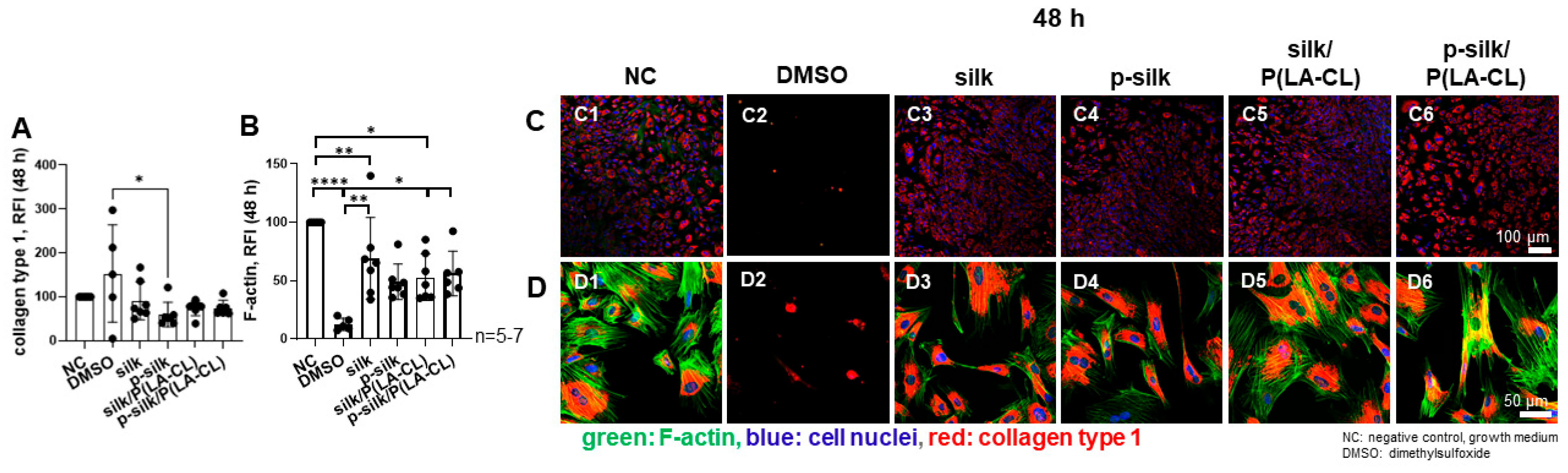
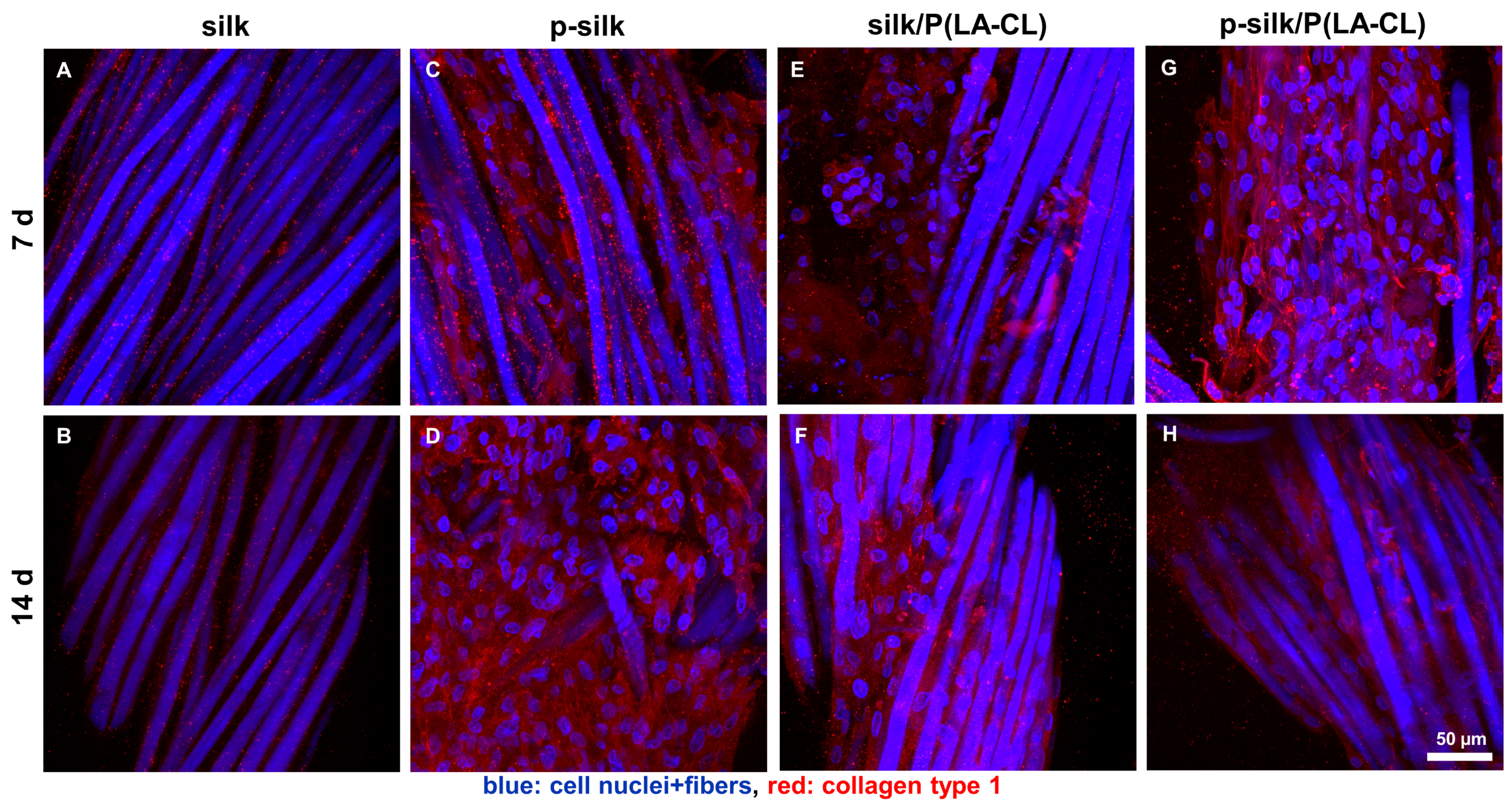
2.4. Expression of Cytoskeletal Components and Collagen Type I on Silk Scaffold Variants
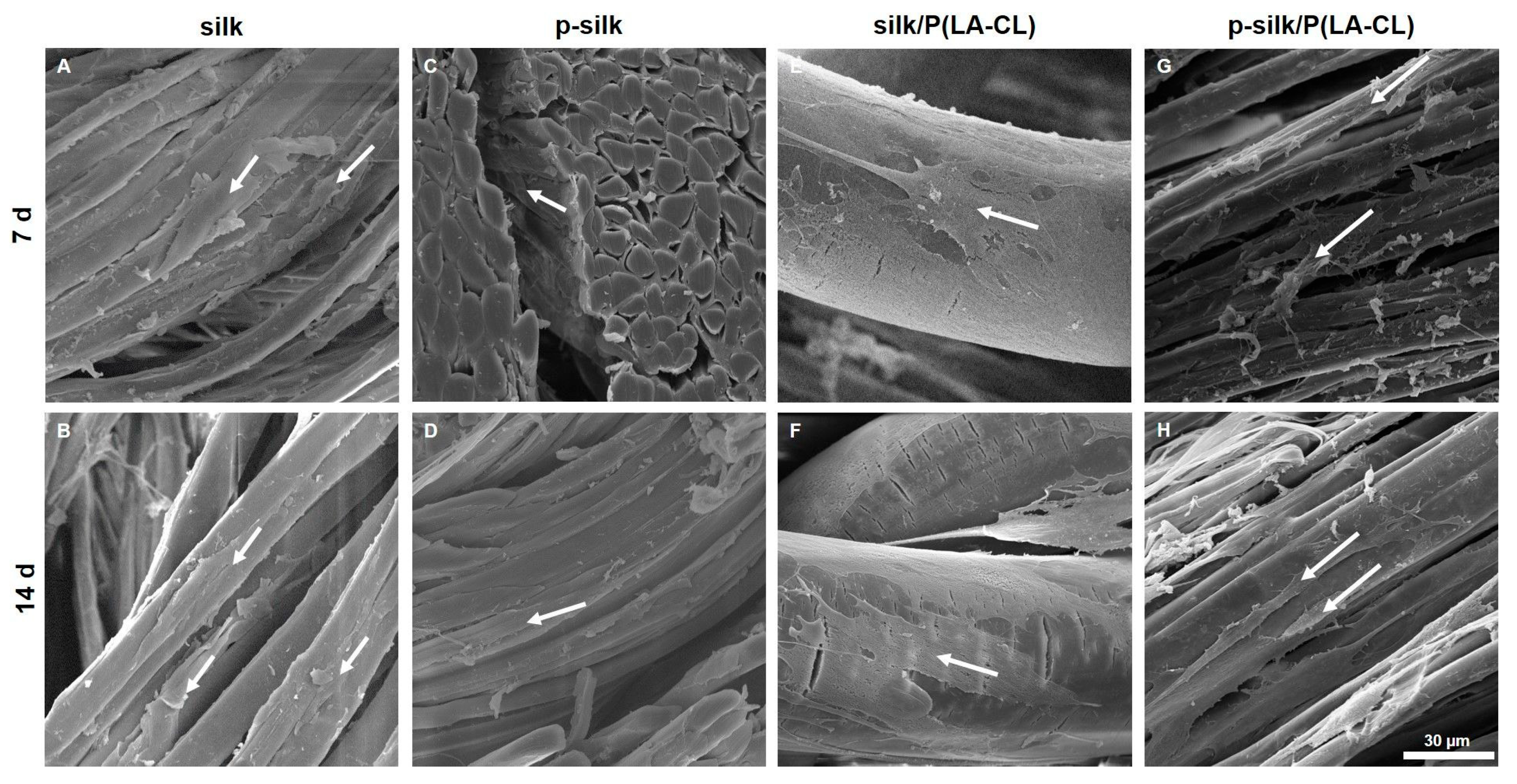
2.5. Ultramorphology of ACL Fibroblasts on Scaffold Variants Depicted by Scanning Electron Microscopy (SEM)
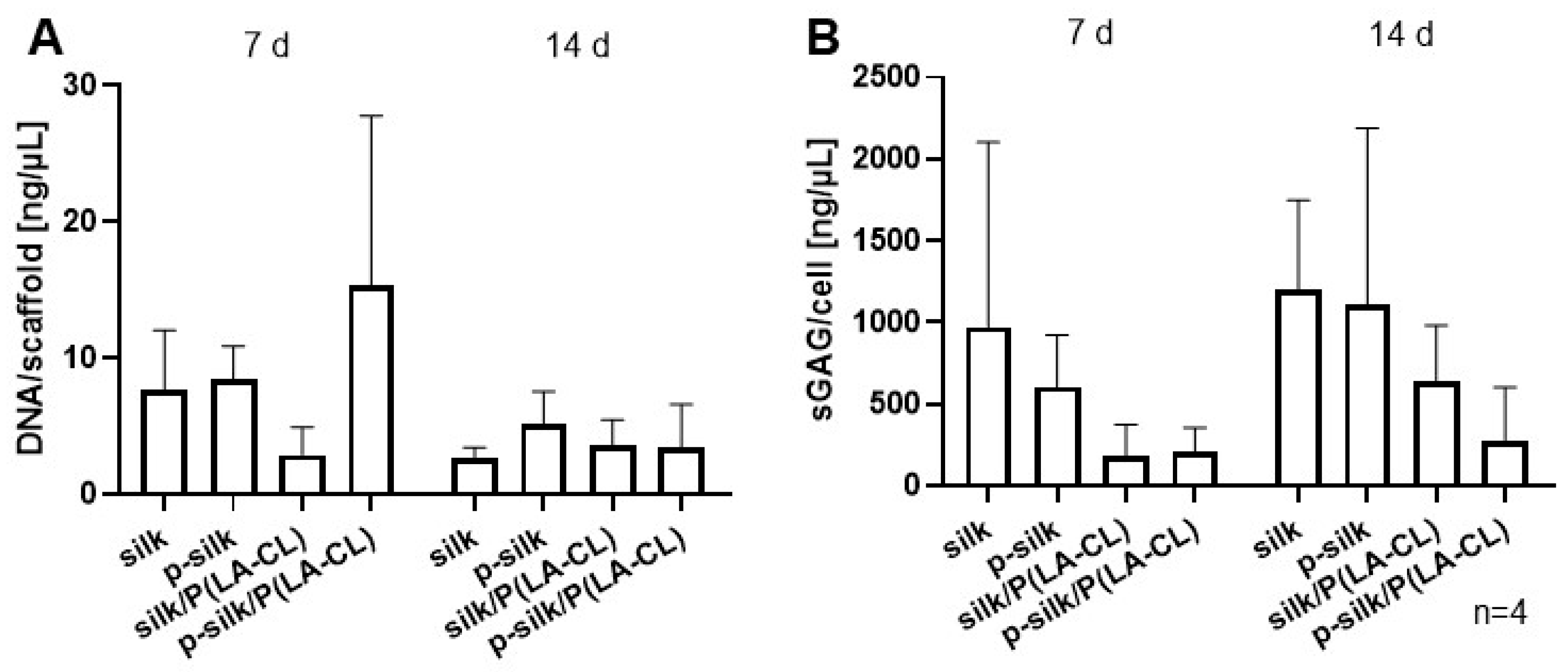
2.6. DNA and Sulfated Glycosaminoglycan Content per Cell During Cultivation Time
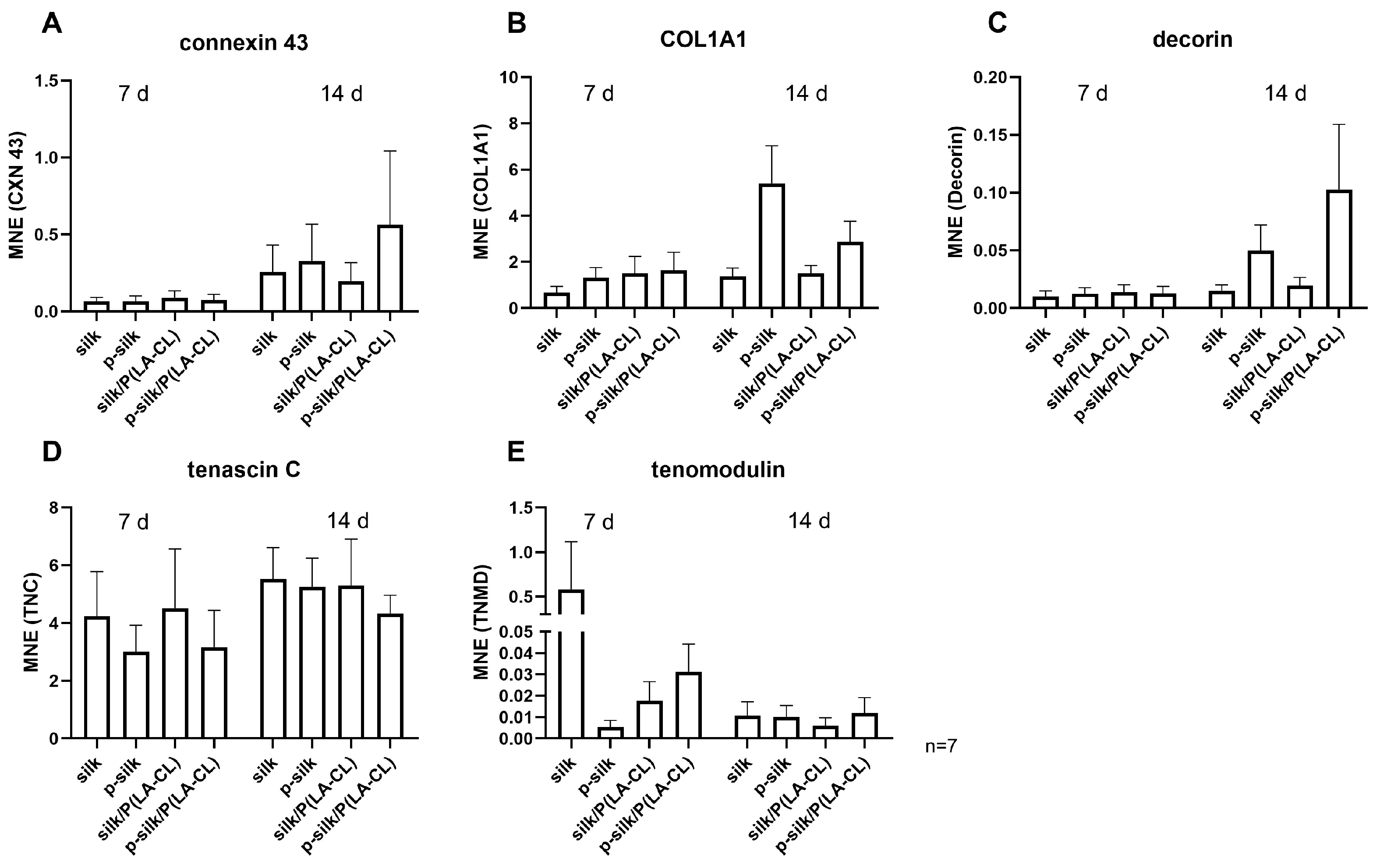
2.7. ACL Fibroblasts Express ACL-Related Genes on Scaffold Variants
3. Discussion
4. Materials and Methods
4.1. Fiber Materials
4.2. Scaffold Manufacture Using Machine Embroidery
4.3. Fiber and Scaffold Characterization
4.3.1. Light Microscopy
4.3.2. Scanning Electron Microscopy
4.3.3. Micro-Computed Tomography (µCT)
4.3.4. Determination of the Porosity of Textile Scaffolds
4.3.5. Uniaxial Tensile Test
4.4. Lapine Cruciate Ligament Fibroblast Isolation
4.5. Cytotoxicity Testing
4.6. Scaffold Seeding
4.7. Life/Death Assay
4.8. Immunofluorescence Labeling
4.9. Measurement of Total DNA and Sulfated Glycosaminoglycan Content
4.10. RNA Isolation
4.11. Gene Expression Analysis
4.12. Statistical Analysis
5. Conclusions
Supplementary Materials
Author Contributions
Funding
Institutional Review Board Statement
Informed Consent Statement
Data Availability Statement
Acknowledgments
Conflicts of Interest
Abbreviations
| 3D | three-dimensional |
| ABI | applied bioscience |
| ACL | anterior cruciate ligament |
| B. mori | Bombyx mori |
| CLSM | confocal laser scanning microscope |
| COL1A1 | collagen type 1 alpha chain |
| CXN43 | Connexin 43 |
| DAPI | 4′,6-diamino-2-phenylindole |
| DCN | decorin |
| DMEM | Dulbecco’s Modified Eagle’s Medium |
| DMMB | dimethylmethylene blue |
| DMSO | dimethylsulfoxide |
| ECM | extracellular matrix |
| EDTA | ethylendiaminetetraacetic acid |
| FBS | fetal bovine serum |
| FDA | fluorescein diacetate |
| GAPDH | glyceraldehyde 3-phosphate dehydrogenase |
| HBSS | Hank‘s balanced salt solution |
| LCL | lapine cruciate ligament |
| MTS | 3-(4,5-dimethylthiazol-2-yl)-5-(3-carboxymethoxyphenyl)-2-(4-sulfophenyl)-2H-tetrazolium inner salt |
| P(LA-CL) | PLA-co-caprolactone |
| PBS | phosphate-buffered saline |
| PI | propidium iodide |
| PLLA | poly-L-lactic acid |
| qRT-PCR | quantitative reverse transcription polymerase chain reaction |
| rpm | rounds per minutes |
| RT | room temperature |
| SD | standard deviation |
| SEM | Scanning electron microscopy |
| TBS | tris buffered saline |
| TE | Tris-EDTA |
| TNC | tenascin C |
| TNMD | tenomodulin |
References
- Murray, M.M. History of ACL treatment and current gold standard of care. In The ACL Handbook: Knee Biology, Mechanics, and Treatment; Springer: Berlin/Heidelberg, Germany, 2013; pp. 19–28. [Google Scholar] [CrossRef]
- Urbanek, O.; Moczulska-Heljak, M.; Wrobel, M.; Mioduszewski, A.; Kolbuk, D. Advanced Graft Development Approaches for ACL Reconstruction or Regeneration. Biomedicines 2023, 11, 507. [Google Scholar] [CrossRef]
- Rathbone, S.; Maffulli, N.; Cartmell, S.H. Most british surgeons would consider using a tissue-engineered anterior cruciate ligament: A questionnaire study. Stem Cells Int. 2012, 2012, 303724. [Google Scholar] [CrossRef]
- Hahn, J.; Schulze-Tanzil, G.; Schröpfer, M.; Meyer, M.; Gögele, C.; Hoyer, M.; Spickenheuer, A.; Heinrich, G.; Breier, A. Viscoelastic Behavior of Embroidered Scaffolds for ACL Tissue Engineering Made of PLA and P(LA-CL) After In Vitro Degradation. Int. J. Mol. Sci. 2019, 20, 4655. [Google Scholar] [CrossRef]
- Riley, T.C.; Mafi, R.; Mafi, P.; Khan, W.S. Knee Ligament Injury and the Clinical Application of Tissue Engineering Techniques: A Systematic Review. Curr. Stem Cell Res. Ther. 2018, 13, 226–234. [Google Scholar] [CrossRef] [PubMed]
- Fung, Y.-C. Biomechanics: Mechanical Properties of Living Tissues; Springer Science & Business Media: Berlin/Heidelberg, Germany, 2013. [Google Scholar]
- Cooper, J.A., Jr.; Sahota, J.S.; Gorum, W.J., 2nd; Carter, J.; Doty, S.B.; Laurencin, C.T. Biomimetic tissue-engineered anterior cruciate ligament replacement. Proc. Natl. Acad. Sci. USA 2007, 104, 3049–3054. [Google Scholar] [CrossRef] [PubMed]
- Teuschl, A.; Heimel, P.; Nurnberger, S.; van Griensven, M.; Redl, H.; Nau, T. A Novel Silk Fiber-Based Scaffold for Regeneration of the Anterior Cruciate Ligament: Histological Results from a Study in Sheep. Am. J. Sports Med. 2016, 44, 1547–1557. [Google Scholar] [CrossRef] [PubMed]
- Aigner, T.B.; DeSimone, E.; Scheibel, T. Biomedical Applications of Recombinant Silk-Based Materials. Adv. Mater. 2018, 30, e1704636. [Google Scholar] [CrossRef]
- Horan, R.L.; Toponarski, I.; Boepple, H.E.; Weitzel, P.P.; Richmond, J.C.; Altman, G.H. Design and characterization of a scaffold for anterior cruciate ligament engineering. J. Knee Surg. 2009, 22, 82–92. [Google Scholar] [CrossRef]
- Fan, H.; Liu, H.; Toh, S.L.; Goh, J.C. Anterior cruciate ligament regeneration using mesenchymal stem cells and silk scaffold in large animal model. Biomaterials 2009, 30, 4967–4977. [Google Scholar] [CrossRef]
- Hahn, J.; Gögele, C.; Schulze-Tanzil, G. Could an Anterior Cruciate Ligament Be Tissue-Engineered from Silk? Cells 2023, 12, 2350. [Google Scholar] [CrossRef]
- Sehnal, F.; Akai, H. Insect silk glands: Their types, development and function, and effects of environmental factors and morphogenetic hormones on them. Int. J. Insect Morphol. Embryol. 1990, 19, 79–132. [Google Scholar] [CrossRef]
- Hassan, M.A.; Basha, A.A.; Eraky, M.; Abbas, E.; El-Samad, L.M. Advancements in silk fibroin and silk sericin-based biomaterial applications for cancer therapy and wound dressing formulation: A comprehensive review. Int. J. Pharm. 2024, 662, 124494. [Google Scholar] [CrossRef] [PubMed]
- Teuschl, A.H.; van Griensven, M.; Redl, H. Sericin removal from raw Bombyx mori silk scaffolds of high hierarchical order. Tissue Eng. Part C Methods 2014, 20, 431–439. [Google Scholar] [CrossRef] [PubMed]
- Laurent, C.; Liu, X.; De Isla, N.; Wang, X.; Rahouadj, R. Defining a scaffold for ligament tissue engineering: What has been done, and what still needs to be done. J. Cell. Immunother. 2018, 4, 4–9. [Google Scholar] [CrossRef]
- Hohlrieder, M.; Teuschl, A.H.; Cicha, K.; van Griensven, M.; Redl, H.; Stampfl, J. Bioreactor and scaffold design for the mechanical stimulation of anterior cruciate ligament grafts. Biomed. Mater. Eng. 2013, 23, 225–237. [Google Scholar] [CrossRef]
- Freeman, J.W.; Woods, M.D.; Laurencin, C.T. Tissue engineering of the anterior cruciate ligament using a braid-twist scaffold design. J. Biomech. 2007, 40, 2029–2036. [Google Scholar] [CrossRef]
- Chen, K.; Sahoo, S.; He, P.; Ng, K.S.; Toh, S.L.; Goh, J.C. A hybrid silk/RADA-based fibrous scaffold with triple hierarchy for ligament regeneration. Tissue Eng. Part A 2012, 18, 1399–1409. [Google Scholar] [CrossRef]
- Ruan, D.; Zhu, T.; Huang, J.; Le, H.; Hu, Y.; Zheng, Z.; Tang, C.; Chen, Y.; Ran, J.; Chen, X.; et al. Knitted Silk-Collagen Scaffold Incorporated with Ligament Stem/Progenitor Cells Sheet for Anterior Cruciate Ligament Reconstruction and Osteoarthritis Prevention. ACS Biomater. Sci. Eng. 2019, 5, 5412–5421. [Google Scholar] [CrossRef]
- Musson, D.S.; Naot, D.; Chhana, A.; Matthews, B.G.; McIntosh, J.D.; Lin, S.T.; Choi, A.J.; Callon, K.E.; Dunbar, P.R.; Lesage, S.; et al. In vitro evaluation of a novel non-mulberry silk scaffold for use in tendon regeneration. Tissue Eng. Part A 2015, 21, 1539–1551. [Google Scholar] [CrossRef]
- Shen, W.; Chen, X.; Hu, Y.; Yin, Z.; Zhu, T.; Hu, J.; Chen, J.; Zheng, Z.; Zhang, W.; Ran, J.; et al. Long-term effects of knitted silk-collagen sponge scaffold on anterior cruciate ligament reconstruction and osteoarthritis prevention. Biomaterials 2014, 35, 8154–8163. [Google Scholar] [CrossRef]
- Gögele, C.; Konrad, J.; Hahn, J.; Breier, A.; Schröpfer, M.; Meyer, M.; Merkel, R.; Hoffmann, B.; Schulze-Tanzil, G. Maintenance of Ligament Homeostasis of Spheroid-Colonized Embroidered and Functionalized Scaffolds after 3D Stretch. Int. J. Mol. Sci. 2021, 22, 8204. [Google Scholar] [CrossRef]
- Kokozidou, M.; Gogele, C.; Pirrung, F.; Hammer, N.; Werner, C.; Kohl, B.; Hahn, J.; Breier, A.; Schropfer, M.; Meyer, M.; et al. In vivo ligamentogenesis in embroidered poly(lactic-co-epsilon-caprolactone)/polylactic acid scaffolds functionalized by fluorination and hexamethylene diisocyanate cross-linked collagen foams. Histochem. Cell Biol. 2023, 159, 275–292. [Google Scholar] [CrossRef] [PubMed]
- Gögele, C.; Hahn, J.; Schulze-Tanzil, G. Anatomical Tissue Engineering of the Anterior Cruciate Ligament Entheses. Int. J. Mol. Sci. 2023, 24, 9745. [Google Scholar] [CrossRef] [PubMed]
- Wang, R.; Wang, Y.; Song, J.; Tian, C.; Jing, X.; Zhao, P.; Xia, Q. A novel method for silkworm cocoons self-degumming and its effect on silk fibers. J. Adv. Res. 2023, 53, 87–98. [Google Scholar] [CrossRef] [PubMed]
- Pantano, M.F.; Berardo, A.; Pugno, N.M. Tightening slip knots in raw and degummed silk to increase toughness without losing strength. Sci. Rep. 2016, 6, 18222. [Google Scholar] [CrossRef]
- Liu, X.; Huang, Q.; Pan, P.; Fang, M.; Zhang, Y.; Yang, S.; Li, M.; Liu, Y. Comparative Study of the Preparation of High-Molecular-Weight Fibroin by Degumming Silk with Several Neutral Proteases. Polymers 2023, 15, 3383. [Google Scholar] [CrossRef]
- Chandini, S. Cocoon filament size deviation in bivoltine breeds/ hybrids of silkworm Bombyx mori. Pharma Innov. J. 2025, 14, 86–88. [Google Scholar]
- Luong, T.-H.; Dang, T.-N.N.; Ngoc, O.P.T.; Dinh-Thuy, T.-H.; Nguyen, T.-H.; Van Toi, V.; Duong, H.T.; Le Son, H. Investigation of the silk fiber extraction process from the vietnam natural bombyx mori silkworm cocoon. In Proceedings of the 5th International Conference on Biomedical Engineering in Vietnam, Ho Chi Minh City, Vietnam, 16–18 June 2014; pp. 325–328. [Google Scholar]
- Boulet-Audet, M.; Buffeteau, T.; Boudreault, S.; Daugey, N.; Pezolet, M. Quantitative determination of band distortions in diamond attenuated total reflectance infrared spectra. J. Phys. Chem. B 2010, 114, 8255–8261. [Google Scholar] [CrossRef]
- Monti, P.; Taddei, P.; Freddi, G.; Asakura, T.; Tsukada, M. Raman spectroscopic characterization of Bombyx mori silk fibroin: Raman spectrum of Silk I. J. Raman Spectrosc. 2001, 32, 103–107. [Google Scholar] [CrossRef]
- Park, J.; Babensee, J.E. Differential functional effects of biomaterials on dendritic cell maturation. Acta Biomater. 2012, 8, 3606–3617. [Google Scholar] [CrossRef]
- Gögele, C.; Hahn, J.; Elschner, C.; Breier, A.; Schröpfer, M.; Prade, I.; Meyer, M.; Schulze-Tanzil, G. Enhanced Growth of Lapine Anterior Cruciate Ligament-Derived Fibroblasts on Scaffolds Embroidered from Poly(l-lactide-co-epsilon-caprolactone) and Polylactic Acid Threads Functionalized by Fluorination and Hexamethylene Diisocyanate Cross-Linked Collagen Foams. Int. J. Mol. Sci. 2020, 21, 1132. [Google Scholar] [CrossRef]
- Gu, H.; Wang, F.; Liu, H.; Printon, K.; Hu, X. Multifunctional silk fibroin–Poly (L-lactic acid) porous nanofibers: Designing adjustable nanopores to control composite properties and biological responses. Mater. Des. 2022, 222, 111053. [Google Scholar] [CrossRef]
- Mahdi Souzani, A.; Rajeshwari, H.R.S.; Selvaganapathy, P.R.; Kishen, A. Impact of 3D collagen-based model and hydrostatic pressure on periodontal ligament fibroblast: A morpho-biochemical analysis. J. Mech. Behav. Biomed. Mater. 2023, 147, 106092. [Google Scholar] [CrossRef] [PubMed]
- Kaunas, R.; Nguyen, P.; Usami, S.; Chien, S. Cooperative effects of Rho and mechanical stretch on stress fiber organization. Proc. Natl. Acad. Sci. USA 2005, 102, 15895–15900. [Google Scholar] [CrossRef] [PubMed]
- Li, Y.; He, M.; Lin, W.; Xiang, Z.; Huang, J.; Xu, P.; Shi, Y.; Wang, H. Responses of bladder smooth muscle to the stretch go through extracellular signal-regulated kinase (ERK)/p90 ribosomal S6 protein kinase (p90RSK)/Nuclear factor-kappaB (NF-kappaB) Pathway. Neurourol. Urodyn. 2019, 38, 1504–1516. [Google Scholar] [CrossRef]
- Chen, S.; He, T.; Zhong, Y.; Chen, M.; Yao, Q.; Chen, D.; Shao, Z.; Xiao, G. Roles of focal adhesion proteins in skeleton and diseases. Acta Pharm. Sin. B 2023, 13, 998–1013. [Google Scholar] [CrossRef]
- Zhu, Y. Gap Junction-Dependent and -Independent Functions of Connexin43 in Biology. Biology 2022, 11, 283. [Google Scholar] [CrossRef]
- Novaretti, J.V.; Astur, D.C.; Casadio, D.; Nicolini, A.P.; de Castro Pochini, A.; Andreoli, C.V.; Ejnisman, B.; Cohen, M. Higher Gene Expression of Healing Factors in Anterior Cruciate Ligament Remnant in Acute Anterior Cruciate Ligament Tear. Am. J. Sports Med. 2018, 46, 1583–1591. [Google Scholar] [CrossRef]
- Shukunami, C.; Yoshimoto, Y.; Takimoto, A.; Yamashita, H.; Hiraki, Y. Molecular characterization and function of tenomodulin, a marker of tendons and ligaments that integrate musculoskeletal components. Jpn. Dent. Sci. Rev. 2016, 52, 84–92. [Google Scholar] [CrossRef]
- Xu, P.; Deng, B.; Zhang, B.; Luo, Q.; Song, G. Stretch-Induced Tenomodulin Expression Promotes Tenocyte Migration via F-Actin and Chromatin Remodeling. Int. J. Mol. Sci. 2021, 22, 4928. [Google Scholar] [CrossRef]
- Gaut, L.; Bonnin, M.A.; Blavet, C.; Cacciapuoti, I.; Orpel, M.; Mericskay, M.; Duprez, D. Mechanical and molecular parameters that influence the tendon differentiation potential of C3H10T1/2 cells in 2D- and 3D-culture systems. Biol. Open 2020, 9, bio047928. [Google Scholar] [CrossRef]
- Wagner, E.R.; Bravo, D.; Dadsetan, M.; Riester, S.M.; Chase, S.; Westendorf, J.J.; Dietz, A.B.; van Wijnen, A.J.; Yaszemski, M.J.; Kakar, S. Ligament Tissue Engineering Using a Novel Porous Polycaprolactone Fumarate Scaffold and Adipose Tissue-Derived Mesenchymal Stem Cells Grown in Platelet Lysate. Tissue Eng. Part A 2015, 21, 2703–2713. [Google Scholar] [CrossRef]
- Shiroud Heidari, B.; Ruan, R.; De-Juan-Pardo, E.M.; Zheng, M.; Doyle, B. Biofabrication and Signaling Strategies for Tendon/Ligament Interfacial Tissue Engineering. ACS Biomater. Sci. Eng. 2021, 7, 383–399. [Google Scholar] [CrossRef]
- Wang, X.; Lin, M.; Kang, Y. Engineering Porous beta-Tricalcium Phosphate (beta-TCP) Scaffolds with Multiple Channels to Promote Cell Migration, Proliferation, and Angiogenesis. ACS Appl. Mater. Interfaces 2019, 11, 9223–9232. [Google Scholar] [CrossRef]
- Kim, Y.J.; Sah, R.L.; Doong, J.Y.; Grodzinsky, A.J. Fluorometric assay of DNA in cartilage explants using Hoechst 33258. Anal. Biochem. 1988, 174, 168–176. [Google Scholar] [CrossRef]
- Schefe, J.H.; Lehmann, K.E.; Buschmann, I.R.; Unger, T.; Funke-Kaiser, H. Quantitative real-time RT-PCR data analysis: Current concepts and the novel “gene expression’s CT difference” formula. J. Mol. Med. 2006, 84, 901–910. [Google Scholar] [CrossRef]






| Group Name | Material for Upper Thread | Material for Lower Thread |
|---|---|---|
| silk | silk, untreated | silk, untreated |
| p-silk | silk, purified | silk, purified |
| silk/P(LA-CL) | silk, untreated | P(LA-CL) |
| p-silk/P(LA-CL) | silk, purified | P(LA-CL) |
| Target | Primary Antibody, Source | Dilution | Secondary Antibody, Source | Dilution |
|---|---|---|---|---|
| collagen type 1 | goat anti human, SouthernBiotech, Birmingham, AL, USA | 1:50 | donkey-anti-goat cy3, Invitrogen, Carlsbad, CA, USA | 1:200 |
| paxillin | mouse-anti-human, BD Biosciences, Toronto, CA | 1:40 | donkey-anti-mouse cy3, Invitrogen, Carlsbad, CA, USA | 1:200 |
| phalloidin-Alexa488 | Phalloidin-iFlour 488, Abcam, Cambridge, UK | 1:100 | - | |
| 4′,6′-diamidino-2-phenylindol (DAPI) | Roche, Mannheim, Germany | 1:100 | - |
| Gene Symbol | Species | Gene Name | Amplicon Length | Assay ID |
|---|---|---|---|---|
| COL1A1 | O. cuniculus | type 1 collagen, alpha 1 chain | 70 | Oc03396073_g1 |
| DCN | Homo sapiens | decorin | 77 | Hs00370384_m1 |
| TNC | O. cuniculus | tenascin C | 61 | Oc06726696_m1 |
| TNMD | O. cuniculus | myodulin | 146 | Oc03399505_m1 |
| CXN43 | O. cuniculus | connexin 43 | 68 | Oc03396056_g1 |
| GAPDH | O. cuniculus | glyceraldehyde-3-phosphate dehydrogenase | 82 | Oc03823402_g1 |
Disclaimer/Publisher’s Note: The statements, opinions and data contained in all publications are solely those of the individual author(s) and contributor(s) and not of MDPI and/or the editor(s). MDPI and/or the editor(s) disclaim responsibility for any injury to people or property resulting from any ideas, methods, instructions or products referred to in the content. |
© 2025 by the authors. Licensee MDPI, Basel, Switzerland. This article is an open access article distributed under the terms and conditions of the Creative Commons Attribution (CC BY) license.
Share and Cite
Majeed, Y.; Gögele, C.; Elschner, C.; Werner, C.; Braun, T.; Hahn, J.; Bernhardt, R.; Krause, U.; Minnich, B.; Schulze-Tanzil, G. Embroidered Silk Fibroin Scaffolds for ACL Tissue Engineering. Int. J. Mol. Sci. 2026, 27, 137. https://doi.org/10.3390/ijms27010137
Majeed Y, Gögele C, Elschner C, Werner C, Braun T, Hahn J, Bernhardt R, Krause U, Minnich B, Schulze-Tanzil G. Embroidered Silk Fibroin Scaffolds for ACL Tissue Engineering. International Journal of Molecular Sciences. 2026; 27(1):137. https://doi.org/10.3390/ijms27010137
Chicago/Turabian StyleMajeed, Yasir, Clemens Gögele, Cindy Elschner, Christian Werner, Tobias Braun, Judith Hahn, Ricardo Bernhardt, Udo Krause, Bernd Minnich, and Gundula Schulze-Tanzil. 2026. "Embroidered Silk Fibroin Scaffolds for ACL Tissue Engineering" International Journal of Molecular Sciences 27, no. 1: 137. https://doi.org/10.3390/ijms27010137
APA StyleMajeed, Y., Gögele, C., Elschner, C., Werner, C., Braun, T., Hahn, J., Bernhardt, R., Krause, U., Minnich, B., & Schulze-Tanzil, G. (2026). Embroidered Silk Fibroin Scaffolds for ACL Tissue Engineering. International Journal of Molecular Sciences, 27(1), 137. https://doi.org/10.3390/ijms27010137