Article

Cytochalasin B Mitigates the Inflammatory Response in Lipopolysaccharide-Induced Mastitis by Suppressing Both the ARPC3/ARPC4-Dependent Cytoskeletal Changes and the Association Between HSP70 and the NLRP3 Inflammasome

1
College of Veterinary Medicine, Gansu Agricultural University, Lanzhou 730070, China
2
Gansu Key Laboratory of Animal Generational Physiology and Reproductive Regulation, Lanzhou 730070, China
3
College of Life Sciences and Biotechnology, Gansu Agricultural University, Lanzhou 730030, China
*
Author to whom correspondence should be addressed.
Int. J. Mol. Sci. 2025, 26(7), 3029; https://doi.org/10.3390/ijms26073029
Submission received: 28 February 2025 / Revised: 13 March 2025 / Accepted: 18 March 2025 / Published: 26 March 2025
(This article belongs to the Special Issue Research on Intracellular Signal Transduction Systems)

Abstract

Cow mastitis is a major challenge in dairy farming, significantly affecting both milk quality and cow health. Cytochalasin B (CB) is a fungal toxin and an actin cytoskeleton depolymerizing agent that exhibits anti-inflammatory and antitumor properties; however, its mechanism in cow mastitis remains unclear. In this study, we systematically evaluated the effects of CB on mastitis using an LPS-induced inflammation model in bovine mammary epithelial cells (MAC-T) and a mouse mastitis model. The techniques employed included Real-time quantitative PCR detecting system (qPCR), Western blot, HE staining, immunofluorescence (IF), and immunohistochemistry (IHC). The results demonstrated that CB significantly alleviated LPS-induced mastitis by downregulating the expression of pro-inflammatory factors IL-1β, TNF-α, and the NLRP3 inflammasome while also reducing cell apoptosis. Further mechanistic investigations revealed that CB mitigates the inflammatory response by inhibiting the expression of ARPC3, ARPC4, and HSP70, thereby disrupting cytoskeletal rearrangement and the activation of the NLRP3 inflammasome. Overall, this study reveals the potential therapeutic role of CB in cow mastitis and provides a theoretical foundation for developing novel intervention strategies.

1. Introduction

Bovine mastitis is one of the most common diseases in the global dairy industry, posing a serious threat to cow health and the quality of dairy products. According to estimates by the Food and Agriculture Organization of the United Nations (FAO, 2020), mastitis causes annual economic losses amounting to billions of dollars worldwide due to reduced milk yield, increased treatment costs, and expenses associated with culling cows [1]. Pathogenic microbial infections are the primary cause of mastitis, with lipopolysaccharide (LPS) from Gram-negative bacteria playing a critical role in triggering the disease by eliciting a robust inflammatory response [2,3]. Although antibiotics are currently the main treatment for bovine mastitis, the increasing prevalence of antibiotic-resistant strains and concerns over antibiotic residues have prompted researchers to explore novel therapeutic strategies.
Cytochalasin B (CB) is a mycotoxin derived from Fusarium species, with a molecular weight of approximately 479.6 Da. As an actin depolymerizer, CB specifically binds to the growing ends of actin filaments, preventing the polymerization of actin monomers and disrupting the dynamic equilibrium of the cytoskeleton [4]. Recent studies have demonstrated that CB possesses significant anti-inflammatory and antitumor properties. Regarding its anti-inflammatory effects, CB attenuates inflammation by inhibiting the migration of inflammatory cells and reducing the release of inflammatory mediators; for example, it plays a crucial role in modulating macrophage-mediated inflammatory responses [5]. Concerning its antitumor activity, CB disrupts the cytoskeletal framework of tumor cells, thereby suppressing their proliferation and invasiveness; for instance, in gliomas, CB induces apoptosis in tumor cells [6], and in hepatocellular carcinoma (HCC), it shows promise as a potential therapeutic agent against this highly invasive tumor [7].
The actin cytoskeleton plays a pivotal role in regulating immune function by orchestrating fundamental cellular processes such as proliferation, differentiation, apoptosis, migration, and signal transduction [8]. Research has shown that remodeling of the actin cytoskeleton is essential not only for the function of immune cells—such as T lymphocytes, B lymphocytes, and macrophages—but also for the broader regulation of the immune system [9]. In the context of inflammation, the pro-inflammatory factor LPS induces dynamic cytoskeletal changes through activation of the PI3K signaling pathway [10,11]. This process relies on actin polymerization mediated by the actin-related protein 2/3 (ARP2/3) complex, which directly affects the migratory and phagocytic capabilities of inflammatory cells, thereby intensifying the inflammatory response [12].
The ARP2/3 complex plays a key role in LPS-induced inflammatory responses. Composed of ARP2, ARP3, and five subunits (ARPC1, ARPC2, ARPC3, ARPC4, and ARPC5), this complex is stably assembled, with ARPC4 being crucial for maintaining its structural integrity and stability through specific interactions with ARPC3 [13,14]. During inflammation, the ARP2/3 complex, activated by neuronal Wiskott–Aldrich syndrome protein (N-WASP), promotes the formation of branched actin structures, thereby enhancing the migratory and phagocytic capabilities of inflammatory cells [12]. Moreover, studies have observed that during bacterial invasion, the expression levels of ARPC3 and ARPC4 increase significantly, and inhibiting their expression can effectively mitigate the host’s inflammatory response [15].
Furthermore, additional critical factors are involved in the molecular mechanisms underlying inflammation. For example, fever—a hallmark of inflammation—is primarily driven by interleukin-1β (IL-1β), whose production depends on the activation of the NLRP3 inflammasome [16]. However, excessive activation of the NLRP3 inflammasome can lead to tissue damage and immune dysfunction, contributing to the onset and progression of inflammatory diseases such as mastitis [17,18]. Heat shock protein HSP70, which functions as a molecular chaperone, interacts with the NLRP3 inflammasome and acts as a negative regulator of its activation. Additionally, HSP70 helps maintain cellular function by participating in clathrin (CLTC)-mediated endocytosis via N-WASP [19,20]. Notably, HSP70 plays dual roles in inflammation: while its elevated expression can suppress apoptosis and enhance host immune responses [21], HSP70 induced by the heat shock response following pro-inflammatory stimuli may exert cytotoxic effects, exacerbating tissue damage [22].
Previous studies by our team have identified a potential interaction mechanism between ARPC3/4 and HSP70 in an LTA-induced bovine mastitis inflammation model [23]. Building on these findings, the current study employs cytochalasin B (CB) as a potential anti-inflammatory agent, using LPS to establish inflammatory models in MAC-T cells and mice. This research investigates the anti-inflammatory mechanisms of CB from two perspectives: first, CB may modulate cytoskeletal rearrangement by inhibiting the expression of ARPC3, ARPC4, and HSP70, thereby alleviating LPS-induced inflammation in bovine mammary epithelial cells; second, CB may disrupt the interaction between HSP70 and the NLRP3 inflammasome, blocking its activation and subsequently attenuating the LPS-induced inflammatory response. Overall, this study aims to provide novel mechanistic insights and potential intervention strategies for the treatment of bovine mastitis.

2. Results

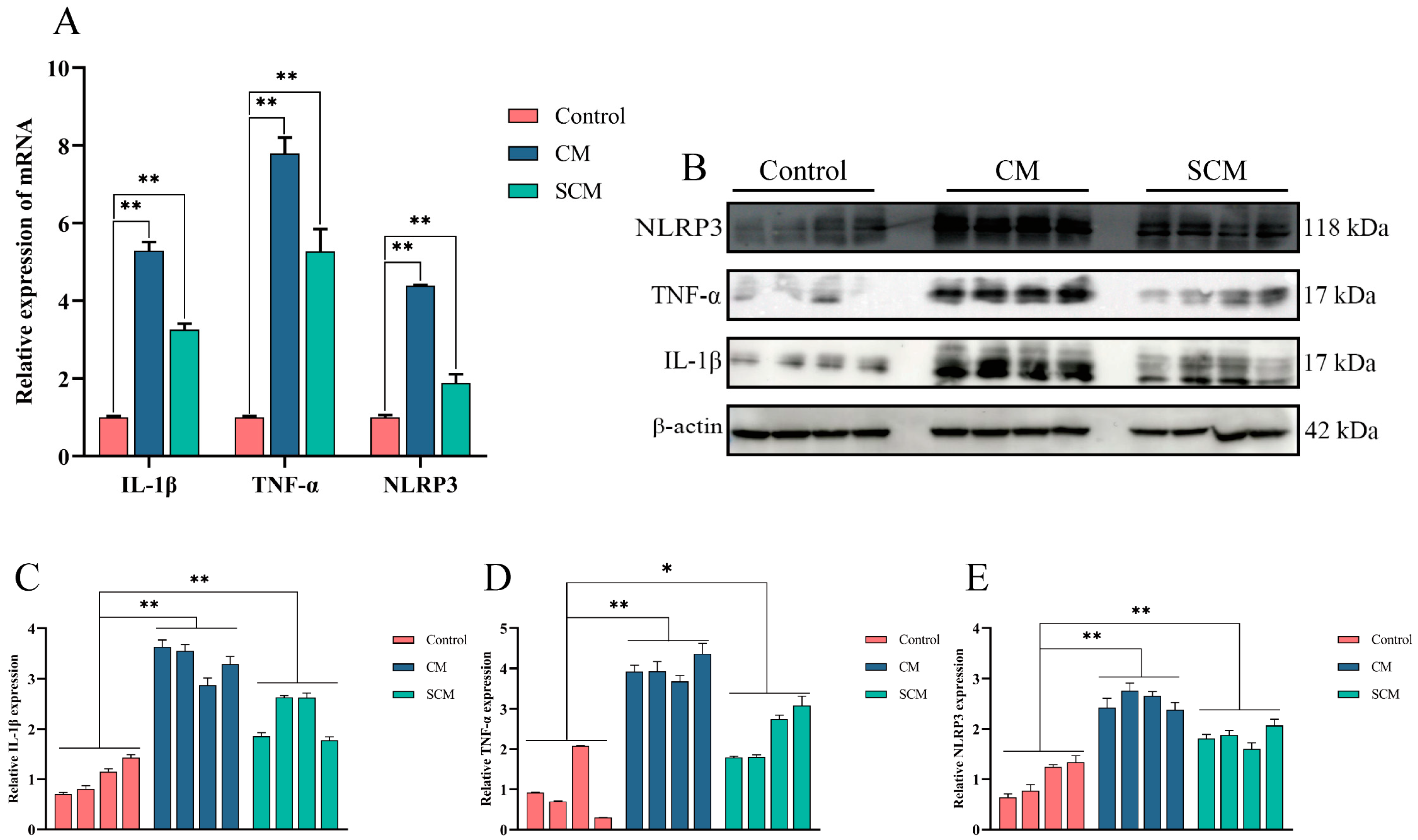
2.1. Expression of Inflammatory Cytokines and Activation of the NLRP3 Inflammasome in Bovine Mastitis Tissue

To assess the immune environment in bovine mammary tissue, qPCR and Western blot were used to evaluate the expression of IL-1β and TNF-α, as well as the activation of the NLRP3 inflammasome, in tissues from healthy cows (control) and those with clinical (CM) and subclinical mastitis (SCM). Compared with the control group, both the CM and SCM groups showed significantly elevated levels of IL-1β, TNF-α, and NLRP3 (Figure 1).

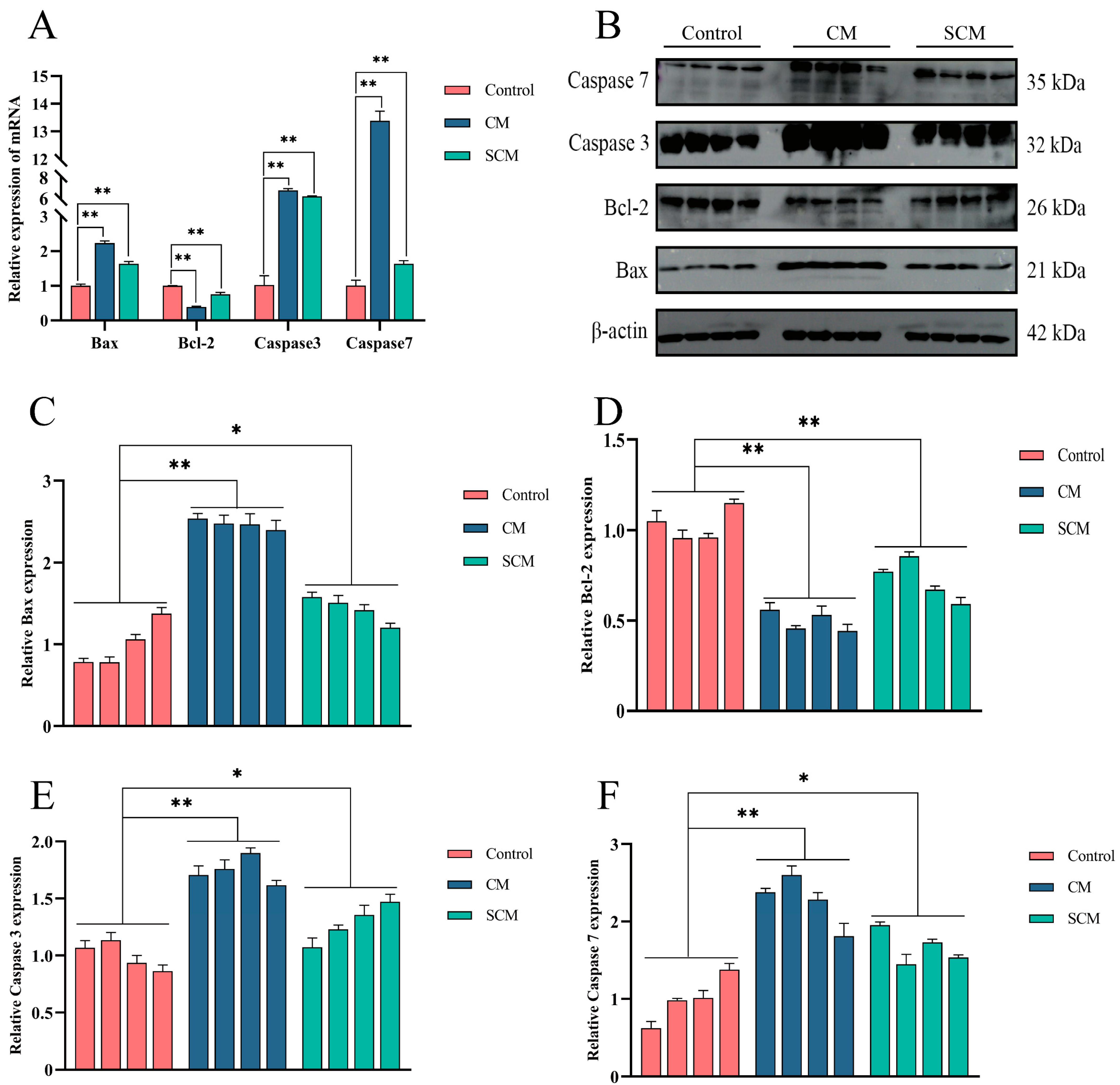
2.2. Increased Apoptosis in Bovine Mastitis Tissue

Given that inflammation is typically accompanied by enhanced apoptosis, qPCR and Western blot were used to measure the expression of Bax, Bcl-2, Caspase 3, and Caspase 7 in mammary tissues from the control, CM, and SCM groups. The results revealed that, relative to the control group, Bcl-2 expression was significantly decreased, while the Bax, Caspase 3, and Caspase 7 levels were markedly increased in both the CM and SCM groups (Figure 2).

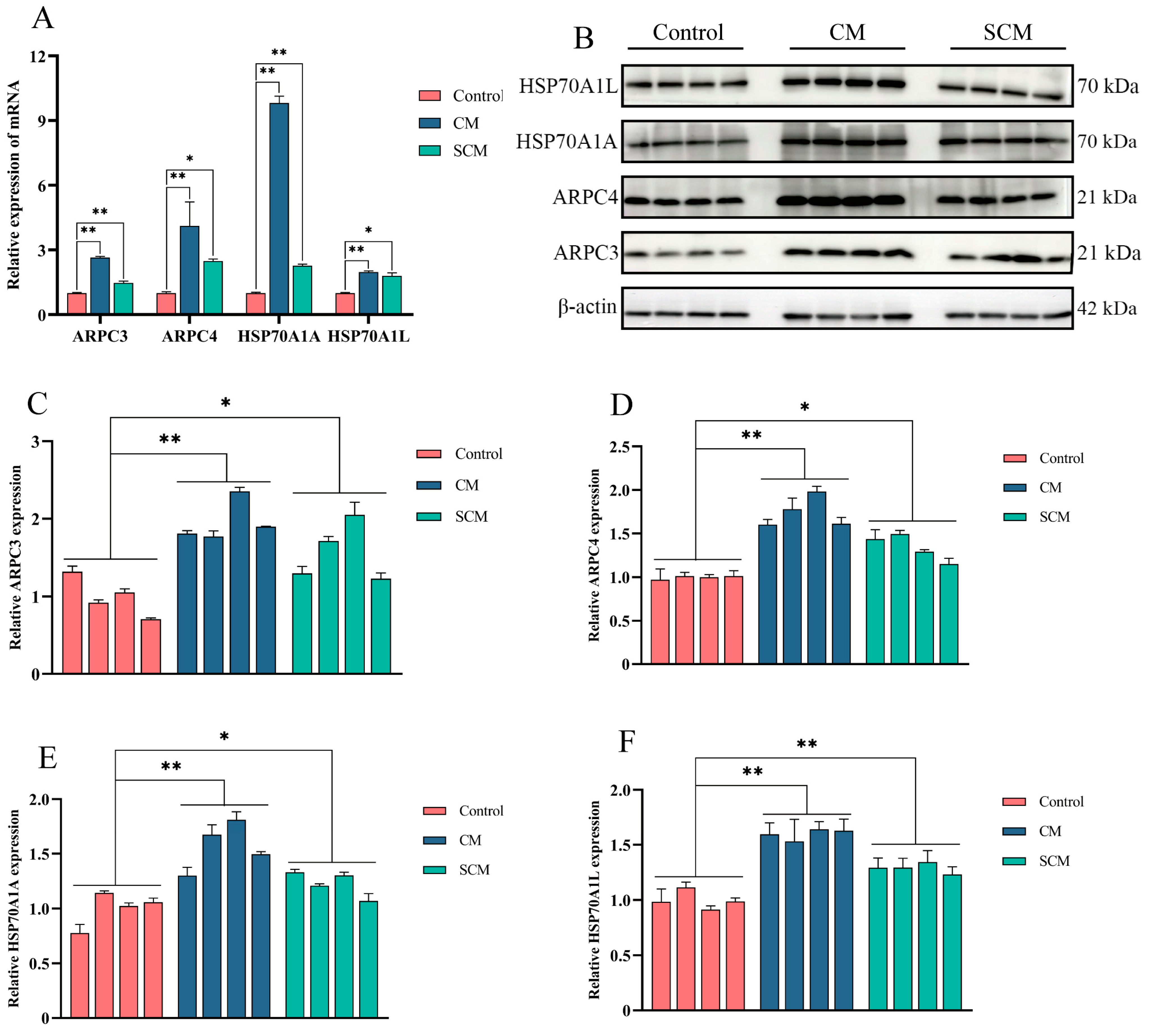
2.3. Enhanced Expression of ARPC3, ARPC4, and HSP70 in Bovine Mastitis Tissue

To examine changes in ARPC3, ARPC4, and HSP70 expression in bovine mastitis, qPCR and Western blot were used to determine the levels of ARPC3, ARPC4, HSP70A1A, and HSP70A1L in tissues from healthy cows (control) and cows with CM and SCM. Both mRNA and protein levels of these markers were significantly elevated in the CM and SCM groups compared with the control group (Figure 3).

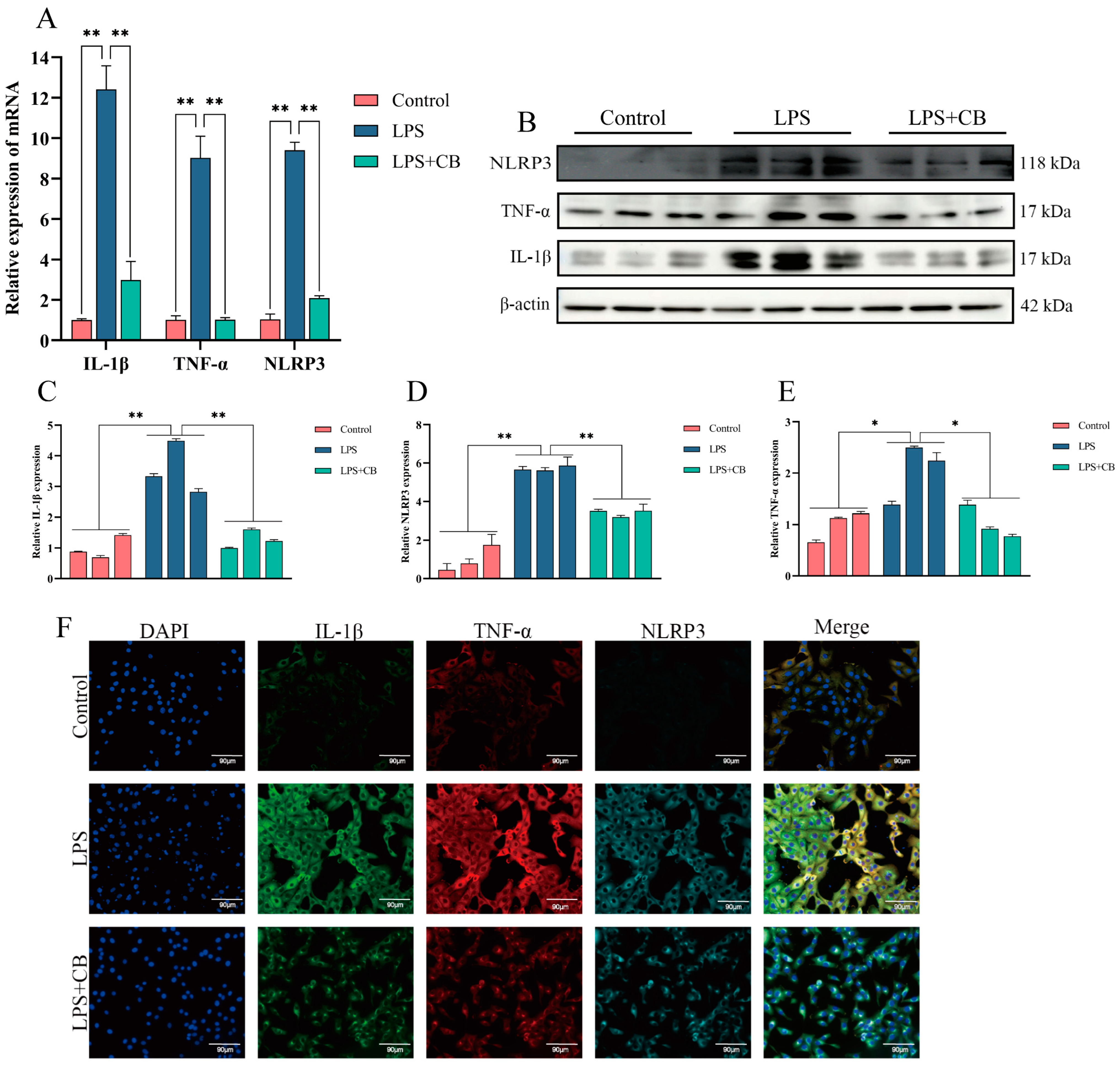
2.4. CB Inhibits LPS-Induced Expression of Inflammatory Cytokines and Activation of the NLRP3 Inflammasome in MAC-T Cells

CB has been shown to have potent anti-inflammatory effects. To investigate its impact on LPS-induced inflammatory responses in MAC-T cells, qPCR and Western blot were used to assess the mRNA and protein expression of IL-1β, TNF-α, and the NLRP3 inflammasome. The LPS + CB group exhibited significantly lower levels of these inflammatory markers compared with the LPS group (Figure 4).

2.5. CB Inhibits LPS-Induced Apoptosis in MAC-T Cells

To explore the role of CB in regulating apoptosis in LPS-stimulated MAC-T cells, qPCR, Western blot, and immunofluorescence (IF) were employed to detect apoptosis-related proteins. The LPS + CB group showed a significant increase in Bcl-2 expression and a significant decrease in Bax, Caspase 3, and Caspase 7 expression compared with the LPS group (Figure 5).

2.6. CB Inhibits LPS-Induced Expression of ARPC3, ARPC4, and HSP70 in MAC-T Cells

To further elucidate the anti-inflammatory mechanism of CB, qPCR, Western blot, and IF were used to examine the expression of ARPC3, ARPC4, HSP70A1A, and HSP70A1L in MAC-T cells. The results demonstrated that the LPS + CB group had a significantly lower expression of these markers compared with the LPS group, indicating that CB effectively suppresses their LPS-induced expression (Figure 6).

2.7. CB’s Effects on LPS-Induced Pathological Damage and Inflammatory Responses in Mouse Mastitis Tissue

To further investigate the anti-inflammatory effects of CB, mouse mammary tissues were subjected to HE staining, immunohistochemistry (IHC), and IF to observe IL-1β, TNF-α, and NLRP3. LPS treatment disrupted tissue structure, as evidenced by increased stroma thickness, rupture, and even atrophy and necrosis of glandular alveoli, along with the detachment of mammary epithelial cells and extensive inflammatory cell infiltration. However, CB treatment markedly ameliorated these pathological changes. Additionally, qPCR and Western blot confirmed that LPS significantly increased IL-1β, TNF-α, and NLRP3 expression in mouse mammary tissues compared with the control group, while CB treatment mitigated these effects (Figure 7). These findings confirm the successful establishment of the mouse mastitis model and demonstrate that CB alleviates mastitis by inhibiting inflammatory cytokine production.

2.8. CB Inhibits LPS-Induced Apoptosis in Mouse Mammary Cells

To further explore CB’s regulatory effects on apoptosis in inflammatory conditions, qPCR, Western blot, IHC, and IF were used to assess apoptosis-related protein expression in mouse mammary cells. The LPS + CB group showed a significant increase in Bcl-2 expression and significant decreases in Bax, Caspase 3, and Caspase 7 expression compared with the LPS group (Figure 8).

2.9. CB Inhibits LPS-Induced Expression of ARPC3, ARPC4, and HSP70 in Mouse Mammary Tissue

To further elucidate the anti-inflammatory mechanism of CB, qPCR, Western blot, IHC, and IF were used to measure the expression of ARPC3, ARPC4, HSP70A1A, and HSP70A1L in mouse mammary tissues. The LPS + CB group exhibited significantly lower levels of these proteins compared with the LPS group, indicating that CB effectively inhibits their LPS-induced expression (Figure 9).

3. Discussion

Bovine mastitis is a major challenge in the dairy industry, severely affecting milk quality and cow health while incurring significant economic losses. Traditionally treated with antibiotics, mastitis management faces issues such as adverse side effects and growing antibiotic resistance, prompting the search for alternative therapies [24,25]. This study systematically investigates the mechanism of cytochalasin B (CB) in LPS-induced mastitis through both in vitro and in vivo experiments. Our results demonstrate that CB significantly alleviates mastitis symptoms by suppressing inflammatory responses, inhibiting apoptosis, and downregulating key molecules including ARPC3, ARPC4, and HSP70, thereby providing a theoretical basis for its potential therapeutic application.
Initially, we assessed the expression of inflammatory cytokines IL-1β, TNF-α, and the activation of the NLRP3 inflammasome in bovine mammary tissues using qPCR and Western blot. Both mRNA and protein levels of these factors were significantly elevated in tissues from cows with clinical (CM) and subclinical mastitis (SCM) compared with healthy controls. This observation aligns with previous research demonstrating that LPS, a critical pathogenic component of Gram-negative bacteria, activates immune regulatory pathways and the NLRP3 inflammasome, thereby promoting IL-1β release and exacerbating inflammatory damage [26,27,28]. Notably, CB treatment in MAC-T cells and mouse mastitis models markedly reduced the expression of IL-1β, TNF-α, and NLRP3, indicating potent anti-inflammatory effects similar to those observed with other natural compounds such as allicin and lentinan [29,30].
Inflammation is frequently accompanied by increased apoptosis, a phenomenon particularly evident in mastitis pathology [31,32]. Our study found that pro-apoptotic proteins Bax, Caspase 3, and Caspase 7 were significantly upregulated, while the anti-apoptotic protein Bcl-2 was downregulated in the CM and SCM groups, indicating heightened apoptosis. In both LPS-induced MAC-T cells and mouse models, CB treatment reversed these changes by upregulating Bcl-2 and downregulating Bax, Caspase 3, and Caspase 7. As an actin depolymerizer, CB may protect cells by disrupting cytoskeletal structures and blocking apoptotic signaling pathways, a mechanism consistent with reports in other inflammatory conditions [33].
To further elucidate CB’s anti-inflammatory mechanism, we examined the expression of ARPC3, ARPC4, and HSP70. Our data revealed that the ARPC3, ARPC4, and HSP70 levels were significantly elevated in the CM and SCM groups but markedly reduced in CB-treated MAC-T cells and mouse models. ARPC3 and ARPC4 are critical components of the ARP2/3 complex, which regulates actin polymerization and cytoskeletal rearrangement, processes that facilitate the migration and phagocytic activity of inflammatory cells [12,34,35]. By downregulating ARPC3 and ARPC4, CB likely disrupts cytoskeletal rearrangement, thereby reducing inflammatory cell migration and attenuating inflammation.
HSP70, a molecular chaperone, plays a complex role in inflammation regulation by interacting with the NLRP3 inflammasome. Although generally considered a negative regulator of NLRP3 activation [19], HSP70 overexpression following pro-inflammatory stimuli can have cytotoxic effects, contributing to apoptosis in endotoxin-exposed cells [36,37]. In our study, CB reduced HSP70 expression, which may decrease its interaction with NLRP3 and further inhibit inflammasome activation. This finding supports the notion that CB exerts its anti-inflammatory effects, at least in part, through the HSP70-NLRP3 axis.
In summary, CB significantly mitigates inflammatory damage in LPS-induced mastitis models by suppressing inflammatory cytokine production, inhibiting apoptosis, and downregulating ARPC3, ARPC4, and HSP70 expression. Its mechanism involves disrupting cytoskeletal rearrangement and blocking NLRP3 inflammasome activation. As a natural product, CB may offer advantages over traditional antibiotics, including lower toxicity and reduced residue risks, aligning with current trends in mastitis treatment research. However, further studies are needed to clarify CB’s specific molecular targets and dose–response relationships, and to optimize its application conditions for translational use in bovine mastitis therapy.

4. Materials and Methods

4.1. Cell Culture and Treatment

Bovine mammary epithelial (MAC-T) cells were obtained from the Chinese Academy of Agricultural Sciences. The cells were cultured in DMEM/F12 medium supplemented (Gibco, Grand Island, NY, USA) with 10% fetal bovine serum (Invigentech, Irvine, CA, USA) in a 37 °C and 5% CO2 incubator. They were maintained in 25 cm2 culture flasks, and experiments were initiated when cell confluence reached 60–70%. For treatment, LPS (Sigma, St. Louis, MO, USA) was dissolved in sterile PBS (Servicebio, Wuhan, China) and applied at a final concentration of 50 µg/mL for 24 h. Cytochalasin B (CB) was purchased from MedChemExpress (MCE, Monmouth Junction, NJ, USA). CB was dissolved in 1 mL of dimethyl sulfoxide (DMSO) (Servicebio, Wuhan, China) to prepare a 1 mg/mL stock solution and used at a final concentration of 500 ng/mL. After the initial 24 h LPS treatment, CB was added to the medium, and the cells were further incubated for an additional 24 h.

4.2. Tissue Sample Preparation

All cows were sourced from a large, standardized dairy farm in Wuzhong, Ningxia, and were all at the same lactation stage. A resident veterinarian conducted diagnoses to exclude other diseases. Udder health was evaluated based on criteria such as redness, swelling, heat, and hardness, along with somatic cell count (SCC) and the Lanzhou Mastitis Test (LMT). Healthy cows exhibited normal udder skin and milk, with SCC values between 7 × 10⁴ and 1 × 10⁵ cells/mL and negative LMT results. Cows with subclinical mastitis, despite lacking overt clinical symptoms, showed abnormal milk test results with SCC values ranging from 2 × 10⁵ to 5 × 10⁵ cells/mL and weakly positive LMT results (+ or ++). In contrast, cows with clinical mastitis displayed clear signs such as udder redness, swelling, and heat, with milk showing watery consistency, clots, or blood; these cows had SCC values between 13 × 105 and 15 × 105 cells/mL and positive LMT results (+++). We selected three healthy Holstein cows (control group, Con, n = 3), three cows with subclinical mastitis (subclinical group, SM, n = 3), and three cows with clinical mastitis (clinical group, CM, n = 3) and sent them to the slaughterhouse. After slaughter, fresh udder tissue from each group was collected under sterile conditions for research. Portions of the tissue were cut into 1.5 cm3 blocks and fixed in sterile, enzyme-free centrifuge tubes containing 4% paraformaldehyde, while the remaining tissue was cut into 1 cm3 blocks and stored in sterile, enzyme-free cryovials at −80 °C for future use.

4.3. Animal Housing and Treatment

Fifty 8-week-old female Kunming mice and twenty-five male Kunming mice were obtained from the Chinese Academy of Agricultural Sciences. After a 7-day acclimation period with free access to food and water, the mice were housed at a 2:1 female-to-male ratio for a 2-day mating period. Eighteen pregnant mice with similar physical conditions were then selected, and following 7 days of lactation, they were randomly assigned to three groups: control, LPS, and CB + LPS. The fourth pair of mammary glands was injected as follows: the LPS and CB + LPS groups received a 50 µL injection of LPS solution (200 µg/mL) to induce mastitis, while the control group was injected with an equal volume of saline. Twenty-four hours after the LPS injection, the CB + LPS group was administered CB (4 mg/kg). After an additional 24 h, the fourth pair of mammary gland tissues was collected under sterile conditions. Tissue samples were divided, with one portion cut into 1.5 cm3 blocks and stored at −80 °C, and the remainder were fixed in 4% formaldehyde for subsequent analyses. This study was approved by the Animal Protection Committee of Gansu Agricultural University.

4.4. RNA Extraction, cDNA Synthesis, and qPCR

Total RNA was isolated from cells and tissues using TRNzol Universal Reagent (G3013, Servicebio, Wuhan, China) according to the manufacturer’s protocol. RNA concentration and purity were measured with a NanoDrop ND-1000 spectrophotometer (Thermo Fisher, San Diego, CA, USA); only samples with an OD260/280 ratio between 1.8 and 2.0 were used. cDNA was synthesized from total RNA using the Evo M-MLV Reverse Transcription Kit (AG11732, Accurate Biotechnology, Changsha, China). Gene expression was quantified with SYBR Green Pro Taq HS Premix (AG11746, Accurate Biotechnology, Changsha, China), using β-actin as the internal reference gene. Relative expression levels were calculated using the 2−ΔΔCT method. mRNA-specific primer sequences are listed in Table 1.

4.5. Western Blot

Total protein was extracted from cells and animal mammary tissues using RIPA lysis buffer (Solarbio, Beijing, China) supplemented with PMSF at a 100:1 ratio. Protein concentration was measured with a BCA assay (PC0020, Solarbio, Beijing, China). The supernatant was mixed with 5× loading buffer, heat-denatured at 98 °C for 15 min, and stored at −80 °C. Western blotting was performed to assess the expression of IL-1β, TNF-α, NLRP3, Bax, Bcl-2, Caspase 3, Caspase 7, ARPC3, ARPC4, HSP70A1A, and HSP70A1L. Equal amounts of protein (50 μg) were separated by SDS-PAGE and transferred to a PVDF membrane. The membrane was washed with 1× PBST, blocked in 5% non-fat milk at room temperature for 2 h, and then incubated overnight at 4 °C with the appropriate primary antibody. The following day, the membrane was washed three times with PBST (10 min each), incubated with the corresponding secondary antibody at 37 °C for 1 h, and washed again three times with PBST. Protein bands were visualized using a chemiluminescent solution with β-actin as the internal control, and band intensities were quantified using Image-J 1.52a software. The antibodies used are listed in Table 2.

4.6. Immunohistochemistry and Immunofluorescence Staining

Mouse mammary tissues preserved in formaldehyde were dehydrated, paraffin-embedded, sectioned into 4 µm slices, and deparaffinized using graded alcohols and xylene before hematoxylin–eosin (HE) staining. After dehydration and coverslipping, the slides were examined under a microscope (Axiocam 208 color, Zeiss, Oberkochen, Germany). For immunohistochemistry (IHC), antigen retrieval was performed with heated citrate buffer and sections were blocked with 5% BSA. The primary antibody was applied overnight at 4 °C, followed by a 1 h incubation with the secondary antibody at 37 °C, then DAB staining and hematoxylin counterstaining were performed, after which the slides were coverslipped for observation. For immunofluorescence (IF), MAC-T cells and mouse mammary tissue sections were stained using a three-marker, four-color multiplex fluorescent kit (AiFang Biological, Changsha, China) per the manufacturer’s instructions and observed with an inverted fluorescence microscope (Revolve Omega, ApexBio, Suzhou, China).

4.7. Statistical Analysis

Western blot bands were quantified using ImageJ software. Statistical analyses were performed with SPSS 25.0, and all data are presented as mean ± standard deviation. A p value < 0.05 (*) was considered statistically significant, while p < 0.01 (**) was deemed highly significant. GraphPad Prism 8.0.2 was used for data visualization.

5. Conclusions

In both the LPS-induced MAC-T cell inflammation model and the mouse mastitis model, CB effectively inhibited inflammatory damage in vitro and in vivo. Furthermore, CB exerts its anti-inflammatory effects by suppressing the expression of ARPC3, ARPC4, and HSP70, thereby disrupting cytoskeletal rearrangement and blocking NLRP3 inflammasome activation. These findings provide a novel theoretical foundation for the potential use of CB in treating bovine mastitis.

Author Contributions

A.W.: methodology, formal analysis, writing—original draft; Y.C.: methodology, formal analysis; B.F.: methodology, formal analysis; J.Z.: methodology, formal analysis; W.B.: methodology, formal analysis; T.Y.: methodology, formal analysis; P.L.: methodology, formal analysis; Z.D.: methodology, formal analysis; T.L.: methodology, formal analysis; Q.Z.: review and editing; Y.H.: review and editing; Y.Z.: conceptualization, funding acquisition; X.Z.: conceptualization, funding acquisition; W.D.: conceptualization, writing—review and editing, funding acquisition. All authors have read and agreed to the published version of the manuscript.

Funding

This study was supported by the fund of the College of Veterinary Medicine, GSAU, Gansu Youth Science and Technology Fund (24JRRA657), Young Doctoral Support Project of Universities in Gansu Province (2024QB-064), and the National Natural Science Foundation of China (No. U21A20262).

Institutional Review Board Statement

The animal study protocol was approved by the Ethics Committee of Gansu Agricultural University (protocol code: GSAU-Eth-LST-2021-003).

Informed Consent Statement

Not applicable.

Data Availability Statement

The datasets analyzed or generated during this study are available from the authors (d.wt2008@163.com).

Conflicts of Interest

The authors declare no conflicts of interest.

References

  1. Ragul, P.; Gopal Dhinakar, R.; Ranjithkumar, D.; Subbarayalu, R.; Mahesh Prabu, E.; Bhavadharani, P.; Veeramani, V.; Arunkumar, N.; Siddarth, P.; Muthupandian, S. Is AMR in Dairy Products a Threat to Human Health? An Updated Review on the Origin, Prevention, Treatment, and Economic Impacts of Subclinical Mastitis. Infect. Drug Resist. 2023, 16, 155–178. [Google Scholar]
  2. Ké, L.; Mingyuan, H.; Lin, Z.; Mengyue, T.; Ming, Y.; Jia, L.; Yeru, L.; Dongmin, Z.; Ruonan, L.; Yuzhong, M. Analysis of antimicrobial resistance and genetic correlations of Escherichia coli in dairy cow mastitis. J. Vet. Res. 2022, 66, 571. [Google Scholar]
  3. Johnzon, C.-F.; Dahlberg, J.; Gustafson, A.-M.; Waern, I.; Moazzami, A.A.; Östensson, K.; Pejler, G. The Effect of Lipopolysaccharide-Induced Experimental Bovine Mastitis on Clinical Parameters, Inflammatory Markers, and the Metabolome: A Kinetic Approach. Front. Immunol. 2018, 9, 1487. [Google Scholar] [CrossRef] [PubMed]
  4. Luca, P.; Provvidenza Maria, A.; Riccardo, T.; Andrea, A.; Giovannamaria, P.; Gregorio, R.; Claudia, C.; Valeria, P.; Nicoletta, C.; Silvia, C.; et al. Cytochalasin B Influences Cytoskeletal Organization and Osteogenic Potential of Human Wharton’s Jelly Mesenchymal Stem Cells. Pharmaceuticals 2023, 16, 289. [Google Scholar] [CrossRef]
  5. Mi-Yeon, K.; Jong-Hoon, K.; Jae Youl, C. Cytochalasin B Modulates Macrophage-Mediated Inflammatory Responses. Biomol. Ther. 2014, 22, 295. [Google Scholar]
  6. Tong, Z.; Liu, N.; Song, H.; Li, J.Q.; Jiang, J.; Zhu, J.; Qi, J. Cytochalasin B inhibits the proliferation of human glioma U251 cells through cell cycle arrest and apoptosis. Genet. Mol. Res. 2014, 13, 10811–10822. [Google Scholar] [CrossRef] [PubMed]
  7. Fábio, V.; Madalena, R.; Ana, B.; Ana, M.A.; José Guilherme, T.; Maria Filomena, B. P-0080 New Options in Hepatocellular Carcinoma Therapy: The Role of Quercetin and Cytochalasin-B. Ann. Oncol. 2012, 23, 54. [Google Scholar]
  8. Serge, M.; Avinash, R.S. The cytoskeleton in cell-autonomous immunity: Structural determinants of host defence. Nat. Rev. Immunol. 2015, 15, 559–573. [Google Scholar]
  9. Richard, J.S.; Virginie, P.H.; Marco, A.P.; Melanie, M.; Rebecca, A.; Marcus, H.; Susanne, M.B.; Jürgen, H.; Robin, A.; Michael, P.W.; et al. HCMV pUL135 Remodels the Actin Cytoskeleton to Impair Immune Recognition of Infected Cells. Cell Host Microbe 2014, 16, 201–214. [Google Scholar]
  10. Jörg, R.; Mary, S.S.; Dan, W.; Thomas, R.; Klaus, L. Phosphoinositide 3-kinase required for lipopolysaccharide-induced transepithelial neutrophil trafficking in the lung. Eur. Respir. J. 2009, 35, 1137–1147. [Google Scholar]
  11. Tao, L.; Liu, Y.; Fan, G.; Zhang, H.; Zong, Y.; Yang, X. GRK6 palmitoylation increasing its membrance translocation promotes LPS-induced inflammation by PI3K/AKT pathway in kuppfer cells. Int. Immunopharmacol. 2023, 117, 109933. [Google Scholar] [PubMed]
  12. Punsiri, M.C.; Caylin, G.W.; Daniel, E.V. Hijacking Host Cell Highways: Manipulation of the Host Actin Cytoskeleton by Obligate Intracellular Bacterial Pathogens. Front. Cell. Infect. Microbiol. 2016, 6, 107. [Google Scholar]
  13. Christopher, T.J.; Thomas, D.P. Crystals of the Arp2/3 complex in two new space groups with structural information about actin-related protein 2 and potential WASP binding sites. Acta Crystallogr. Sect. F Struct. Biol. Commun. 2015, 71, 1161–1168. [Google Scholar]
  14. Xiong, H.; Han, X.; Cai, L.; Zheng, H. Natural polysaccharides exert anti-tumor effects as dendritic cell immune enhancers. Front. Oncol. 2023, 13, 1274048. [Google Scholar]
  15. Pan, M.H.; Xu, R.; Zhang, Y.; Yin, L.; Li, R.; Wen, D.; Lu, S.; Gao, Y.; Zhao, X.; Wei, Q.; et al. The Impact of Arp2/3 Complex Inhibition on Cytoskeleton Dynamics and Mitochondrial Function during Goat Oocyte Meiosis. Animals 2023, 13, 263. [Google Scholar] [CrossRef]
  16. Susannah, B.; Lori, B.; Matthew, D.M.; Carla, A.P.; James, L.M.; Hal, M.H. Divergence of IL-1, IL-18, and cell death in NLRP3 inflammasomopathies. J. Clin. Investig. 2013, 123, 4695–4705. [Google Scholar]
  17. Paul, K.; Kieran, G.M.; Cliona, O.F. Non-canonical Inflammasome-Mediated IL-1β Production by Primary Endometrial Epithelial and Stromal Fibroblast Cells Is NLRP3 and Caspase-4 Dependent. Front. Immunol. 2019, 10, 102. [Google Scholar]
  18. Hu, X.; Li, D.; Wang, J.; Guo, J.; Li, Y.; Cao, Y.; Zhang, N.; Fu, Y. Melatonin inhibits endoplasmic reticulum stress-associated TXNIP/NLRP3 inflammasome activation in lipopolysaccharide-induced endometritis in mice. Int. Immunopharmacol. 2018, 64, 101–109. [Google Scholar]
  19. Pierre, M.; Angélique, C.; Valentin, D.; Lionel, A.; Carmen, G.; François, G.; Cédric, R. HSP70 is a negative regulator of NLRP3 inflammasome activation. Cell Death Dis. 2019, 10, 256. [Google Scholar]
  20. Dong, W.; Chen, Y.; Zhang, Q.; Zhao, X.; Liu, P.; He, H.; Lu, T.; He, Y.; Du, X.; Hu, J.; et al. Effects of lipoteichoic and arachidonic acids on the immune-regulatory mechanism of bovine mammary epithelial cells using multi-omics analysis. Front. Vet. Sci. 2022, 9, 984607. [Google Scholar]
  21. Yang, H.; Zhang, X.; Li, H.; Ye, Y.; Li, Z.; Han, X.; Hu, Y.; Zhang, C.; Jiang, Y. Heat Shock 70 kDa Protein Cognate 3 of Brown Planthopper Is Required for Survival and Suppresses Immune Response in Plants. Insects 2022, 13, 299. [Google Scholar] [CrossRef]
  22. Yu, C.; Tracy, S.V.; Peter, P.L.; Earl, G.N.; Currie, R.W. Heat Shock Paradox and a New Role of Heat Shock Proteins and their Receptors as Anti-Inflammation Targets. Inflamm. Allergy Drug Targets 2007, 6, 91–100. [Google Scholar]
  23. Fang, B.; Yang, T.; Chen, Y.; Duan, Z.; Hu, J.; Wang, Q.; He, Y.; Zhang, Y.; Dong, W.; Zhang, Q.; et al. Activation of ARP2/3 and HSP70 Expression by Lipoteichoic Acid: Potential Bidirectional Regulation of Apoptosis in a Mastitis Inflammation Model. Biomolecules 2024, 14, 901. [Google Scholar] [CrossRef]
  24. Lara, T.-O.; Mariana, L.; Patricia, D.R. Fighting antibiotic resistance in the local management of bovine mastitis. Biomed. Pharmacother. 2024, 170, 115967. [Google Scholar]
  25. Winnie, M.; Abdullah Ibn, M.; Bharat, P.; Tobenna, A.; Agnes, K.N. Antibiotic Resistant Enterobacteriaceae in Milk Alternatives. Foods 2021, 10, 3070. [Google Scholar] [CrossRef] [PubMed]
  26. Meysam, N.; Yongkang, G.; Gerhard, A.; Humblot, P.; Erik, B.R. Gene Networks and Pathways Involved in LPS-Induced Proliferative Response of Bovine Endometrial Epithelial Cells. Genes 2022, 13, 2342. [Google Scholar] [CrossRef] [PubMed]
  27. Omar, B.; Dong, X.; Alfred, L.R.; Anna, C.; Juan, J.L. Innate immune responses induced by lipopolysaccharide and lipoteichoic acid in primary goat mammary epithelial cells. J. Anim. Sci. Biotechnol. 2017, 8, 842–851. [Google Scholar]
  28. François, G.; Lionel, A.; Antoine, T.; Laetitia, A.; Yuting, M.; Carla, O.; Karim, V.; Theocharis, P.; Grégoire, M.; Evelyn, U.; et al. Activation of the NLRP3 inflammasome in dendritic cells induces IL-1β–dependent adaptive immunity against tumors. Nat. Med. 2009, 15, 1170–1178. [Google Scholar]
  29. Che, H.Y.; Zhou, C.H.; Lyu, C.C.; Meng, Y.; He, Y.T.; Wang, H.Q.; Wu, H.Y.; Zhang, J.B.; Yuan, B. Allicin Alleviated LPS-Induced Mastitis via the TLR4/NF-KB Signaling Pathway in Bovine Mammary Epithelial Cells. Int. J. Mol. Sci. 2023, 24, 3805. [Google Scholar]
  30. Yin, H.; Xue, G.; Dai, A.; Wu, H. Protective Effects of Lentinan Against Lipopolysaccharide-Induced Mastitis in Mice. Front. Pharmacol. 2021, 12, 755768. [Google Scholar]
  31. Li, Y.; Zhu, Y.; Chu, B.; Liu, N.; Chen, S.; Wang, J. Lactobacillus rhamnosus GR-1 Prevents Escherichia coli-Induced Apoptosis Through PINK1/Parkin-Mediated Mitophagy in Bovine Mastitis. Front. Immunol. 2021, 12, 715098. [Google Scholar]
  32. Chen, W.; Liu, Y.; Zhang, L.; Gu, X.; Liu, G.; Shahid, M.; Gao, J.; Ali, T.; Han, B. Nocardia cyriacigeogica from Bovine Mastitis Induced In vitro Apoptosis of Bovine Mammary Epithelial Cells via Activation of Mitochondrial-Caspase Pathway. Front. Cell. Infect. Microbiol. 2017, 7, 194. [Google Scholar]
  33. Campbell, W.G.; Kathryn, R.A. The actin cytoskeleton: A key regulator of apoptosis and ageing? Nat. Rev. Mol. Cell Biol. 2005, 6, 583–589. [Google Scholar]
  34. Yadaiah, M.; Chao, Y.; Małgorzata, B.; Kelley, A.B.; Adam, Z.; Grzegorz, R.; Tatyana, S.; Roberto, D. PICK1 is implicated in organelle motility in an Arp2/3 complex–independent manner. Mol. Biol. Cell 2015, 26, 1308–1322. [Google Scholar]
  35. Katrin, G.; Pathmanaban, R.; Azim Dheghani, A.; Marcel, H.S.; Gilles, G.; Martín, S.; Martina, S. More than the “Killer Trait”: Infection with the Bacterial Endosymbiont Caedibacter taeniospiralis Causes Transcriptomic Modulation in Paramecium Host. Genome Biol. Evol. 2018, 10, 646–656. [Google Scholar]
  36. Susan, L.D.; Timothy, G.B.; Cobb, J.P. The heat shock paradox: Does NF-KB determine cell fate? FASEB J. 2001, 15, 270–274. [Google Scholar]
  37. Timothy, G.B.; Patricia, A.A.; Elise, H.S.; Gregory, B.B. Induction of heat shock response leads to apoptosis in endothelial cells previously exposed to endotoxin. Am. J. Physiol. Heart Circ. Physiol. 1993, 265, H165–H170. [Google Scholar]
Figure 1. The expression of inflammatory cytokines and the activation of inflammasome in dairy cow mastitis tissue. (A) qPCR was used to detect the expression of inflammatory cytokines and NLRP3 inflammasome mRNA levels. (BE) Western blot was used to detect the expression of inflammatory cytokines and NLRP3 inflammasome protein levels. *, p < 0.05; **, p < 0.01.
Ijms 26 03029 g001
Figure 2. The apoptosis of inflammatory cells in dairy cow mammary gland was increased. (A) The mRNA levels of Caspase 3, Caspase 7, Bax, and Bcl-2 were detected by qPCR. (BF) Western blot was used to detect the expression of Caspase 3, Caspase 7, Bax, and Bcl-2. *, p < 0.05; **, p < 0.01.
Ijms 26 03029 g002
Figure 3. The expression of ARPC3, ARPC4, and HSP70 in mastitis tissues of dairy cows was enhanced. (A) The mRNA levels of ARPC3, ARPC4, HSP70A1A, and HSP70A1L were detected by qPCR. (BF) Western blot was used to detect the expression of ARPC3, ARPC4, HSP70A1A, and HSP70A1L. *, p < 0.05; **, p < 0.01.
Ijms 26 03029 g003
Figure 4. CB inhibited LPS-induced expression of inflammatory cytokines and activation of NLRP3 inflammasome in MAC-T cells. (A) qPCR was used to detect the expression of inflammatory cytokines IL-1β, TNF-α, and NLRP3 inflammasome mRNA levels. (BE) Western blot was used to detect the expression of inflammatory cytokines IL-1β, TNF-α, and NLRP3 inflammasome protein levels. (F) IF was used to detect the expression of inflammatory cytokines IL-1β, TNF-α, and NLRP3 inflammasome protein. Scale bar: 90 μm (white line segment). *, p < 0.05; **, p < 0.01.
Ijms 26 03029 g004
Figure 5. CB inhibited LPS-induced apoptosis of MAC-T cells. (A) The mRNA levels of Caspase 3, Caspase 7, Bax, and Bcl-2 were detected by qPCR. (BF) Western blot was used to detect the expression of Caspase 3, Caspase 7, Bax, and Bcl-2. (G) The expression of Caspase 3, Caspase 7, Bax, and Bcl-2 protein was detected by IF. Scale bar: 90 μm (white line segment). *, p < 0.05; **, p < 0.01.
Ijms 26 03029 g005
Figure 6. CB inhibited LPS-induced expression of ARPC3, ARPC4, and HSP70 in MAC-T cells. (A) The expression of ARPC3, ARPC4, HSP70A1A, and HSP70A1L mRNA levels was detected by qPCR. (BF) Western blot was used to detect the expression of ARPC3, ARPC4, HSP70A1A, and HSP70A1L. (G) The expression of ARPC3, ARPC4, HSP70A1A, and HSP70A1L protein was detected by IF. Scale bar: 90 μm (white line segment). *, p < 0.05; **, p < 0.01.
Ijms 26 03029 g006
Figure 7. The effect of CB on the pathological damage of mammary gland in mice with LPS-induced mastitis and the effect of CB on the expression of inflammatory cytokines and the activation of NLRP3 inflammasome in mice with LPS-induced mastitis. (A) qPCR was used to detect the expression of inflammatory cytokines IL-1β, TNF-α, and NLRP3 inflammasome mRNA levels. (BE) Western blot was used to detect the expression of inflammatory cytokines IL-1β, TNF-α, and NLRP3 inflammasome protein levels. (F) HE was used to observe the effect of CB on the pathological damage of mammary gland tissue in mice with LPS-induced mastitis. Scale bar: 100 μm (black line segment). (G,H) IHC and IF were used to detect the expression of inflammatory cytokines IL-1β, TNF-α, and NLRP3 inflammasome protein. Scale bar: 90 μm (white line segment),100 μm (black line segment). **, p < 0.01.
Ijms 26 03029 g007
Figure 8. CB inhibits LPS-induced apoptosis of mouse mammary gland cells. (A) The mRNA levels of Caspase 3, Caspase 7, Bax, and Bcl-2 in mouse mammary gland were detected by qPCR. (BF) Western blot was used to detect the expression of Caspase 3, Caspase 7, Bax, and Bcl-2 protein in breast tissue of mice. (G) IHC was used to detect the expression of Caspase 3, Caspase 7, Bax, and Bcl-2 protein in mouse breast tissue. Scale bar: 100 μm (black line segment). (H) IF was used to detect the expression of Caspase 3, Caspase 7, Bax, and Bcl-2 protein in mouse breast tissue. Scale bar: 90 μm (white line segment). *, p < 0.05; **, p < 0.01.
Ijms 26 03029 g008
Figure 9. CB inhibited LPS-induced expression of ARPC3, ARPC4, and HSP70 in mouse mammary gland. (A) qPCR was used to detect the expression of ARPC3, ARPC4, HSP70A1A, and HSP70A1L mRNA in mouse breast tissue. (BF) Western blot was used to detect the expression of ARPC3, ARPC4, HSP70A1A, and HSP70A1L protein in mouse breast tissue. (G) IHC was used to detect the expression of ARPC3, ARPC4, HSP70A1A, and HSP70A1L proteins. Scale bar: 100 μm (black line segment). (H) IF was used to detect the protein expression of ARPC3, ARPC4, HSP70A1A, and HSP70A1L. Scale bar: 90 μm (white line segment). **, p < 0.01.
Ijms 26 03029 g009
Table 1. The information of all primers.
SpeciesGeneGenBank NO.Sequence (5′–3′)Length
MouseIL-1βNM_008361.4GCCACCTTTTGACAGTGATGAG135
ATGTGCTGCTGCGAGATTTG
TNF-αNM_013693.3AAACCACCAAGTGGAGGAGC120
ACAAGGTACAACCCATCGGC
NLRP3NM_145827.4ATTACCCGCCCGAGAAAGG83
CATGAGTGTGGCTAGATCCAAG
BAXNM_007527.4CACTAAAGTGCCCGAGCTGA96
CAGCCACCCTGGTCTTGG
Bcl-2NM_009741.5GAACTGGGGGAGGATTGTGG194
GCATGCTGGGGCCATATAGT
Caspase 3NM_009810.3GGAGCAGCTTTGTGTGTGTG242
AGCCTCCACCGGTATCTTCT
Caspase 7NM_007611.3GAGGAGGACCACAGCAACTC238
CGTCAATGTCGTTGATGGGC
ARPC3NM_019824.4GCCATTTATGCCAAGCCTGC151
TCACAAAGCAAGTCCACCACT
ARPC4NM_026552.3CTGCCACTCTCCGCCCCTAC126
TGCTACTCCTGACTTCGACCTCTG
HSP70A1ANM_005345.6GACAAGTGCCAGGAGGTCAT155
CCGAAGCCCCCAGCC
HSP70A1LNM_013558.2GACGCCAACGGTATCCTGAA176
TTGGCAGCGATTTTCTCCCT
β-actinNM_007393.5GGCTGTATTCCCCTCCATCG154
CCAGTTGGTAACAATGCCATGT
BovineIL-1βNM_174093.1TCCGACGAGTTTCTGTGTGA206
ATACCCAAGGCCACAGGAAT
TNF-αNM_173966.3TTGTTCCTCACCCACACCAT239
CCAAAGTAGACCTGCCCAGA
NLRP3NM_001102219.1GGCACCTTTCTTCCATGGCT219
ACCCGGTCAGAGTCCAGAAA
BAXNM_173894.1GAGATGAATTGGACAGTAACA118
TTGAAGTTGCCGTCAGAA
Bcl-2NM_001166486.1ATGACCGAGTACCTGAAC79
CATACAGCTCCACAAAGG
Caspase 3NM_001077840.1AGTGGTGCTGAGGATGAC135
ACAAAGAGCCTGGATGAA
Caspase 7XM_002698509.6GAAATTCAGCCTGCTTCGCC110
CCCCCTAAAATGGGCTGTCA
ARPC3NM_001034271.2TTTTCGTTGGGGTGGAGACO138
TCTCTAGGGGCAGGTCCTTI
ARPC4NM_001076163.1CCGTGTCTCTGTGAAGTCGT103
ATCTAATGCCCACCCTGACC
HSP70A1ANM_203322.3AGTGCCAGGAGGTGATTTCC100
ATGGGGTTACACACCTGCTC
HSP70A1LNM_001167895.1GCCAAGAACCAGGTAGCCAT149
ATTACCTTGGGCTTGCCTCC
β-actinAY141970.1CAACCGTGAGAAGATGACCCA293
TGTCACGGACGATTTCCGCTC
Table 2. The information of all antibodies.
NameManufacturerCat.NO.
IL-1βAffinityAF4006
TNF-αAffinityAF7014
NLRP3Proteintech27458-1-AP
BAXProteintech50599-2-IG
Bcl-2Proteintech26593-1-AP
Caspase 3Biossbs-0081R
Caspase 7Biossbsm-60304R
ARPC3Proteintech14652-1-AP
ARPC4Proteintech10930-1-AP
HSP70A1AProteintech10995-1-AP
HSP70A1LProteintech13970-1-AP
β-actinProteintech66009-1-IG
Disclaimer/Publisher’s Note: The statements, opinions and data contained in all publications are solely those of the individual author(s) and contributor(s) and not of MDPI and/or the editor(s). MDPI and/or the editor(s) disclaim responsibility for any injury to people or property resulting from any ideas, methods, instructions or products referred to in the content.

