Article

Cytoprotective Compounds in the Primate Eye: Baseline Metabolomic Profiles of Macaca fascicularis Ocular Tissues

by
Maxim V. Fomenko
1,
Lyudmila V. Yanshole
1,
Vadim V. Yanshole
1,
Elena Y. Radomskaya
2,
Dmitry V. Bulgin
2,
Renad Z. Sagdeev
1 and
Yuri P. Tsentalovich
1,*
1
Laboratory of Proteomics and Metabolomics, International Tomography Center SB RAS, Institutskaya 3a, Novosibirsk 630090, Russia
2
Research Institute of Medical Primatology, Mira Str. 177, s. Vesyoloe, Sochi 354376, Russia
*
Author to whom correspondence should be addressed.
Int. J. Mol. Sci. 2025, 26(22), 10816; https://doi.org/10.3390/ijms262210816
Submission received: 8 October 2025 / Revised: 31 October 2025 / Accepted: 5 November 2025 / Published: 7 November 2025
(This article belongs to the Special Issue Animal Models for Human Diseases)

Abstract

Nonhuman primates are often considered as the best animal models for studying human ophthalmological diseases, but the metabolomic composition of primate ocular tissues remains largely unknown. In this work, we performed NMR-based quantitative metabolomic analysis of crab-eating macaque (Macaca fascicularis) serum, aqueous (AH) and vitreous (VH) humors, and lens. We determined the concentrations of a total 94 compounds in these tissues, 13 of which play important cytoprotective roles. The obtained metabolomic profiles represent the baseline metabolomes of blood and eye tissues characteristic of young healthy M. fascicularis adults. The obtained data indicate that antioxidants ascorbate and ergothioneine are actively pumped from blood into AH with the use of specific transporters, and there is an active transport against the concentration gradient of amino acids from AH into the lens. The comparison of metabolomic profiles of M. fascicularis and human ocular tissues shows a very high degree of similarity at the qualitative level, while the quantitative compositions of cytoprotective compounds (antioxidants, osmolytes, and ultraviolet filters) in M. fascicularis and human lenses differ. Despite these differences, from the metabolomic viewpoint, M. fascicularis are much better models of human diseases than rodents, which are often used in studies of eye disorders.

Graphical Abstract

1. Introduction

Currently, study of the pathogenesis of various human diseases is often based on a comparison of transcriptomic, proteomic, and metabolomic profiles of pathological and normal tissues [1,2,3,4,5]. In particular, differences in metabolomic compositions indicate alterations in certain metabolic pathways, which can help to develop disease diagnosis methods and disease treatment strategies. The problem is that some human tissues, in particular ocular tissues, are difficult to obtain. The sampling of control human ocular tissues is often either difficult or impossible, and the majority of human metabolomic ophthalmological studies are based on a use of tissues (lens, cornea, and aqueous (AH) and vitreous (VH) humors) obtained from cadavers several hours post-mortem [6,7,8,9,10], though some of these tissues can also be obtained as post-operational material [6,10,11,12]. Post-mortem tissues might often be unreliable for metabolomic studies, since anaerobic reactions and cell lysis proceeding due to the disruption of the cell energy status and osmoregulation cause fast and strong post-mortem changes in the metabolomic composition of biological tissues and fluids [13,14,15,16,17].
An alternative way of studying human eye diseases is the use of experimental animals. Rodents—mice and rats—are the laboratory animals most often used in ophthalmological studies, for lenticular studies in particular [18,19,20,21]. However, the comparison of metabolomic compositions of human and animal lenses indicate that for human ophthalmological diseases, rodents present rather poor models [8,9,20]. The lens nucleus and its internal cortex consist of fiber cells with minimal metabolic activity, with a high abundance of structural lenticular proteins (crystallins), and without the protein turnover. The crystallins produced in the lens in the prenatal period and early childhood remain in the lens until the end of life. The protection of these long-lived proteins and the fiber cells themselves from oxidative and osmotic stresses and from ultraviolet (UV) radiation almost completely rely on metabolites—antioxidants, osmolytes, and molecular UV filters. These compounds either enter the lens from the surrounding aqueous humor, or are synthesized in the metabolically active lens epithelial cells. Previous studies [8,9,20] show that the sets of cytoprotective compounds in human and rat lenses differ significantly. This indicates that it is extremely desirable to find an animal model with a lens metabolomic composition similar to that of the human lens.
Monkeys and apes are the species genetically closest to humans. Since the anatomical structures and functional capabilities of human and monkey eyes are similar, nonhuman primates are often considered as the best animal models for studying human eye diseases [22]. Primates have been successfully used for studying ocular diseases and development methods of their treatment [23,24,25,26], including amblyopia, cataract, and glaucoma. However, at the present, the metabolomic composition of nonhuman primate ocular tissues remains largely unknown. We found only occasional references on the presence and abundance of individual metabolites in the monkey ocular tissues, such as UV filters in the monkey lens [27,28]. In this work, we obtained the quantitative metabolomic profiles of four tissues from a crab-eating macaque (Macaca fascicularis), including the blood plasma, lens, and aqueous and vitreous humors. The obtained quantitative data can be considered as baseline metabolomic profiles of blood and ocular tissues characteristic of young healthy M. fascicularis adults, and can be further used for metabolomic studies of the influence of various factors (aging and diseases) on metabolic processes in the body of monkeys. We also compared our results with previously published metabolomic data on human tissues to establish similarities and differences between metabolomes of ocular tissues of humans and nonhuman primates, and to validate the suitability of nonhuman primates as models for human ophthalmological diseases from the biochemical viewpoint.

2. Results

2.1. Identification and Quantitation of Metabolites in 1H NMR Spectra

Examples of the obtained NMR spectra of metabolomic extracts from serum, AH, VH, and lens of M. fascicularis with signal assignment are given in Figures S1–S4. In these figures, unrecognized signals are denoted as s109, s151, m232, and so on, where the letter corresponds to the signal multiplicity (“s” for singlet, “d” for doublet, “t” for triplet, and “m” for multiplet), and three digits shows the chemical shift. For example, s151 corresponds to a singlet at 1.51 ppm. In general, the obtained spectra are qualitatively similar to the spectra of human serum, AH, VH, and lens reported earlier by our and other groups [8,11,29,30,31]. Metabolite identification was performed according to their NMR spectra available in databases and in the literature [5,8,9,11,30,31]. We successfully assigned almost all signals in the NMR spectra of serum, AH, and lens. The literature data on VH metabolomic composition and on NMR spectra of compounds present in VH are significantly more limited. For this reason, several signals in the VH spectra (in particular, signals in 4.0–4.6 ppm and 1.9–2.1 ppm regions) remained unassigned. Very likely, these signals correspond to carbohydrates, probably formed due to degradation of hyaluronic acid present in VH in high amounts.
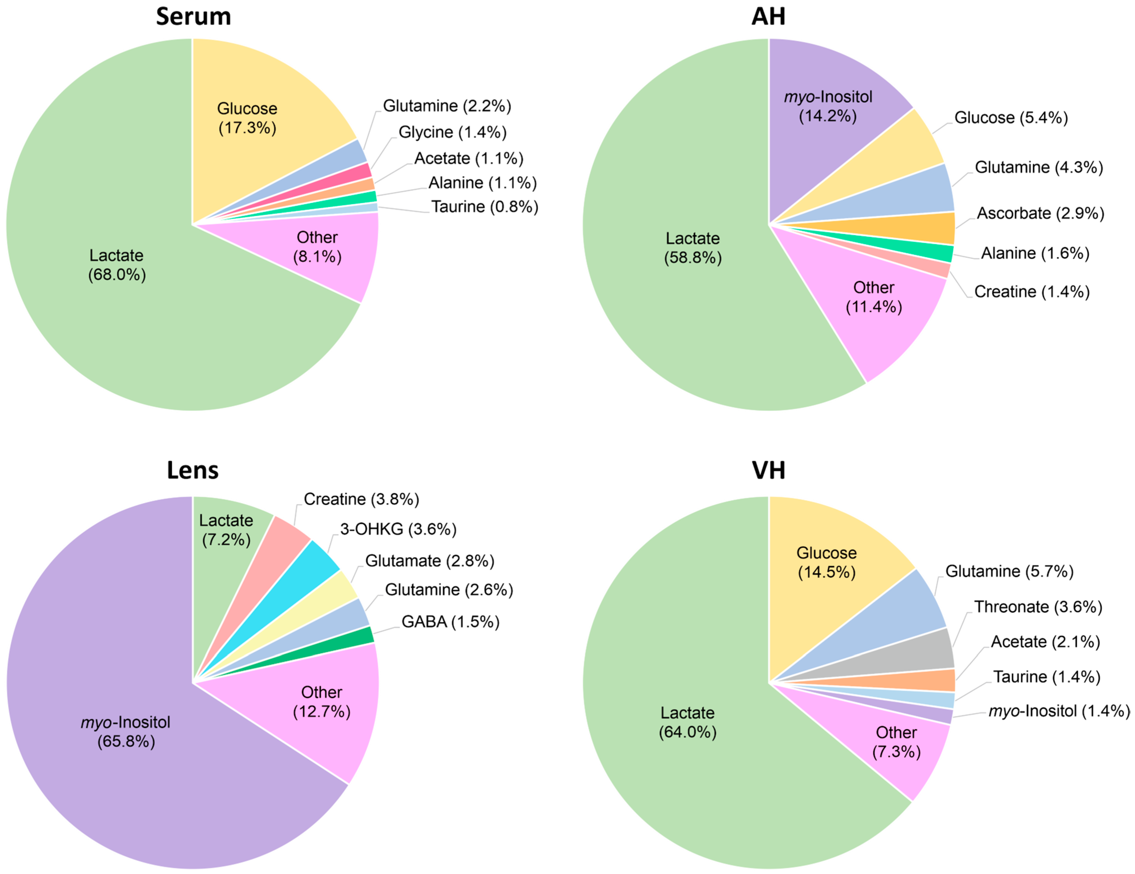
The concentrations of metabolites in the serum, AH, VH, and lens (in µM for fluids and in nmol per gram of wet tissue for the lens) were calculated by the integration of the NMR signals relative to the internal standard DSS followed by normalization to the tissue volume (or weight). In total, together with UV filters (see below), we determined the concentrations of 94 compounds, including 62 compounds in serum, 59 in AH, 45 in VH, and 67 in lens (Figure 1). The obtained quantitative metabolomic data for cytoprotective compounds—osmolytes, antioxidants, and UV filters—are given in Table 1, and the complete set of quantitative metabolomic data is presented in Table S1. A comparison of the most abundant metabolites in M. fascicularis serum, AH, VH, and lens is shown in Figure 2. Previous studies [32] showed that a single extraction according to protocol used in this work recovered approximately 90% of the metabolites. Therefore, the data in Table 1 and Table S1 should be considered as an underestimation of actual metabolite concentrations by 10%.

2.2. Identification and Quantitation of Molecular UV Filters by LC-MS and LC-OD

NMR spectra reveal only two UV filters in the M. fascicularis lens, 3-hydroxykynurenine O-β-d-glucoside (3-OHKG), and kynurenine (KN). To identify and quantify less abundant UV filters, we performed combined LC-OD and LC-MS study of macaque lenses. The UV-Vis chromatogram recorded at 360 nm for the M. fascicularis lens extract (Figure S5 and Table S2) is similar to the chromatogram reported for the human lens [33]. The signal assignment was performed according to the mass spectra obtained by LC-MS method with the use of the same column and under the same chromatographic conditions. We identified eight UV filters in the macaque lens: 3-OHKG, KN, 3-hydroxykynurenine (3-OHKN), 3-hydroxykynurenine O-β-d-diglucoside (3-OHKDG), 4-(2-amino-3-hydroxyphenyl)-4-oxobutanoic acid O-β-d-glucoside (AHBG), 4-(2-amino-3-hydroxyphenyl)-4-oxobutanoic acid O-β-d-diglucoside (AHBDG), deaminated 3-OHKG (4-(2-amino-3-hydroxyphenyl)-4-oxocrotonic acid O-β-d-glucoside, 3-OHCKAG), and previously not reported UV filter—methylated 3-OHKG (Me-3-OHKG, C17H24N2O9). Three detected UV filters remained unidentified. The major properties of UV filter compounds have been described in previous publications [33,34,35,36,37]. The quantification of UV filters was performed by their absorption spectra, assuming that all kynurenine-derived molecules have the same absorption coefficients at the UV absorption maximum near 360 nm, ε (360) = 4.5 × 10³ M−1 cm−1 [9,38]. The only exception is 3-OHCKAG, for which the value ε (410) = 1.7 × 10³ M−1 cm−1 was taken [9,34]. The most abundant UV filter in the M. fascicularis lens is 3-OHKG, which accounts to approximately 90% of the total UV filter absorbance at 360 nm, followed by AHBG and KN.

2.3. Comparison of In Vivo and Post-Mortem Blood Samples

Samples of blood, lens, AH, and VH from M. fascicularis were taken 15–20 min post-mortem. The goal of the preliminary stage of the study was to estimate the scale of post-mortem metabolomic changes occurring in these tissues. For this purpose, we compared the metabolomic profiles of M. fascicularis blood taken in vivo (seven individuals) and post-mortem (six individuals). It is known [13,15,16,39,40] that the primary post-mortem event affecting blood metabolomic composition is the disruption of the majority of energy-dependent metabolic processes, TCA cycle, purine catabolism, and functioning of Na+/K+ pumps, in particular. The disruption of TCA cycle and purine catabolism causes fast decay of pyruvate and significant elevation of fumarate, succinate, and hypoxanthine levels [13,15]. The dysfunction of Na+/K+ pumps leads to the increase in the intracellular osmotic pressure, cell lysis, and cytoplasm leakage into intercellular space, greatly elevating the levels of amino acids and other metabolites in the blood plasma [15,16,40,41]. In our measurements, we found that in post-mortem samples, the concentration of pyruvate decreased almost threefold, while the levels of fumarate, succinate, hypoxanthine, and xanthine increased by factors of 2.5, 2, 1.7, and 2.7, respectively (Table S1). At the same time, concentrations of the majority of amino acids did not change. These observations indicate that under our conditions, the post-mortem metabolomic changes in the blood are at the very beginning, and the cell lysis has not started yet. In recent reports [15,16], we demonstrated that the post-mortem changes in the ocular tissues proceed significantly more slowly than in the blood. Therefore, one can assume that the quantitative metabolomic compositions of AH, VH, and lens of M. fascicularis reported in the present work are very close to the in vivo metabolomic profiles of these tissues.

2.4. Serum Metabolomic Composition

The major constituents of the M. fascicularis serum are amino acids and organic acids; they represent 43 out of 62 compounds detected in the serum in the present work. The most abundant amino acids are proteinogenic amino acids alanine, glutamine, glycine, and lysine with concentrations between 200 and 700 µM. Among all metabolites, the highest level was found for lactate (up to 20 mM) followed by glucose (4–6 mM). We also detected small quantities of nitrogenous bases and their derivatives, including allantoin, AMP, creatinine, hypoxanthine, inosine, uridine, and xanthine. Typically, these compounds are abundant in cytoplasm, and their presence in the blood plasma might be at least partly attributed to the beginning of post-mortem processes occurring during either the post-mortem interval (for post-mortem samples) or the sample preparation (for in vivo samples).

2.5. AH Metabolomic Composition

Qualitatively, the metabolomic profiles of the blood serum and AH measured in the present work are rather similar; however, we observed some distinct quantitative differences. In particular, AH contains much lower levels of glycine, acetate, glucose, mannose, and betaine, while the concentrations of methionine, pyroglutamate, GABA, malonate, pyruvate, myo-inositol, ascorbate, ergothioneine, 3-OHKG, and AMP are much higher.

2.6. Lens Metabolomic Composition

For majority of proteinogenic amino acids, their levels in the lens are approximately twofold higher than that in AH. One should take into account that the structural lens proteins, crystallins, constitute about 40% of the lens tissue. Therefore, one can conclude that the concentrations of amino acids in the protein-free intracellular space of the lens is at least three- or fourfold higher than in AH. High amino acid demand in the lens should be attributed to the non-stop synthesis of crystallins in differentiating lens cells. Most likely, the elevated amino acid concentrations in the lens are produced by transport proteins pumping amino acids from AH against the concentration gradients [42,43,44].
An important feature of the lens metabolome is a high abundance of compounds providing the cellular protection: osmolytes, antioxidants, and UV filters. In the M. fascicularis lens, the major osmolyte is myo-inositol, the major UV filter—3-OHKG, and the major antioxidants—GSH, ascorbate, and ergothioneine.

2.7. VH Metabolomic Composition

The composition of amino acids in the M. fascicularis VH is poorer than that in other ocular tissues; we reliably detected and quantified only 11 proteinogenic amino acids, and their concentrations are lower than that in serum, AH, and lens. The most abundant metabolites in VH are lactate and glucose, which constitute more than 70% of the total metabolite abundance, followed by glutamine and threonate. As we mentioned above, a number of signals in the NMR spectrum of VH correspond to unknown compounds. Previous NMR studies of rabbit, porcine, and human VH [15,16,45] do not help to assign these signals, and their correct attribution requires a separate study. This study should include chromatographic separation of metabolomic extracts from VH into fractions, each of which contains only a few metabolites, followed by NMR and MS characterization of unknown compounds. We plan to conduct such a study in the near future.

3. Discussion

In this work, we have measured the quantitative metabolomic compositions of M. fascicularis serum and ocular tissues, AH, VH, and lens. Earlier, the metabolomic studies were performed for biological fluids of nonhuman primates, including blood, urine, milk, sperm, and cerebrospinal fluid [46,47,48,49,50,51,52,53], and the data for blood are in a good agreement with our results. Detailed quantitative metabolomic profiles of ocular tissues are published in this work for the first time.

3.1. Comparison of M. fascicularis Serum and AH Metabolomic Profiles

The Volcano plot in Figure 3 shows the comparison of metabolomic compositions of M. fascicularis AH and serum. AH is secreted by the ciliary processes through both the active secretion and the passive diffusion/ultrafiltration of blood plasma [54], and the metabolomic compositions of plasma and AH are rather similar [11].
The difference between the metabolite concentrations in serum and AH is determined by three major factors: (a) An active pumping into AH of metabolites playing key roles in the homeostasis of ocular tissues, the eye lens in particular; these pumps are located at the blood-ocular barrier; (b) a fast metabolite consumption by intraocular tissues, leading to the decrease in their concentrations in AH as compared to blood; (c) a metabolite secretion from ocular tissues into AH leading to the increase in their concentrations in AH as compared to blood. The comparison of the metabolomic data for serum and AH shows that the levels of several compounds in AH is much higher, including ascorbate, ergothioneine, 3-OHKG, myo-inositol, pyroglutamate, GABA, and AMP. Ascorbate and ergothioneine are exogenous antioxidants; the cells of primates do not synthesize these compounds, and primates obtain them only with food. However, the level of ergothioneine in AH (46 nmol/g) is higher than that in serum (3 nmol/g) by an order of magnitude; the concentration of ascorbate in AH is 590 nmol/g, while in the blood it is below the NMR detection limit. These observations clearly indicate an active transport of ascorbate and ergothioneine into AH. The pumping of ascorbate from blood into AH has been shown many years ago [11,55,56]; here, we demonstrate that the same mechanism operates for ergothioneine as well. Earlier, the existence of the ergothioneine-specific transporter (OCTN1) was discovered by Gründemann et al. [57], and the accumulation of ergothioneine in the eye was shown for mice [58,59,60]. The existence of the mechanism of the ergothioneine accumulation inside the eye indicates the importance of this compound for normal eye functioning. In contrast, UV filter 3OHKG and osmolyte myo-inositol are produced in the metabolically active lens epithelial monolayer and the outer cortex. The concentrations of these compounds in the lens are very high (Table 1), and their presence in AH should be attributed to the diffusion from the lens. Pyroglutamate, GABA, and AMP participate in many metabolic cycles, and their enhanced levels in AH as compared to blood may originate from the diffusion from different ocular tissues, including the lens.
The levels of glycine, glucose, mannose, and betaine in AH are much lower than in blood (Table S1). These metabolites are actively used in intraocular tissues for energy production and for intracellular synthesis, and their low concentrations in AH can be attributed to the fast consumption by the lens and other ocular tissues.

3.2. Comparison of Metabolomic Profiles of Human and M. fascicularis Tissues

3.2.1. Metabolomic Profiles of Serum

The similarity of metabolomic compositions of human and nonhuman primate serum has been shown in previous publications [50], and the data obtained in the present work confirm this statement. The comparison of metabolomic data on M. fascicularis serum (Table S1) with the published data on human serum [5] shows that the concentrations of the majority of amino acids, organic acids, sugars, amines, and other compounds are similar. We found significant differences for only a few metabolites. In particular, the levels of lactate, myo-inositol, and N-acetylcarnitine in M. fascicularis serum are significantly higher. This is also in a good agreement with the previous report [50].

3.2.2. Metabolomic Profiles of AH

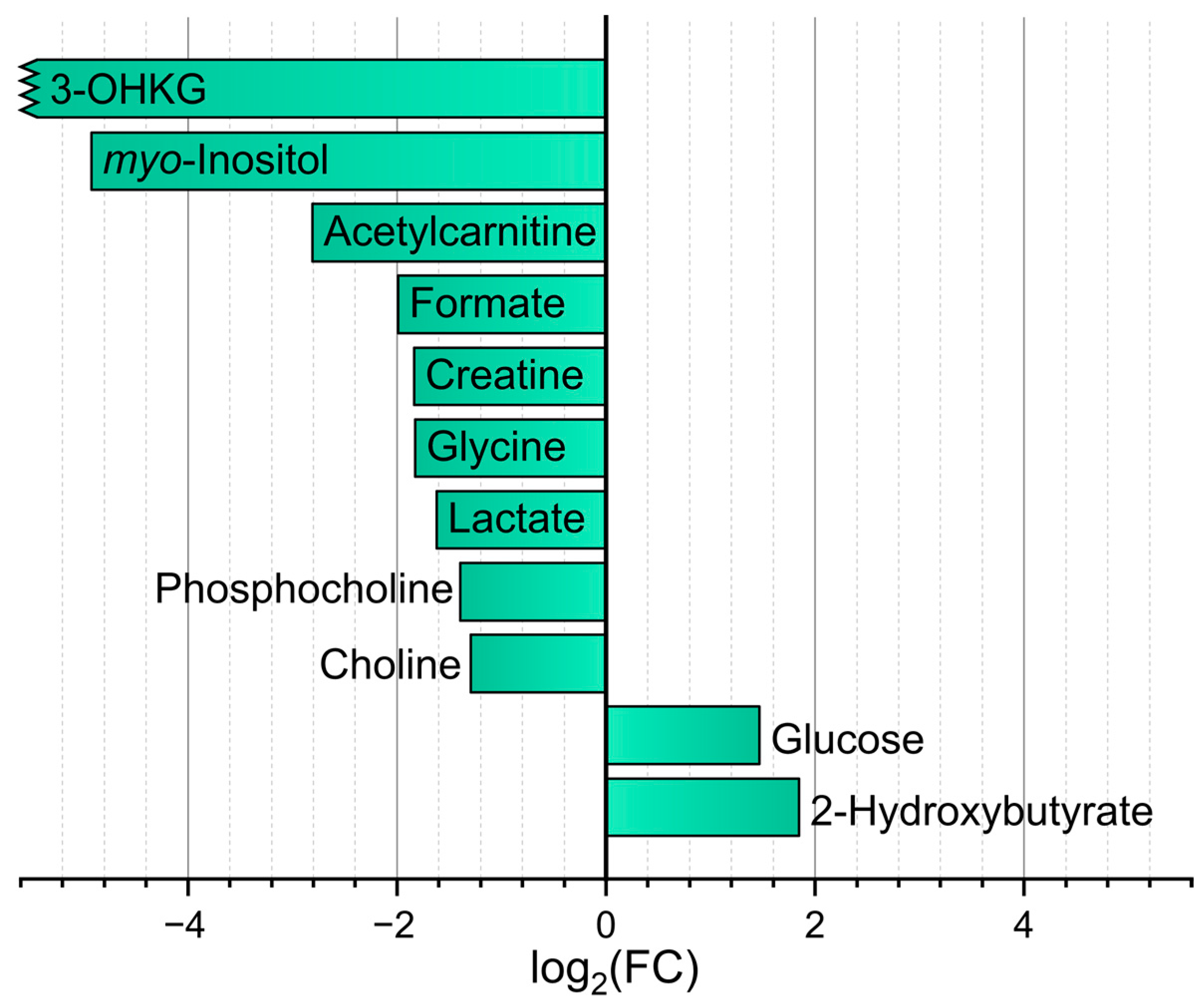
With the metabolomic difference between human and M. fascicularis, AH is somewhat more pronounced. We compared the data obtained in the present work (Table 1 and Table S1) with the recently published [10] AH metabolomic profile of young (average age of 31 years) human adults. In that work, AH samples were collected in vivo during corneal transplantation operations. Significant (fold change > 2, p < 0.01 (FDR)) difference was found for 11 metabolites (Figure 4). Among them, two metabolites have higher abundance in the human AH, and nine in the M. fascicularis AH. It should be noted that the levels of myo-inositol and 3-OHKG in the M. fascicularis lens is much higher than that in the human lens, and the higher abundance of these compounds in M. fascicularis AH as compared to human AH most likely corresponds to their diffusion from the lens.

3.2.3. Metabolomic Profiles of Lens

From a biomedical viewpoint, the comparison of M. fascicularis and human lenses is the most interesting one. Due to the ethical restrictions, normal human lenses with the short post-mortem interval (PMI) are practically unavailable, and the majority of ophthalmological experimental studies (especially the cataract-related studies) are performed with the use of experimental animals, usually rodents. For that reason, we decided to compare the metabolomic profile of M. fascicularis lens with the previously obtained metabolomic data on the human and rat eye lenses [9,20]. The data in the paper [9] are given for normal human lenses with the post-mortem intervals of 7–17 h. Apparently, significant metabolomic changes did occur during the PMI, including the depletion of energy metabolites, lactate accumulation, changes in concentrations of compound from the TCA cycle, and elevation of amino acid levels due to the protein hydrolysis [13,15,16,39,40,61]. Therefore, we restricted our comparison to compounds protecting the lens tissue against the cataract development.
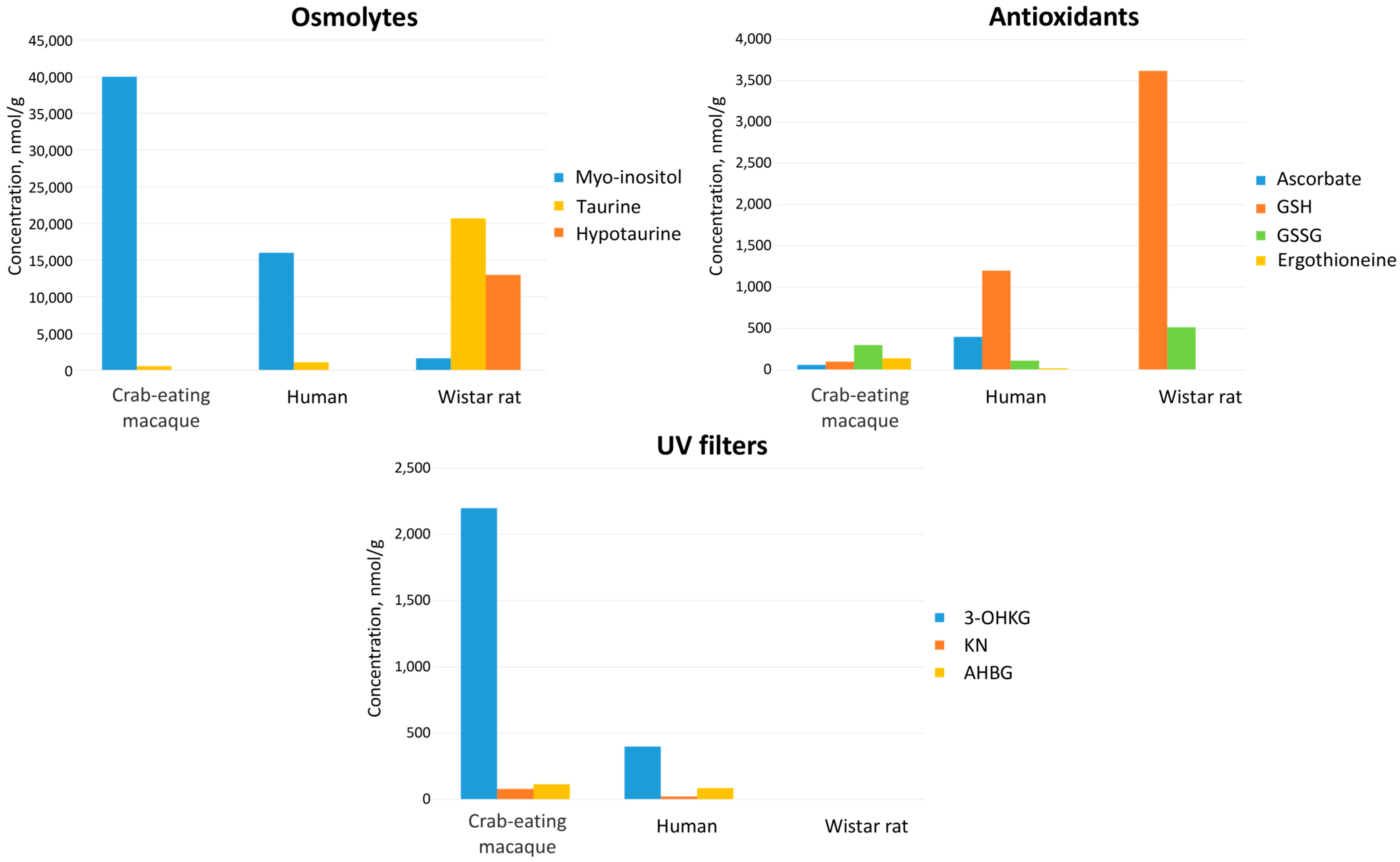
Cataract is the most common age-related eye disease. It is generally accepted that the main cause of the cataract development is the oxidative stress leading to the post-translational modifications of crystallins, their insolubilization, aggregation, and precipitation [62]. Violation of the correct laying of fiber cells in the lens may also contribute to the cataractogenesis [63]. Due to very low metabolic activity in the lens nucleus, the defense of the lens cells against UV radiation, oxidative, and osmotic stresses rely on cytoprotective molecules—UV filters, antioxidants, and osmolytes. Figure 5 shows the graphical representation of the cytoprotective compound compositions in the human, M. fascicularis, and rat lenses. Qualitatively, the sets of cytoprotective metabolites in the human and M. fascicularis lenses are very similar, and differ significantly from that in the rat lens. The main antioxidants in the both human [9] and M. fascicularis lenses are reduced glutathione (GSH), ascorbate, and ergothioneine, while in the rat lens, only GSH is present at the significant level [20], and its abundance is approximately threefold higher than that in the human lens. The major osmolytes in the rat lens are taurine and hypotaurine, while in the human and M. fascicularis lenses it is myo-inositol. The human and M. fascicularis lenses contain high concentrations of 3-OHKG and other kynurenine derivatives acting as UV filters, while the rat lens does not contain UV filters.
Nevertheless, quantitatively, the metabolomes of the human and M. fascicularis lenses differ. The levels of ascorbate and GSH in the human eye lens are almost an order of magnitude higher than that in the M. fascicularis lens, while the concentration of ergothioneine is almost an order of magnitude lower. The abundance of the major lens osmolyte, myo-inositol, in the M. fascicularis lens is almost three times higher than that in the human lens, and the level of the major UV filter, 3-OHKG, is approximately five times higher. This difference, at least in part, could be attributed to the age-related factor. It has been reported that 3-OHKG concentration in the human lens decreases with age [33], and myo-inositol concentration decreases in rat [64] and presumably in human lens [65,66]. The human lenses in work [9] were from individuals after middle age, while the lenses studied in the present work were taken from young (four-year-old) monkeys. In addition to that, the human lens contains a substantial amount of GSH-3-OHKG—a product of the GSH addition to 3-OHCKAG [33,35,67]—which, in turn, is the product of 3-OHKG deamination [34,68,69]. In the M. fascicularis lens, we did not detect this compound. This difference can be attributed to two major factors. First, the formation of GSH-3-OHKG in the human lens occurs mostly after middle age [33], again pointing at the age-related factor. Perhaps, in lenses of old macaques, GSH-3-OHKG is also present in detectable amounts. Second, the GSH level in the M. fascicularis lens is much lower than that in the human lens, so the probability to form GSH-3-OHKG adduct is lower. Nevertheless, it is difficult to judge from the data obtained whether the observed quantitative metabolomic differences between M. fascicularis and human lenses should be attributed to the different ages, lifestyles, and feeding behaviors, or if they are genetically fixed.

4. Materials and Methods

4.1. Sample Collection

All experiments involving animals were performed in accordance with the ARVO Statement for the Use of Animals in Ophthalmic and Vision Research and the European Union Directive 2010/63/EU on the protection of animals used for scientific purposes, and approved by bioethics committee of the Research Institute of Medical Primatology (Record 80/1 from 2 December 2021).
AH, VH, lens, and plasma were obtained from six 4-year-old crab-eating macaque (Macaca fascicularis) species (3 female and 3 male); in vivo plasma was obtained from seven other 4-year-old species (3 female and 4 male). In vivo and post-mortem blood samples were collected by venipuncture using a syringe with a 21G needle. Within 10–15 min after the blood collection, blood samples were centrifuged (3000× g, 10 min), and the plasma obtained was transferred into separate vials. The plasma samples were immediately frozen and kept at −70 °C until analysis.
Euthanasia was performed by intravenous administration of 5.0 mL of Anestofol 5% (Interpharm, Moscow, Russia) with preliminary general anesthesia by intravenous injection of 0.10 mL/kg of Xila (2% xylazine) (Interchemie Werken “de Adelaar” BV, Castenray, The Netherlands) and 0.05 mL/kg of Zoletil® (Virbac Sante Animale, Carros, France). The samples of AH were taken from the anterior chamber by the cornea puncture with a 21G needle. Then, the cornea was cut along the iris border, the lens was extracted, and VH was collected with the 21G needle. The samples of AH, VH, and lens were frozen and kept at −70 °C.

4.2. Sample Preparation

Sample preparation for all biological fluids and tissues was performed according to the procedures described in refs [15,16,61]. Typical lens weight was 90–100 mg, typical plasma volume was 300 µL, VH volume was 600 µL, and AH volume was 70–200 µL. Lenses were placed in glass vials and homogenized with a TissueRuptor II homogenizer (Qiagen, Venlo, The Netherlands). Biological fluids and lens homogenate were subjected to liquid–liquid extraction in methanol–chloroform–water mixture. Metabolomic extracts were collected, divided into two parts for NMR (2/3) and LC–MS (1/3) analyses, and lyophilized.

4.3. NMR Measurements

1H NMR spectra were obtained as described in Ref. [61]. Briefly, we re-dissolved the extracts for NMR measurements in 600 μL of D2O containing 2 × 10⁻⁵ M sodium 4,4-dimethyl-4-silapentane-1-sulfonic acid (DSS) as an internal standard and 50 mM deuterated phosphate buffer (pH 7.2). We performed the NMR measurements at the Center of Collective Use “Mass spectrometric investigations” SB RAS with the use of an NMR spectrometer AVANCE III HD 700 MHz (Bruker BioSpin, Ettlingen, Germany) equipped with a 16.44 Tesla Ascend cryomagnet. The spectra were obtained with the use of zgpr sequence with the water signal suppression, 10 s relaxation delay, 6.7 s acquisition time, 70° detection pulse, and 64 scans. Preliminary measurements showed that for most metabolites, the T1 relaxation time ranged from 1.5 to 4 s. The longest relaxation times were found for histidine (7.6 s) and acetate (6.2 s) protons. Therefore, under our experimental conditions (total scan time 16.7 s, and 70° detection pulse) correct concentration values can be expected for all metabolites.

4.4. LC-MS and LC-OD Measurements

We performed liquid chromatography with mass spectrometric (LC-MS) and UV-Vis (LC-OD) measurements simultaneously on two instrumental setups with the same HPLC conditions. The extracts for LC-MS and LC-OD analyses were re-dissolved in 100 μL of 10 mM ammonium formate and 0.1% formic acid (v/v) solution in H2O. The LC separation was performed on an Intensity Solo 2 C18 column (2 × 100 mm, 2 µm, 100 Å, Bruker Daltonics) with a pre-column. The LC conditions were as follows: mobile phase A—10 mM ammonium formate in H2O, mobile phase B—acetonitrile, both with the presence of formic acid 0.1%; flow 0.2 mL/min; the gradient was 5% B (0–3 min), 5–95% B (3–20 min), 95% B (20–25 min), 95–5% B (25–25.5 min), and 5% B (25.5–35 min); the injection volume was 10 μL.
The LC-OD setup consisted of an LC 1200 HPLC system (Agilent Technologies, Santa Clara, CA, USA), equipped with a quaternary pump, an autosampler, and a diode array detector (DAD). The chromatograms were monitored at 220, 260, 280, 360, and 450 nm and the analysis of LC-OD data was performed using either ChemStation revision B.04.03 software (Agilent Technologies, Santa Clara, CA, USA) for these fixed wavelengths or OpenChrom v.1.5.0 software (Lablicate GmbH, Hamburg, Germany) for other wavelengths from LC-DAD data.
The LC-MS setup consisted of an Elute UHPLC chromatograph (a binary pump with HPG, an autosampler, and a column thermostat) with an Impact II high-resolution electrospray quadrupole time-of-flight (ESI-Q-TOF) mass spectrometer (Bruker Daltonics, Germany). Mass spectra were recorded in the positive ion mode over the 50–1500 m/z range with 3 Hz scan rate. Each LC-MS chromatogram contained a calibration segment in the void, where sodium formate clusters used as a calibrant were supplied by a syringe pump connected to an ESI source via a divert valve. The typical resolution was ca. 30,000 and the accuracy <1 ppm. The data obtained were analyzed using DataAnalysis 5.3 software (Bruker Daltonics, Germany).
LC-MS and LC-OD setups were used only for identification and quantification of UV filters. The quantification was performed by their absorption at 360 nm using kynurenine as a chemical standard, assuming that these kynurenine derivatives have the same absorption coefficients at 360 nm (ε360 = 4.5 × 10³ M−1 cm−1).

4.5. Data Analysis

The phase and baseline corrections as well as signal integration in NMR spectra were performed using MestReNova v.12 software (Mestrelab Research S.L., A Coruna, Spain). Metabolite identification was based on chemical shifts and multiplicities for individual compounds obtained from the HMDB (https://hmdb.ca, accessed on 13 November 2023) and AMDB (https://amdb.online, accessed on 13 November 2023) databases, Chenomx NMR Suite v.8 (Chenomx Inc., Edmonton, AB, Canada), and from our previous reports on lens and other tissues’ metabolomic composition [8,9,16,20,61]. The concentrations of metabolites in the samples were determined by the peak area integration relative to the internal standard DSS, and then recalculated into metabolite concentrations in the tissue (for the lens, in nmoles per gram of the tissue wet weight, and for AH, VH, and serum, in nmoles per milliliter).
Statistical treatment of metabolomics data was performed at the MetaboAnalyst 5.0 web-platform (https://www.metaboanalyst.ca, accessed on 13 November 2023, [70]). A Volcano plot was built with the following parameters: Mann–Whitney U-test with the false discovery rate (FDR) correction of p-values, and the threshold for p-value < 0.01 and fold change (FC) above 2.

5. Conclusions

The results obtained in the present work indicate that the metabolic processes in the ocular tissues of humans and nonhuman primates proceed in a similar way. First, the qualitative metabolomic compositions of such tissues as serum, AH, and lens are very similar. In particular, the set of lenticular cytoprotective compounds—antioxidants, osmolytes, and UV filters—are practically the same for both humans and macaques. Second, there is an active transport of exogenous antioxidants ascorbate and ergothioneine from blood into AH, greatly enhancing their concentrations in the ocular tissues. Third, the concentrations of the majority of proteinogenic amino acids in the lenses of humans and M. fascicularis are significantly higher than that in AH. This indicates an active pumping of amino acids into lens against the concentration gradient for both species. Finally, similar combinations of kynurenine-like molecules show that the mechanisms of the synthesis and degradation of UV filters in the lens of human and nonhuman primates are the same. Therefore, from a biochemical viewpoint, nonhuman primates are excellent models for studying human ophthalmological diseases.

Supplementary Materials

The following supporting information can be downloaded at https://www.mdpi.com/article/10.3390/ijms262210816/s1.

Author Contributions

Conceptualization, methodology, writing—original draft preparation, project administration, data curation, Y.P.T.; supervision, R.Z.S.; formal analysis, M.V.F. and L.V.Y.; visualization, M.V.F.; investigation, M.V.F., L.V.Y., V.V.Y. and Y.P.T.; writing—review and editing, L.V.Y., R.Z.S. and V.V.Y.; resources, E.Y.R. and D.V.B. All authors have read and agreed to the published version of the manuscript.

Funding

This research was funded by Russian Science Foundation, grant number 25-14-00035, https://rscf.ru/en/project/25-14-00035/ (accessed on 7 September 2025).

Institutional Review Board Statement

The study was conducted in accordance with the ARVO Statement for the Use of Animals in Ophthalmic and Vision Research and the European Union Directive 2010/63/EU on the protection of animals used for scientific purposes, and with ethical approval from the Research Institute of Medical Primatology (Record 80/1 from 2 December 2021).

Informed Consent Statement

Not applicable.

Data Availability Statement

Raw NMR spectra, description of specimens and samples, metabolite concentrations, and the preliminary metabolomic analysis is available at our Animal Metabolite Database repository, Experiment ID 273 (https://amdb.online/amdb/experiments/273/, accessed on 7 September 2025) and at the MetaboLights repository, study identifier MTBLS8943 (https://www.ebi.ac.uk/metabolights/MTBLS8943/, accessed on 7 September 2025). All other data are available from the corresponding author upon request.

Acknowledgments

Authors thank the Ministry of Science and Higher Education of the RF for access to NMR and LC-MS equipment.

Conflicts of Interest

The authors declare no conflicts of interest.

Abbreviations

The following abbreviations are used in this manuscript:
(U)HPLC(ultra)high performance liquid chromatography
3-OHCKAG4-(2-amino-3-hydroxyphenyl)-4-oxocrotonic acid O-β-d-glucoside
3-OHKDG3-hydroxykynurenine O-β-d-diglucoside
3-OHKG3-hydroxykynurenine O-β-d-glucoside
3-OHKN3-hydroxykynurenine
AHaqueous humor
AHBDG4-(2-amino-3-hydroxyphenyl)-4-oxobutanoic acid O-β-d-diglucoside
AHBG4-(2-amino-3-hydroxyphenyl)-4-oxobutanoic acid O-β-d-glucoside
AMPadenosine monophosphate
DSSsodium 4,4-dimethyl-4-silapentane-1-sulfonate
FCfold change
FDRfalse discovery rate
GABAgamma-aminobutyric acid
GSHglutathione
KNkynurenine
LC-MSliquid chromatography with mass spectrometric detection
LC-ODliquid chromatography with optical detection
Me-3-OHKGmethylated 3-OHKG
NMRnuclear magnetic resonance
PMIpost-mortem interval
SDstandard deviation
TCAtricarboxylic acid cycle
UVultraviolet
VHvitreous humor

References

  1. Madsen, R.; Lundstedt, T.; Trygg, J. Chemometrics in Metabolomics—A Review in Human Disease Diagnosis. Anal. Chim. Acta 2010, 659, 23–33. [Google Scholar] [CrossRef]
  2. Emwas, A.-H.M.; Salek, R.M.; Griffin, J.L.; Merzaban, J. NMR-Based Metabolomics in Human Disease Diagnosis: Applications, Limitations, and Recommendations. Metabolomics 2013, 9, 1048–1072. [Google Scholar] [CrossRef]
  3. Karczewski, K.J.; Snyder, M.P. Integrative Omics for Health and Disease. Nat. Rev. Genet. 2018, 19, 299–310. [Google Scholar] [CrossRef]
  4. Suhre, K.; Arnold, M.; Bhagwat, A.M.; Cotton, R.J.; Engelke, R.; Raffler, J.; Sarwath, H.; Thareja, G.; Wahl, A.; DeLisle, R.K.; et al. Connecting Genetic Risk to Disease End Points through the Human Blood Plasma Proteome. Nat. Commun. 2017, 8, 14357, Erratum in Nat. Commun. 2017, 8, 15345. [Google Scholar] [CrossRef]
  5. Wishart, D.S.; Guo, A.; Oler, E.; Wang, F.; Anjum, A.; Peters, H.; Dizon, R.; Sayeeda, Z.; Tian, S.; Lee, B.L.; et al. HMDB 5.0: The Human Metabolome Database for 2022. Nucleic Acids Res. 2021, 50, D622–D631. [Google Scholar] [CrossRef]
  6. Kryczka, T.; Ehlers, N.; Nielsen, K.; Wylegala, E.; Dobrowolski, D.; Midelfart, A. Metabolic Profile of Keratoconic Cornea. Curr. Eye Res. 2013, 38, 305–309. [Google Scholar] [CrossRef] [PubMed]
  7. Kryczka, T.; Wylęgała, E.; Dobrowolski, D.; Midelfart, A. NMR Spectroscopy of Human Eye Tissues: A New Insight into Ocular Biochemistry. Sci. World J. 2014, 2014, 546192. [Google Scholar] [CrossRef] [PubMed]
  8. Tsentalovich, Y.P.; Verkhovod, T.D.; Yanshole, V.V.; Kiryutin, A.S.; Yanshole, L.V.; Fursova, A.Z.; Stepakov, D.A.; Novoselov, V.P.; Sagdeev, R.Z. Metabolomic Composition of Normal Aged and Cataractous Human Lenses. Exp. Eye Res. 2015, 134, 15–23. [Google Scholar] [CrossRef]
  9. Yanshole, V.V.; Yanshole, L.V.; Snytnikova, O.A.; Tsentalovich, Y.P. Quantitative Metabolomic Analysis of Changes in the Lens and Aqueous Humor under Development of Age-Related Nuclear Cataract. Metabolomics 2019, 15, 29. [Google Scholar] [CrossRef]
  10. Snytnikova, O.A.; Yanshole, L.V.; Iskakov, I.A.; Yanshole, V.V.; Chernykh, V.V.; Stepakov, D.A.; Novoselov, V.P.; Tsentalovich, Y.P. Quantitative Metabolomic Analysis of the Human Cornea and Aqueous Humor. Metabolomics 2017, 13, 152. [Google Scholar] [CrossRef]
  11. Snytnikova, O.A.; Khlichkina, A.A.; Yanshole, L.V.; Yanshole, V.V.; Iskakov, I.A.; Egorova, E.V.; Stepakov, D.A.; Novoselov, V.P.; Tsentalovich, Y.P. Metabolomics of the Human Aqueous Humor. Metabolomics 2017, 13, 5. [Google Scholar] [CrossRef]
  12. Pietrowska, K.; Dmuchowska, D.A.; Krasnicki, P.; Bujalska, A.; Samczuk, P.; Parfieniuk, E.; Kowalczyk, T.; Wojnar, M.; Mariak, Z.; Kretowski, A.; et al. An Exploratory LC-MS-Based Metabolomics Study Reveals Differences in Aqueous Humor Composition between Diabetic and Non-Diabetic Patients with Cataract. Electrophoresis 2018, 39, 1233–1240. [Google Scholar] [CrossRef] [PubMed]
  13. Donaldson, A.E.; Lamont, I.L. Biochemistry Changes That Occur after Death: Potential Markers for Determining Post-Mortem Interval. PLoS ONE 2013, 8, e82011. [Google Scholar] [CrossRef]
  14. Palmiere, C.; Mangin, P. Urea Nitrogen, Creatinine, and Uric Acid Levels in Postmortem Serum, Vitreous Humor, and Pericardial Fluid. Int. J. Leg. Med. 2015, 129, 301–305. [Google Scholar] [CrossRef]
  15. Zelentsova, E.A.; Yanshole, L.V.; Snytnikova, O.A.; Yanshole, V.V.; Tsentalovich, Y.P.; Sagdeev, R.Z. Post-Mortem Changes in the Metabolomic Compositions of Rabbit Blood, Aqueous and Vitreous Humors. Metabolomics 2016, 12, 172. [Google Scholar] [CrossRef]
  16. Zelentsova, E.A.; Yanshole, L.V.; Melnikov, A.D.; Kudryavtsev, I.S.; Novoselov, V.P.; Tsentalovich, Y.P. Post-Mortem Changes in Metabolomic Profiles of Human Serum, Aqueous Humor and Vitreous Humor. Metabolomics 2020, 16, 80. [Google Scholar] [CrossRef]
  17. Locci, E.; Stocchero, M.; Noto, A.; Chighine, A.; Natali, L.; Napoli, P.E.; Caria, R.; De-Giorgio, F.; Nioi, M.; d’Aloja, E. A 1H NMR Metabolomic Approach for the Estimation of the Time since Death Using Aqueous Humour: An Animal Model. Metabolomics 2019, 15, 76. [Google Scholar] [CrossRef]
  18. Zigler, J.S.; Bodaness, R.S.; Gery, I.; Kinoshita, J.H. Effects of Lipid Peroxidation Products on the Rat Lens in Organ Culture: A Possible Mechanism of Cataract Initiation in Retinal Degenerative Disease. Arch. Biochem. Biophys. 1983, 225, 149–156. [Google Scholar] [CrossRef]
  19. Son, H.-Y.; Kim, H.; Kwon, Y.H. Taurine Prevents Oxidative Damage of High Glucose-Induced Cataractogenesis in Isolated Rat Lenses. J. Nutr. Sci. Vitaminol. 2007, 53, 324–330. [Google Scholar] [CrossRef]
  20. Yanshole, V.V.; Snytnikova, O.A.; Kiryutin, A.S.; Yanshole, L.V.; Sagdeev, R.Z.; Tsentalovich, Y.P. Metabolomics of the Rat Lens: A Combined LC-MS and NMR Study. Exp. Eye Res. 2014, 125, 71–78. [Google Scholar] [CrossRef] [PubMed]
  21. Tan, S.Z.; Mullard, G.; Hollywood, K.A.; Dunn, W.B.; Bishop, P.N. Characterisation of the Metabolome of Ocular Tissues and Post-Mortem Changes in the Rat Retina. Exp. Eye Res. 2016, 149, 8–15. [Google Scholar] [CrossRef]
  22. Mustari, M.J. Nonhuman Primate Studies to Advance Vision Science and Prevent Blindness. ILAR J. 2017, 58, 216–225. [Google Scholar] [CrossRef] [PubMed]
  23. Rasmussen, C.A.; Kaufman, P.L. Primate Glaucoma Models. J. Glaucoma 2005, 14, 311. [Google Scholar] [CrossRef] [PubMed]
  24. Willoughby, C.L.; Fleuriet, J.; Walton, M.M.; Mustari, M.J.; McLoon, L.K. Adaptation of Slow Myofibers: The Effect of Sustained BDNF Treatment of Extraocular Muscles in Infant Nonhuman Primates. Investig. Ophthalmol. Vis. Sci. 2015, 56, 3467–3483. [Google Scholar] [CrossRef]
  25. Lambert, W.S.; Carlson, B.J.; Ghose, P.; Vest, V.D.; Yao, V.; Calkins, D.J. Towards A Microbead Occlusion Model of Glaucoma for a Non-Human Primate. Sci. Rep. 2019, 9, 11572. [Google Scholar] [CrossRef]
  26. Liu, Z.; Liow, S.S.; Lai, S.L.; Alli-Shaik, A.; Holder, G.E.; Parikh, B.H.; Krishnakumar, S.; Li, Z.; Tan, M.J.; Gunaratne, J.; et al. Retinal-Detachment Repair and Vitreous-like-Body Reformation via a Thermogelling Polymer Endotamponade. Nat. Biomed. Eng. 2019, 3, 598–610. [Google Scholar] [CrossRef]
  27. Gaillard, E.R.; Zheng, L.; Merriam, J.C.; Dillon, J. Age-Related Changes in the Absorption Characteristics of the Primate Lens. Investig. Ophthalmol. Vis. Sci. 2000, 41, 1454–1459. [Google Scholar]
  28. Gaillard, E.R.; Merriam, J.; Zheng, L.; Dillon, J. Transmission of Light to the Young Primate Retina: Possible Implications for the Formation of Lipofuscin. Photochem. Photobiol. 2011, 87, 18–21. [Google Scholar] [CrossRef]
  29. Barba, I.; Garcia-Ramírez, M.; Hernández, C.; Alonso, M.A.; Masmiquel, L.; García-Dorado, D.; Simó, R. Metabolic Fingerprints of Proliferative Diabetic Retinopathy: An 1H-NMR–Based Metabonomic Approach Using Vitreous Humor. Investig. Ophthalmol. Vis. Sci. 2010, 51, 4416–4421. [Google Scholar] [CrossRef]
  30. Nagana Gowda, G.A.; Raftery, D. Quantitating Metabolites in Protein Precipitated Serum Using NMR Spectroscopy. Anal. Chem. 2014, 86, 5433–5440. [Google Scholar] [CrossRef] [PubMed]
  31. Gowda, N.G.A.; Gowda, Y.N.; Raftery, D. Expanding the Limits of Human Blood Metabolite Quantitation Using NMR Spectroscopy. Anal. Chem. 2015, 87, 706–715. [Google Scholar] [CrossRef]
  32. Snytnikova, O.A.; Khlichkina, A.A.; Sagdeev, R.Z.; Tsentalovich, Y.P. Evaluation of Sample Preparation Protocols for Quantitative NMR-Based Metabolomics. Metabolomics 2019, 15, 84. [Google Scholar] [CrossRef]
  33. Bova, L.M.; Sweeney, M.H.; Jamie, J.F.; Truscott, R.J. Major Changes in Human Ocular UV Protection with Age. Investig. Ophthalmol. Vis. Sci. 2001, 42, 200–205. [Google Scholar]
  34. Tsentalovich, Y.P.; Snytnikova, O.A.; Forbes, M.D.E.; Chernyak, E.I.; Morozov, S.V. Photochemical and Thermal Reactivity of Kynurenine. Exp. Eye Res. 2006, 83, 1439–1445. [Google Scholar] [CrossRef]
  35. Taylor, L.M.; Andrew Aquilina, J.; Jamie, J.F.; Truscott, R.J.W. UV Filter Instability: Consequences for the Human Lens. Exp. Eye Res. 2002, 75, 165–175. [Google Scholar] [CrossRef]
  36. Sherin, P.S.; Grilj, J.; Tsentalovich, Y.P.; Vauthey, E. Ultrafast Excited-State Dynamics of Kynurenine, a UV Filter of the Human Eye. J. Phys. Chem. B 2009, 113, 4953–4962. [Google Scholar] [CrossRef]
  37. Tsentalovich, Y.P.; Sherin, P.S.; Kopylova, L.V.; Cherepanov, I.V.; Grilj, J.; Vauthey, E. Photochemical Properties of UV Filter Molecules of the Human Eye. Investig. Ophthalmol. Vis. Sci. 2011, 52, 7687–7696. [Google Scholar] [CrossRef] [PubMed]
  38. Tsentalovich, Y.P.; Snytnikova, O.A.; Sherin, P.S.; Forbes, M.D.E. Photochemistry of Kynurenine, a Tryptophan Metabolite: Properties of the Triplet State. J. Phys. Chem. A 2005, 109, 3565–3568. [Google Scholar] [CrossRef] [PubMed]
  39. Donaldson, A.E.; Lamont, I.L. Estimation of Post-Mortem Interval Using Biochemical Markers. Aust. J. Forensic Sci. 2014, 46, 8–26. [Google Scholar] [CrossRef]
  40. Donaldson, A.E.; Lamont, I.L. Metabolomics of Post-Mortem Blood: Identifying Potential Markers of Post-Mortem Interval. Metabolomics 2015, 11, 237–245. [Google Scholar] [CrossRef]
  41. Cotran, R.S.; Kumar, V.; Robbins, S.L. Cellular Injury and Cellular Death, 5th ed.; W.B. Saunders Company: Philadelphia, PA, USA, 1994. [Google Scholar]
  42. Marcantonio, J.; Duncan, G. Amino Acid Transport and Protein Synthesis in Human Normal and Cataractous Lenses. Curr. Eye Res. 1987, 6, 1299–1308. [Google Scholar] [CrossRef] [PubMed]
  43. Lim, J.; Lorentzen, K.A.; Kistler, J.; Donaldson, P.J. Molecular Identification and Characterisation of the Glycine Transporter (GLYT1) and the Glutamine/Glutamate Transporter (ASCT2) in the Rat Lens. Exp. Eye Res. 2006, 83, 447–455. [Google Scholar] [CrossRef]
  44. Knöpfel, E.B.; Vilches, C.; Camargo, S.M.R.; Errasti-Murugarren, E.; Stäubli, A.; Mayayo, C.; Munier, F.L.; Miroshnikova, N.; Poncet, N.; Junza, A.; et al. Dysfunctional LAT2 Amino Acid Transporter Is Associated With Cataract in Mouse and Humans. Front. Physiol. 2019, 10, 688. [Google Scholar] [CrossRef]
  45. Elmi, A.; Ventrella, D.; Laghi, L.; Carnevali, G.; Zhu, C.; Pertile, G.; Barone, F.; Benfenati, F.; Bacci, M.L. 1H NMR Spectroscopy Characterization of Porcine Vitreous Humor in Physiological and Photoreceptor Degeneration Conditions. Investig. Ophthalmol. Vis. Sci. 2019, 60, 741–747. [Google Scholar] [CrossRef]
  46. Hung, P.; Froenicke, L.; Lin, C.Y.; Lyons, L.A.; Miller, M.G.; Pinkerton, K.E.; VandeVoort, C.A. Effects of Environmental Tobacco Smoke in Vivo on Rhesus Monkey Semen Quality, Sperm Function, and Sperm Metabolism. Reprod. Toxicol. 2009, 27, 140–148. [Google Scholar] [CrossRef]
  47. Lin, C.-Y.; Hung, P.; VandeVoort, C.A.; Miller, M.G. 1H NMR to Investigate Metabolism and Energy Supply in Rhesus Macaque Sperm. Reprod. Toxicol. 2009, 28, 75–80. [Google Scholar] [CrossRef] [PubMed]
  48. Rezzi, S.; Martin, F.-P.J.; Shanmuganayagam, D.; Colman, R.J.; Nicholson, J.K.; Weindruch, R. Metabolic Shifts Due to Long-Term Caloric Restriction Revealed in Nonhuman Primates. Exp. Gerontol. 2009, 44, 356–362. [Google Scholar] [CrossRef]
  49. Rao, R.; Ennis, K.; Oz, G.; Lubach, G.R.; Georgieff, M.K.; Coe, C.L. Metabolomic Analysis of Cerebrospinal Fluid Indicates Iron Deficiency Compromises Cerebral Energy Metabolism in the Infant Monkey. Neurochem. Res. 2013, 38, 573–580. [Google Scholar] [CrossRef]
  50. O’Sullivan, A.; He, X.; McNiven, E.M.S.; Hinde, K.; Haggarty, N.W.; Lönnerdal, B.; Slupsky, C.M. Metabolomic Phenotyping Validates the Infant Rhesus Monkey as a Model of Human Infant Metabolism. J. Pediatr. Gastroenterol. Nutr. 2013, 56, 355. [Google Scholar] [CrossRef] [PubMed]
  51. Zheng, S.; Tan, W.; Li, X.; Li, B.; Gong, B.; Pyle, W.G.; Wu, J.; Li, L.; Luo, T.; Zou, Y.; et al. Aged Monkeys Fed a High-Fat/High-Sugar Diet Recapitulate Metabolic Disorders and Cardiac Contractile Dysfunction. J. Cardiovasc. Trans. Res. 2021, 14, 799–815. [Google Scholar] [CrossRef]
  52. Crook, A.; De Lima Leite, A.; Payne, T.; Bhinderwala, F.; Woods, J.; Singh, V.K.; Powers, R. Radiation Exposure Induces Cross-Species Temporal Metabolic Changes That Are Mitigated in Mice by Amifostine. Sci. Rep. 2021, 11, 14004. [Google Scholar] [CrossRef]
  53. Hasegawa, Y.; Zhang, Z.; Taha, A.Y.; Capitanio, J.P.; Bauman, M.D.; Golub, M.S.; Van de Water, J.; VandeVoort, C.A.; Walker, C.K.; Slupsky, C.M. Impact of Maternal Obesity on the Gestational Metabolome and Infant Metabolome, Brain, and Behavioral Development in Rhesus Macaques. Metabolites 2022, 12, 764. [Google Scholar] [CrossRef]
  54. Civan, M.M.; Macknight, A.D.C. The Ins and Outs of Aqueous Humour Secretion. Exp. Eye Res. 2004, 78, 625–631. [Google Scholar] [CrossRef]
  55. DiMattio, J. A Comparative Study of Ascorbic Acid Entry into Aqueous and Vitreous Humors of the Rat and Guinea Pig. Investig. Ophthalmol. Vis. Sci. 1989, 30, 2320–2331. [Google Scholar]
  56. Delamere, N.A. Ascorbic Acid and the Eye. In Subcellular Biochemistry: Ascorbic Acid: Biochemistry and Biomedical Cell Biology; Harris, J.R., Ed.; Subcellular Biochemistry; Springer: Boston, MA, USA, 1996; pp. 313–329. ISBN 978-1-4613-0325-1. [Google Scholar]
  57. Gründemann, D.; Harlfinger, S.; Golz, S.; Geerts, A.; Lazar, A.; Berkels, R.; Jung, N.; Rubbert, A.; Schömig, E. Discovery of the Ergothioneine Transporter. Proc. Natl. Acad. Sci. USA 2005, 102, 5256–5261. [Google Scholar] [CrossRef]
  58. Tang, R.M.Y.; Cheah, I.K.-M.; Yew, T.S.K.; Halliwell, B. Distribution and Accumulation of Dietary Ergothioneine and Its Metabolites in Mouse Tissues. Sci. Rep. 2018, 8, 1601. [Google Scholar] [CrossRef]
  59. Halliwell, B.; Cheah, I.K.; Tang, R.M.Y. Ergothioneine—A Diet-Derived Antioxidant with Therapeutic Potential. FEBS Lett. 2018, 592, 3357–3366. [Google Scholar] [CrossRef] [PubMed]
  60. Halliwell, B.; Tang, R.M.Y.; Cheah, I.K. Diet-Derived Antioxidants: The Special Case of Ergothioneine. Annu. Rev. Food Sci. Technol. 2023, 14, 323–345. [Google Scholar] [CrossRef] [PubMed]
  61. Fomenko, M.V.; Yanshole, L.V.; Tsentalovich, Y.P. Stability of Metabolomic Content during Sample Preparation: Blood and Brain Tissues. Metabolites 2022, 12, 811. [Google Scholar] [CrossRef] [PubMed]
  62. Truscott, R.J. Age-Related Nuclear Cataract-Oxidation Is the Key. Exp. Eye Res. 2005, 80, 709–725. [Google Scholar] [CrossRef]
  63. Berthoud, V.M.; Gao, J.; Minogue, P.J.; Jara, O.; Mathias, R.T.; Beyer, E.C. Connexin Mutants Compromise the Lens Circulation and Cause Cataracts through Biomineralization. Int. J. Mol. Sci. 2020, 21, 5822. [Google Scholar] [CrossRef]
  64. Wada, E.; Tsumita, T. Ageing and Compositional Changes of Rat Lens. Mech. Ageing Dev. 1984, 27, 287–294. [Google Scholar] [CrossRef]
  65. Heys, K.R.; Friedrich, M.G.; Truscott, R.J.W. Free and Bound Water in Normal and Cataractous Human Lenses. Investig. Ophthalmol. Vis. Sci. 2008, 49, 1991. [Google Scholar] [CrossRef] [PubMed]
  66. Serebryany, E.; Chowdhury, S.; Woods, C.N.; Thorn, D.C.; Watson, N.E.; McClelland, A.A.; Klevit, R.E.; Shakhnovich, E.I. A Native Chemical Chaperone in the Human Eye Lens. eLife 2022, 11, e76923. [Google Scholar] [CrossRef]
  67. Kopylova, L.V.; Snytnikova, O.A.; Chernyak, E.I.; Morozov, S.V.; Tsentalovich, Y.P. UV Filter Decomposition. A Study of Reactions of 4-(2-Aminophenyl)-4-Oxocrotonic Acid with Amino Acids and Antioxidants Present in the Human Lens. Exp. Eye Res. 2007, 85, 242–249. [Google Scholar] [CrossRef] [PubMed]
  68. Bova, L.M.; Wood, A.M.; Jamie, J.F.; Truscott, R.J. UV Filter Compounds in Human Lenses: The Origin of 4-(2-Amino-3-Hydroxyphenyl)-4-Oxobutanoic Acid O-Beta-D-Glucoside. Investig. Ophthalmol. Vis. Sci. 1999, 40, 3237–3244. [Google Scholar]
  69. Kopylova, L.V.; Snytnikova, O.A.; Chernyak, E.I.; Morozov, S.V.; Forbes, M.D.E.; Tsentalovich, Y.P. Kinetics and Mechanism of Thermal Decomposition of Kynurenines and Biomolecular Conjugates: Ramifications for the Modification of Mammalian Eye Lens Proteins. Org. Biomol. Chem. 2009, 7, 2958–2966. [Google Scholar] [CrossRef]
  70. Pang, Z.; Chong, J.; Zhou, G.; de Lima Morais, D.A.; Chang, L.; Barrette, M.; Gauthier, C.; Jacques, P.-É.; Li, S.; Xia, J. MetaboAnalyst 5.0: Narrowing the Gap between Raw Spectra and Functional Insights. Nucleic Acids Res. 2021, 49, W388–W396. [Google Scholar] [CrossRef]
Figure 1. Venn diagram for metabolites detected in the M. fascicularis serum, AH, VH, and lens.
Ijms 26 10816 g001
Figure 2. Most abundant metabolites in the M. fascicularis serum, AH, lens, and VH.
Ijms 26 10816 g002
Figure 3. Volcano plot for metabolites in M. fascicularis AH and serum. Thresholds are indicated as dashed lines for the FC > 2 and p < 0.01 (FDR-corrected). Red dots indicate higher metabolite concentration in AH, blue dots indicate higher concentrations in serum.
Ijms 26 10816 g003
Figure 4. Approximate ratio of metabolite concentrations for compounds with statistically significant difference (FC > 2, p < 0.01 (FDR)) in human [11] to that in macaque (this work) AH. Bars to the left indicate higher levels in macaque, and to the right, in human. Jagged bar ends indicate ratio > 30.
Ijms 26 10816 g004
Figure 5. Comparison of levels of osmolytes, antioxidants, and UV filters in the lenses of macaque (this work), human [9], and rat [20].
Ijms 26 10816 g005
Table 1. Metabolite concentrations in M. fascicularis serum, AH, lens, and VH. Values are presented as mean ± standard deviation (SD).
MetaboliteSerum In Vivo, µMSerum Post-Mortem, µMAH Post-Mortem, µMLens Post-Mortem, nmol/gVH Post-Mortem, µM
Osmolytes
Taurine109 ± 21246 ± 25108 ± 24570 ± 160190 ± 190
myo-Inositol92 ± 11110 ± 402900 ± 110040,000 ± 5000180 ± 40
Antioxidants
AscorbateND 1ND590 ± 15063 ± 2583 ± 9
Ergothioneine0.7 ± 1.03.0 ± 2.946 ± 21140 ± 60ND
GSHNDNDND83 ± 22ND
GSSGNDNDND320 ± 70ND
Molecular UV filters
3-OHKGNDND160 ± 602200 ± 700ND
3-OHCKAG 2NDNDND1.8 ± 0.9ND
3-OHKDG 2NDNDND0.7 ± 0.4ND
3-OHKN 2NDNDND4.9 ± 1.1ND
AHBDG 2NDNDND5.8± 2.8ND
AHBG 2NDNDND109 ± 18ND
KNNDNDND80 ± 50ND
Me-3-OHKG 2NDNDND9.4 ± 1.1ND
1 ND—not detected. The value is below the NMR limit of quantification (LOQ). 2 Concentration value is determined by LC-OD method.
Disclaimer/Publisher’s Note: The statements, opinions and data contained in all publications are solely those of the individual author(s) and contributor(s) and not of MDPI and/or the editor(s). MDPI and/or the editor(s) disclaim responsibility for any injury to people or property resulting from any ideas, methods, instructions or products referred to in the content.